ORIGINAL ARTICLE
Immunolocalization of AQP5 in resting and stimulated normal labial glands and in Sj ogren’s syndrome €
V Gresz1, A Horvath2, I Gera2, S Nielsen3, T Zelles4
1Department of Oral Diagnostics, Semmelweis University, Budapest;2Department of Periodontology, Semmelweis University, Budapest, Hungary;3The Water and Salt Research Center and Institute of Anatomy, University of Aarhus, Aarhus, Denmark;
4Department of Oral Biology, Semmelweis University, Budapest, Hungary
OBJECTIVE: In our current work,in vivoexamination of AQP5 distribution in labial salivary glands following stimulation of secretion has been carried out in normal individuals and in patients with Sj€ogren’s syndrome.
SUBJECTS AND METHODS: For this study, we selected five patients with primary Sj€ogren’s syndrome (mean age 62.4±10.6 s.d. years) diagnosed in accordance with the European Cooperative Community classification cri- teria. There were five patients (mean age 27±2.5 s.d.
years) in the control group. The subcellular distribution of AQP5 in human labial gland biopsies was determined with light and immunoelectron microscopy before and 30 min after administration of oral pilocarpine.
RESULTS: In unstimulated control and Sj€ogren’s labial glands, AQP5 is about 90% localized in the apical plasma membrane, with only rarely associated gold par- ticles with intracellular membrane structures. We have found no evidence of pilocarpine-induced changes in localization of AQP5 in either healthy individuals or patients with Sj€ogren’s syndrome.
CONCLUSIONS: Our studies indicate that neither Sj€ogren’s syndrome itself, nor muscarinic cholinergic stimulation in vivocaused any significant changes in the distribution of AQP5 in the labial salivary gland cells.
Oral Diseases(2015)21, e114–e120
Keywords:aquaporin-5; oral pilocarpine; salivary secretion;
Sj€ogren’s syndrome
Introduction
Sj€ogren’s syndrome (SS) is an autoimmune disease char- acterized by progressive infiltration of exocrine glands by
mononuclear cells, mainly resulting in decreased secre- tions of salivary glands (xerostomia) and lacrimal glands (xerophthalmia) (Carpenter et al, 2000; Jonsson et al, 2001; Fox, 2005). In human minor and major salivary glands, AQP5 is abundantly localized to the apical plasma membrane domains of acinar cells and not detected in the duct cells (Greszet al, 2001).
In the symptomatic treatment for xerostomia, oral pilo- carpine has been used to increase salivary secretion (Fox et al, 1991, 2001; Rhodus, 1997; Vivino et al, 1999;
Nyarady et al, 2006). At 30, 60, and 90 min after drug intake, salivary flow rate significantly increased with a peak salivary flow occurring approximately 60 min after drug intake. The main adverse effect was sweating. Five- milligram pilocarpine tablets/day improved symptoms significantly.
Examination of AQP5 distribution in intracellular struc- tures and apical plasma membrane of the human labial sa- livary gland cells following stimulation of secretion have not previously been performed in vivo. Discrepancies in the literature regarding AQP5-localization in human sali- vary tissue were challenging to us (Ishikawa et al, 1998, 1999, 2000, 2002, 2004, 2005; Tadaet al, 1999; Ishikawa and Ishida, 2000; Beroukas et al, 2001; Steinfeld et al, 2001, 2002; Steinfeld and Delporte, 2002; Waterman et al, 2002, 2003; Matsuzaki et al, 2003; Gresz et al, 2004; Li et al, 2004, 2006; Delporte and Steinfeld, 2006;
Xiaoet al, 2011). The aim of this study was to determine the effect of oral pilocarpine on the localization of AQP5 in human labial minor salivary glands of normal and patients with SS using light microscopy and immunogold electron microscopy with different types of anti-human AQP5 antibodies. To show the earliest events in salivary secretion, a stimulation time of 30 min was chosen.
According to the data in the literature, in our current work, we chose a time point where the concentration of AQP5 is the highest at the place of its basic function, that is, at the place of water secretion, namely at the apical membrane. This was 30 min after stimulation with pilo- carpine. At 90 min, a decrease in secretion has already been observed (Vivinoet al, 1999).
Correspondence: Veronika Gresz, DMD, PhD, Department of Oral Diag- nostics, Semmelweis University, Szentkiralyi utca 47, H-1088 Budapest, Hungary. Tel: +3613171044, Fax: +3614591500 /ext. 59160,
E-mail: greszveronika@gmail.com
Received 13 December 2013; revised 13 February 2014; accepted 13 March 2014
All rights reserved www.wiley.com
Materials and methods Diagnosis of patients with SS
In accordance with the World Medical Association Decla- ration of Helsinki (version 2002), the study was performed and all participants signed informed consent forms. The procedures were approved by the Semmelweis University Regional Committee of Science and Research Ethics (TU- KEB 153/2012). The trial was accomplished by the Good Clinical Practice norms. Symptoms were assessed by ques- tionnaires. Every conditions where the use of pilocarpine would be contraindicated were excluded. Five patients with primary SS (mean age 62.410.6 s.d. years, range 53–74 years) were selected and diagnosed according to the European Cooperative Community classification crite- ria (Vitali et al, 2002). Keratoconjunctivitis sicca and xerostomia were evaluated. After measuring whole-mouth salivary flow rates, labial salivary gland biopsies were taken. Less than 0.4 ml/min of unstimulated salivary flow rate was considered as salivary hypofunction and less or equal than 0.1 ml/min as xerostomia (Skopouli et al, 1989). The diagnosis of SS was confirmed by positive test results (Table 1).
The controlfive patients (mean age 272.5 s.d. years, range 24–31 years) were having treatment for mucocele (mucous cyst) inside the lower lip and had no evidence of SS.
Labial gland biopsy and salivary stimulation with oral pilocarpine
Labial gland biopsies were made as in Daniels for routine diagnosis of Sj€ogren’s syndrome (Daniels, 1984, 1986).
The biopsy samples were diagnosed by an expert in pri- mary SS pathology in a blinded manner. A positive diag- nosis of SS was made with focus scores >1 (Vitali et al, 2002). After unstimulated labial glands had been removed from both sets of patients, salivary secretion was induced by oral pilocarpine (5 mg tablet, Salagen, pilocarpinhydro- chlorid, Novartis Pharma GmbH, 90327 N€urnberg, OGYI- T-5479) (Wynn, 1996; Salagen tablets, 1997) for 30 min, and then additional labial glands were excised. Finally, the wound was subsequently closed with single sutures (non- absorbable polyamide monofilament 6.0, Braun AG, Ger- many). After uneventful healing, the stitches were removed at 4–7 days postoperative.
Antibodies
Previously characterized, affinity-purified polyclonal anti- bodies to human AQP5 were used in our immunohisto- chemical studies: One was provided by Professor Peter Agre (Johns Hopkins University, Baltimore, USA), and the other one was developed against the 20 amino acids of the C-terminal of human AQP5 (that is different from mouse and rat C-terminals).
Immunohistochemical procedure for light microscopy (Gresz et al, 2004)
Briefly biopsy samples of individual labial glands were transferred into cold 4% paraformaldehyde (in 0.1 M PBS, pH 7.4), stored for 24 h or more, dehydrated in ethanol and xylene, and embedded in paraffin. Two-micrometer-thick sections were dewaxed and rehydrated. Immunoperoxidase labeling was performed. After overnight incubation with primary antibody at 4°C, sections were rinsed and labeling was visualized with goat anti-rabbit secondary antibodies conjugated with horseradish peroxidase. After washing in PBS, sections were counterstained in Mayers hematoxylin for 1 min, rinsed, dehydrated, and mounted. Photomico- graphs were taken with an Eclipse E-600 (Nikon, Melville, NY, USA) TMS phase-contrast microscope.
Immunohistochemical procedure for immunoelectron microscopy (Gresz et al, 2004)
For immunoelectron microscopy, the samples were prepared and treated as in our previous studies described in details before (Nielsenet al, 1995, 1997; Yasuiet al, 1999; Kwon et al, 2000; Greszet al, 2004). Briefly, after freeze-substitu- tion, equilibration over 3 days in 0.5% uranyl acetate–meth- anol, and rinsing in methanol, the samples were infiltrated with Lowicryl HM20 followed by UV polymerization. Ul- trathin Lowicryl HM20 sections were pretreated, rinsed, and labeled with anti AQP-5 antibody diluted 1:200, and incu- bated overnight at 4°C. Sections were then rinsed and incu- bated at room temperature for an hour with goat anti-rabbit IgG conjugated to gold particles of 10 nm diameter. After staining with uranyl acetate and lead citrate, sections were examined in Philips CM100 electron microscope (Eindho- ven, the Netherlands). Quantitation of the electron micro- graphs was performed. Labial glands from 5 to 5 patients from each group were investigated blindly. The number of gold particles associated with the apical membrane or with intracellular structures was determined per cell.
Results Normal subjects
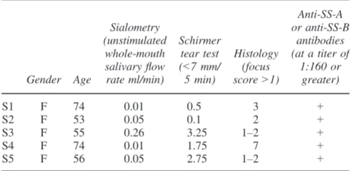
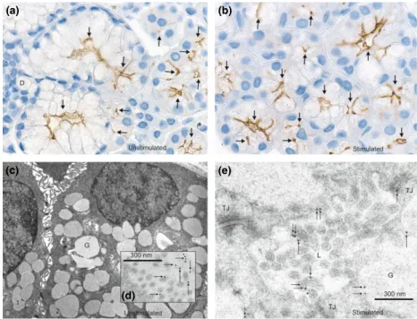
Immunoperoxidase labeling with human AQP5 antibodies was performed on paraffin sections of human labial sali- vary glands. They did not cross-react with rat salivary gland tissue (data not shown) and they gave the same labeling pattern (Figure 1). AQP5 labeling was observed apically in the acinar cells including the membranes of the secretory canaliculi in a punctate appearance (Figure 1a).
No AQP5 labeling was present in the ducts of the labial glands (e.g., D in Figure 1a).
The subcellular localization of AQP5 in human labial sal- ivary glands was further investigated by immunoelectron
Table 1Diagnostics of the patients with Sj€ogren’s syndrome
Gender Age
Sialometry (unstimulated whole-mouth salivaryflow rate ml/min)
Schirmer tear test (<7 mm/
5 min)
Histology (focus score>1)
Anti-SS-A or anti-SS-B
antibodies (at a titer of
1:160 or greater)
S1 F 74 0.01 0.5 3 +
S2 F 53 0.05 0.1 2 +
S3 F 55 0.26 3.25 1–2 +
S4 F 74 0.01 1.75 7 +
S5 F 56 0.05 2.75 1–2 +
SS, Sj€ogren’s syndrome.
e115
microscopy. A quantitative assessment of immunogold labeling on sections fromfive different patients showed that gold particles were few in number in intracellular structures [9.9 0.8% (mean s.e.m.)], they were mainly associ- ated with the apical plasma membranes (Figure 1c, d), especially with the microvilli [90.1 0.8% (mean s.e.m.)].
Salivary secretion was stimulated by administration of a pilocarpine tablet (5 mg) for 30 min. Light microscope immunohistochemistry (Figure 1b) and immunoelectron microscopy (Figure 1e) revealed that there were no signifi- cant changes in the distribution of AQP5. On average, the level of intracellular labeling remained almost the same (9.6 0.8%), and the other gold particles (90.4 0.8%) were associated with the apical plasma membrane in a punctate appearance that can be a result of clustering of AQP5, around the area of the microvilli. Thus, in control human labial salivary glands, no indication of changes can be observed in distribution of AQP5 after stimulation with oral pilocarpine.
Patients with SS
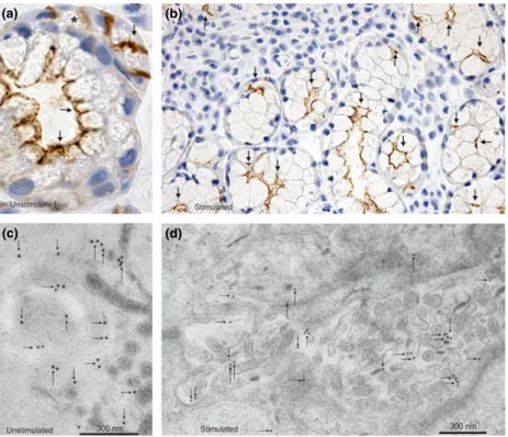
Distribution and localization of AQP5 in human labial minor salivary glands were the same in patients with SS as in the controls. We have used the same human AQP5 antibodies as on normal subjects and they gave the same labeling pattern also in patients with SS (Figure 2). AQP5 was definitely localized in the apical plasma membranes of acinar cells (Figure 2a). Thus, in patients with Sj€ogren’s syndrome, almost the same subcellular localization pattern
of AQP5 was found: 91.81.4% (mean s.e.m.) of the gold particles were present in the apical plasma membrane, and only 8.21.4% (mean s.e.m.) at the intracellular structures (Figure 2c).
The muscarinic stimulation for 30 min significantly increased saliva production, as it was nearly undetectable without stimulation (data not shown). Immunohistochemis- try demonstrated that even after pilocarpine treatment, the AQP5 labeling remained localized to the apical plasma membrane of the acinar cells (Figure 2b). It was con- firmed by immunoelectron microscopy: 91.9 0.5%
(means.e.m.) of gold particles were present in the api- cal plasma membrane, including the microvilli, and 8.10.5% (means.e.m.) were present in the intracel- lular compartments (Figure 2d).
Discussion
In this study, we have tested the effect of oral pilocarpine on AQP5 distribution in human labial salivary gland biop- sies of control and patients with Sj€ogren’s syndrome before dosing and 30 min after drug exposure with light microscopy and immunogold electron microscopy.
Corresponding with our previous results on human sali- vary glands (Gresz et al, 2001), AQP5 labeling was clearly apical at the acinar membrane domains, and no AQP5 labeling was present in the ducts of the labial glands (e.g., D in Figure 1a). In line with the results of Beroukas et al, (2001) (Waterman et al, 2002, 2003), AQP5 was localized apically in the acinar cells of patients
(a) (b)
(c) (e)
(d)
Figure 1AQP5 distribution in human labial salivary glands of normal subjects by immunohistochemistry. Immunoperoxidase light microscopical and immunogold electron microscopical localization of AQP5 under unstimulated (a, c, d) and stimulated (b,e) conditions of salivary secretion. (a) In unstimulated normal human labial glands, AQP5 labeling is associated exclusively with the apical membrane of acinar cells (arrows), and it is absent in the ducts (D). (c) Low magnification shows two adjacent cells of a human labial gland acinus. (d) Gold particles are associated with microvilli of the apical membrane and of intercellular secretory canaliculi (arrows) in the acini. (b) Immunolocalization of AQP5 30 min after administration of oral pilo- carpine. The localization pattern is similar to that in normal tissues (panel a). (e) Immunogold electron microscopic localization of AQP5 after 30-min oral pilocarpine administration. Compared with control tissues (Panelsc,d), the subcellular localization of AQP5 has not changed. Immunogold labeling can be observed on microvilli of the acinar lumen (arrows). Cells labeled with 1 and 2 are two adjacent cells; G, secretory granule; L, lumen; N, nucleus; TJ, tight junction. Magnifications:9200 (a);9260 (b);98.200 (c)
e116
with Sj€ogren’s syndrome. However, our finding for the distribution and density of AQP5 in human labial minor salivary glands was contrast to the results in other papers (Steinfeld et al, 2001; Steinfeld et al, 2002; Steinfeld and Delporte, 2002; Xiao et al, 2011) They claim that AQP5 labeling was present also at the basolateral membrane of acinar cells in patients with SS (Steinfeld et al, 2001, 2002), not to say that a more recent study reported about surprising localizations of AQP5 in human labial glands (Xiao et al, 2011), where an abundance of staining at nearly every structure of human labial glands including different types of ducts was shown in controls. However, the possibility of non-specific immunohistochemical bind- ings arises, for their immunohistochemical results particu- larly claiming that there is a change in distribution of AQP5 in the labial glands of patients with SS.
Reacting on the contradictions, Beroukas et al, and Watermanet al, published a paper (Watermanet al, 2002) and then a longer letter (Waterman et al, 2003) and con- firmed our results (Gresz et al, 2001). They concluded that basolateral staining of AQP5 in salivary acini is non- specific background staining, and they were unable to demonstrate any altered AQP5 expression in Sj€ogren’s salivary glands.
The contradictory data may have been caused by the use of different AQP5 antibodies, because anti-rat AQP5 antibody does not cross-react with the human AQP5 pro- tein (Steinfeld et al, 2002; Delporte and Steinfeld, 2006).
We have tested different human antibodies, and our results
have confirmed an exclusive apical labeling of AQP5 in Sj€ogren’s labial minor salivary glands.
Another controversial issue is the possibility that the distribution of AQP5 is altered by trafficking to the apical membrane. In vitro experiments using gland slices and cultured cells supported this idea (Ishikawa et al, 1998, 1999, 2000, 2002, 2004, 2005; Tada et al, 1999; Ishikawa and Ishida, 2000; Li et al, 2004, 2006). We tested this hypothesis in vivo in rat salivary glands, following stimu- lation or inhibition of salivary secretion in a previous study: we have not found evidence of AQP5 transloca- tion, AQP5 was mainly localized in the apical membrane structures of the acini and of the intercalated ducts of the rat submandibular gland, and there was no difference between controls and treated subjects (Gresz et al, 2004).
The only difference was the clustering of AQP5 at the apical membrane, especially at the microvilli in stimulated salivary secretion. It could be explained by the intensive exocytosis, where clustering seems to be an important step for apical sorting of proteins (Hannan et al, 1993;
Weisz and Rodriguez-Boulan, 2009). Matsuzaki et al, (2003, 2006, 2012) have also performed immunoelectron microscopy in rodent salivary glands to clarify whether AQP5 is localized intracellularly; however, they were not able to detect apparent labeling in the cytoplasm, so their results also suggested that AQP5 translocation is unlikely to occur.
Similar to our findings in rat submandibular gland (Gresz et al, 2004), following pilocarpine administration,
(a) (b)
(c) (d)
Figure 2AQP5 distribution in human labial salivary glands of Sj€ogren’s syndrome (SS) patients by immunohistochemistry. Immunoperoxidase light microscopical and immunogold electron microscopical localization of AQP5 under unstimulated (a,c) and stimulated (b,d) conditions of salivary secre- tion. (a) Large magnification of a mucous acinus with a serous demilune (marked with*) of a SS patient under resting conditions. AQP5 is associated with the apical membrane domains (arrows) and is similar to control patients (Figure 1). (c) Electron microscopy shows extensive immunogold labeling in the apical plasma membrane, especially on microvilli of unstimulated Sj€ogren’s labial glands. (b,d) Immunolocalization of AQP5 30 min after oral pilocarpine administration. The subcellular localization of AQP5 has not changed as it can be detected by light (b) or immunogold electron microscopy (d). AQP5 is localized basically in the apical membrane, especially on the microvilli of the acinar lumen (arrows). Magnifications:9400 (a);9150 (b)
e117
the subcellular distribution of AQP5 did not change in human labial minor salivary glands, either. Our immuno- electron microscopic findings supported our light micro- scopical results showing that the density of gold particles remained very sparse in intracellular structures both in normal and in Sj€ogren’s groups. There was no evidence of AQP5 trafficking after 30-min simulation of salivary secretion with oral pilocarpine. This suggests that during stimulated secretion, the accelerated turnover of the apical membrane happens without movement of AQP5; thus, an anchoring mechanism is responsible for retaining AQP5 in the apical membrane. This could cause clustering of AQP5 labeling after stimulation with pilocarpine, espe- cially at the microvilli.
In a study, Nielsen et al, suggested that AQP5 is regu- lated differently in the acinar and interlobular ducts (Ishik- awa et al, 2005). They proposed that AQP5 translocation to the apical membrane and dissociation from lipid rafts takes place at the apical plasma membrane of the interlob- ular ducts of the rat parotid glands as it arrived there.
Lipid rafts are small, dynamic membrane microdomains that compartmentalize cellular processes, influencing membrane protein and receptor trafficking.
The different authors focus mainly on the distribution and translocation of AQP5 in unstimulated and stimulated salivary glands and in Sj€ogren’s syndrome and diabetes, as well. In the past 10 years, there have been many publi- cations about the role of AQP5 in salivary secretion, relat- ing to its distribution and translocation. We consider the contradictory data originated from the diversity of the experiments, which were based mainly on rats and mice, and relatively few of them were carried out on humans.
The contradictions are multiplied by the species differ- ences, by the type of the salivary glands investigated, by the different cell types within the glands, and by the dif- ferences in unstimulated and stimulated conditions. Not surprisingly Peter Agre, the Nobel Prize winner and father of AQP-research emphasized‘Man Is not a rodent: . . .’in a title of a paper (King and Agre, 2001).
It is generally accepted that AQP5 is essential for the movement of water into secretion (Ma et al, 1999). We know almost nothing about the mechanism of secretion of AQP5 protein itself into the saliva. The correlation between mRNA and protein levels of AQP5 is not neces- sarily parallel. In the parotid glands of diabetic rats, an increase in AQP5 mRNA and a decrease in AQP5 protein levels were measured (Wanget al, 2011). The cellular dis- tribution of AQP5 has been investigated by many workers, but there are little data about expression, degradation, and trafficking of AQP5 or any connection between them (Hoffert et al, 2000; Azlina et al, 2010). One wonders whether the channels are always open and working. As there are normal amounts in SS glands, it seems that the rate of secretion is not dependent on its presence. (Do not forget that most of all salivary secretion is dependent on nerve impulses!). A recent study about AQP5 trafficking in Sj€ogren’s syndrome clearly indicated that autoantibod- ies against M3 muscarinic receptors inhibit AQP5 translo- cation from intracellular compartments to the apical membrane and may lead to decreased salivary secretion (Leeet al, 2013).
In summary, our studies indicate that AQP5 labeling is basically associated with the microvilli and the intercellu- lar secretory canaliculi of the acini in human labial minor salivary glands, including resting conditions. Neither SS itself, nor muscarinic cholinergic stimulation resulted in any significant changes in distribution of AQP5 after 30- min stimulation of salivary secretion. The present AQP5 research leads us to believe that in a few seconds after stimulation, the possible changes in AQP5-distribution is not a result of higher level protein synthesis or transloca- tion, but a result of a cytoskeletal reorganization and retention of AQP5 in the apical membrane of the acini.
The mechanism is so far undefined, but most likely it is a critical step in the huge acinar water transport during stim- ulated salivary secretion. The poor flow of saliva in SS is not caused by reduced presence of AQP5.
Acknowledgements
We thank Inger Merete S. Paulsen, Zhila Nikrozi, and Ida Maria Jalk for excellent technical assistance and for Dr. Jeppe Praetori- us for developing a human AQP5 antibody. The study was sup- ported by the Danish National Research Foundation and by the Human Frontier Science Program, Karen Elise Jensen Founda- tion, the European Commission (QRLT-2000-00987 and QLRT- 2000-00778), and the Danish Medical Research Council.
Author contributions
Veronika Gresz (Department of Oral Diagnostics, Semmelweis University) is the corresponding author, and she did the immuno- histochemical experiments, light microscopy, and electron microscopy. Istvan Gera and Attila Horvath from the Department of Periodontology, Semmelweis University, presented the patients, and they were responsible for the diagnosis of Sj€ogren’s syndrome, they have made sialometry, Schirmer test, and labial gland biopsy. Soren Nielsen, the head of The Water and Salt Research Center and Institute of Anatomy, University of Aarhus, was the leader and supervisor of the immunohistochemical exper- iments, light microscopy and electron microscopy. Tivadar Zelles (Department of Oral Biology, Semmelweis University) has updated knowledge in thefield and in the relevant literature, and he was advising and editing the manuscript.
References
Azlina A, Javkhlan P, Hiroshima Yet al(2010). Roles of lyso- somal proteolytic systems in AQP5 degradation in the sub- mandibular glands of rats following chorda tympani parasympathetic denervation. Am J Physiol Gastrointest Liver Physiol299: G1106–G1117.
Beroukas D, Hiscock J, Jonsson R, Waterman SA, Gordon TP (2001). Subcellular distribution of aquaporin 5 in salivary glands in primary Sjogren’s syndrome.Lancet358: 1875–1876.
Carpenter GH, Proctor GB, Pankhurst CL, O’Donohue J, Scott D, Hunnable MP (2000). Sialochemical markers of salivary gland involvement with Sj€ogren’s syndrome secondary to rheumatoid arthritis and primary biliary cirrhosis. J Oral Pathol Med29: 452–459.
Daniels TE (1984). Labial salivary gland biopsy in Sj€ogren’s syndrome: assessment as a diagnostic criterion in 362 suspected cases.Arthritis Rheum27: 147–156.
Daniels TE (1986). Salivary histopathology in diagnosis of Sj€ogren’s syndrome.Scand J Rheumatol Suppl61: 36–43.
e118
Delporte C, Steinfeld S (2006). Distribution and roles of aquapo- rins in salivary glands. Review. Biochim Biophys Acta 1758:
1061–1070.
Fox RI (2005). Sj€ogren’s syndrome [review]. Lancet 366: 321–
331.
Fox PC, Atkinson JC, Macynski AA et al (1991). Pilocarpine treatment of salivary gland hypofunction and dry mouth (xero- stomia).Arch Intern Med151: 1149–1152.
Fox RI, Konttinen Y, Fisher A (2001). Use of muscarinic agon- ists in the treatment of Sjogren’s syndrome. Clin Immunol 101: 249–263.
Gresz V, Kwon TH, Hurley PTet al (2001). Identification and localization of aquaporin water channels in human salivary glands. Am J Physiol Gastrointest Liver Physiol 281: G247– G254.
Gresz V, Kwon TH, Gong H et al (2004). Immunolocalization of AQP-5 in rat parotid and submandibular salivary glands after stimulation or inhibition of secretionin vivo.Am J Phys- iol Gastrointest Liver Physiol287: G151–G161.
Hannan LA, Lisanti MP, Rodriguez-Boulan E, Edidin M (1993).
Correctly sorted molecules of a GPI-anchored protein are clus- tered and immobile when they arrive at the apical surface of MDCK cells.J Cell Biol120: 353–358.
Hoffert JD, Leitch V, Agre P, King LS (2000). Hypertonic induction of aquaporin-5 expression through an ERK-depen- dent pathway.J Biol Chem275: 9070–9077.
Ishikawa Y, Ishida H (2000). Aquaporin water channel in sali- vary glands.Jpn J Pharmacol83: 95–101.
Ishikawa Y, Eguchi T, Skowronski MT, Ishida H (1998). Acetyl- choline acts on M3 muscarinic receptors and induces the trans- location of aquaporin5 water channel via cytosolic Ca2+
elevation in rat parotid glands.Biochem Biophys Res Commun 245: 835–840.
Ishikawa Y, Skowronski MT, Inoue N, Ishida H (1999). Alpha (1)-adrenoceptor-induced trafficking of aquaporin-5 to the api- cal plasma membrane of rat parotid cells. Biochem Biophys Res Commun265: 94–100.
Ishikawa Y, Skowronski MT, Ishida H (2000). Persistent increase in the amount of aquaporin-5 in the apical plasma membrane of rat parotid acinar cells induced by a muscarinic agonist SNI-2011.FEBS Lett477: 253–257.
Ishikawa Y, Iida H, Ishida H (2002). The muscarinic acethylco- line receptor-stimulated increase in aquaporin-5 levels in the apical plasma membrane in rat parotid acinar cells is coupled with activation of nitric oxide/cGMP signal transduction. Mol Pharmacol61: 1423–1434.
Ishikawa Y, Inoue N, Zhenfang Y, Nakae Y (2004). Molecular mechanisms and drug development in aquaporin water channel diseases: the translocation of aquaporin-5 from lipid rafts to the apical plasma membranes of parotid glands of normal rats and the impairment of it in diabetic or aged rats.J Pharmacol Sci96: 271–275.
Ishikawa Y, Yuan Z, Inoue N et al (2005). Identification of AQP5 in lipid rafts and its translocation to apical membranes by activation of M3 mAChRs in interlobular ducts of rat paro- tid gland.Am J Physiol Cell Physiol289: C1303–C1311.
Jonsson R, Haga HJ, Gordon T (2001). Sj€ogren’s syndrome. In:
Koopman WJ, ed. Arthritis and allied conditions: a textbook of rheumatology. 14th edn. Lippincott Williams & Wilkins:
Philadelphia, pp. 1736–1759.
King LS, Agre P (2001). Man Is not a rodent: aquaporins in the airways.Am J Respir Cell Mol Biol24: 221–223.
Kwon TH, Pushkin A, Abuladze N, Nielsen S, Kurtz I (2000).
Immunoelectron microscopic localization of NBC3 sodium- bicarbonate cotransporter in rat kidney. Am J Physiol Renal Physiol278: F327–F336.
Lee BH, Gauna AE, Perez G et al (2013). Autoantibodies against muscarinic type 3 receptor in Sj€ogren’s syndrome inhi- bit aquaporin 5 trafficking.PLoS One8: e53113.
Li J, Ha YM, Ku NY, Choi SY, Lee SJ, Oh SB (2004). Inhibi- tory effects of autoantibodies on the muscarinic receptors in Sjogren’s syndrome.Lab Invest84: 1430–1438.
Li J, Lee S, Choi SY et al(2006). Effects of pilocarpine on the secretory acinar cells in human submandibular glands.Life Sci 79: 2441–2447.
Ma T, Song Y, Gillespie A, Carlson EJ, Epstein CJ, Verkman AS (1999). Defective secretion of saliva in transgenic mice lacking aquaporin-5 water channels.J Biol Chem274: 20071–
20074.
Matsuzaki T, Tajika Y, Suzuki T, Aoki T, Hagiwara H, Takata K (2003). Immunolocalization of the water channel, aquapo- rin-5 (AQP5), in the rat digestive system. Arch Histol Cytol 66: 307–315.
Matsuzaki T, Ablimit A, Suzuki T, Aoki T, Hagiwara H, Takata K (2006). Changes of aquaporin 5-distribution during release and reaccumulation of secretory granules in isoproterenol-trea- ted mouse parotid gland.J Electron Microsc55: 183–189.
Matsuzaki T, Susa T, Shimizu K et al (2012). Function of the membrane water channel aquaporin-5 in the salivary gland.
Acta Histochem Cytochem45: 251–259.
Nielsen S, Pallone T, Smith BL, Christensen EI, Agre P, Mauns- bach AB (1995). Aquaporin-1 water channels in short and long loop descending thin limbs and in descending vasa recta in rat kidney.Am J Physiol268: F1023–F1037.
Nielsen S, King LS, Christensen BM, Agre P (1997). Aquaporins in complex tissues II. Subcellular distribution in respiratory and glandular tissues of rat.Am J Physiol273: C1549–C1561.
Nyarady Z, Nemeth A, Ban Aet al(2006). A randomized study to assess the effectiveness of orally administered pilocarpine during and after radiotherapy of head and neck cancer. Anti- cancer Res26: 1557–1562.
Rhodus NL (1997). Oral pilocarpine HCL stimulates labial (minor) salivary gland flow in patients with Sjogren’s syn- drome.Oral Dis3: 93–98.
Salagen tablets. Physicians’ desk reference (1997). 51st edn.
Medical Economics Books: Montvale, NJ, pp. 1546–1547.
Skopouli FN, Siouna-Fatourou HI, Ziciadis C, Moutsopoulos HM (1989). Evaluation of unstimulated whole salivaflow rate and stimulated parotid flow as confirmatory tests for xerosto- mia.Clin Exp Rheumatol7: 127–129.
Steinfeld SD, Delporte C (2002). Distribution of salivary aquapo- rin-5 in Sj€ogren’s syndrome.Lancet359: 1777–1778.
Steinfeld S, Cogan E, King LS, Agre P, Kiss R, Delporte C (2001). Abnormal distribution of aquaporin-5 water channel protein in salivary glands from Sj€ogren’s syndrome patients.
Lab Invest81: 143–148.
Steinfeld SD, Appelboom TA, Delporte C (2002). Treatment with infliximab restores normal aquaporin 5 distribution in minor salivary glands of patients with Sj€ogren’s syndrome.
Arthritis Rheum46: 2249–2251.
Tada J, Sawa T, Yamanaka Net al(1999). Involvement of vesi- cle-cytoskeleton interaction in AQP5 trafficking in AQP5- gene-transfected HSG cells. Biochem Biophys Res Commun 266: 443–447.
Vitali C, Bombardieri S, Jonsson R et al (2002). Classification criteria for Sjogren’s syndrome: a revised version of the Euro- pean criteria proposed by the American-European Consensus Group.Ann Rheum Dis61: 554–558.
Vivino FB, Al-Hashimi I, Khan Z, LeVeque FG, Salisbury PL 3rd, Tran-Johnson TK (1999). Pilocarpine tablets for the treat- ment of dry mouth and dry eye symptoms in patients with Sjogren syndrome: a randomized, placebo-controlled, fixed-
e119
dose, multicenter trial. P92-01 Study Group. Arch Intern Med 159: 174–181.
Wang D, Yuan Z, Inoue N, Cho G, Shono M, Ishikawa Y (2011). Abnormal subcellular localization of AQP5 and down- regulated AQP5 protein in parotid glands of streptozotocin- induced diabetic rats.Biochim Biophys Acta1810: 543–554.
Waterman SA, Beroukas D, Hiscock J, Jonsson R, Gordon TP (2002). Distribution of salivary aquaporin-5 in Sj€ogren’s syn- drome [letter].Lancet359: 1778.
Waterman SA, Beroukas D, Hiscock J, Jonsson R, Gordon TP (2003). Aquaporins in primary Sj€ogren’s syndrome: comment on the articles by Steinfeld et al.Arthritis Rheum48:1167–1168.
Weisz OA, Rodriguez-Boulan E (2009). Apical trafficking in epi- thelial cells: signals, clusters and motors. J Cell Sci 122:
4253–4266.
Wynn RL (1996). Oral pilocarpine (Salagen)–a recently approved salivary stimulant.Gen Dent44: 29–30.
Xiao L, Ng TB, Feng YB et al (2011). Dendrobium candidum extract increases the expression of aquaporin-5 in labial glands from patients with Sj€ogren’s syndrome. Phytomedicine 18:
194–198.
Yasui M, Kwon TH, Knepper MA, Nielsen S, Agre P (1999).
Aquaporin-6: an intracellular vesicle water channel protein in renal epithelia.Proc Natl Acad Sci USA96: 5808–5813.
e120