Digital method and content
development of the hungarian higher education in dentistry in Hungarian,
German and English
Dr. Bán Ágnes, Dr. Benke Beáta, Dr. Blazsek József, Dr. Bori Erzsébet, Dr.
Frank Dorottya, Dr. Fulmer András, Dr. Gaszner Balázs, Dr. Gelencsér Gábor, Dr. Gurdán Zsuzsanna, Dr. Herényi Gejza, Dr. Hollósy Tibor, Dr. Jász Máté, Dr.
Kádár Kristóf, Dr. Kerémi Beáta, Dr. Kiss Gábor, Dr. Krajczár Károly, Dr.
Lempel Edina, Dr. Lohinai Zsolt, Dr. Mandel Iván, Dr. Marada Gyula, Dr. Molnár Bálint, Dr. Muzsek Zsófia Katalin, Dr. Nagy Balázs, Dr. Nagy Dávid, Dr. Nemes Bálint, Dr. Németh Dániel, Dr. Németh Zsolt Ferenc, Dr. Orsi Enikő, Dr. Ottóffy- Kende Dóra, Dr. Rostási-Szabó Judit, Dr. Sándor Balázs, Dr. Szalma József, Dr.
Szántó Ildikó, Dr. Szombath Dezső, Dr. Tóth Pál, Dr. Vajda Katalin, Dr. Varga
Gábor, Dr. Várnai Katalin, Dialóg Campus Kiadó
Digital method and content development of the hungarian higher education in dentistry in Hungarian, German and English
by Dr. Bán Ágnes, Dr. Benke Beáta, Dr. Blazsek József, Dr. Bori Erzsébet, Dr. Frank Dorottya, Dr. Fulmer András, Dr. Gaszner Balázs, Dr. Gelencsér Gábor, Dr. Gurdán Zsuzsanna, Dr. Herényi Gejza, Dr. Hollósy Tibor, Dr. Jász Máté, Dr. Kádár Kristóf, Dr. Kerémi Beáta, Dr. Kiss Gábor, Dr. Krajczár Károly, Dr. Lempel Edina, Dr. Lohinai Zsolt, Dr. Mandel Iván, Dr. Marada Gyula, Dr. Molnár Bálint, Dr. Muzsek Zsófia Katalin, Dr. Nagy Balázs, Dr. Nagy Dávid, Dr. Nemes Bálint, Dr. Németh Dániel, Dr. Németh Zsolt Ferenc, Dr. Orsi Enikő, Dr. Ottóffy-Kende Dóra, Dr. Rostási-Szabó Judit, Dr. Sándor Balázs, Dr. Szalma József, Dr. Szántó Ildikó, Dr. Szombath Dezső, Dr. Tóth Pál, Dr. Vajda Katalin, Dr. Varga Gábor, Dr. Várnai Katalin
Publication date 2014
Copyright © 2014 Pécsi Tudományegyetem, Semmelweis Egyetem, Dialóg Campus Kiadó
Copyright 2014., Dr. Bán Ágnes, Dr. Benke Beáta, Dr. Blazsek József, Dr. Bori Erzsébet, Dr. Frank Dorottya, Dr. Fulmer András, Dr.
Gaszner Balázs, Dr. Gelencsér Gábor, Dr. Gurdán Zsuzsanna, Dr. Herényi Gejza, Dr. Hollósy Tibor, Dr. Jász Máté, Dr. Kádár Kristóf, Dr.
Kerémi Beáta, Dr. Kiss Gábor, Dr. Krajczár Károly, Dr. Lempel Edina, Dr. Lohinai Zsolt, Dr. Mandel Iván, Dr. Marada Gyula, Dr. Molnár Bálint, Dr. Muzsek Zsófia Katalin, Dr. Nagy Balázs, Dr. Nagy Dávid, Dr. Nemes Bálint, Dr. Németh Dániel, Dr. Németh Zsolt Ferenc, Dr.
Orsi Enikő, Dr. Ottóffy-Kende Dóra, Dr. Rostási-Szabó Judit, Dr. Sándor Balázs, Dr. Szalma József, Dr. Szántó Ildikó, Dr. Szombath Dezső, Dr. Tóth Pál, Dr. Vajda Katalin, Dr. Varga Gábor, Dr. Várnai Katalin
Table of Contents
Digital method and content development of the hungarian higher education in dentistry in Hungarian, German and English ... lxv Preface ... lxvii
1. 1. Oral biology ... 1
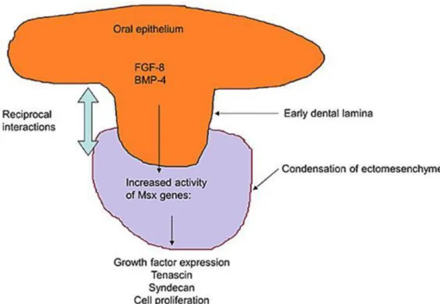
1. 1.1. Development of tooth germ – Gabor Varga ... 1
1.1. Test – Development of tooth germ (answers) ... 9
2. 1.2. Fibers and extracellular matrix of hard tissues – Gabor Varga ... 9
2.1. Test – Fibers and extracellular matrix of hard tissues (answers) ... 17
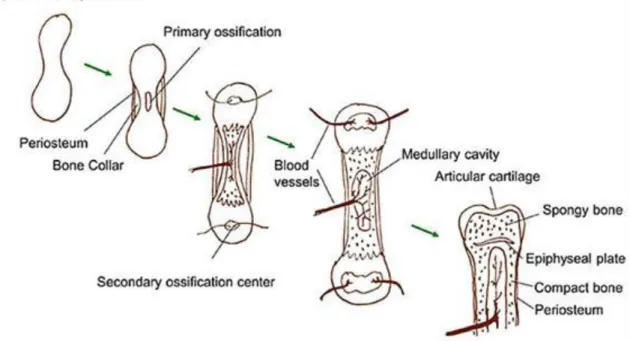
3. 1.3. Osteogenesis – Gabor Varga ... 18
3.1. Test – Osteogenesis (answers) ... 25
4. 1.4. Dentinogenesis and disturbances; formation of primary-, secondary- and tertiary dentin; dentin permeability – Gabor Varga ... 26
4.1. Test – Dentinogenesis and disturbances; formation of primary-, secondary- and tertiary dentin; dentin permeability (answers) ... 33
5. 1.5. Amelogenesis – Gabor Varga ... 34
5.1. Test – Amelogenesis (answers) ... 41
6. 1.6. Mineral composition of enamel and dentine. Bioapatites – Beata Keremi ... 42
6.1. Mineral components of dental enamel, dentin and cementum ... 42
6.2. Features of crystallization ... 44
6.2.1. Homogeneous nucleation ... 45
6.2.2. Heterogenous nucleation ... 45
6.2.3. Factors influencing crystallization ... 45
6.3. Spatial structure of apatite crystals ... 45
6.4. Trace element effects on apatite crystals ... 47
6.5. Test - Mineral composition of enamel and dentine. Bioapatites (answers) ... 47
7. 1.7. Calcium homeostasis. Dental aspects of calcium and phosphate metabolism disorders – Dezso Szombath ... 48
7.1. Hyper- and hypocalcemia ... 48
7.2. Dental aspects of vitamin D metabolism ... 51
7.3. Congenital hypophosphatasia ... 52
7.4. Test – Calcium homeostasis (answers) ... 52
8. 1.8. Formation of hard tissues, mineralization, bone resorption and osteoclasts – Gábor Varga 53 8.1. Test – Formation of hard tissues, mineralization, bone resorption and osteoclasts (answers) ... 60
9. 1.9. Cementogenesis – Balint Molnar ... 60
9.1. Acellular fibrillar cementum ... 60
9.2. Cellular, fibrillar cementum ... 61
9.3. Process of Cementogenesis ... 62
9.4. Cemento-dentinal junction ... 62
9.5. Cementoneogenesis, periodontal regeneration ... 63
9.6. Test – Cementogenesis (answers) ... 63
10. 1.10. Pathomechanism of bleeding and its relation to dentistry – Katalin Varnai ... 64
10.1. Test – Pathomechanism of bleeding and its relation to dentistry (answers) ... 69
11. 1.11. Tooth eruption and tooth movement – Balint Nemes ... 70
11.1. Tooth eruption ... 70
11.1.1. Cellular processes ... 70
11.1.2. Molecular background ... 71
11.1.3. Eruption of the deciduous and permanent teeth ... 71
11.1.4. Abnormalities of tooth eruption ... 72
11.2. Tooth movement ... 74
11.2.1. Active tooth movement: Basic rules ... 74
11.3. Test – Tooth eruption and tooth movement (answers) ... 75
12. 1.12. The morphology and function of salivary glands – Gabor Varga ... 76
12.1. Test – The morphology and function of salivary glands (answers) ... 83
13. 1.13. Salivary gland electrolyte, water and protein secretion – Gabor Varga ... 84
13.1. Test – Salivary gland electrolyte, water and protein secretion (answers) ... 88
14. 1.14. Oral function and diagnostic role of secreted saliva – Gabor Varga ... 89
14.1. Test – Oral function and diagnostic role of secreted saliva (answers) ... 94
15. 1.15. Dental stem cells for dental research – Gabor Varga ... 95
15.1. Test – Dental stem cells for dental research (answers) ... 104
16. 1.16. Nutrition and oral health; Characterization of oral tissues and function in elderly – Gábor Varga ... 105
16.1. Test – Nutrition on oral health (answers) ... 110
17. 1.17. Pathophysiology of chewing – Mate Jasz ... 110
17.1. Test – Chewing (answers) ... 112
18. 1.18. Pathophysiology of inflammation – Beata Keremi ... 113
18.1. Inflammation ... 113
18.2. Features of acute inflammation ... 114
18.2.1. Cells involved in acute inflammation ... 114
18.2.2. Two main phases of acute inflammation ... 115
18.2.3. Cytokines ... 119
18.2.4. Chemokines ... 120
18.2.5. Oral aspects of inflammation ... 121
18.3. Test – Pathophysiology of inflammation (answers) ... 122
19. 1.19. Structural and functional characteristic of dental pulp, blood supply to the oral tissues, pulpal pain and inflammation – Gábor Varga ... 122
19.1. Test – Structural and functional characteristics of dental pulp, blood supply to the oral tissues, pulpal pain and inflammation (answers) ... 129
20. 1.20. Radiation, oral symptoms associated with radiotherapy – Kristof Kadar ... 129
20.1. Biological effect of ionizing radiation ... 129
20.2. Oral effects of therapeutic irradiation ... 132
20.2.1. Osteoradionecrosis ... 132
20.2.2. Salivary dysfunction after radiotherapy ... 133
20.2.3. Oral mucositis ... 133
20.3. Test – Ionizing radiation and the oral effects therapeutic irradiation (answers) . 136 21. 1.21. Gene therapy and gene polymorphysms in dentistry – Gabor Varga ... 137
21.1. Test – Gene therapy and gene polymorphism in dentistry (answers) ... 147
22. 1.22. Pathomechanisms in oral cancer – Gábor Varga ... 148
22.1. Test – Mechanisms of tumor formation and oral cancer (answers) ... 161
23. 1.23. Oral sensation: Taste and smelling – Jozsef Blazsek ... 162
23.1. Test – Oral sensation: Taste and smelling (answers) ... 169
24. 1.24. Gingival sulcus and crevicular fluid – Zsolt Lohinai ... 169
24.1. Gingival sulcus ... 169
24.2. Gingival crevicular fluid (GCF) ... 171
24.3. Biological width ... 171
24.4. Test – Gingival sulcus and crevicular fluid (answers) ... 172
25. 1.25. Oral aspects of salt and water household disturbancies – Jozsef Blazsek ... 173
25.1. Test – Oral aspects of salt and water household disturbances (answers) ... 181
26. 1.26. Oral aspects of gastric and pancreatic disorders – Gabor Varga ... 182
26.1. Test – Oral aspects of gastric and pancreatic disorders (answers) ... 194
27. 1.27. Oral aspects of acid-base regulation – Jozsef Blazsek ... 195
27.1. Test – Oral aspects of acid-base regulation (answers) ... 200
28. 1.28. Oral aspects of kidney disorders – Gabor Varga ... 201
28.1. Test – Oral aspects of kidney disorders (answers) ... 212
29. 1.29. Pathophysiology of liver – Beata Keremi ... 212
29.1. Major functions of the liver ... 213
29.2. Bilirubin metabolism ... 216
29.3. Icteruses ... 217
29.4. Forms of hepatitis ... 218
29.4.1. Viral forms of hepatitis ... 218
29.4.2. Chronic hepatitis ... 220
29.5. Cirrhosis ... 221
29.6. Portal hypertension ... 221
29.6.1. Ascites ... 222
29.6.2. Portocaval shunt ... 223
29.7. Liver failure ... 223
29.8. Liver tumors ... 224
29.9. Pathomechanisms of liver diseases ... 224
29.10. Liver function problems ... 225
29.11. Test – Pathophysiology of liver and oral aspects (answers) ... 225
30. 1.30. Cardiac insufficiency and shock – oral aspects – Kristof Kadar ... 226
30.1. Heart failure (central circulatory failure) ... 226
30.1.1. Symptoms of heart failure ... 227
30.1.2. The underlying causes of heart failure ... 228
30.1.3. Aggravating factors in heart failure: ... 228
30.1.4. The basic pathomechanisms of heart failure ... 228
30.1.5. Neurohormonal response ... 229
30.2. Circulatory shock (peripheral circulatory failure) ... 230
30.2.1. Types of shock ... 231
30.2.2. Pathomechanism of shock ... 232
30.2.3. Organ damage in shock – shock organs ... 234
30.3. Test – Central and peripheral circulatory failure (answers) ... 235
31. 1.31. Oral aspects of hypertension – Gabor Varga ... 236
31.1. Test – Oral aspects of hypertension (answers) ... 244
32. 1.32. Oral aspects of protein metabolism and energy balance – Gabor Varga ... 244
32.1. Test – Oral aspects of protein metabolism and energy balance (answers) ... 254
33. 1.33. Oral aspects of carbohydrate metabolism and diabetes – Beata Keremi ... 255
33.1. Types ... 255
33.1.1. Type 1 DM ... 255
33.1.2. Type 2 DM ... 255
33.1.3. Secondary Diabetes Mellitus ... 256
33.1.4. Gestational diabetes mellitus ... 256
33.2. Symptoms ... 256
33.3. Consequences ... 256
33.4. Oral manifestation ... 257
33.5. Diagnosis ... 259
33.5.1. Blood glucose analysis – from fingertip capillary blood ... 259
33.5.2. Measurement of glycosylated hemoglobin ... 259
33.6. Treatment ... 259
33.7. Metabolic X syndrome ... 259
33.8. Test – Oral aspects of carbohydrate metabolism and diabetes (answers) ... 260
34. 1.34. Oral aspects of lipid metabolism – Beata Keremi ... 262
34.1. Classification of lipids ... 262
34.2. Lipids in nutrition ... 262
34.3. Digestions and absorption of lipids ... 263
34.4. Lipoproteins ... 263
34.4.1. Structure of lipoproteins ... 263
34.4.2. Functions of lipoproteins ... 264
34.4.3. Types of lipoproteins ... 265
34.5. Steps of lipid metabolism ... 265
34.6. Dyslipidemias ... 268
34.6.1. Types of hyperlipoproteinemias ... 268
34.7. Therapy of lipid metabolism disturbances ... 269
34.8. Test – Oral aspects of lipid metabolism (answers) ... 270
35. 1.35. Oral aspects of atherosclerosis – Beata Keremi ... 271
35.1. Atherosclerosis ... 271
35.1.1. Risk factors in the development of atherosclerosis ... 275
35.1.2. Development of atherosclerotic plaque ... 276
35.2. Theories for atherosclerosis development ... 278
35.3. Oral implications of atherosclerosis – or what is common in dental and atherosclerotic plaques? ... 281
35.4. Test – Oral aspects of atherosclerosis (answers) ... 281
36. 1.36. Pain sensation – oral aspects – Gabor Varga ... 282
36.1. Test – Pain sensation – oral aspects (answers) ... 289
37. 1.37. Oral aspects of endocrine disorders – Dezso Szombath ... 290
37.1. Growth hormone ... 290
37.2. Adrenal cortex ... 292
37.3. Thyroid ... 293
37.4. Test – Oral aspects of endocrine disorders (answers) ... 294
38. 1.38. Disorders of respiratory functions – Beata Keremi ... 295
38.1. Lung defense ... 295
38.2. Lung function ... 296
38.3. Assessing of respiratory function ... 296
38.3.1. Static lung volume ... 297
38.3.2. Gas exchange ... 298
38.4. Lung disease ... 298
38.4.1. Dyspnoea ... 298
38.4.2. Cough ... 300
38.4.3. Sputum ... 300
38.4.4. Haemoptysis ... 301
38.4.5. Pain ... 301
38.4.6. Wheeze ... 302
38.4.7. Stridor ... 302
38.5. Test – Disorders of respiratory functions (answers) ... 302
39. 1.39. Practices: Oral-Clearance – Beata Keremi ... 303
39.1. Factors determining oral clearance rate ... 303
39.1.1. Flow rate of saliva secretion ... 303
39.1.2. Swallowing habit ... 304
39.1.3. Breathing habit ... 304
39.1.4. General state of health ... 305
39.1.5. Food consumed ... 305
39.2. Modeling of oral clearance ... 305
39.3. Phases of oral clearance ... 306
39.4. Alterations associated with ageing ... 307
39.5. Fluoride clearance ... 307
39.6. Bacterial clearance ... 307
39.7. Test – Practices: Oral-Clearance (answers) ... 307
40. 1.40. Practices: Oral defense mechanisms – Jozsef Blazsek ... 308
40.1. Oral specific immunity ... 317
40.2. Test – Practices: Oral defense mechanisms (answers) ... 319
41. 1.41. Practices: Analysis of dental plaque – Gabor Kiss ... 320
41.1. Composition ... 320
41.2. Location ... 320
41.3. Mechanism of plaque formation ... 320
41.4. Dental plaque as biofilm ... 322
41.5. Pathogenicity of plaque ... 323
41.6. Dental calculus ... 323
41.7. Test – Practices: Analysis of dental plaque (answers) ... 324
42. 1.42. Practices: Salivary secretion – Gabor Varga ... 325
42.1. Test – Practices: Salivary secretion (answers) ... 330
43. 1.43. Practices: Chewing – Mate Jasz ... 331
43.1. Backward movements of the mandible in the sagittal plane ... 332
43.2. Backward movements of the mandible in the frontal plane ... 332
43.3. The backward movements of the mandible in the horizontal plane ... 333
43.4. Test – Practices: Chewing (answers) ... 333
44. 1.44. Practices: PCR technique in dental research – Erzsebet Bori ... 334
44.1. Test – Practices: PCR technique in dental research (answers) ... 353
45. 1.45. Practices: Investigation methods of mineralised tissues – Kristof Kadar ... 353
45.1. Quantitative methods ... 354
45.1.1. Investigation of surface hardness ... 354
45.1.2. Surface profilometry ... 355
45.1.3. Microradiography ... 356
45.1.4. Fluorescence based methods ... 356
45.2. Qualitative and semiquantitative methods ... 356
45.3. Test – Practices: Investigation methods of mineralised tissues (answers) ... 357
46. 1.46. The structure and development of dental deposits. Dental plaque and calculus. Scaling, polishing, professional and home care oral hygiene – Balazs Sandor ... 358
46.1. The structure and development of dental deposits ... 358
46.1.1. Acquired pellicle (salivary pellicle) ... 358
46.1.2. Dental plaque ... 359
46.1.3. Materia alba ... 361
46.1.4. Debris (food debris) ... 361
46.1.5. Calculus (tartar) ... 361
46.2. Oral hygiene ... 363
46.2.1. Professional oral hygienic care ... 363
46.2.2. Home care oral hygiene ... 370
47. 1.47. The development of the carious lesion. De- and remineralization – Balazs Sandor 381 47.1. Definition of dental caries (decay) ... 381
47.2. Structure of the tooth enamel ... 382
47.3. Demineralization ... 383
47.3.1. The process of demineralization ... 383
47.3.2. Reducing the chance of demineralization ... 384
47.4. Remineralization ... 385
47.4.1. The process of remineralization ... 385
47.4.2. The use of remineralizing solutions ... 385
47.5. Caries ... 385
47.5.1. The histopathological progression of caries ... 385
47.5.2. Main bacteria involved in the formation of dental caries ... 387
47.5.3. The role of saliva in the formation of caries ... 387
47.5.4. Clinical classification of caries ... 388
47.6. Acid etching ... 391
48. 1.48. The effect of fluorides and calcium, toxic considerations. Local and systemic fluoride administration – Balazs Sandor ... 393
48.1. Features of fluoride ... 393
48.2. Toxic effects of fluoride ... 394
48.2.1. Acute fluoride intoxication ... 394
48.2.2. Chronic fluoride intoxication ... 395
48.3. Systemic fluoridation ... 396
48.3.1. Fluoridation of water ... 396
48.3.2. Salt fluoridation ... 397
48.3.3. Milk fluoridation ... 397
48.3.4. Fluoride tablets ... 397
48.4. Local fluoridation ... 398
48.4.1. Materials of home local fluoridation ... 399
48.4.2. Proffesional treatment ... 400
49. 1.49. Fissure sealing – Balazs Sandor ... 401
49.1. Epidemiology ... 402
49.2. Definition of pit and fissure sealing ... 402
49.3. Basis of indication for pit and fissure sealing ... 403
49.4. Technique of pit and fissure sealing with resin based pit and fissure sealing materials 404 49.5. Minimal invasive /extended pit and fissure sealing ... 407
49.6. Possible side effects ... 409
50. 1.50. Effect of feeding pattern to the oral diseases – Balazs Sandor ... 411
50.1. Basic terms ... 411
50.1.1. The transport of food ... 411
50.1.2. BMI (body mass index) ... 412
50.1.3. Food guide pyramid, nutritional components ... 412
50.2. Typical effects of food components ... 413
50.2.1. Proteins ... 413
50.2.2. Carbohydrates ... 414
50.2.3. Fats and oils ... 415
50.2.4. Vitamins, minerals and trace elements, water ... 416
50.2.5. Physical characteristics of foods ... 417
50.2.6. Utilization of sugar substitutes ... 417
50.2.7. Snacking diet ... 420
50.2.8. Breastfeeding ... 421
50.2.9. Bad habits: smoking ... 421
50.2.10. Erosion ... 422
50.2.11. Osteoporosis ... 422
50.2.12. Diet for patients wearing dentures ... 422
50.2.13. Effects of physical and mental retardation, severe systemic diseases, patients requiring continuous care, effects of sugar containing medecines ... 423
51. 1.51. Compex prevention concerning age, dental specialities and general diseases – Balazs Sandor ... 423
51.1. Health education ... 423
51.1.1. Methodological Principles of Health Education ... 423
51.1.2. Health Educational Methods ... 424
51.1.3. Practical Opportunities for Health Education ... 424
51.1.4. Places of Health Education ... 424
51.2. Childhood ... 424
51.2.1. Dental Prevention Required During Pregnancy ... 425
51.2.2. Dental Prevention in Early Childhood (1-3 years) ... 427
51.2.3. In the case of kindergarten and elementary school children (3-6 years) 427
51.2.4. Dental Prevention at School (age group between 7-14) ... 428
51.2.5. Dental Prevention of Secondary School Children ... 429
51.3. Adulthood ... 429
51.4. Elderly Age groups ... 429
51.4.1. Old-age Deformation ... 430
51.4.2. Measurable Parameters of Daily Activity ... 431
51.4.3. Goals of the Maintenance of Oral Health ... 431
51.5. Tesztkérdések az 1.46–51 fejezetekhez ... 432
52. 1.52. Test questions–answers 1–45. ... 434
52.1. I.1. Development of tooth germ – Gabor VARGA ... 434
52.2. I.2. Fibers and extracellular matrix of hard tissues - Gabor VARGA ... 434
52.3. I.3. Osteogenesis – Gabor VARGA ... 434
52.4. I.4. Dentinogenesis and disturbances; formation of primary-, secondary- and tertiary dentin; dentin permeability – Gabor VARGA ... 435
52.5. I.5. Amelogenesis – Gabor VARGA ... 435
52.6. I.6. Test - Mineral composition of enamel and dentine. Bioapatites – Beata KEREMI 435 52.7. I.7. Test - Calcium homeostasis – Dezso SZOMBATH ... 435
52.8. I.8. Formation of hard tissues, mineralization, bone resorption and osteoclasts – Gábor VARGA ... 436
52.9. I.9. Test - Cementogenesis – Balint MOLNAR ... 436
52.10. I.10. Test - Pathomechanism of bleeding and its relation to dentistry – Katalin VARNAI ... 436
52.11. I.11 Test - Tooth eruption and tooth movement – Balint NEMES ... 437
52.12. I.12. The morphology and function of salivary glands - Gabor VARGA ... 437
52.13. I.13. Salivary gland electrolyte, water and protein secretion - Gabor VARGA 437 52.14. I.14. Oral function and diagnostic role of secreted saliva - Gabor VARGA ... 437
52.15. I.15. Dental stem cells for dental research - Gabor VARGA ... 438
52.16. I.16. Nutrition on oral health – Gábor VARGA ... 438
52.17. I.17. Test – Chewing – Mate JASZ ... 438
52.18. I.18. Test – Pathophysiology of inflammation – Beata Keremi ... 438
52.19. I.19. Structural and functional characteristics of dental pulp, blood supply to the oral tissues, pulpal pain and inflammation – Gábor VARGA ... 439
52.20. I.20. Test - Ionizing radiation and the oral effects therapeutic irradiation – Kristof KADAR ... 439
52.21. I.21. Gene therapy and gene polymorphism in dentistry – Gábor VARGA ... 439
52.22. I.22. Mechanisms of tumor formation and oral cancer – Gábor VARGA ... 439
52.23. I.23. Test - Oral sensation: Taste and smelling – József BLAZSEK ... 440
52.24. I.24. Test - Gingival sulcus and crevicular fluid – Zsolt LOHINAI ... 440
52.25. I.25. Test - Oral aspects of salt and water household disturbances – József BLAZSEK 440 52.26. I.26. Oral aspects of gastric and pancreatic disorders – Gábor VARGA ... 440
52.27. I.27. Test - Oral aspects of acid-base regulation – József BLAZSEK ... 441
52.28. I.28. Oral aspects of kidney disorders – Gábor VARGA ... 441
52.29. I.29. Test - Pathophysiology of liver and oral aspects – Beata KEREMI ... 441
52.30. I.30. Test - Central and peripheral circulatory failure – Kádár Kristóf ... 441
52.31. I.31. Oral aspects of hypertension – Gábor VARGA ... 442
52.32. I.32. Oral aspects of protein metabolism and energy balance – Gábor VARGA 442 52.33. I.33. Test - Diabetes mellitus and its oral aspects – Beata KEREMI ... 442
52.34. I.34. Test - Lipid metabolism – Beata Keremi ... 442
52.35. I.35. Test - Atherosclerosis – Beata Keremi ... 443
52.36. I.36. Pain sensation – oral aspects – Gábor VARGA ... 443
52.37. I.37. Test - Oral aspects of endocrine disorders – Dezso SZOMBATH ... 443
52.38. I.38. Test - Disorders of respiratory functions – Beata KEREMI ... 443
52.39. I.39. Test - Oral clearance – Beata Keremi ... 444
52.40. I.40. Test - Practice: Oral defense mechanisms – József Blazsek ... 444
52.41. I.41. Test - Practices: Analysis of dental plaque – Gabor KISS ... 444
52.42. I.42. Practice: Salivary secretion – Gábor VARGA ... 445
52.43. I.43. Test - Practice: Chewing – Mate JASZ ... 445
52.44. I.44. Test - Practices: PCR technique in dental research – Erzsebet BORI ... 445
52.45. I.45. Test - Practice: Investigation methods of mineralised tissues – Kristof KADAR 445 2. 2. Pediatric dentistry and orthodontics ... 447
1. 2.1. Diagnostic procedures in Pediatric Dentistry. Medication therapy. Developmental diseases – Balazs Sandor ... 447
1.1. Diagnostic procedures in pediatric dentistry ... 447
1.1.1. Patient history ... 447
1.1.2. Extraoral examination ... 447
1.1.3. Intraoral examination ... 448
1.1.4. Imaging ... 450
1.1.5. Other diagnostic possibilities ... 451
1.1.6. Special needs ... 452
1.2. Pharmacological management of pain, fever and anxiety ... 453
1.2.1. Systemic fever and pain relief ... 453
1.2.2. Local anesthetics ... 455
1.2.3. Behavior management (sedation) ... 459
1.2.4. Antibiotic treatment ... 460
1.3. Developmental diseases and their treatment ... 460
1.4. Suggestions for therapy in particular dental developmental disorders ... 463
2. 2.2. Cariologic lesions and consequent diseases in primary dentition – Balazs Sandor .... 465
2.1. Characteristics of primary dentition ... 465
2.2. The pathological progression of caries ... 467
2.3. Treatment of primary tooth caries ... 468
2.3.1. Incipient caries ... 469
2.3.2. Superficial and moderate caries ... 469
2.3.3. Profound caries regarding its localization ... 469
2.3.4. Complicated caries ... 471
2.4. ECC (Early Childhood Caries) ... 471
2.4.1. First steps of treatment ... 471
2.4.2. Mild lesions effecting small surfaces ... 471
2.4.3. Advanced ECC ... 471
2.5. Consequent diseases of primary tooth caries ... 471
2.5.1. Consequent diseases of the pulp: inflammation of the pulp (pulpitis) ... 471
2.5.2. Consequent diseases of the periradicular region (complicated gangrena) 472 2.6. Endodontic treatment in primary dentition ... 473
2.6.1. When is endodontic treatment needed ... 473
2.6.2. Extraction or endodontic treatment ... 473
2.6.3. Indications and contraindications ... 473
2.6.4. Treatment options ... 474
3. 2.3. Traumatic lesions in primary dentition – Balazs Sandor ... 480
3.1. Epidemiology and etiology of dental traumas ... 480
3.2. The classification of dental injuries (Table 1.) ... 482
3.3. Injuries to the deciduous teeth ... 483
3.4. Patient examination ... 484
3.5. Treatment of traumatic lesions in primary dentition according to the diagnosis .. 486
3.6. Pathologic sequel of trauma to the teeth ... 495
3.7. Prevention of traumas ... 495
4. 2.4. Cariologic lesions and consequent diseases in young permanent dentition – Balazs Sandor 496 4.1. Sequence of eruption ... 496
4.2. Principles of treatment of young permanent teeth dentition ... 497
4.2.1. Stepwise or total caries removal ... 498
4.3. Treatment of consequent diseases ... 498
4.3.1. Apexogenesis ... 499
4.3.2. Apexification ... 501
5. 2.5. Traumatic cases in young permanent dentition – Balazs Sandor ... 505
5.1. Young permanent teeth ... 505
5.2. Endodontic treatment in young permanent teeth ... 505
5.3. Treatment and diagnosis of injuries to the young prmanent dentitions ... 506
5.4. Prevention of traumas ... 519
5.5. Test ... 520
6. 2.6. Most common dental and jaw disorders – Zsuzsanna Gurdan ... 522
6.1. Etiology of orthodontic anomalies ... 522
6.2. Classification of dentition disorders ... 528
7. 2.7. The diagnostic methods and treatment plan of orthodontic anomalies – Zsuzsanna Gurdan 534 7.1. Medical history ... 534
7.2. Clinical examination ... 537
7.3. Functional analysis ... 538
7.4. Examination of functional anomalies ... 538
7.5. Photo analysis ... 538
7.6. Radiographic examinations ... 538
7.7. Computed tomography ... 543
7.8. Model analysis ... 544
8. 2.8. The methods of orthodontic treatments – Zsuzsanna Gurdan ... 546
9. 2.9. Complex orthodontic cases – Judit Rostasi-Szabo ... 550
9.1. Cooperation with pediatric dentistry ... 551
9.2. Cooperation with prosthetic dentistry ... 553
9.3. Cooperation with periodontology ... 556
9.4. Cooperation with dentoalveolar and maxillofacial surgery ... 557
10. 2.10. Cleft patients – Judit Rostasi-Szabo ... 559
10.1. Etiology ... 559
10.2. Incidence ... 560
10.3. The Classifications of Clefts ... 560
10.4. The therapy of cleft patients ... 561
10.5. The complex therapy prescribed for children with cleft lip and cleft palate ... 564
10.6. The Orthodontic Therapy of Patients with Cleft Lip and Palate ... 568
11. 2.11. Screening, controlling – Zsuzsanna Gurdan ... 571
11.1. Definition of retention ... 571
11.2. Etiology ... 571
11.3. Factors influencing the prevalence of relapse ... 571
11.4. Planning of the retention phase ... 572
11.5. Duration of retention time ... 573
11.6. Types of retention appliances ... 573
11.7. Malocclusions requiring the usage of a removable retention appliance ... 575
11.8. Malocclusions requiring fixed retention appliances ... 576
11.9. Malocclusions that requires no retention appliances ... 576
11.10. Active retention appliances ... 576
11.11. Retention possibilities, systems for different occlusion problems ... 576
11.12. Postretentional phase ... 579
3. 3. Reconstructive dentistry ... 580
1. 3.1. The Modern Concept of Endodontics, Examination Methodes, Treatment Plan – Edina Lempel ... 580
1.1. The Modern Concept of Endodontics ... 580
1.1.1. Diseases of the pulp ... 580
1.1.2. The endodontic treatment ... 581
1.2. Endodontic examination methodes, treatment plan (http://www.sld.cu/galerias/pdf/endodoncia_parte_2.pdf) ... 583
2. 3.2. Pulp Diseases and Their Diagnosis – Edina Lempel ... 584
2.1. The healthy pulp ... 584
2.2. Reversible pulpitis (hyperaemia pulpae) ... 585
2.3. Cracked tooth syndrome ... 585
2.4. Irreversible pulpitis ... 586
2.4.1. Asymptomatic irreversible pulpitis (Pulpitis chronica) ... 586
2.4.2. Symptomatic irreversible pulpitis (Pulpitis acuta) ... 589
2.5. Pulpal necrosis (gangraena pulpae) ... 591
2.6. Acute periapical periodontitis (periodontitis periapicalis acuta, symptomatic periapical periodontitis) ... 592
2.7. Acute periapical abscess (abscessus periapicalis acuta, symptomatic periapical periodontitis) ... 594
2.8. Chronic periapical periodontitis (periodontitis periapicalis chronica, asymptomatic periapical periodontitis) ... 596
2.9. Periodonto-Endodontic Interrelationships ... 601
2.9.1. Primary endodontic lesions ... 601
2.9.2. Primary endodontic lesions with secondary periodontal involvement ... 602
2.9.3. Primary periodontal lesions ... 604
2.9.4. Primary periodontal lesions with secondary endodontic involvement ... 604
3. 3.3. Endodontic Examination Methods, Differential Diagnosis of the Oro-Facial Pains – Edina Lempel ... 606
3.1. Endodontic examination methods ... 606
3.1.1. Anamnesis ... 606
3.1.2. Extraoral examination ... 607
3.1.3. Intraoral examination ... 607
3.1.4. Imaging tests ... 612
3.1.5. Laboratory diagnostic methodes ... 616
3.2. Differential diagnosis of oro-facial and dental pain ... 616
3.2.1. Acute disorders with facial pain ... 616
3.2.2. Chronic disorders with facial pain ... 618
4. 3.4. Pathology of pulp and periapical tissues – Edina Lempel ... 619
4.1. The healthy pulp ... 619
4.1.1. Anatomic features affecting inflammation of the dental pulp ... 621
4.1.2. Histopathologic classification of pulpal diseases ... 621
4.2. Regressive changes of the pulp ... 622
4.2.1. Vascular degeneration of odontoblasts ... 622
4.2.2. Hyalinic degeneration of the pulp ... 622
4.2.3. Lipidic degeneration of the pulp ... 622
4.2.4. Reticular atrophy ... 622
4.2.5. Pathological calcification ... 622
4.3. Inflammatory diseases of the pulp ... 622
4.3.1. Reversible pulpitis (hyperaemia pulpae) ... 622
4.3.2. Cracked tooth syndrome ... 624
4.3.3. Irreversible pulpitis ... 624
4.3.4. Pulpal necrosis (gangraena pulpae) ... 626
4.4. Inflammatory diseases of the periapical tissues ... 626
4.4.1. Acute Inflammatory Periapical Diseases ... 626
5. 3.5. NiTi rotary systems and usage of supplementary electronic devices, and operating microscope in endodontic treatment – Dora Ottoffy-Kende ... 634
5.1. Rotary NiTi preparation instruments ... 634
5.1.1. Features of the NiTi rotary instruments ... 634
5.1.2. The preparation rules ... 637
5.2. Electronic endodontic devices ... 640
5.2.1. Endodontic motors ... 640
5.2.2. Apexlocators ... 640
5.2.3. Laser instruments ... 641
5.2.4. Ultrasonic preparation devices ... 641
5.2.5. Electronic devices for heat guttapercha techniques ... 644
5.3. Operating microscopes (http://www.aae.org/uploadedFiles/Publications_and_Research/Guidelines_and_Position_Stat ements/microscopesstatement.pdf) ... 645
6. 3.6. Working length determination of the root canal – Dora Ottoffy-Kende ... 647
6.1. Influential factors of the working length determination ... 648
6.2. Methods of working length determination ... 648
6.2.1. Methods of radiological working length determination (needle control x-ray) 649 6.2.2. Electric working length determination ... 651
7. 3.7. Principles of root canal preparation, difficulties and common failures – Dora Ottoffy-Kende 654 7.1. Principles of root canal preparation ... 654
7.1.1. Basic rules of mechanical preparation of the root canal, basic preparation strategies ... 654
7.1.2. The principals of chemical preparation and disinfection of the root canal 658 7.2. Preparation mishaps ... 661
8. 3.8. Step-back technique, step-down technique, double-flared technique, balanced-force technique – Edina Lempel ... 666
8.1. Step-back technique (Clem 1969) ... 666
8.2. Step down technique (Goering 1982) ... 668
8.3. Double-flared technique (Fava 1984) ... 669
8.4. Balanced-force technique (Roan 1985) ... 670
9. 3.9. Lateral compaction, vertical compaction. Removing of root canal filling – Dora Ottoffy- Kende ... 672
9.1. Procedures of root canal filling ... 672
9.1.1. Cold lateral condensation technique ... 672
9.1.2. Warm vertical condensation ... 679
9.1.3. Other thermoplastic root canal filling procedures ... 684
9.2. Removing of the root canal filling ... 686
10. 3.10. Components of dental composites, their effects on color – Edina Lempel ... 687
10.1. Structure of composites ... 688
10.1.1. Organic resin matrix ... 689
10.1.2. Inorganic filler particles ... 691
10.1.3. Initiators and accelerators ... 694
10.1.4. Pigments ... 695
10.1.5. Co-iniciators ... 695
10.1.6. Inhibitors ... 695
10.1.7. Coupling agents ... 695
11. 3.11. Polymerization stress, layering techniques in molars – Edina Lempel ... 695
11.1. The process of the polymerization ... 696
11.1.1. Influencing factors of the polymerization kinetics ... 696
11.2. Polymerization stress ... 696
11.2.1. Influencing factors of degree of shrinkage or stress ... 697
11.2.2. Possibilities to decrease the stress ... 698
12. 3.12. Direct veneers with different techniques – Edina Lempel ... 709
12.1. The features of tooth color ... 709
12.1.1. The color of objects ... 709
12.1.2. The color of teeth ... 710
12.1.3. Other optical features of the tooth ... 714
12.1.4. Dentin-effect ... 714
12.1.5. Enamel-effect ... 715
12.1.6. Dentin-enamel effect ... 716
12.2. Direct composite veneers ... 716
12.2.1. Preparation ... 716
12.2.2. Reconstruction techniques for direct veneers ... 717
13. 3.13. Dental ceramics – Edina Lempel ... 738
13.1. Classification according to the composition ... 741
13.1.1. Silicate ceramics ... 741
13.1.2. Oxide ceramics ... 743
13.2. Classification according to the odontotechnological procedure ... 744
13.2.1. Laminated ceramic ... 744
13.2.2. Glass infiltrating procedure ... 747
13.2.3. Moulded (cast) ceramics ... 747
13.2.4. Pressed ceramics (http://www.roedentallab.com/downloads/emaxpressdata.pdf) 748 13.2.5. CAD/CAM technique (http://www.edmclaren.com/Pubs/PDFs/CADCAM%20Update%20Technologies%20an d%20Materials%20and%20Clinical%20Perspectives.pdf) ... 754
13.2.6. Sonoerosion procedure ... 758
13.3. Surface treatment of ceramics ... 758
13.3.1. Methods for surface treatment ... 758
13.3.2. Treated by silane (Silanization) ... 759
13.3.3. Cementing (luting) materials ... 759
13.3.4. Indications of ceramic restorations ... 761
13.3.5. Contraindications of ceramic restorations ... 761
14. 3.14. Fabrication of indirect – ceramic and composite – tooth coloured restorations – Dora Ottoffy-Kende ... 762
14.1. Definition ... 762
14.1.1. Inlay ... 762
14.1.2. Onlay ... 762
14.1.3. Overlay ... 762
14.2. Indications and contraindications ... 762
14.2.1. Indications ... 762
14.2.2. Contraindications ... 762
14.3. Advantages and disadvantages of indirect tooth coloured restorations ... 763
14.3.1. Advantages ... 763
14.3.2. Disadvantages ... 763
14.4. Materials and methods of making indirect tooth coloured inlay onlay and overlay 764 14.4.1. Materials and laboratory process of making indirect ceramic inlays and onlays 764 14.4.2. Materials and methods of making indirect composite inlays and onlays 777 14.5. Comparison of indirect ceramic and composite restorations ... 778
14.6. Success rate of indirect ceramic and composite inlays and onlays. Often failures and options of correction ... 778
14.6.1. Steps of the repair of ceramic inlays ... 779
14.6.2. Steps of the repair of composite inlays ... 779
14.7. Making of indirect ceramic and composite inlays and onlays clinical steps and laboratory process ... 779
14.7.1. Preparation ... 779
14.7.2. Try in and correction ... 781
14.7.3. Cementation ... 782
14.8. Summary ... 786
15. 3.15. Ceramic veneers – Dora Ottoffy-Kende ... 786
15.1. Definition ... 786
15.2. Indications and contarindications of making ceramic veneers ... 786
15.2.1. Indications ... 786
15.2.2. Contraindications ... 787
15.3. Materials and laboratory process producing ceramic veneers ... 787
15.3.1. The attributes of indirect veneer technique ... 787
15.3.2. Materials for making ceramic veneers ... 788
15.4. Options of preparation of ceramic veneers ... 789
15.4.1. Classification according to preparation depth ... 789
15.4.2. Classification according to preparation design ... 789
15.4.3. Conventional veneer preparation steb by step ... 794
15.5. Succes rate of ceramic veneers and the influencing factors ... 795
15.5.1. Influencing factors of sucess rate ... 795
15.6. Making of ceramic veneer ... 795
15.6.1. Steps of imiging ... 795
15.6.2. Preparation (conventional) ... 795
15.6.3. Shade selection, bite registration ... 796
15.6.4. Making of veneer with layering or heat pressed ceramic process based on conventional impression ... 796
15.6.5. Making of ceramic veneer with CAD/CAM technique ... 797
15.6.6. Veneer assessment on casts ... 797
15.6.7. Try in ... 797
15.6.8. Cementation ... 797
15.6.9. Finishing and polishing ... 798
15.7. Summary ... 799
16. 3.16. Smile design – Dora Ottoffy-Kende ... 799
16.1. Definition ... 799
16.2. Principles of smile design ... 799
16.2.1. Psychological factors ... 799
16.2.2. Physiological factors ... 800
16.2.3. Functional factors ... 800
16.2.4. Esthetic factors ... 800
16.3. The procedure of the smile design ... 800
16.3.1. Typology of the presonality ... 800
16.3.2. Analysis of the functional, physiological and esthetic factors ... 801
16.3.3. Principels of esthetic rules ... 805
16.3.4. The main parameters of the smile ... 816
16.3.5. The procedure of the smile design from step by step ... 816
16.4. Summary ... 817
17. 3.17. Classification of periodontal diseases – Ivan Mandel ... 819
17.1. Diagnosis of periodontal diseases ... 819
17.1.1. Medical history ... 819
17.1.2. Physical examination ... 820
17.1.3. X-ray examination ... 824
17.1.4. Laboratory tests ... 824
17.2. Classification of periodontal diseases ... 825
17.2.1. Gingival diseases ... 825
17.2.2. Chronic periodontitis ... 826
17.2.3. Aggressive periodontitis ... 827
17.2.4. Periodontitis as a manifestation of systemic diseases ... 828
17.2.5. Necrotizing periodontal diseases ... 828
17.2.6. Abscesses of the periodontium ... 829
17.2.7. Periodontitis associated with endodontic lesions ... 830
17.2.8. Developmental or acquired deformities and conditions ... 831
18. 3.18. Microbiology and immunopathogenesis of the periodontal inflammation – Ivan Mandel 832 19. 3.19. The role of local and systemic risk factors in periodontitis – Ivan Mandel ... 839
19.1. Definition of a risk factor ... 839
19.2. Local factors ... 839
19.2.1. Iatrogenic risk factors ... 839
19.2.2. Natural risk factors ... 841
19.3. Systemic risk factors ... 847
19.3.1. Non-modifiable systemic risk factors ... 847
19.3.2. Modifiable systemic risk factors ... 848
20. 3.20. Morphology of periodontal attachment loss – Ivan Mandel ... 848
20.1. Examination of attachment loss ... 849
20.2. Gingivitis ... 849
20.3. Chronic periodontitis ... 849
20.4. Aggressive periodontitis ... 850
20.4.1. Types of osseous defects ... 850
20.5. Furcation involvement ... 860
20.6. Gingival recession ... 861
20.7. Dehiscence and fenestration ... 863
21. 3.21. Repair and regeneration of the periodontium – Ivan Mandel ... 863
21.1. Periodontal repair ... 863
21.2. Periodontal regeneration ... 864
21.2.1. Criteria required for periodontal regeneration ... 864
21.2.2. Guided Tissue Regeneration techniques ... 864
22. 3.22. Guidelines for the treatment of periodontal diseases – Ivan Mandel ... 874
22.1. Diagnosis based treatment plan ... 874
22.2. Causal therapy ... 876
22.3. Correctional therapy ... 881
22.4. Maintenance (supportive) therapy ... 884
22.5. Antibiotics in periodontal therapy ... 886
22.5.1. Systemic antibiotics ... 886
22.5.2. Local antibiotics ... 886
23. 3.23. Periodontal surgery – Ivan Mandel ... 888
23.1. Basic surgical terminology ... 889
23.1.1. Types of incisions ... 889
23.1.2. Types of surgical flaps ... 892
23.2. Periodontal pocket surgery ... 895
23.2.1. Gingivectomy/gingivoplasty ... 895
23.2.2. Apically repositioned flap surgery ... 896
23.2.3. Root resection surgery ... 898
23.2.4. Subgingival curettage (‗closed curettage‘) ... 899
23.2.5. Excisional new attachment procedure (ENAP) ... 900
23.2.6. Modified Widman flap (‗open curettage‘, ‗access flap‘) ... 900
23.2.7. Regenerative periodontal surgery ... 901
23.3. Periodontal plastic surgery ... 902
23.3.1. Surgical recession covering ... 903
23.3.2. Widening of the attached gingiva ... 906
23.3.3. Frenectomy ... 906
23.3.4. Papilla augmentation ... 907
23.3.5. Surgical crown lengthening ... 907
23.3.6. Correction of edentulous ridge defects ... 907
24. 3.24. Oral mucosa diseases I. (Classification, patient examination, differential diagnosis) – Ivan Mandel ... 908
24.1. Classification ... 908
24.2. Steps of establishing a diagnosis ... 908
25. 3.25. Oral mucosa diseases II. (Ulcerative, vesiculobullous and infectious diseases) – Ivan Mandel ... 912
25.1. Ulcerative lesions of the oral mucosa ... 912
25.1.1. Primary ulcers ... 912
25.1.2. Behcet‘s syndrome ... 914
25.2. Vesiculobullous diseases ... 914
25.2.1. Erythema Exsudativum Multiforme (EEM) ... 914
25.2.2. Pemphigus vulgaris ... 917
25.2.3. Pemphigus vegetans ... 919
25.2.4. Bullous Pemphigoid ... 920
25.3. Infectious diseases ... 921
25.3.1. The most common bacterial infections ... 921
25.3.2. The most common viral infections ... 921
25.3.3. The most common Candida infections ... 922
26. 3.26. Oral mucosa diseases III. (The most common precancerous lesions and oral manifestations of systemic diseases) – Ivan Mandel ... 923
26.1. Premalignancies ... 923
26.1.1. Praecancerous lesions ... 924
26.1.2. Praecancerous conditions ... 928
26.2. Oral mucosal manifestations of systemic diseases ... 933
26.2.1. Neuroendocrine diseases ... 933
26.2.2. Metabolic and nutritional diseases ... 934
26.2.3. Oral manifestations of gastrointestinal diseases ... 937
26.2.4. Oral manifestation of hematological diseases ... 938
4. 4. Oral surgery ... 941
1. 4.1. Clinical Anatomy – Gabor Gelencser ... 941
1.1. Mandible ... 941
1.2. Musculature ... 941
1.3. Inferior Alveolar Nerve ... 942
1.4. Midface ... 943
2. 4.2. Etiology – Gabor Gelencser ... 943
3. 4.3. Fracture Types – Gabor Gelencser ... 944
3.1. Open and closed fractures ... 944
3.2. Other Fracture Types ... 944
3.3. Fracture with dislocation and without dislocation ... 944
4. 4.4. Localisation – Gabor Gelencser ... 945
4.1. The Le-Fort classification ... 945
4.2. Calssification due to the localisation ... 945
4.3. Classification due to the original or changed occlusion ... 945
4.4. Localisation and distribution according to age ... 945
5. 4.5. Diagnostic Methods – Gabor Gelencser ... 945
5.1. Extraoral Examination ... 946
5.1.1. Examination of the ear ... 946
5.1.2. Examination of the eye region ... 946
5.1.3. Examination of the nose ... 947
5.1.4. Examination of the midface ... 947
5.1.5. Extraoral examination of the lower jaw ... 947
5.2. Intraoral Examination ... 947
6. 4.6. Clinical Symptoms – Eniko Orsi ... 948
6.1. Signs that refers to fracture ... 948
6.2. Mandible Fractures ... 948
6.2.1. Parasymphyseal – symphyseal Fractures ... 949
6.2.2. Corpus Fracture ... 949
6.2.3. Angulus Fracture ... 950
6.2.4. Ramus fracture ... 951
6.2.5. Fracture of he processus muscularis ... 951
6.2.6. Fracture of the condylar process ... 951
6.3. Fractures of the midface ... 952
6.3.1. Fractures which do not affect the occlusion ... 952
6.3.2. Fractures which affect the occlusion ... 954
7. 4.7. Radiological Diagnostics – Eniko Orsi ... 958
7.1. Radiographies ... 959
7.1.1. Orthopantomography ... 959
7.1.2. Condylar radiograph ... 959
7.1.3. Occipitomentale exposure ... 960
7.1.4. Axial exposure ... 961
7.1.5. Exposure from lateral direction ... 961
7.2. CT ... 962
8. 4.8. Treatment – Eniko Orsi ... 964
8.1. Treatments before and nowadays ... 964
8.2. Reconstruction with miniplates ... 965
8.3. Immobilisation and bone healing ... 966
8.3.1. Secundary bone healing ... 967
8.3.2. Primer bone healing ... 967
9. 4.9. Conservative Treatment – Eniko Orsi ... 967
9.1. Observation ... 967
9.2. Consrvative treatments ... 968
9.3. Different alternative treatments ... 969
10. 4.10. Surgical Treatment – Eniko Orsi ... 969
10.1. Anasthetic conditions ... 969
10.2. Fractures in childhood ... 969
10.3. Malar Fracture In elderly ... 970
10.4. Fracture Line And The Teeth ... 970
10.5. Treatments Of Mandible Fractures ... 971
10.6. Midface Fractures ... 977
10.6.1. Midface Fractures Which Do Not Influence The Occlusion ... 977
10.6.2. Occlusion Influental Fractures ... 981
10.7. Complications of the not well treated cases ... 985
10.8. Plan of a complicated multiple fracture ... 985
10.9. Summary ... 985
11. 4.11. Surgery of the periapical space – Jozsef Szalma ... 986
11.1. The indications and contraindications of root-end resections ... 986
11.2. Preoperative diagnostics and differential diagnostics ... 989
11.3. The surgical steps of resection ... 989
12. 4.12. The etiology of sialolithiasis, the structure of sialoliths and their surgical treatment – Jozsef Szalma ... 993
12.1. The suspected mechanism of stone formation ... 996
12.2. Involvement of the glands ... 996
12.3. The surgical therapy of sialolithiasis ... 996
12.3.1. The submandibular gland ... 996
12.3.2. Parotid gland ... 1001
13. 4.13. Dento-alveolar surgical considerations of the maxillary sinus – Jozsef Szalma .. 1001
13.1. Odontogenic sinusitis ... 1001
13.1.1. Etiology ... 1001
13.1.2. Symptoms ... 1003
13.1.3. Classification ... 1004
13.1.4. Diagnostics ... 1004
13.2. Therapeutic considerations in sinusitis ... 1006
13.2.1. Conservative treatment ... 1006
13.2.2. Surgical therapy ... 1006
13.3. The closure techniques of antro-oral communications ... 1006
13.3.1. The etiology of sinus perforation ... 1006
13.3.2. Symptoms and diagnostics of sinus perforation ... 1006
13.3.3. The surgical care of sinus perforation ... 1007
5. 5. Oral Radiology ... 1008
1. 5.1. Intraoral x-ray anatomy of the lower jaw – Daniel Nemeth ... 1008
1.1. Anatomy of dental x-ray images ... 1008
1.2. Intaroral radiograms ... 1008
1.3. The radiograms of the teeth ... 1008
1.4. The lower jaw ... 1010
2. 5.2. Intraoral X-ray anatomy of the upper jaw – Gyula Marada ... 1018
2.1. Anatomy of the maxilla ... 1018
3. 5.3. The anatomy of the panoramic X-ray – Gyula Marada ... 1029
3.1. The maxilla ... 1030
3.2. The structures of the mandible and the neck ... 1039
3.3. Airways and soft structure ... 1043
3.4. Not anatomical structures on panoramic radiograms ... 1046
3.5. The phantom views ... 1046
4. 5.4. The successfull panoramic x-ray exposure – Gyula Marada ... 1047
4.1. The panoramic specifity ... 1047
4.2. The correct position of the patient ... 1048
5. 5.5. The anatomy of the CBCT images – Gyula Marada ... 1054
5.1. Consumption of the CBCT ... 1055
5.2. Axial slices ... 1056
5.3. Coronal segments ... 1059
5.4. Saggital slices ... 1062
5.5. Transaxial slices ... 1065
5.6. Airways ... 1067
6. 5.6. Test ... 1071
6. 6. Prosthodontics ... 1074
1. 6.1. Treatment plan, anamnesis – Beata Benke ... 1074
1.1. Clinical examination ... 1074
1.1.1. The examination process ... 1074
1.2. The causes of tooth loss ... 1075
1.3. Aftermath of tooth loss ... 1075
2. 6.2. The anatomy of edentulous upper jaw – Beata Benke ... 1076
2.1. Structures on the hard palate ... 1076
2.2. Soft palate (palatum molle) ... 1077
2.3. Maxillary frena ... 1077
2.4. The maxillary tuberosity (Tuber alveolare maxillae (1)) ... 1077
2.5. Buccal vestibule ... 1078
2.6. Ridge forms ... 1078
3. 6.3. The anatomy of edentulous lower jaw – Beata Benke ... 1079
3.1. Retromolar region ... 1079
3.2. Tuberculum masseter fissure ... 1080
3.3. Lingual pocket ... 1080
3.4. Mandibular accessories recessus (recessus mandibulae accessorius) ... 1080
3.5. Buccinator pocket ... 1081
3.6. Torus mandibularis ... 1081
3.7. The floor of the mouth ... 1082
3.7.1. Sublingual area ... 1082
3.7.2. Paralingual area ... 1082
3.7.3. The types of the floor of the mouth ... 1082
3.8. Edentulous ridge/arch forms on the lower jaw ... 1082
3.9. Frena ... 1083
3.10. Types of the mucosa ... 1083
4. 6.4. Information of the patient, treatment plan – Beata Benke ... 1083
4.1. Adaptation ... 1083
4.1.1. Limitations of denture ... 1083
4.1.2. Adaptation to chewing depends on ... 1084
4.2. Speaking skills ... 1084
4.3. Hygiene ... 1084
4.4. Continuing care ... 1085
5. 6.5. Steps of complete denture fabrication – Zsofia Muzsek ... 1085
6. 6.6. Impressions – Zsofia Muzsek ... 1085
6.1. Preliminary Edentulous Impressions ... 1085
6.2. Selection of stocktray ... 1086
6.3. Patient preparation ... 1086
6.4. Impression taking ... 1086
6.5. Disinfection ... 1087
6.6. Evaluation of the alginate impression ... 1087
6.7. Diagnostic cast ... 1087
6.8. Tissue conditioning ... 1087
6.9. Custom tray ... 1088
6.10. Border moulding ... 1089
6.11. Functional impression ... 1090
6.11.1. Impressions with zinc oxide eugenol impression material ... 1090
6.11.2. Impression with silicone impression material ... 1090
6.12. The functional movements (1), (2), (3), (4) ... 1091
6.12.1. Functional movement of the upper jaw ... 1091
6.12.2. Functional movement of the lower jaw ... 1091
6.13. Beading and Boxing ... 1093
6.14. Relief (1), (2), (3), (4) ... 1094
7. 6.7. Theoretical background of Gnathology – Zsofia Muzsek ... 1094
7.1. The resting position of the mandible ... 1095
7.2. Intercuspid position (ICP) ... 1095
7.3. Retral contact position (RCP) ... 1095
7.4. Centric relation (CR) ... 1095
7.5. Central occlusion (CO) ... 1095
7.6. Movements of the mandible ... 1095
8. 6.8. Recording the VDO (vertical dimension of occlusion)/Determination of occlusal height – Zsofia Muzsek ... 1096
8.1. Determination of the vertical dimension ... 1097
8.2. Determining the horizontal dimension of the occlusal height, spatule probe(1), (2), (3), fixation ... 1100
8.3. Application of the Gothic arch ... 1100
8.4. Preparation of samples for tooth alignment ... 1102
8.4.1. Marking of lower gypsum cast ... 1102
8.4.2. Marking the upper master cast ... 1102
9. 6.9. Tooth arrangement – Zsofia Muzsek ... 1103
9.1. Choosing posterior teeth ... 1104
9.2. Rules of tooth arrangement ... 1104
9.3. Concepts of tooth setup ... 1105
9.3.1. Lingualized occlusal scheme ... 1105
9.3.2. The monoplane occlusion (1), (2) ... 1105
9.3.3. Bilateral balanced occlusion ... 1106
10. 6.10. Try-in – Daniel Nemeth ... 1107
10.1. Primary try-in ... 1108
10.1.1. Vertical dimension ... 1108
10.1.2. Horizontal dimension ... 1108
10.1.3. Position of teeth ... 1108
10.1.4. Excentric connections ... 1108
10.1.5. Aesthetics ... 1108
10.1.6. Phonetics ... 1109
10.1.7. Base plate contours ... 1109
10.1.8. Opinion of the patient ... 1109
10.2. Last try-in ... 1109
11. 6.11. Processing – Daniel Nemeth ... 1110
12. 6.12. Remounting – Daniel Nemeth ... 1110
13. 6.13. Problems after the delivering – Daniel Nemeth ... 1114
13.1. Single complete dentures ... 1114
14. 6.14. Repair of the denture – Gyula Marada ... 1116
15. 6.15. Denture Pain and Looseness – Gyula Marada ... 1118
15.1. Diagnosing Denture Problems ... 1118
16. 6.16. Copy denture – Gyula Marada ... 1119
17. 6.17. Neutral zone – Gyula Marada ... 1120
7. 7. Dental anatomy ... 1122
1. 7.1. Bones of the chewing organ – Balazs Gaszner, Tibor Hollosy, Pal Toth ... 1122
1.1. Maxilla ... 1122
1.2. Mandible ... 1126
2. 7.2. Temporomandibular joint – Balazs Gaszner, Tibor Hollosy, Pal Toth ... 1131
2.1. Components of TMJ ... 1132
2.1.1. Articular surfaces ... 1132
2.1.2. Articular disc ... 1137
2.1.3. Joint capsule ... 1138
2.1.4. Ligaments ... 1139
2.2. Movements at TMJ ... 1140
2.2.1. Rotational movement (depression and elevation, opening and closing the mouth) 1140
2.2.2. Translational movement of TMJ ... 1141
2.2.3. Grinding or lateral movements (side to side excursion) ... 1142
2.3. Vascularization and innervation ... 1142
2.3.1. Blood supply ... 1142
2.3.2. Sensory innervation ... 1143
2.4. Clinical correlates ... 1143
2.4.1. Arthritis and ankylosis ... 1143
2.4.2. Trauma ... 1143
2.4.3. TMJ dislocation ... 1143
2.4.4. Bruxism ... 1143
3. 7.3. Muscles of facial expression and muscles of mastication – Balazs Gaszner, Tibor Hollosy, Pal Toth ... 1144
3.1. Muscles of facial expression ... 1144
3.1.1. Muscles around the orbit (orbital group) ... 1144
3.1.2. Muscles around the nose (nasal group) ... 1144
3.1.3. Muscles around the ear (auricular group) ... 1145
3.1.4. Muscles around the mouth (oral group) ... 1145
3.2. Muscles of mastication ... 1147
3.2.1. Masseter muscle ... 1147
3.2.2. Temporalis muscle ... 1148
3.2.3. Medial pterygoid muscle ... 1149
3.2.4. Lateral pterygoid muscle ... 1150
3.3. Other muscles functionally associated with mastication ... 1150
4. 7.4. Vascular and nerve supply to the head and neck – Balazs Gaszner, Tibor Hollosy, Pal Toth 1150 4.1. Arterial supply to the head and neck ... 1150
4.1.1. Branches of the external carotid artery ... 1151
4.1.2. Branches of the subclavian artery ... 1155
4.2. Venous drainage of the head and neck ... 1156
4.2.1. Internal jugular vein ... 1156
4.2.2. External jugular vein ... 1156
4.3. Nerve supply to the head and neck ... 1157
4.3.1. Innervation of the face ... 1157
4.3.2. Innervation of the neck ... 1157
4.3.3. Cervical plexus ... 1158
4.3.4. Nerve supply to the viscera of the head and neck ... 1158
5. 7.5. The oral cavity – Balazs Gaszner, Tibor Hollosy, Pal Toth ... 1158
5.1. Oral fissure ... 1158
5.2. Oral vestible ... 1159
5.3. Oral cavity proper ... 1160
5.3.1. Oral diaphragm ... 1160
5.3.2. Hard palate ... 1164
5.3.3. Soft palate ... 1165
5.3.4. Oropharyngeal isthmus (isthmus faucium or fauces) ... 1167
5.4. Tongue ... 1167
6. 7.6. Anatomy of the teeth – Gyula Marada ... 1171
6.1. Teeth of the upper-jaw ... 1171
6.1.1. Upper central incisor ... 1171
6.1.2. Upper lateral incisor ... 1172
6.1.3. The upper canine ... 1173
6.1.4. Upper first premolar ... 1174
6.1.5. Upper second premolar ... 1175
6.1.6. Upper first molar ... 1175
6.1.7. Upper second molar ... 1176
6.2. Teeth of the lower-jaw ... 1177
6.2.1. Lower central incisor ... 1177
6.2.2. Second lower incisor ... 1178
6.2.3. Lower canine ... 1178
6.2.4. Lower first premolar ... 1179
6.2.5. Lower second premolar ... 1180
6.2.6. Lower first molar ... 1180
6.2.7. Lower second molar ... 1181
7. 7.7. Anatomy of the periodontium – Balazs Sandor ... 1181
7.1. Gingiva ... 1182
7.1.1. Attached gingiva ... 1183
7.1.2. Marginal gingiva ... 1183
7.1.3. Clinical morphology of the gingiva ... 1183
7.1.4. Histology of the gingiva ... 1184
7.1.5. Blood and nerve supply of the gingiva ... 1187
7.2. Periodontal ligament (PDL) ... 1187
7.3. Cementum ... 1189
7.3.1. The composition of cementum ... 1189
7.3.2. Cementoenamel junction ... 1189
7.4. Alveolar process (processus alveolaris) ... 1191
7.5. Blood and nerve supply of the periodontium ... 1193
7.5.1. Blood supply ... 1193
7.5.2. Nerve supply: second and third branch of trigeminal nerve ... 1193
7.5.3. Lymphatic drainage ... 1194
7.6. Questions ... 1194
8. 7.8. Morphology and histology of the salivary glands – Balazs Gaszner, Tibor Hollosy, Pal Toth 1195 8.1. Big salivary glands ... 1195
8.1.1. Parotid gland ... 1195
8.1.2. Submandibular gland ... 1197
8.1.3. The sublingual gland ... 1200
8.2. Minor salivary gland ... 1202
9. 7.9. Anatomy of the maxillary sinus – Balazs Gaszner, Tibor Hollosy, Pal Toth ... 1204
9.1. Description of the maxillary sinus ... 1205
9.2. Development of the maxillary sinus ... 1210
9.3. Clinical considerations ... 1211
10. 7.10. Test questions (multiple choice) ... 1211
A. Index ... 1214
List of Figures
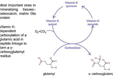
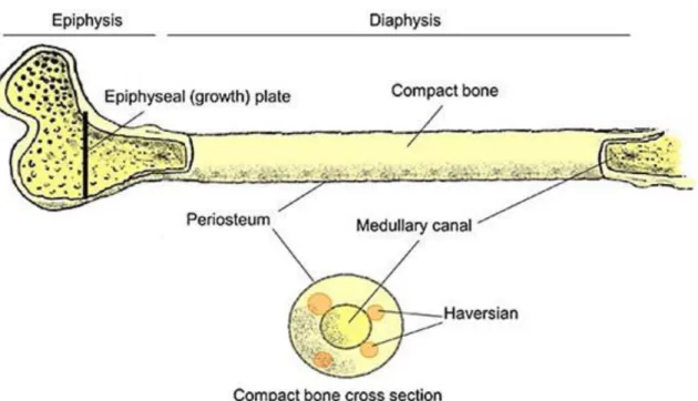
1.1. Figure 1. – Jawbone in cross section ... 1 1.2. Figure 2. – Tooth development ... 1 1.3. Figure 3. – Tooth development – details 1 ... 1 1.4. Figure 4. – Tooth development – details 2 ... 2 1.5. Figure 5. – Section of tooth – enamel and dentine formation ... 3 1.6. Figure 6. – Enamel organ and dental papilla ... 4 1.7. Figure 7. – Basal membrane divide ameloblasts and odontoblasts ... 5 1.8. Figure 8. – Control of tooth shape – ectomesenchymal dominance ... 5 1.9. Figure 9. – Effects of differentation and growth factors ... 6 1.10. Figure 10. – Components of the regulation ... 6 1.11. Figure 11. – A model of the molecular regulation of tooth development from initiation to crown morphogenesis ... 7 1.12. Figure 12. – Oligodontia in a human patient with hypohydrotic ectodermal dysplasia (HED) – The ectodysplasin gene is crucial for tooth development ... 8 1.13. Figure 13. – Molar longitudinal section ... 8 1.14. Figure 1. – Extracellular matrix of hard tissues ... 10 1.15. Figure 2. – Most important protein components of bone and dentin ... 10 1.16. Figure 3. – Collagen – three polypeptide chains forming a rope ... 10 1.17. Figure 4. – Types and distribution of collagen ... 11 1.18. Figure 5. – Structure of procollagen ... 11 1.19. Figure 6. – Overview of collagen biosynthesis ... 12 1.20. Figure 7. – Hydroxylation during collagen biosynthesis ... 12 1.21. Figure 8. – Stages in collagen synthesis – rope formation ... 13 1.22. Figure 9. – Enzymatic cleavage of collagen by mammalian collagenases ... 13 1.23. Figure 10. – Interactions between hydroxyapatite crystals and ionic substances ... 14 1.24. Figure 11. – Disorder frequency of amino acid chains of proteins participating in various biological functions ... 14 1.25. Figure 12. – Sialic acid, a major constituent of sialoproteins ... 15 1.26. Figure 13. – Structure of proteoglycans ... 15 1.27. Figure 14. – Formation of γ-carboxyglutamyl residues ... 16 1.28. Figure 15. – The most important amino acids in hard tissue phosphoproteins ... 16 1.29. Figure 16. – Some possible functions of proteins of hard tissue matrices affecting mineralization 17
1.30. Figure 1. – Bone ... 18 1.31. Figure 2. – Bone anatomy ... 18 1.32. Figure 3. – Macromorphological structure of bone ... 19 1.33. Figure 4. – Osteocytes in compact bone ... 19 1.34. Figure 5. – Major constituents of bone ... 20 1.35. Figure 6. – Major cellular and matrix constituents of bone ... 20 1.36. Figure 7. – Bone formation 1 – intramembranous ossification ... 21 1.37. Figure 8. – Bone formation 2 – Endochondral ossification ... 21 1.38. Figure 9. – Endochondral ossification: steps for bone replacement of cartilage ... 22 1.39. Figure 10. – Ossification: long bones continue to grow and elongate (lengthen) though adolescence 22
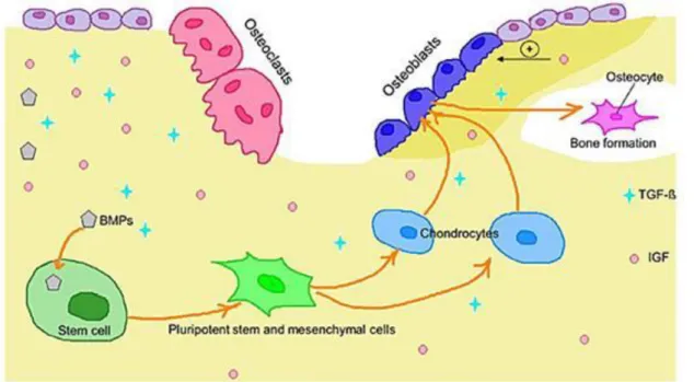
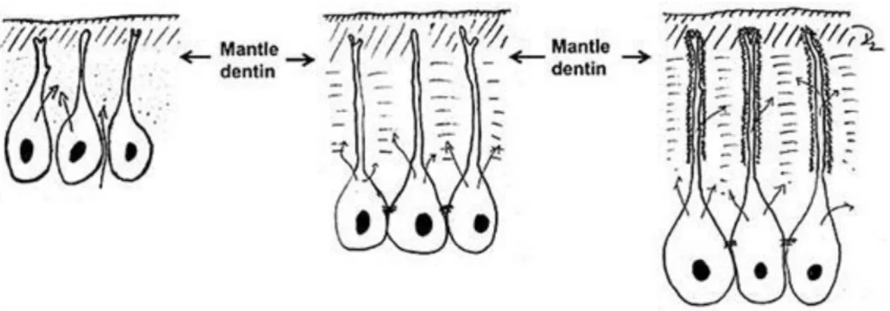
1.40. Figure 11. – Mesengenic process ... 23 1.41. Figure 12. – Main regulatory factors of osteogenesis ... 24 1.42. Figure 13. – Bone formation is regulated by bioactive peptides such as BMPs and also by other growth factors ... 24 1.43. Figure 14. – Role of the extracellular matrix in bone formation and metabolism ... 24 1.44. Figure 15. – Bone response to injury ... 25 1.45. Figure 1. – Constituents of dentin ... 26 1.46. Figure 2. – (A) Primary, (B) secondary and (C) tertiary or reparative dentin ... 26 1.47. Figure 3. – Differentiation of odontoblasts ... 27 1.48. Figure 4. – Formation of mantle dentin during the early phase of mineralization ... 27 1.49. Figure 5. – Dentin is produced by odontoblasts ... 28 1.50. Figure 6. – Mature secretory odontoblast ... 28
1.51. Figure 7. – Involvement of hard tissue proteins in mineral formation ... 29 1.52. Figure 8. – Dentinogenesis Imperfecta ... 29 1.53. Figure 9. – Collagen fibers around tubules ... 30 1.54. Figure 10. – Components of dentin ... 30 1.55. Figure 11. – The empty dentin tubules provide the basis for permeability longitudinal section 30 1.56. Figure 12. – Permeability: the number and the diameter change depending on the dentin tubules 31
1.57. Figure 13. – Neuronal network of pulp/dentin ... 31 1.58. Figure 14. – Hagen–Poiseuille equation – fluid movement – basis of the hydrodynamic theory 32 1.59. Figure 15. – Increase of outward fluid movements from the pulp during inflammation ... 32 1.60. Figure 16. – Diffusion – Fick‘s 2nd law ... 32 1.61. Figure 17. – In a caries lesion, cariogenic bacteria invade the dentinal tubules, demineralizing sclerotic and peritubular dentin in the process ... 32 1.62. Figure 18. – Dentine sensitivity ... 33 1.63. Figure 19. – Dentine hypersensivity – Treatment ... 33 1.64. Figure 1. – The arrangement of ameloblasts during enamel formation ... 34 1.65. Figure 2. – Amelogenesis ... 34 1.66. Figure 3. – Formal and structural changes of ameloblasts during enamel formation ... 35 1.67. Figure 4. ... 35 1.68. Figure 5. – Secretory ameloblasts – formation of prismatic enamel (PE) and interprismatic enamel (IPE) ... 36 1.69. Figure 6. – Parallel running crystallites (Kr) in the early phase of enamel development ... 37 1.70. Figure 7. – Maturation ameloblast phenotypes ... 37 1.71. Figure 8. – Hypothetic model for pH regulation by ruffle ended ameloblasts to neutralize liberated H+ ... 38 1.72. Figure 9. – Cross sectional arrangement of enamel cristal rods (prisms) ... 38 1.73. Figure 10 . – Structure of the matured enamel ... 38 1.74. Figure 11. – Amelogenesis – list of enamel proteins ... 39 1.75. Figure 12. – Concept of the role of amelogenins in the mineralization of enamel ... 39 1.76. Figure 13. – Role of ameloblastin in the regulation of ameloblast function ... 40 1.77. Figure 14. – Amelogenesis imperfecta ... 40 1.78. Figure 15. – Structure of the X-chromosomal copy of the human amelogenin gene ... 41 1.79. Table 1. – Hard tissue composition of teeth and bone. Data correspond to 100 g dry weight 42 1.80. Table 2. – Ionic radius. Ionic radius of ions incorporated into apatite crystals ... 43 1.81. Figure 1. – Structure of hydroxil-, fluoro- and chlorapatite. The figure shows the locations of hydroxid-, fluoride- and chloride ions between Ca2+ ions in the apatite crystals. It can be observed that F-, an anion with smaller ionic radius, is easily incorporated between Ca2+ ions , and may thus lead to a more stable crystal structure. Several favorable properties of fluoro-apatite are explained by more stable crystal structure (see in preventive dentistry) ... 43 1.82. Table 3. – Types of calcium phosphates. In the table, naturally occuring types of calcium phosphates are shown along with an indication of their occurrence. The different types are likely to interconvert into each other through a maturation process. Calculus contains almost all calcium phosphate compounds in addition to the amorphous form ... 44 1.83. Table 4. – Most important infuencing factors of crystallization ... 45 1.84. 6. ábra ... 46 1.85. Figure 3. – Structure of the unit cell. The unit cell is the basic building block of the apatite crystals.
It has a characteristic flattened cubic shape and the base plate is rhomb ... 46 1.86. Table 1. – Consequences of hypercalcemia ... 48 1.87. Table 2. – Causes of hypercalcemia ... 48 1.88. Table 3. – Primary hyperparathyroidism (1‰ frequency) ... 49 1.89. Table 4. – Pathogenesis of hypoparathyroidism ... 50 1.90. Figure 1. – Patomechanism of hypoparathyreosis ... 50 1.91. Table 5. – Conditions that lead to hypocalcemia ... 51 1.92. Figure 2. – Patomechanism of renal osteodystrophy ... 51 1.93. Figure 1. – Major cell types of bone ... 53 1.94. Figure 2. – Scematic representation of osteoblasts and osteoclasts ... 54 1.95. Figure 3. – Osteoclast differentiation and interaction with osteoblasts ... 54 1.96. Figure 4. – Osteoclast structure ... 55 1.97. Figure 5. – Process of bone metabolism: Continuous rebuilding ... 55