The hemispheric lateralization of sleep spindles in humans
R´obert B´odizs1,2,3*†, Ferenc Gombos2†, Péter P. Ujma1,3, Sára Szakadát1, Piroska Sándor1, Péter Simor6,7, Adrián P´otári6, Boris Nikolai Konrad4, Lisa Genzel8, Axel Steiger5, Martin Dresler4,5
and Ilona Kovács2
1Institute of Behavioural Sciences, Semmelweis University, Budapest, Hungary
2Department of General Psychology, Pázmány Péter Catholic University, Budapest, Hungary
3National Institute of Clinical Neurosciences, Budapest, Hungary
4Donders Institute for Brain, Cognition and Behaviour, Nijmegen, The Netherlands
5Max Planck Institute of Psychiatry, Munich, Germany
6Department of Cognitive Science, Budapest University of Technology and Economics, Budapest, Hungary
7Nyíro Gyula Hospital, National Institute of Psychiatry and Addictions, Budapest, Hungary˝
8Centre for Cognitive and Neural Systems, University of Edinburgh, Edinburgh, United Kingdom (Received: September 27, 2016; accepted: January 27, 2017)
Females and males differ in several features of their spindle oscillations, as well as in the hemispheric lateralization of their neurocognitive processes. In addition, the hemispheric lateralization of cognitive functions was shown to vary in an age-dependent manner. In spite of the above knowledge, data on the hemispheric lateralization of these oscillatory phenomena are scarce and no sex differences or age effects in the hemispheric lateralization of sleep spindles were reported. Here, we aim tofill this gap by the description of the hemispheric lateralization of sleep spindles in healthy human subjects. Data sets from three research groups were unified (N= 251, age range: 4−69 years, 122 females) in this retrospective multicenter study. The amplitude, density, and duration of slow (frontally dominant) and fast (centroparietally dominant) spindles were analyzed using the individual adjustment method. Hemispheric lateraliza- tion was quantified by the (L−R)/mean (L, R) index. Orbitofronto-temporo-occipital and parietal fast sleep spindle measures are left lateralized, while prefrontal spindle amplitude is characterized by right hemispheric dominance. Left lateralization of fast spindle density and duration in the temporal and orbitofrontal regions, respectively, increases as a function of age in males, but not in females. In turn, females are characterized by higher left hemispheric dominance in occipitally measured fast spindle durations as compared with males. Sleep spindles are asymmetrically distributed over the two hemispheres. This phenomenon is sexually dimorphic and region-specific perhaps indexing sex differences in neurocognitive architectures.
Keywords: sleep spindles; sigma activity; hemispheric lateralization; sexual dimorphism; gender differences;
temporal lobe
HIGHLIGHTS
– Frontal sleep spindle amplitude is right lateralized – Posterior fast spindle density, duration & amplitude
are left lateralized
– Temporal slow sleep spindle duration is left lateralized – Left dominance of fast spindle density/duration
increases with age in males
INTRODUCTION
Sleep spindles are episodes of mid-frequency (between wakefulness-related alpha and rapid eye movement-sleep specific beta waves) oscillatory electroencephalogram (EEG) activities emerging on the background of irregular, colored noise-like or slow-wave (0.1−4 Hz) activity of non- rapid eye movement (NREM) sleep (De Gennaro and Ferrara, 2003; B´odizs, Körmendi, Rig ´o, and Lázár, 2009;
Loomis, Harvey, and Hobart, 1935a,1935b). Spindles were shown to be associated with enhanced offline neuroplas- ticity (Lüthi, 2014), reflecting neurocognition (Fogel and Smith, 2011), and individual differences in anatomical– microstructural (white matter) features (Piantoni et al., 2013). Recent intracranial and magnetoencephalographic recordings revealed that the majority of sleep spindles are local phenomena (Nir et al., 2011;Andrillon et al., 2011;
Dehghani, Cash, and Halgren, 2011). Moreover, the topography of the correlations revealing associations between spindles and cognitive performance indicates function-specific localizations or hemispheric lateraliza- tion in healthy subjects (B ´odizs, Lázár, and Rig ´o, 2008;
Nishida and Walker, 2007), as well as in typically and atypically developing humans (Selvitelli, Krishnamurthy,
* Correspondence: R´obert B´odizs, Ph.D., Institute of Behavioural Sciences, Semmelweis University, Nagyvárad tér 4, H-1089 Budapest, Hungary, E-mail:bodizs.robert@med.semmelweis‑univ.hu
†These authors contributed equally to this work.
This is an open-access article distributed under the terms of the Creative Commons Attribution License, which permits unrestricted use, distribution, and reproduction in any medium for non-commercial purposes, provided the original author and source are credited.
© 2017 The Author(s)
ORIGINAL PAPER Sleep Spindles & Cortical Up States 1(1), pp. 42–54 (2017)
DOI: 10.1556/2053.01.2017.002 First published online March 08, 2017
Herzog, Schomer, and Chang, 2009; Doucette, Kurth, Chevalier, Munakata, and LeBourgeois, 2015). In addition, sleep spindles were shown to vary significantly with age, the most conspicuousfinding being a decrease in spindle density, duration, and amplitude in the aged, perhaps reflecting diminishing neural plasticity and/or sleep quality (Nicolas, Petit, Rompré, and Montplaisir, 2001;Martin et al., 2013).
The functional specialization of the two hemispheres is a basic finding in human neuropsychology (Herve, Zago, Petit, Mazoyer, and Tzourio-Mazoyer, 2013). The neuroan- atomical localization of language, spatial cognition, and other cognitive functions were shown to be hemispherically asymmetric (Badzakova-Trajkov, Corballis, and Haberling, 2015). Some reports suggest that the hemispheric laterali- zation of cognitive functions is different in males and females, with males usually having higher scores of hemi- spheric specialization/lateralization (Draca, 2010), while others conclude that no verbal function-related sex differ- ences in hemispheric lateralization are present (Sommer, Aleman, Somers, Boks, and Kahn, 2008). Peripubertal testosterone levels affect cortical maturation in the left hemisphere more than in the right hemisphere in males, whereas the opposite held true for females (Nguyen et al., 2013). Several reports support the view that hemispheric lateralization of cognitive functions depends on age.
Findings suggest that left hemisphere specialization of expressive language is already established in young children and persists through adulthood (Paquette et al., 2015), while others emphasize a graded hemispheric specialization during ontogenetic development (Behrmann and Plaut, 2015). The Hemispheric Asymmetry Reduction in Older Adults (HAROLD) model predicts an age-related decrease in the hemispheric lateralization of cognitive functions. It is suggested that the asymmetry reduction reflects a compen- satory function or a dedifferentiation process (Cabeza, 2002). Another model on age-related changes in hemi- spheric asymmetry is the right hemisphere hemi-aging hypothesis, predicting an accelerated aging of the right hemisphere as compared with the left (Dolcos, Rice, and Cabeza, 2002). The above models were never tested from the perspective of sleep-related neural activity.
Reports on the hemispheric asymmetry of sleep slow waves were published in the early 2000s (Sekimoto et al., 2000; Achermann, Finelli, and Borbély, 2001; Ferrara, De Gennaro, Curcio, Cristiani, and Bertini, 2002). Despite the indirect indications of the relevance of hemispheric asymmetry in sleep spindles, only scarce reports were found in the literature, with no assessment of sex differences at all.
Significant left>right differences in the spectral power at bins specific to spindle frequency activity were observed in bipolar fronto-central, centro-parietal, and parieto-occipital derivation pairs (Roth, Achermann, and Borbély, 1999).
This finding was later partially replicated by period ampli- tude analysis of linked-mastoid-referred all-night EEGs of NREM sleep: the number of sigma waves (>6 cycles of 11.4–16.7 Hz oscillatory activity) over the left parietal region was shown to be significantly higher than the sigma wave count over the right parietal area in 15 healthy male volunteers (Sekimoto et al., 2005). However, a reverse pattern was found over the frontal region: frontal sigma waves were more frequent over the right hemisphere as
compared with the left one (Sekimoto et al., 2005). Further- more, preliminary findings indicating the left hemispheric dominance of fast sleep spindle densities and durations in prefrontal and parietal regions were already reported during the validation of the individual adjustment method (IAM) of sleep spindle analysis (B ´odizs et al., 2009). Together, these findings suggest that besides the well-known anteroposterior differences (Gibbs and Gibbs, 1951; De Gennaro and Ferrara, 2003; B´odizs et al., 2009; Lüthi, 2014), sleep spindles may also exhibit observable hemispheric asymme- try. Moreover, scarce findings indicate that the spindle asymmetries might depend on the derivation used (mono- polar vs. bipolar), and perhaps on the region analyzed. No clear indication of age effects or potential sex differences are available in these reports, as researchers did not involve children, adolescents, older adults, and females in their samples (Roth et al., 1999; Sekimoto et al., 2005) or did not analyze sex effects (B´odizs et al., 2009). In sum, it is reasonable to assume that spindles are asymmetrically distributed over the cerebral hemispheres in humans. Fur- thermore, we hypothesize that the left–right asymmetry of sleep spindles reflect the age-dependent changes and sex differences in hemispheric lateralization of cognitive func- tions. Last, we hypothesize that sex differences in the hemispheric lateralization of sleep spindles emerge during ontogenetic development around puberty/early adolescence.
METHODS
Here we conduct a retrospective, multicenter polysomno- graphy study based on available all-night recordings of the Max Planck Institute of Psychiatry (Munich, Germany), the Psychophysiology and Chronobiology Research Group of the Semmelweis University (Budapest, Hungary), and the Laboratory of Developmental Neuroscience at the General Psychology Department of Pázmány Péter Catholic Univer- sity (Budapest, Hungary).
Subjects
Data sets from three research groups were unified to examine the polysomnographic data of 251 healthy parti- cipants (Mage = 25.73 years, SDage = 12.23 years, age range: 4–69 years, 122 females, see Table 1and Supple- mentary Fig. S1). Below the age of 9 years, the statistical variable “Age” was defined with two decimal places to increase accuracy during periods of rapid development (e.g., 6 years and 3 months equaled 6.25 years). Handedness data were available for a subgroup of adult subjects (N = 84) in the form of the points derived from the Edinburgh Handedness Inventory (Oldfield, 1971).
According to semi-structured interviews with experi- enced psychiatrists or psychologists, all subjects were healthy, had no history of neurologic or psychiatric disease, and were free of any current drug effects, excluding contra- ceptives in females. Consumption of small habitual doses of caffeine (maximum two cups of coffee until noon), but no alcohol, was allowed. Six male and two female subjects were light-to-moderate smokers (self-reported), and the rest of the subjects were non-smokers.
Table1.Detailsoftherecordingproceduresofdifferentsubsamples SubsampleOriginalsetting/aim
Number ofsubjects (females)Agerange (years)
AvailableEEG derivations(10–20 system)Recording apparatusPrecision (bit)Hardwareprefiltering (Hz)Samplingrate (Hz/channel)Recording softwareReferences MPIPa–ILab,sleep,andIQ95(43)18–69Fp1,Fp2,Fpz,AF1, AF2,F3,F4,Fz,F7, F8,C3,C4,Cz,P3, P4,Pz,T3,T4,T5, T6,O1,O2
Comlab32Digital SleepLab80.53–70250Brainlab3.3Ujmaetal.(2014) MPIPa–II20(12)Fp1,Fp2,F3,F4,C3, C4,P3,P4,O1,O2Ujmaetal.(2014) PPCUb–IHome/Williamssyndrome study(controlsincluded here)
20(14)6–28Fp1,Fp2,Fpz,F3,F4, F7,F8,Fz,C3,C4, Cz,T3,T4,T5,T6, P3,P4,Pz,O1,O2, Oz SD-LTM32BS (MicromedLtd., Italy) 220.15–250(plus<463.3Hz digitalanti-alising filteringbefore downsamplingfrom 4096to1024Hz) 1024Brain-Quick SystemPlus (Micromed)
B´odizs,Gombos,and Kovács(2012) PPCUb–IIHome/adolescentsleep23(12)15–22B´odizs,Gombos, Ujma,andKovács (2014) SUc–ILab/sleep&IQ,sleepspindle methodology,wake–sleep transitionanalysis
49(19)17–55Fp1,Fp2,F3,F4,F7, F8,Fz,C3,C4,Cz, T3,T4,T5,T6,P3, P4,O1,O2 FlatStyleSLEEPLa MontHeadbox, HBX32-SLP preamplifier(La MontMedical)
120.5–70249Datalab(Medcare)B´odizs,Sverteczki, andMészáros (2008),Ujmaetal. (2014) SUc–IILab/nightmarestudy(controls includedhere)16(7)19–21Fp1,Fp2,F3,F4,Fz, F7,F8,C3,C4,Cz, P3,P4,Pz,T3,T4, T5,T6,O1,O2
Brain-QuickBQ132S (Micromed)120.33–1,500(plus<450Hz anti-aliasingdigital filteringbefore downsamplingfrom 4096to1024Hz) 1024System98 (Micromed)Simor,Horváth, Ujma,Gombos, andB´odizs(2013) SUc–IIILab/home/children’s dreaming29(15)3.84–8.42Brain-QuickBQ132S/ SDLTM32BS (Micromed)
12/220.33–1,500/0.15–250 (plus<450/<463.3Hz anti-aliasingdigital filteringbefore downsamplingfrom4096 to1024Hz) System98/System PlusEvolution (Micromed)
Ujma,Sándor, Szakadát,Gombos, andB´odizs(2016) a MaxPlanckInstituteofPsychiatry,Munich,Germany.b PázmányPéterCatholicUniversity,Budapest,Hungary.c SemmelweisUniversity,Budapest,Hungary.
Sleep recordings
Sleep was recorded for two consecutive nights by standard polysomnography, including EEG according to the 10–20 system (Jasper, 1958), as well as electro- oculography, bipolar submental electromyography, and electrocardiography. EEG electrodes were re-referenced to the mathematically linked mastoids [(A1+A2)/2]. Impe- dances for the EEG electrodes were kept <8 kΩ. Signals were collected, prefiltered, amplified, and digitized at dif- ferent sampling rates using different recording apparatus in the different subsamples (Table 1). Sleep EEG recordings for the second nights spent in the laboratory were manually scored on a 20-s basis by applying standard criteria (Iber et al., 2007). Epochs with artifacts were removed on a 4-s basis by visual inspection of all recorded channels (includ- ing polygraphy). The EEG derivations contaminated by persistent, long-lasting artifacts were removed from our analyses. These latter derivations as well as the ones missing in a subgroup of our subjects were treated as missing data.
The polysomnographic records used in this study were the second nights. (First night records were discarded to control the so-calledfirst night effect.)
Although the recording apparatuses are of several types, it has to be mentioned that spindle laterality is a technically neutral measure, as it is based on the inter-hemispheric differences derived within the same technical setting. Thus, in contrast to absolute amplitude values, hemispheric later- alization indices are reliable measures in multicenter studies.
Quantitative EEG analyses
Sleep spindles of all-night NREM sleep were analyzed by the IAM (B´odizs et al., 2009; Ujma et al., 2015). This method is based on the average amplitude spectra of (in this case all-night) N2 and N3 sleep. The frequency criteria of slow and fast sleep spindles are derived from the individual- specific peaks of these spectra (between 9 and 16 Hz), based on the inflexion points. The slow- and the fast sleep spindle frequencies are tested for frontal- and centro-parietal domi- nance, respectively. The amplitude criteria for slow- and fast spindles are determined in individual- and derivation- specific manner by multiplying the number of intra-spindle frequency bins with the mean amplitude spectrum values corresponding to lower and upper frequency limits. The EEG is then band-passfiltered for individual slow and fast sleep spindle frequencies using a fast Fourier transformation filtering method and the precise envelopes of the filtered signals calculated. EEG segments corresponding to the envelopes transcending the amplitude criteria for at least 0.5 s are considered spindles (a scheme of the detection is provided in Fig. 1 of Ujma et al., 2015). In fact, these segments are contributing to the individual- and derivation- specific lower and higher frequency spectral peaks between 9 and 16 Hz. Based on the IAM approach, individual- and derivation-specific densities (spindles×min−1), durations (s), and amplitudes (μV) of slow, frontally dominant and fast, centroparietally dominant sleep spindles were deter- mined. Although these measures are not fully statistically
Fig. 1.The hemispheric lateralization of different sleep spindle features in healthy human subjects. A: EEG locations (dark teal–left, tawny–right). B: Left (Fp1) and right (Fp2) frontopolar samples of stage 2 sleep EEG traces. Highlighted periods (black rectangles) exemplify hemispheric asymmetries in sleep spindles. Vertical gray lines indicate seconds. C: The hemispheric lateralization of sleep spindle
densities. D: The hemispheric lateralization of sleep spindle durations. E: The hemispheric lateralization of sleep spindle amplitudes.
Horizontal bars denote 95% confidence intervals of the mean hemispheric lateralization indices: (Left−Right)/mean (Left, Right). Vertical dotted black lines indicate zero values (0= no hemispheric lateralization). Overall means of absolute left and right values are seen over the
horizontal bars indicating significant lateralization effects
independent (Supplementary Table S1), we followed a descriptive approach in our current paper, by analyzing density, duration, and amplitude in separate statistical models. We followed this approach because spindle den- sity, duration, and amplitude were shown to depend in part on different neurophysiological mechanisms, related to long term potentiation (Werk, Harbour, and Chapman, 2005), thalamic inhibition/corticothalamic feedback (Bon- jean et al., 2011;Barth ´o et al., 2014) and thalamocortical recruitment/globality (Dempsey and Morison, 1941;
Andrillon et al., 2011), respectively.
Statistics
The hemispheric lateralization of sleep spindle features (density, amplitude, and duration for both slow and fast subtypes) was analyzed by calculating the hemispheric lateralization indices between homologous derivation pairs as follows:
Spindle laterality index= ðL−RÞ=meanðL, RÞ
where L = left and R = right.
Hemispheric asymmetry was tested by one-samplet-tests with the null hypothesis that the population mean is equal to 0 (no hemispheric laterality). False discovery rate was controlled by the Benjamini–Hochberg method (Benjamini and Hochberg, 1995). We report the uncorrected signifi- cance values (p) for the t-tests surviving Benjamini– Hochberg correction. For those variables revealing a signif- icant hemispheric lateralization in the whole sample, we went on to analyze sex and age effects, as well as their interactions by general linear models (GLMs). In the next step, we revealed the directions and sources of the interac- tion effects in our GLMs by Pearson product-moment correlation coefficients as follows: in cases where significant main effects of age or “sex×age”-type interaction effects emerged, Pearson correlation coefficients between age and the respective spindle measures (left, right, and asymmetry index) were calculated for the whole sample (in case of age main effect) or separately for females and males (in case of sex×age interaction effect). In the former case, the sign of the significant correlation indicates the direction of the age effect (“+”for increase and“–”for decrease), while in the latter case females and males could be compared in terms of their age-related decrease, increase, or stability in spindle asymmetries by the Fisher’sr-to-ztransformation method.
A change (increase or decrease) in spindle asymmetry can emerge from three potential sources: a change in one of the two hemispheres, i.e., left or right or a change in both hemispheres. To test the three potential sources mentioned above, the age-associated changes in left and right spindle measures were compared by the Fisher’sr-to-ztransforma- tion method.
Last, but not least we examined if different spindle asymmetries between males and females are specific to different age groups. Hence, we categorized our subjects as follows: children (age<10 years;N = 31, 15 females), teenagers (10 years≤age<20 years;N = 36, 18 females), young adults (20 years ≤ age<40 years; N = 150, 75 females) and middle-aged adults (age ≥ 40 years;
N = 34, 14 females). Females and males were compared in terms of their spindle asymmetry measures with a signif- icant sex×age interaction effect in the GLMs in each of the above age groups by factorial ANOVAs (sex×age group) with Fisher’s least squares differences post-hoc tests (see Supplementary Tables S2–S5 for descriptive statistics of hemispheric laterality indices in different age groups).
Due to the variable number of N(caused by artifactual derivations or missing channels in some subsamples), we will provide the number of observations or the degrees of freedom (df) while presenting the outcomes of statistical tests throughout the manuscript.
Ethics
Adult participants or the parents of the underage participants signed informed consent for the participation in the study according to the Declaration of Helsinki. The individual research protocols used in this multilaboratory study were approved by the local ethical committees, namely the ethical boards of the Medical Faculty of the Ludwig Maximilian University, the Semmelweis University, and the Pázmány Péter Catholic University.
RESULTS
In general, more fast, than slow sleep spindle measures (12 vs. 2) were shown to be characterized by asymmetric hemispheric distributions. Sleep spindle densities and dura- tions were left lateralized predominantly in temporal and parietal regions, while spindle amplitudes were region- dependent right or left lateralized as follows (Fig.1). Hand- edness did not correlate significantly with the hemispheric lateralization of sleep spindles.
Sleep spindle densities
Overall hemispheric lateralization. Significant left hemi- spheric dominance of fast sleep spindle densities in the middle and posterior temporal (tT3–T4(FastSpiDens) = 5.49;
df = 230; p<10−6 and tT5–T6(FastSpiDens) = 3.82; df= 231; p = .000169, respectively), as well as parietal (tP3–P4(FastSpiDens)= 3.10;df = 250;p= .0021) derivation pairs was found (Fig.1C).
Age and sex effects in hemispheric lateralization.Middle and posterior temporal regions were found to be character- ized by age-related changes in left hemispheric dominance of fast sleep spindle dominances in males, but not females (Fig. 2A and B). Thus, the GLM results indicate an age-related change in the left hemispheric dominance of middle temporal fast sleep spindle densities in males, but not in females (Fig. 2A): the main effect of age (F = 8.64;
df = 1, 227;p = .0036), as well as a sex×age interaction (F = 3.92; df = 1, 227;p= .048) were significant. Like- wise, posterior temporal left hemispheric dominance was characterized by age-related change in males, but not females (Fig. 2B): age (F = 9.79;df = 1, 228;p= .001) and sex×age (F = 4.68; df= 1, 228; p= .031) effects were found to be significant. No significant age, sex, or age×sex effects were found for the hemispheric
46 |Sleep Spindles & Cortical Up States 1(1), pp. 42–54 (2017) B´odizs et al.
lateralization of parietal fast sleep spindle densities.
A supplementary GLM analysis with age and sex as predictors and the lateralization indices of all spindle fea- tures as within-subject dependent variables revealed an increased left lateralization of temporal spindle density (irrespective of spindle type) in males as compared with females (Supplementary Analyses).
Left and right hemispheric findings. To clarify if the changing asymmetry in males derives from increasing/
decreasing left/right hemispheric spindling or just the change in relative dominance of one of the hemispheres, Pearson product-moment correlation coefficients between age and temporal fast spindle measures were calculated.
Fast sleep spindle densities of the male subgroup uniformly decreased with age over both the left (rAge_vs_T3(FastSpiDens)
= −.48;N = 122;p<10−6) and the right (rAge_vs_T4(Fast- SpiDens) = −.51; N = 122; p<10−6) temporal regions.
There was no significant difference between these correla- tions (z = .31;p = .75;Fig.2E,Supplementary Fig. S2).
However, as seen in the significant age×sex effect in the GLM, the left–right lateralization index (left hemispheric dominance) significantly increased with age in males (rAge_vs_T3–T4(FastSpiDens) = .28; N = 122; p = .0012;
Fig.2A). The overall picture was similar for the posterior temporal derivations of our male subjects; however, the absolute values of the correlations reflecting
age-associated decreases in fast sleep spindle densities were somewhat lower (rAge_vs_T5(FastSpiDens) = −.32;
rAge_vs_T6(FastSpiDens) = −.40; rAge_vs_T5–T6(FastSpiDens) = .30; N = 122; p= .0002, .000003, and .0006, respec- tively;Fig.2F). Age-associated decreases in left and right middle and posterior temporal fast sleep spindle decreases were of similar magnitude in females (rAge_vs_T3(FastSpiDens) =
−.40, rAge_vs_T4(FastSpiDens) = −.46, rAge_vs_T5(FastSpiDens)
= −.37, rAge_vs_T6(FastSpiDens)= −.39, N = 110; p = .00001,
10−6, .00004, and .00001, respectively; Fig. 2C and D).
Nevertheless, no significant age-related changes in temporally derived fast sleep spindle asymmetries were found in females (rAge_vs_T3–T4(FastSpiDens) = .07, rAge_vs_T5–T6(FastSpiDens) = .07,N = 110,p= .45 in both cases; Fig.2Aand B).
Age groups. Sex differences in the left hemispheric dominance of middle temporal (T3–T4) fast sleep spindle densities are non-significant in children (F = 1.16;N = 14 andN = 16 for females and males, respectively;p = .27) and teenagers (F = 0.36; N = 18 for both females and males; p = .54), but are significantly higher in young (N = 68) and middle-aged (N = 20) adult males as com- pared with young (N = 63) and middle-aged (N = 14) adult females, respectively [F = 7.44 (p = .0068) and F = 13.64 (p = .0002) for young and middle-aged adults, respectively;Fig. 2A]. Similarly, no sex differences in the Fig. 2.Hemispheric asymmetry of temporally recorded fast sleep spindle densities as a function of age, sex, and left/right absolute values.
A: Age-related changes in the left hemispheric dominance of middle temporal fast sleep spindle densities in females (♀, red) and males (♂♂, blue). Horizontal dotted line indicates 0 value (no lateralization). Note the age-dependence of left hemispheric dominance in males, but not females. Gray area indexes the age range (≥20 years) characterized by significant male>female left hemispheric asymmetry. B: Age-related changes in the left hemispheric dominance of posterior temporal fast sleep spindle density in females (♀, red) and males (♂♂, blue). Horizontal dotted line indicates 0 value (no lateralization). Note the age-dependence of left hemispheric dominance in males, but not females. C: The age-dependent decreases in left (T3, dark teal) and right (T4, tawny) middle temporal fast sleep spindle densities of females. D: The age- dependent decreases in left (T5, dark teal) and right (T6, tawny) posterior temporal fast sleep spindle densities of females. E: The age- dependent decreases in left (T3, dark teal) and right (T4, tawny) middle temporal fast sleep spindle densities of males. F: The age-dependent decreases in left (T5, dark teal) and right (T6, tawny) posterior temporal fast sleep spindle densities of males. Uniform age-dependent
decrease in sleep spindle densities are seen over both hemispheres and in both sexes (panels C–F). *p<.05; **p<.01; ***p<.001
left hemispheric dominance of posterior temporal (T5–T6) fast sleep spindle densities are seen in children (F = 0.45;
N = 15 and N = 16 for females and males, respectively;
p = .50) and teenagers (F = 0.15;N = 18 for both sexes;
p = .69). However, in contrast to the middle temporal fast spindle density asymmetries, sex differences in the left hemispheric dominance of posterior temporal fast sleep spindle densities are non-significant in young (F = 2.45;
N = 63 and N = 68 for females and males, respectively;
p = .11) and middle-aged (F = 2.85;N = 14 andN = 20 for females and males, respectively;p = .09) adults either (Fig. 2B).
Interim summary on the laterality of sleep spindle den- sity.Fast sleep spindle density is left lateralized in temporal and parietal regions (Fig.1C). The left hemispheric domi- nance in middle and posterior temporal fast sleep spindle densities is characterized by age-associated increases in males, but not in females (Fig. 2AandB, Supplementary Fig. S2). Absolute fast sleep spindle densities uniformly decrease as a function of age over both left and right temporal regions in both sexes (Fig. 2C–F). Left lateralization of middle temporal fast sleep spindle density is increased in young (20–40 years) and middle- aged (40–69 years) males as compared with females (Fig. 2A).
Sleep spindle durations
Overall hemispheric lateralization. Measures of fast sleep spindle durations were left lateralized in several derivation pairs, including orbitofronto-temporal (tF7–F8(FastSpiDur) = 3.37; df = 228; p = .00086; tT3–T4(FastSpiDur) = 3.28;
df = 230; p = .0011;tT5–T6(FastSpiDur) = 5.19; df = 231;
p<10−6), parietal (tP3–P4(FastSpiDur) = 3.62; df= 250;
p = .00035), and occipital (tO1–O2(FastSpiDur) = 3.13;
df = 250; p = .0019) regions. In addition, slow sleep spindle durations of the middle temporal derivation pair (tT3–T4(SlowSpiDur) = 3.07; df = 230; p = .0023) were significantly left lateralized as well (Fig.1D).
Age and sex effects in hemispheric lateralization. Age- related changes in the hemispheric lateralization of orbito- frontal and occipital fast sleep spindle durations were different in males as compared with females. GLMs revealed a significant sex×age interaction effect for the hemispheric laterality of fast sleep spindle durations in the F7–F8 (F = 7.19; df= 1, 228; p= .007; Fig. 3A) and O1–O2 (F = 7.69; df= 1, 250; p= .005; Fig. 3B) deri- vation pairs. No other main effects or interactions were revealed for the hemispheric asymmetries in sleep spindle durations. A GLM analysis with age and sex as predictors and all spindle lateralization indices as within-subject
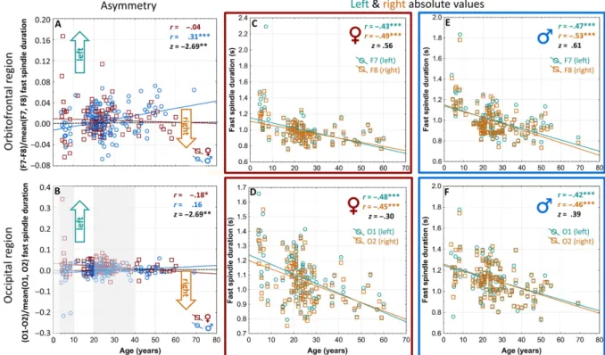
Fig. 3.Hemispheric asymmetry of orbitofrontally and occipitally recorded fast sleep spindle durations as a function of age, sex, and left/right absolute values. A: Age-related changes in the left hemispheric dominance of orbitofrontal fast sleep spindle durations in females (♀, red) and males (♂♂, blue). Horizontal dotted line indicates 0 value (no lateralization). Note the age-dependence of left hemispheric dominance in males, but not females. B: Age-related changes in the left hemispheric dominance of occipital fast sleep spindle durations in females (♀, red) and males (♂♂, blue). Horizontal dotted line indicates 0 value (no lateralization). Note the age-dependence of left hemispheric dominance in females, but not males. Gray areas indicate the age ranges (<10 years and 20–40 years) characterized by significant group effects (females>
males). C: The age-dependent decreases in left (F7, dark teal) and right (F8, tawny) orbitofrontal fast sleep spindle durations of females.
D: The age-dependent decreases in left (O1, dark teal) and right (O2, tawny) occipital fast sleep spindle durations of females. E: The age- dependent decreases in left (F7, dark teal) and right (F8, tawny) orbitofrontal fast sleep spindle durations of males. F; The age-dependent decreases in left (O1, dark teal) and right (O2, tawny) occipital fast sleep spindle durations of males. Uniform age-dependent decrease in sleep
spindle durations are seen over both hemispheres and in both sexes (panels C–F). *p<.05; **p<.01; ***p<.001
48 |Sleep Spindles & Cortical Up States 1(1), pp. 42–54 (2017) B´odizs et al.
dependent variables revealed that occipital spindle duration is more left lateralized in females as compared with males, irrespective of spindle type (slow or fast; see Supplementary Analyses).
Left and right hemispheric findings. As in the case of sleep spindle densities, we aimed to unravel the sources of age-dependent changes in asymmetric spindle durations by analyzing the correlations of age with the appropriate measures at derivations F7, F8, O1, O2, as well as with the laterality indices between homologous derivation pairs (Fig. 3C–F). Sleep spindle durations age-dependently decreased in the left (rAge_vs_F7(FastSpiDur) = −.47; N = 120; p<10−6) and the right (rAge_vs_F8(FastSpiDur)= −.53;
N = 120; p<10−6) orbitofrontal regions of males (Fig.3E). There was no significant difference between these correlations (z = .61;N = 120;p = .27).
In case of the age-associated changes in occipital fast sleep spindle durations, however, a different pattern emerged. In this case, the age×sex interaction effect was driven by decreasing the left hemispheric dominance in occipital fast sleep spindle durations in females (rO1–O2(FastSpiDur) = −.18; N = 122; p = .042; Fig. 3B).
The effect was not due to different age-associated changes in the left or the right occipital derivations of females, as these values were similar (rO1(FastSpiDur) = −.48;rO2(FastSpi- Dur) = −.45; N = 122 and p<10−6 for both) and not significantly different (z = 0.3;p= .76; Fig.3D).
Age groups.The hemispheric asymmetry of orbitofrontal (F7–F8) fast sleep spindle durations did not significantly differ in females and males among children (F = 3.07;
N = 14 and N = 15 for females and males, respectively;
p = .08), teenagers (F = 0.24; N = 18 and N = 17 for females and males, respectively; p= .62), young adults (F = 0.79; N = 63 and N = 68 for females and males, respectively;p = .37), and middle-aged adults (F = 3.69;
N = 14 and N = 20 for females and males, respectively;
p = .055). As regarding the sex differences in the hemi- spheric asymmetry of occipitally derived (O1–O2) fast sleep spindle durations, females (N = 15) were more left later- alized than males (N = 16) during childhood (F = 32.45;
p<10−6, Fig. 3B), but no difference in teenagers (F = 0.67; N = 18 for both females and males; p = .41) could be observed. Furthermore, young adult females (N = 75) were more left lateralized than males (N = 75;
F = 4.20; p = .041), but again, no sex difference among the middle-aged (N = 14 vs. 20 females and males, respec- tively) was evident (F = 0.37;p = .54;Fig.3B).
Interim summary on the laterality of sleep spindle dura- tion. Orbitofrontal, temporal, parietal, and occipital fast sleep spindle durations, as well as middle temporal slow sleep spindle durations were left lateralized (Fig. 1D). The left lateralization of orbitofrontal fast sleep spindle duration is age-dependently increasing in males, but not in females (Fig. 3A). In turn, the left hemispheric dominance of occipital fast sleep spindle duration is age-dependently decreasing exclusively in females (Fig. 3B). Females are characterized by a higher degree of left lateralization of their fast sleep spindle durations during childhood and young adulthood (Fig. 3B). Fast sleep spindle durations are uni- formly and age-dependently decreasing over both hemi- spheres and regions (Fig. 3C–F).
Sleep spindle amplitudes
Overall hemispheric lateralization. Sleep spindle amplitudes were significantly right lateralized in the frontopolar-prefrontal regions (tFp1–Fp2(SlowSpiAmp) =
−2.66; df= 249; p = .0082 and tF3–F4(FastSpiAmp) =
−3.68; df= 250; p= .00028 for slow and fast sleep spindle amplitudes, respectively,Fig.1E). In turn, fast sleep spindle amplitudes in the posterior temporal (tT5–T6(FastSpiAmp)
= 4.28, df = 231, p= .00002), parietal (tP3–P4(FastSpiAmp) = 5.09, df = 250, p= .000001), and occipital (tO1–O2(Fas-
tSpiAmp) = 2.62,df = 250,p = .0092) derivation pairs were characterized by significant left lateralization (Fig.1E).
Age and sex effects in hemispheric lateralization. Fron- topolar-prefrontal right hemispheric lateralization of sleep spindle amplitudes was not significantly dependent on age and/or sex. However, the posterior temporal left hemi- spheric dominance of fast sleep spindle amplitude increased as a function of age (main effect of age: F = 3.97; df = 1, 230;p = .047). This latter effect did not depend on sex.
In addition, occipital fast sleep spindle amplitudes were shown to be more left lateralized in females as compared with males (main effect of sex: F = 4.39; df= 1, 230;
p = .037).
Left and right hemispheric findings. The age versus posterior temporal fast sleep spindle amplitude correlation was non-significant for the left hemisphere (rAge_vs_T5(Fast- SpiAmp) = −.09; N = 232; p= .16), and negative for the right hemisphere (rAge_vs_T6(FastSpiAmp) = −.13; N = 232;
p = .04) in the whole sample (for both females and males).
The difference between these correlations was not signifi- cant (z= .43;p= .66). Indeed, the left–right dominance in posterior temporal fast sleep spindle amplitudes slightly and age-dependently increased in our subjects (rAge_vs_T5–T6 (FastSpiAmp) = .13; N = 232; p = .03), which seem to be at least partially explained by right hemispheric decline in fast spindle amplitudes.
Age groups.Hemispheric lateralization in the amplitudes of posterior temporal fast sleep spindles was not different between female and male subgroups of any age group.
However, the left hemispheric dominance in the amplitudes of occipitally derived fast sleep spindles was significantly higher in female children as compared with male children (p = .002). This finding was similar to the observed female>male difference in occipital fast sleep spindle durations (Fig. 3B).
Interim summary on the laterality of sleep spindle amplitude. Posterior temporal, parietal, and occipital fast sleep spindle amplitude is left lateralized, whereas frontal sleep spindle amplitude (both slow and fast) is right later- alized (Fig.1E). The left hemispheric dominance of poste- rior temporal fast sleep spindle amplitude increases slightly as a function of age, irrespective of sex. Left lateralization of occipital fast sleep spindle amplitudes is higher in females as compared with males.
DISCUSSION
Here, we aimed to provide a detailed analysis on the hemispheric asymmetry of sleep spindles as measured by
the IAM approach in a large sample of subjects involving both females and males of a broad age range. We hypothe- sized that sleep spindles are asymmetrically distributed over the cerebral hemispheres and are more left lateralized in males than in females. Moreover, we hypothesized that the sex differences in the hemispheric asymmetry of sleep spindles are age-dependent, emerging around puberty.
Overall hemispheric lateralization of sleep spindle measures
Our hypotheses were partially supported. Several sleep spindle features are asymmetrically distributed over the two hemispheres. Fast sleep spindles over the orbitofrontal, temporal, parietal, and occipital cortices are characterized by significant left hemispheric dominance in terms of density, duration, and/or amplitude (Fig. 1). In turn, sleep spindle amplitudes measured over the prefrontal cortices are characterized by right hemispheric dominance (Fig. 1E).
Thesefindings cohere with the above-mentioned reports on asymmetric spindle frequency Fourier spectra and wave count over the two hemispheres (Sekimoto et al., 2005;
Roth et al., 1999). Region specificity was reported in one of the above studies (Sekimoto et al., 2005): right hemispheric dominance of sigma wave count is highly coherent with our finding of right lateralized sleep spindle amplitudes in the prefrontal region. The well-known uneven distribution of different sleep spindle types (slow and fast) over the rostro- caudal axis of the brain (Gibbs and Gibbs, 1951; De Gennaro and Ferrara, 2003; B ´odizs et al., 2009; Lüthi, 2014) can be complemented with our present findings, indicating the putative relevance of analyzing the hemi- spheric lateralization measures of spindles in different age and gender groups, as well as in various neuropsychiatric conditions. The anterior–posterior and left–right differences in sleep spindles cohere with the concept of local sleep spindling (Nir et al., 2011;Andrillon et al., 2011;Dehghani, Cash, Rossetti, Chen, and Halgren, 2010;Piantoni, Halgren, and Cash, 2016). Moreover, our results add further support for the concept of local–in this case hemispheric-specific– sleep regulation (Achermann et al., 2001; Ferrara et al., 2002), which should be generalized beyond the rhythmic neural activity related to the slow-wave frequency domain and should incorporate the spindle oscillations as well.
Hemispheric asymmetries in sleep spindling might reflect different experience-dependent pressures for offline plastic- ity and network reorganizations during sleep in the two hemispheres. The difference in these needs might stem from differences in cognitive demands of the two hemispheres during information processing in wakefulness and online behavioral control. The functional relevance of the laterali- zation of regionally specific sleep spindling in offline improvement of lateralized motor skills was explicitly proven in a well-controlled experimental setting (Nishida and Walker, 2007). As regarding trait-like effects, it was suggested that tonic (resting state) asymmetries in the alpha rhythm reflects subjects’cognitive styles (Furst, 1976;Glass and Butler, 1977). Ourfindings might be interpreted in the same framework. Left hemispheric dominance of fast sleep spindles could reflect the preferential reliance on left hemisphere-related cognitive functions.
Our findings on the hemispheric lateralization of sleep spindle density, duration, and amplitude are characterized by similar topography (Fig. 1). Thus, in spite of the avail- able evidence of specific neurophysiological processes con- tributing to distinct sleep spindle features (Werk et al., 2005;
Bonjean et al., 2011; Barth ´o et al., 2014; Dempsey and Morison, 1941;Andrillon et al., 2011), our results indicate the commonalities in these indices. Further studies are needed to unravel the reliability of composite measures, like spindle intensity/activity in contrast to the distinct features analyzed in the current work.
Based on the availability of handedness data in a sub- group of our adult subjects, we cannot infer any specific relationship between hand preference and sleep spindle lateralization in humans. It deserves mention that the stron- gest effects in spindle lateralization consist of a significant left hemispheric dominance of the temporal and orbitofron- tal regions. The left temporal and orbitofrontal regions are closely connected to the receptive and productive language regions of the brain, namely, the Wernicke’s and the Broca’s areas (Ardila, Bernal, and Rosselli, 2016). Thus, language lateralization could indeed influence spindle lateralization in humans. Moreover, this would explain the non-significant correlation of handedness and spindle asymmetry, as lan- guage lateralization of the majority of both right- and left- handers is left hemispheric (96% and 76%, respectively).
Only a minority of the left-hander subjects are characterized by a weak (8%) or strong (2%) right hemispheric language dominance (Pujol, Deus, Losilla, and Capdevila, 1999).
Taken the relatively low frequency (10%) of left-handers in non-selected populations (Hardyck and Petrinovich, 1977) (including the 13% value in our current sample), the abovefindings and assumption of language lateralization as being the basis of spindle lateralization would imply a non- significant correlation between handedness and spindle laterality. Although testable, there is no direct evidence supporting this hypothesis yet.
Age- and sex-related differences in the hemispheric lateralization of sleep spindle measures
In addition to unraveling the region-specificity, spindle feature-related (density, duration, and amplitude) and frequency-dependent (slow vs. fast) dimensions of the hemispheric lateralization of sleep spindling, we detected sex-specific, age-associated increases in the left hemispheric dominance of fast sleep spindle densities and durations over the temporal and orbital frontal cortices, respectively. Tak- ing into account the cross-sectional nature of our data, these findings indicate that older males are characterized by more pronounced left temporal/orbitofrontal dominance of fast sleep spindling. There are two potential explanations for these effects. First, the developmental view suggests an enhancement of left hemispheric dominance in males related to maturational, hormonal (puberty), as well as experience- dependent, network reorganizational effects. A viable empirical basis for the latter could be thefinding of Nishida and Walker (2007), revealing the functional involvement of spindle asymmetry in the efficient consolidation of asym- metrically represented experiences. The other non-exclusive possibility is a cohort effect: older participants could be
50 |Sleep Spindles & Cortical Up States 1(1), pp. 42–54 (2017) B´odizs et al.
former subjects of educational and parenting systems that are significantly different from the recent ones and could have an effect of enhancing the sex differences in cognitive strategies/hemispheric lateralization, albeit there is no avail- able literature supporting such a hypothesis.
The sexual dimorphism in the hemispheric lateralization of sleep spindles was strikingly region-specific. In contrast to the above-discussed age-dependent increase in left hemispheric dominance of orbitofrontal/temporal fast sleep spindling in males, occipital fast spindles were left hemi- spherically dominant in females. Moreover, the strong left hemispheric dominance of occipital fast spindling was predominantly evident in female children and decreased rapidly and significantly with age (Fig.3B).
The hypothesis on the puberty-related disjunction of spindle-related hemispheric laterality measures of females and males was not clearly supported by our data. Although age-dependent changes in laterality measures are clearly present and sometimes significantly different among females and males, the largest sex-related differences are usually seen in young/older adults (Fig. 2) or children (Fig. 3). In contrast to the findings on the developmental trajectories of brain maturation/cortical thinning (De Bellis et al., 2001; Nguyen et al., 2013), we could not reveal a sharp female/male difference in the hemispheric laterality indices of sleep spindles in teenagers. Thisfinding suggests that in addition to sex, processes of long-term, experience- dependent plasticity may shape the hemispheric lateraliza- tion of sleep spindles in humans. Although anteroposterior gradients and frequency components of spectralfingerprints describing individual-specific sleep spindle features were shown to be strongly genetically determined (De Gennaro et al., 2008), no such evidence was found provided for the lateralization of spindles. Future studies are needed to reveal the genetic and experience-dependent causes of cerebral asymmetric sleep spindling in humans.
Findings on the age-related changes of hemispheric lateralization of cognitive functions in children and adoles- cents are controversial: views on early established (Paquette et al., 2015) and graded asymmetry (Behrmann and Plaut, 2015) were published. Our findings on the hemispheric lateralization of sleep spindles are mixed in this regard:
asymmetries revealed for the temporal areas are age- dependently increasing (graded asymmetry; Figs.2A andB and 3A), while frontal and occipital asymmetries are age- independent or early established (Fig.3B), respectively.
Theories on aging and hemispheric lateralization are assuming an age-associated decrease or increase in cerebral hemispheric dominance. The HAROLD model would pre- dict an age-related decrease in the hemispheric lateralization of sleep spindles (Cabeza, 2002). Whatever may be the reason for this assumed asymmetry reduction is, here we report evidence for an opposite process during sleep: hemi- spheric asymmetry of temporal and orbitofrontal fast sleep spindling is greater in the aged, indicating increasing left hemispheric involvement in sleep spindle generation/main- tenance and perhaps increasing reliance on left hemispheric offline neural plasticity in males. In turn occipital spindling is left-hemispherically dominant in females. The age-related change in the left hemispheric dominance of occipital fast spindling is paralleled by an opposite process in males.
Thus, the age-related decrease in hemispheric asymmetry predicted by the HAROLD model was partially supported in terms of the occipitally derived fast sleep spindles in females (Fig. 3B).
Another model on age-related changes in hemispheric asymmetry is the right hemisphere hemi-aging hypothesis predicting an accelerated aging of the right hemisphere (Dolcos et al., 2002). A steeper age-related decline in right hemispheric fast sleep spindling could indeed account for ourfinding of increased left spindle dominance in the aged males. This was not evidenced, however. Fast sleep spindle densities and durations uniformly decreased over both hemi- spheres in the aged. It has to be mentioned that posterior temporal fast sleep spindle amplitudes declined significantly in the right but not in the left hemisphere; however, the difference between the two correlations was not significant.
Altogether, ourfindings on age-related decreases in spindle measures are coherent with the literature (Nicolas et al., 2001;Martin et al., 2013). However, ourfinding on the age- associated increase in the left temporal and orbitofrontal dominance of fast sleep spindle density (generation/initia- tion probability) and duration (maintenance) in males was not reported before.
Looking at the scatterplots, one can discern a high and age-dependently increasing dispersion of the male data on temporal fast spindle asymmetry (left hemispheric domi- nance). It is evident that the sex×age effect in spindle laterality is driven by an extreme left hemispheric male subgroup and/or a higher global variation of spindle laterality in males as compared with females. This type of difference is a common finding of sexual dimorphism for many pheno- typic traits (Lehre, Lehre, Laake, and Danbolt, 2009).
When we look at the data derived from the occipital cortex, a completely different picture emerges. Females are characterized by higher left hemispheric dominance in occipitally measured fast sleep spindle amplitudes as com- pared with males. Moreover, females, but not males, are characterized by an age-associated decrease in occipitally derived, left hemisphere dominant fast sleep spindle dura- tions. That is, young females seem to be characterized by left hemispheric dominance in fast sleep spindling, which may be a case of region-specific and sexually dimorphic spindle lateralization (Fig. 3; Supplementary Analyses).
Last, there is evidence for a right lateralization of frontal sleep spindle amplitudes (Fig.1E). Although the cause of this right lateralization in sleep spindle amplitudes over the frontal lobes is unknown and a theoretical framework explaining this effect is lacking, it is worth noting that one of the two studies analyzing the hemispheric lateralization of sleep spindles already reported a similar effect (Sekimoto et al., 2005).
Given the well-established reciprocal relationship between spindles and delta activity (De Gennaro and Ferrara, 2003), the right lateralization of frontal sleep spindle amplitudes could be the reflection of the preferential left lateralization of slow-wave activity as measured in the frontal lobes after prolonged wakefulness (Achermann et al., 2001).
Limitations
Despite the high number of subjects and wide age range, our study has some limitations to be mentioned. One is the
cross-sectional nature of our data. There is no possibility to discriminate age effects and cohort effects from such data.
Moreover, the pubertal ages (10–14 years) are scarcely represented in this sample, which might decrease the reli- ability of our statistical models. Also handedness data is only partially available in our subjects. Moreover, the technical non-homogeneity of our data could partly influ- ence the absolute amplitude-related outcomes of this study.
In addition, the differences in sleeping environments (home and different laboratories) could modulate spindle laterality as well. Some of the sex differences in different age groups show considerable fluctuation, with trend statistics. Later studies have to consider these effects using more robust approaches (e.g., Bayesian analyses), and this not entirely logical progression of effects/non-effects with age have to be analyzed in more detail. Last, but not least the sleep stages N2 and N3, as well as the successive sleep cycles might have specific importance in unrevealing the causes and correlates of the hemispheric lateralization of different sleep spindle features. Sleep stage and cycle effects were not addressed in the present study, but will be part of a follow- up publication.
CONCLUSION
We conclude that sleep spindles are asymmetrically distrib- uted over the two hemispheres. This phenomenon is sexu- ally dimorphic and region-specific perhaps indexing sex differences in neurocognitive architectures.
Authors’ contribution: RB, PPU, and MD designed the study; RB, SS, PirS, PétS, BNK, and MD acquired data;
RB, PPU, FG, SS, and AP analyzed data; all authors contributed to the writing of the manuscript.
Conflict of interest: The authors declare no conflict of interest.
Acknowledgements:This study was supported by the Hun- garian Medical Research Council (ETT-162/2003) and the Hungarian National Research Fund (OTKATS-049785, OTKA-NF60806, and OTKA-NK104481), the Hungarian Brain Research Program (KTIA_NAP_13-1-2013-0001), as well as the general budgets of the Max Planck Institute of Psychiatry, the Institute of Behavioural Sciences, Sem- melweis University, and the Department of General Psy- chology, Pázmány Péter Catholic University. Péter P.
Ujma was supported by the ÚNKP-16-4 New National Excellence Program of the Ministry of Human Capacities.
Péter Simor was supported by the European Union and the State of Hungary, co-financed by the European Social Fund in the framework of TAMOP 4.2.4 A/-11-1-2012- 0001 “National Excellence Program” and by the János Bolyai Research Scholarship of the Hungarian Academy of Sciences. We would like to thank the German Mensa and Mensa HungarIQa for the help they provided in subject
recruitment. We also thank András Vargha and Emese Krist ´of for her assistance during the preparation of this manuscript.
REFERENCES
Achermann P, Finelli LA, and Borbély AA (2001). Unihemi- spheric enhancement of delta power in human frontal sleep EEG by prolonged wakefulness. Brain Res 913(2):220–223.
doi:10.1016/S0006-8993(01)02796-2
Andrillon T, Nir Y, Staba RJ, Ferrarelli F, Cirelli C, Tononi G, and Fried I (2011). Sleep spindles in humans: insights from intra- cranial EEG and unit recordings. J Neurosci 31(49):
17821–17834. doi:10.1523/JNEUROSCI.2604-11.2011 Ardila A, Bernal B, and Rosselli M (2016). How localized are
language brain areas? A review of Brodmann areas involve- ment in oral language.Arch Clin Neuropsychol31(1):112–122.
doi:10.1093/arclin/acv081
Badzakova-Trajkov G, Corballis MC, and Haberling IS (2015).
Complementarity or independence of hemispheric specializa- tions? A brief review. Neuropsychologia 93(Pt B):386–393.
doi:10.1016/j.neuropsychologia.2015.12.018
Barth´o P, Slézia A, Mátyás F, Faradzs-Zade L, Ulbert I, Harris KD, and Acsády L (2014). Ongoing network state controls the length of sleep spindles via inhibitory activity. Neuron 82(6):1367–1379. doi:10.1016/j.neuron.2014.04.046 Behrmann M and Plaut DC (2015). A vision of graded hemispheric
specialization. Ann N Y Acad Sci 1359:30–46. doi:10.1111/
nyas.12833
Benjamini Y and Hochberg Y (1995). Controlling the false dis- covery rate: a practical and powerful approach to multiple testing.J R Statist Soc Ser B57(1):289–300. Retrieved from http://www.jstor.org/stable/2346101
B´odizs R, Gombos F, and Kovács I (2012). Sleep EEGfingerprints reveal accelerated thalamocortical oscillatory dynamics in Williams syndrome. Res Dev Disabil 33(1):153–164.
doi:10.1016/j.ridd.2011.09.004
B´odizs R, Gombos F, Ujma PP, and Kovács I (2014). Sleep spindling andfluid intelligence across adolescent development:
sex matters. Front Hum Neurosci 8:952. doi:10.3389/
fnhum.2014.00952
B´odizs R, Körmendi J, Rig´o P, and Lázár AS (2009). The individual adjustment method of sleep spindle analysis: methodological improvements and roots in thefingerprint paradigm.J Neurosci Methods178:205–213. doi:10.1016/j.jneumeth.2008.11.006 B´odizs R, Lázár AS, and Rig´o P (2008). Correlation of visuospatial
memory ability with right parietal EEG spindling during sleep. Acta Physiol Hung 95(3):297–306. doi:10.1556/
APhysiol.95.2008.3.5
B´odizs R, Sverteczki M, and Mészáros E (2008). Wakefulness- sleep transition: emerging electroencephalographic similarities with the rapid eye movement phase. Brain Res Bull 76(1–2):85–89. doi:10.1016/j.brainresbull.2007.11.013 Bonjean M, Baker T, Lemieux M, Timofeev I, Sejnowski T, and
Bazhenov M (2011). Corticothalamic feedback controls sleep spindle duration in vivo. J Neurosci 31(25):9124–9134.
doi:10.1523/JNEUROSCI.0077-11.2011
52 |Sleep Spindles & Cortical Up States 1(1), pp. 42–54 (2017) B´odizs et al.