The transcriptional activity of hepatocyte nuclear factor 4 alpha is inhibited via
phosphorylation by ERK1/2
Borba´la Vető1,2☯, Do´ ra Bojcsuk3☯, Caroline Bacquet1¤, Judit Kiss1, Szabolcs Sipeki4, Ludovic Martin5, La´szlo´ Buday1,4, Ba´lint L. Ba´lint3, Tama´s Ara´nyi1,5*
1 Institute of Enzymology, Research Center for Natural Sciences, Hungarian Academy of Sciences, Budapest, Hungary, 2 Doctoral School of Molecular Medicine, Semmelweis University, Budapest, Hungary, 3 Genomic Medicine and Bioinformatic Core Facility, Department of Biochemistry and Molecular Biology, Medical Faculty, University of Debrecen, Debrecen, Hungary, 4 Department of Medical Chemistry, Molecular Biology and Pathobiochemistry, Semmelweis University, Budapest, Hungary, 5 CNRS UMR 6214, INSERM U1083, University of Angers, Angers, France
☯These authors contributed equally to this work.
¤ Current address: Amazon Regional University IKIAM, Tena, Ecuador
*aranyi.tamas@ttk.mta.hu
Abstract
Hepatocyte nuclear factor 4 alpha (HNF4α) nuclear receptor is a master regulator of hepa- tocyte development, nutrient transport and metabolism. HNF4αis regulated both at the transcriptional and post-transcriptional levels by different mechanisms. Several kinases (PKA, PKC, AMPK) were shown to phosphorylate and decrease the activity of HNF4α. Acti- vation of the ERK1/2 signalling pathway, inducing proliferation and survival, inhibits the expression of HNF4α. However, based on our previous results we hypothesized that HNF4αis also regulated at the post-transcriptional level by ERK1/2. Here we show that ERK1/2 is capable of directly phosphorylating HNF4αin vitro at several phosphorylation sites including residues previously shown to be targeted by other kinases, as well. Further- more, we also demonstrate that phosphorylation of HNF4αleads to a reduced trans-activa- tional capacity of the nuclear receptor in luciferase reporter gene assay. We confirm the functional relevance of these findings by demonstrating with ChIP-qPCR experiments that 30-minute activation of ERK1/2 leads to reduced chromatin binding of HNF4α. Accordingly, we have observed decreasing but not disappearing binding of HNF4αto the target genes.
In addition, 24-hour activation of the pathway further decreased HNF4αchromatin binding to specific loci in ChIP-qPCR experiments, which confirms the previous reports on the decreased expression of the HNF4a gene due to ERK1/2 activation. Our data suggest that the ERK1/2 pathway plays an important role in the regulation of HNF4α-dependent hepatic gene expression.
a1111111111 a1111111111 a1111111111 a1111111111 a1111111111
OPEN ACCESS
Citation: VetőB, Bojcsuk D, Bacquet C, Kiss J, Sipeki S, Martin L, et al. (2017) The transcriptional activity of hepatocyte nuclear factor 4 alpha is inhibited via phosphorylation by ERK1/2. PLoS ONE 12(2): e0172020. doi:10.1371/journal.
pone.0172020
Editor: Laszlo Tora, Institute of Genetics and Molecular and Cellular Biology, FRANCE
Received: November 23, 2016 Accepted: January 30, 2017 Published: February 14, 2017
Copyright:©2017 Vetőet al. This is an open access article distributed under the terms of the Creative Commons Attribution License, which permits unrestricted use, distribution, and reproduction in any medium, provided the original author and source are credited.
Data Availability Statement: All relevant data are within the paper and its Supporting Information files.
Funding: The work was supported by grants from the MedinProt Program of the Hungarian Academy of Sciences (LB and TA). BLB is a Szodoray Fellow of the University of Debrecen, Medical Faculty, in which his research was funded by an Internal Research University Grant entitled, “Dissecting the genetic and epigenetic components of gene expression regulation in the context of 1000
Introduction
Hepatocyte nuclear factor 4α(HNF4α) is a protein that was first identified as an activator of theTransthyretin gene expressed upon binding to acis-regulatory element [1]. The transcrip- tion factor (TF) was then purified [2], and the analysis of the gene showed that it is a member of the nuclear receptor superfamily. Later studies demonstrated its fundamental role in several processes [3]. Accordingly, the HNF4αknockout mouse model is embryonic lethal since the TF is required for gastrulation and liver development [4].
In adults, HNF4αis expressed in the liver, pancreas, intestines and the kidney [5]. It par- ticipates in the regulation of a multitude of genes, in addition, it is considered as a master regulator in hepatocytes [6]. Genes of lipid and glucose metabolism [7], transporters and tran- scription factors are among the most important targets of the HNF4αprotein [8,9]. Although it reacts to environmental stresses, such as fasting or feeding, it has long been considered as an orphan receptor [10].
Crystallization studies have shown, however, that it can be co-crystallized as a homodimer with long-chain fatty acids in its ligand-binding pocket [11]. Furthermore, the ligand seemed to be a constitutive activator, suggesting that HNF4αis constitutively active [12]. Since several functional studies have demonstrated that the transcription factor is not constitutively activat- ing its target genes, the investigation of post-translational modifications and the search for interacting partners has begun [13,14]. These latter experiments revealed the interaction of HNF4αwith different transcription factors (e.g. farnesoid X receptor FXR) [15], co-activators and co-repressors [15,16].
The study of potential post-translational modifications has identified several acetylation, ubiquitilation and phosphorylation sites. Mass spectrometry analyses of post-translational modifications by phosphopeptide mapping showed that HNF4αcan be phosphorylated in the hepatoma-derived HepG2 cell line at several sites [13]. Different kinase cascades were also shown to phosphorylate the protein at specific residues. Protein kinase C (PKC) has the most important inhibitory effect by phosphorylating serine 78 [17]. Protein kinase A (PKA) [18]
and p38 [19] were also shown to phosphorylate the protein. However, these latter results seem to be controversial. Finally, AMP activated kinase (AMPK) targets serine 313 [20] and the phosphorylation of this amino acid residue was also shown to decrease the activity of the tran- scription factor, however, to a lesser extent than PKC [17].
ABCC6is a gene of the ATP-binding cassette (ABC) transporter family. It encodes a protein mainly expressed in the basolateral membrane of hepatocytes, which transports an unknown substrate from the cells to the bloodstream. Loss-of-function recessive mutations of the gene lead to the development of ectopic soft tissue calcification and the fragmentation of elastic fibres. The resulting syndrome is calledPseudoxanthoma elasticum(PXE), characterized by dermatologic, ocular and cardiovascular symptoms (reviewed in [21]).
We have identified severalcis-regulatory elements at the gene promoter and a primate- specific sequence in the first intron of the gene [22]. We have also shown that a transcription factor network including HNF4α, CCAAT/enhancer binding protein (CeBP)αandβbind thesecis-regulatory elements [23]. Our results clearly suggested that HNF4αorchestrates this network and it is responsible for the tissue- and cell-type specific expression ofABCC6 [22,24].
While investigating the transcriptional regulation of the humanABCC6gene in human cell lines, we observed that the activation of several kinase cascades (PKC, AMPK and ERK1/2) inhibits the expression of the geneviaHNF4α[24]. In our model system, HNF4αwas intro- duced in a eukaryotic expression plasmid and the gene expression was not sensitive to ERK1/
2. Therefore, in the present study we investigated whether HNF4αis a direct target of ERK1/2
genomes project”. This work was also supported by the Orsza´gos Tudoma´nyos Kutata´si Alapprogramok (OTKA, Hungarian National Research Fund) through grants K100638, K104227, and NN 110115. The funders had no role in study design, data collection and analysis, decision to publish, or preparation of the manuscript.
Competing interests: The authors have declared that no competing interests exist.
phosphorylation. Here we show that ERK1/2 directly phosphorylates HNF4α, which decreases thetrans-activating capacity of the transcription factor.
Results
We have shown that the expression ofABCC6is inhibited by ERK1/2 kinaseviaHNF4α[24].
It has also been reported that ERK1/2 can decrease HNF4αexpression [25]. However, in our experimental system we did not investigate the effect of ERK1/2 on the endogenousHNF4a gene, but we rather used a plasmid-based eukaryotic expression system, which was not sensi- tive to ERK1/2. Thus, we hypothesized that ERK1/2 directly inhibits the HNF4αprotein.
HNF4αis phosphorylated by ERK1/2
In order to confirm this hypothesis, we carried outin vitrophosphorylation assay on HNF4α by ERK1. We usedin vitrotranslated human recombinant HNF4αprotein—not decorated with any post-translational modifications–in fusion with GST-tag at N-terminal, ERK1 kinase and radioactively labelled [γ-32P] ATP. The samples were run on SDS-PAGE and subjected to autoradiography. As shown onFig 1, ERK1 kinase is capable of autophosphorylation, [26], but it also phosphorylated HNF4α, as indicated by a band in the middle lane.
Phosphopeptide mapping of detected phosphorylation sites
Subsequently, we asked which serine/threonine residue(s) are phosphorylated. For this, the above described phosphorylation assay was performed in duplicates, where one of the samples was labelled only with non-radioactive phosphate. This ERK1-phosphorylated HNF4αsample was cut out from the gel and subjected to mass spectrometry analysis. We have identified a great number of phosphorylated amino acid residues, although, if two sites were adjacent, the method we used could not distinguish between them (Fig 2andTable 1). Affected phosphory- lation sites appear in the DNA binding domain, the hinge, the ligand-binding domain and the C-terminus of HNF4α, as well. In spite of the numerous positions identified, interestingly, the previously reported main phosphorylation site, the S87 residue of the human protein (corre- sponding to rat S78) has not been found in our experiment (seeDiscussion) [17]. We have concluded that ERK1/2 is capable of phosphorylating human HNF4αat multiple sites, which occur at previously described (S138/T139, S142/S143, S147/S148, S151, T166/S167, S313) and new positions reported here first (S95, S262/S265, S451, T457/T459). Moreover, our results show that the ERK1-targeted positions overlap with the phosphorylation sites of other kinases, for instance PKA, p38 and AMPK (seeIntroductionandDiscussion).
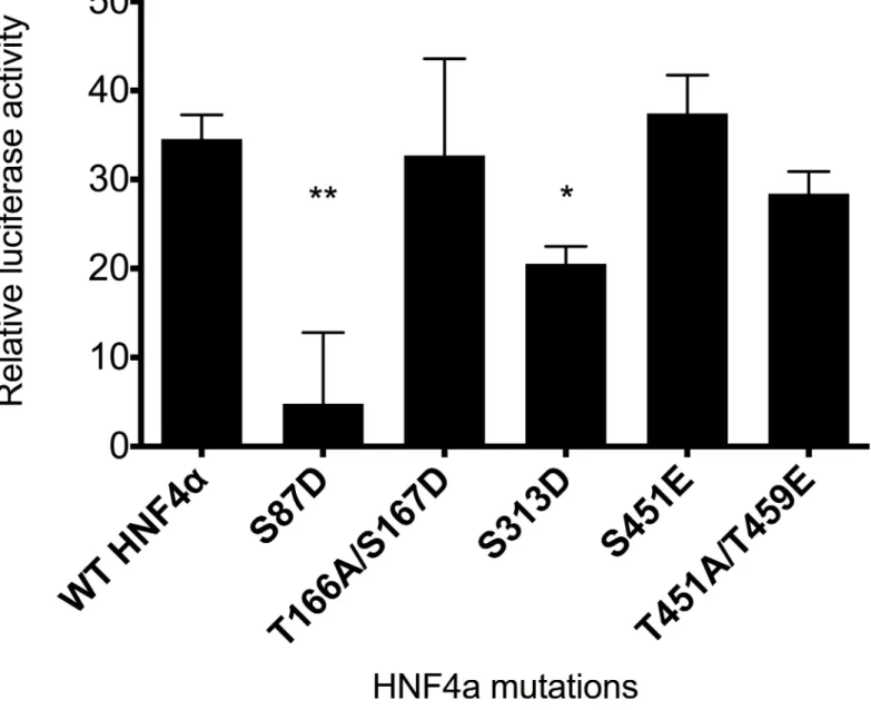
HNF4αphosphorylation via ERK1/2 inhibits ABCC6 transcriptional activity This great number of amino acid residues targeted by ERK1 phosphorylation raised the ques- tion of the functional relevance of the observed phenomenon. In order to answer this question, several phosphorylation sites were selected and investigated in luciferase reporter gene assay.
Five phosphorylation sites were chosen for further examination. Some of them were already studied previously, while we also investigated residues detected for the first time in the previ- ous experiment. Mutations were designed for the following serine or threonine phosphoryla- tion sites creating phosphomimetic (glutamate or aspartate) or neutral (alanine) mutants:
S87D, T166A/S167D, S313D, S451E, T457A/T459E and S451E/T457A/T459E triple mutant. If two phosphorylation sites were adjacent or in very close proximity, both were mutated.
Serine 87 mutation to aspartate was chosen because this site is undoubtedly a target for PKC phosphorylation, which abolishes HNF4αactivity [17]. Thus, this site served as a positive
control in our experiments. T166/S167 is a site, which was described to be phosphorylated by p38αor p38βMAP kinases [13,19], therefore also suggesting an important role of these two adjacent sites. The logic in our mutational screen was similar to that observed in the literature, i.e. to change only one site into a phosphomimetic mutation and mutate the other to a neutral one, if there were two adjacent phosphorylation sites. Accordingly, T166 was mutated into a neutral amino acid (alanine), whereas S167 became a phosphomimetic mutant (aspartate).
The site S313D is the target of phosphorylation by AMPK [14]. Finally, we newly identified the sites S451 and T457/T459. Therefore, we designed a neutral/phosphomimetic double mutant, in this case the phosphomimetic mutant being glutamate. Lastly, S451/T457/T459 were mutated in order to see the effect of a triple mutant, by including the former S451E mutant. Thus, we intended to examine if these C-terminal amino acids take part in transcrip- tional regulation when phosphorylated.
Fig 1. In vitro phosphorylation assay of HNF4αby ERK1 kinase. The phosphorylation assay was carried out with in vitro translated human recombinant HNF4αprotein, ERK1 kinase and radioactively labelled [γ-32P] ATP. HNF4αdoes not contain any post-translational modifications, but it has a GST-tag at N-terminal. The samples were run on SDS-PAGE and subjected to autoradiography. ERK1 kinase is capable of autophosphorylation.
doi:10.1371/journal.pone.0172020.g001
After creating all the mutants, co-transfection was performed with a luciferase reporter gene under the control ofABCC6promoter and different HNF4αmutants into HeLa cells lacking endogenous HNF4αexpression. The obtained results were normalized first for the background noise, then for transfection efficiency by the co-transfected control reporter vec- tor, which essentially represents transfection efficiency. As shown onFig 3, wild type HNF4α results in similar activity to T166A/S167D, S451E and T457A/T459E, and S451E/T457A/
T459E (data not shown), therefore, we regard these residues as not being responsible for implementing the effect of phosphorylation on transcriptional activity or gene expression in this assay. However, both S87D (used as positive control) and S313D have significant inhibi- tory effect onABCC6promoter activity: they decrease the activity to approximately 15% and 55%, respectively, compared to control. In conclusion, the phosphorylation site S313 targeted by both ERK1 and AMPK is responsible to have an inhibitory effect on transcription.
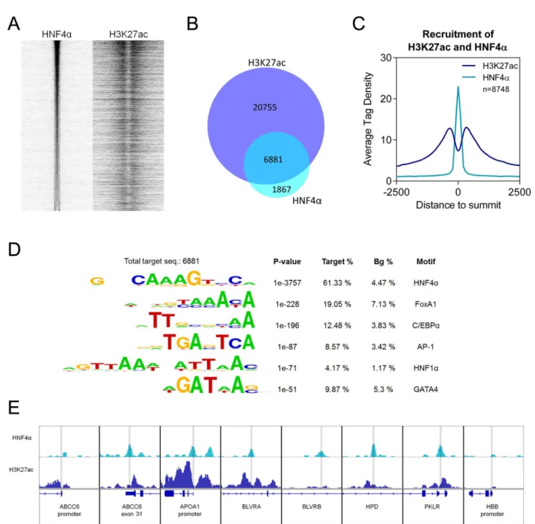
HNF4αand H3K27ac histone mark overlap in ChIP-Seq
In order to further confirm the functional relevance of HNF4αphosphorylation by ERK1/2, we carried out a cell-based assay, which reflects appropriately the endogenous intracellular processes. First, we screened control HepG2 cells for active HNF4αbinding sites prior to selecting some genomic target loci to investigate the effect of ERK1/2 on HNF4αbinding.
To detect active HNF4αbinding sites in the HepG2 cells at the genome-wide level, we car- ried out chromatin immunoprecipitation followed by sequencing (ChIP-seq) with an antibody
Fig 2. Phosphorylated sites on HNF4αprotein by ERK1 kinase detected by mass spectrometry. Non-radioactively phosphorylated HNF4αby ERK1/2 was cut out from the polyacrilamid gel. Tryptic peptide fragments were analysed by mass spectrometry. Affected phosphorylation sites (indicated by lollipops) appear in the DNA binding domain (DBD), the hinge, the ligand-binding domain (LBD) and the C-terminus of HNF4α, as well. The phosphorylated amino acid residues can be attributed to either previously described sites (S138/
T139, S142/S143, S147/S148, S151, T166/S167, S313) or new positions reported first here (S95, S262/S265, S451, T457/T459).
doi:10.1371/journal.pone.0172020.g002
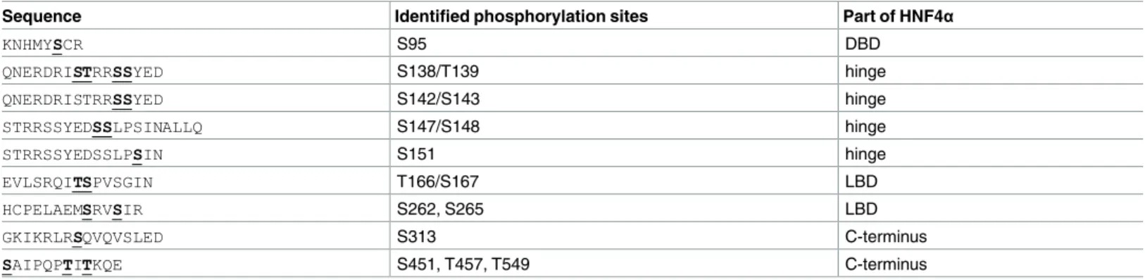
Table 1. Phosphorylated amino acid residues identified by mass spectrometry. In vitro phosphorylated HNF4αwas subjected to mass spectrometry analysis. The fragments containing the phosphorylated amino acid residues correspond to phosphorylation sites on the HNF4αprotein. Detected phosphory- lation sites (serine/threonine residues) in the cryptic fragments are marked bold and underlined. Affected phosphorylation sites appear in the DNA binding domain (DBD), the hinge, the ligand-binding domain (LBD) and the C-terminus of HNF4α, as well.
Sequence Identified phosphorylation sites Part of HNF4α
KNHMYSCR S95 DBD
QNERDRISTRRSSYED S138/T139 hinge
QNERDRISTRRSSYED S142/S143 hinge
STRRSSYEDSSLPSINALLQ S147/S148 hinge
STRRSSYEDSSLPSIN S151 hinge
EVLSRQITSPVSGIN T166/S167 LBD
HCPELAEMSRVSIR S262, S265 LBD
GKIKRLRSQVQVSLED S313 C-terminus
SAIPQPTITKQE S451, T457, T549 C-terminus
doi:10.1371/journal.pone.0172020.t001
against HNF4αand—in a parallel experiment on chromatin prepared from the same HepG2 sample—with an antibody against acetylated histone 3 lysine 27 (H3K27ac). The H3K27ac covalent modification of the chromatin indicates active regulatory regions, which are often enhancers.
From the ChIP-seq sample immunoprecipitated with HNF4αantibody, 8,748 transcription factor binding sites (TFBSs) could be identified (S1 Table). We have plotted these sites on a read distribution plot and we have sorted them based on tag density. Then we have matched the signal of the active histone mark H3K27ac (Fig 4A).Fig 4Billustrates on a Venn diagram that over 75% of HNF4αpeaks overlap with H3K27ac peaks.
Fig 3. Luciferase assay measuring ABCC6 promoter activity of HNF4αphosphomimetic mutants in HeLa cells. Mutations were designed for serine or threonine phosphorylation sites creating phosphomimetic (glutamate or aspartate) or neutral (alanine) mutants. For luciferase assays, triple co- transfection was performed with the phACCC6(-332/+72)Luc construct composed of the ABCC6 promoter fragment, pcDNA5-FRT/TO plasmid encoding HNF4αvariants and pRL-TK Renilla luciferase Control Reporter Vector. Luciferase activity was measured 48 hours after transfection. Relative luciferase activity was calculated by normalizing for background noise and for transfection efficiency by the co-transfected control reporter vector. The error bars represent S.D. Tukey-HSD test was performed. Significance versus WT HNF4 alpha is indicated by asterisks:*p<0.05;**p<0.01.
doi:10.1371/journal.pone.0172020.g003
We have also plotted the average tag density of the HNF4αpeaks and the H3K27ac signals, indicating that the HNF4αTFBSs represent a Gaussian distribution, whereas histone signals show the peak-valley-peak shape (Fig 4C). We have also observed that 451 peaks overlap with the TSS (+1 is part of the binding site) and in these cases, the average tag density of H3K27ac is
Fig 4. (A) Read distribution plot of HNF4αand H3K27ac upon vehicle treatment, relative to the 8,748 HNF4αpeaks in 2-kb frames. Peaks are sorted according to HNF4αtag density. (B) Area-proportional Venn diagram illustrating the overlap between H3K27ac regions and HNF4αpeaks.
(C) Histogram shows the average tag density of vehicle-treated HNF4αpeaks and H3K27ac signals at the sites of 8,748 HNF4αbinding sites. (D) Motif enrichment of 8,748 HNF4αpeaks. The P value and target and background (Bg) percentages are included for each motif. (E) IGV snapshot of HNF4αand H3K27ac ChIP-seq coverage representing eight selected genomic regions upon vehicle treatment. HBB promoter: negative control region. The interval scale is 50 in both cases. Peaks, highlighted with grey lines, represent the sites of the investigated HNF4α-target genomic regions.
doi:10.1371/journal.pone.0172020.g004
twice as high as in the case of the distribution centered to HNF4 alpha peaks (Fig 4CandS1 Fig). Furthermore, 33% and 71% of the HNF4 alpha peaks are within +/- 5 kb and +/- 30 kb from the TSS, respectively. These results indicate that the predicted TFBSs follow the expected typical patterns and confirm that these are real TFBSs without significant non-specific binding.
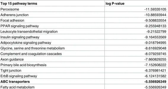
To further validate the predicted TFBSs, we applied motif enrichment analysis on these regions. As expected, the consensus motif of HNF4α(61.33%) and its cooperative factors, such as forkhead box A1 (FoxA1) and C/EBPα, are enriched. Motif of the activator protein 1 (AP- 1), HNF1 homeobox A (HNF1α) and GATA binding protein 4 (GATA4) were also identified (Fig 4D). By annotating the 8,748 HNF4αTFBSs, many of them are located near genes related to PPAR and insulin signalling or fatty acid metabolism. Furthermore, when we verified the pathways enriched for genes with HNF4αbinding sites, the membrane transport-related ABC-transporter genes also appeared (Table 2). Therefore, we selected the following target genes for further experiments:4-hydroxyphenylpyruvate dioxygenase(HPD),Pyruvate kinase, liver and red blood cell(PKLR). We have also selected theABCC6gene since we and others have reported that HNF4αbinds the promoter of this gene [24,27].Apolipoprotein A1 (APOA1) is a well-known target for HNF4α, therefore we also included it in the investigation.
Indeed, our ChIP-seq results also showed that it is bound by HNF4αtogether with the pres- ence of the H3K27ac, suggesting that they have active regulatory regions in HepG2 cells. More- over, it is listed as a gene having a role in PPAR signalling in the KEGG pathways obtained from our ChIP-seq data. Finally, we have identifiedBiliverdin A(BLVRA) andBiliverdin B (BLVRB), which are closely connected to heme oxygenase in heme catabolism, a known target of HNF4α, which plays a role in anti-oxidative and anti-inflammatory defence mechanisms.
We have also selected a negative control region: theβ-globinpromoter, which was devoid of acetylation and HNF4αbinding (Fig 4E).
ERK1/2 activation leads to reduced binding of HNF4αto specific genomic regions
In the following experiment we used the selected genomic target regions to investigate the dynamics of HNF4αbinding to our genes of interest upon ERK1/2 induction in HepG2 cells.
Table 2. Top 15 biological pathways related to the 8,748 HNF4αbinding sites. Data derived from KEGG database.
Top 15 pathway terms log P-value
Peroxisome -11.59335105
Adherens junction -10.88593944
Focal adhesion -9.508833554
PPAR signaling pathway -9.255948133
Leukocyte transendothelial migration -9.21522799
Insulin signaling pathway -9.164553569
Adipocytokine signaling pathway -9.018794995
Glycine, serine and threonine metabolism -8.616929048
Complement and coagulation cascades -8.079259745
Axon guidance -7.860829255
Primary bile acid biosynthesis -7.152608222
Tight junction -6.376981421
ErbB signaling pathway -6.124131582
ABC transporters -5.556926349
Fatty acid metabolism -5.556926349
doi:10.1371/journal.pone.0172020.t002
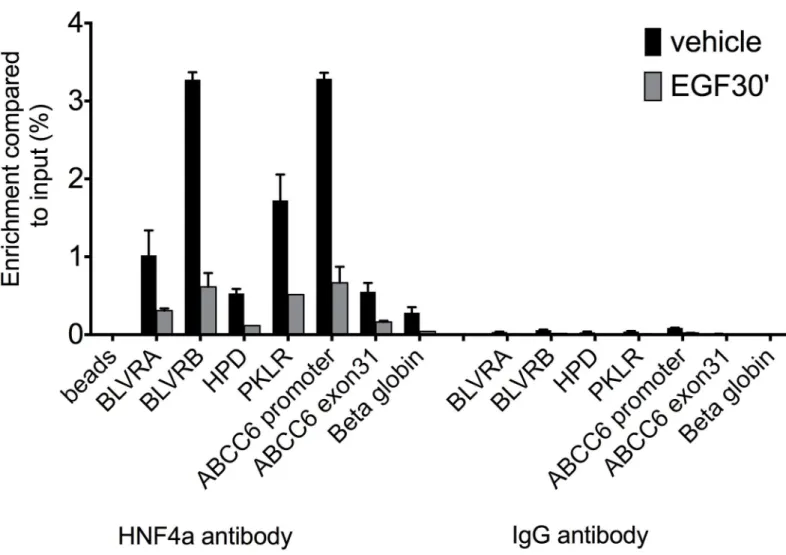
Since phosphorylation of proteins is a fast process, and happens within minutes [28], we decided to perform short-term (30 minutes) treatment of HepG2 cells with epidermal growth factor (EGF), which activates the ERK1/2 signalling cascade after binding to its receptor.
In our ChIP-qPCR experiments, enrichments of immunoprecipitated target fragments by anti-HNF4αantibody were compared to the input fraction or to the selected negative control region (β-globin) for TF occupancy, where no binding of the protein of interest is expected.
Similarly, we systematically performed another negative control, immunoprecipitation with IgG, used as non-specific antibody. We carried out 7 independent experiments under the same conditions in HepG2 cells and as hypothesized, 30 minutes treatment led to reduced HNF4αbinding to the selected target sites, as shown onFig 5andS2 Fig. We evaluated the effect of short-term EGF treatment by performing a one-sample t-test after normalization of the immunoprecipitated fraction from the treated cells for each target gene to their respective con- trols. Accordingly, highly significant effect was observed after short treatment (p<0.004). More precisely, after 30 minutes treatment, based on our ChIP-qPCR data, there is a substantial loss
Fig 5. ChIP-qPCR results showing HNF4αoccupancy on seven genomic regions of hepatic HNF4αtarget genes. Immunoprecipitation of chromatin from HepG2 cells was performed with anti-HNF4αor IgG antibody untreated or treated with EGF for 30 mins. Enrichment was compared to % input (y axis). HNF4αbinding (black columns) is decreased after short EGF treatment (grey). BLVR A: Biliverdin A, BLVR B: Biliverdin B, HPD:
4-hydroxyphenylpyruvate dioxygenase, PKLR: Pyruvate kinase, liver and RBC, ABCC6: ATP-binding cassette subfamily C, member 6 and APOA1:
Apolipoprotein A1. Beta globin: negative control region. Beads represent immunoprecipitation performed without any antibody. S.D. is indicated on the figure. Results of a representative experiment (n = 7) are shown.
doi:10.1371/journal.pone.0172020.g005
of HNF4αbinding compared to the control case for the individual target genes, shown onS2 Table. This effect was further enhanced by 24h EGF treatment. Collectively, our data suggest that ERK1/2 phosphorylates HNF4αbothin vitroandin vivo, leading to the reduced DNA binding capacity of the transcription factor at target loci.
Discussion
We have shown previously that HNF4αregulatesABCC6gene expression. We have also dem- onstrated that ERK1/2 activation inhibits HNF4α-dependentABCC6expression. We hypothe- sized that HNF4αis phosphorylated by ERK1/2 leading to its reducedtrans-activational capacity in addition to the reduced expression of theHNF4agene. Here we have demonstrated that ERK1 is able to phosphorylate HNF4αat several serine and threonine residues. We have also shown that phosphorylation of HNF4αinhibits itstrans-activational capacity in reporter gene assay and its chromatin binding activity as determined by ChIP-qPCR confirming the physiological relevance of our findings.
HNF4αplays a major role in hepatic development and it is a master gene regulator in dif- ferentiated hepatocytes. It regulates thousands of genes playing important roles in glucose, lipid and amino acid metabolism, bile acid synthesis, detoxification and inflammation. Our results are in harmony with these findings. In our ChIP-seq experiments, in non-treated HepG2 cells we found almost 9000 genomic HNF4αbinding sites associated with 5500 genes.
More than 60% of them are canonical HNF4αelements, but binding motifs for FoxA1, C/EBPαand HNF1αwere also enriched in the immunoprecipitated DNA fraction. These sequences are known to be bound by TFs interacting with HNF4α, thereby strengthening the validity of our findings. CEBPαhas been previously reported as being part of a complex with HNF4αforming together an intricate regulatory network of hepatocyte gene expression [22, 29]. HNF1αis a transcription factor known to interact with HNF4α, where HNF4αenhances HNF1α-mediated activation of hepatic transcription [30]. Our data also indicated that we identified actively transcribed HNF4α-regulated genes since those sites were also occupied by the H3K27ac histone mark, a hallmark of active genes [31] (Fig 4B). Finally, we were able to identify typical HNF4αtarget genes in the immunoprecipitated fraction. Furthermore, the KEGG pathway analysis of all identified HNF4αtarget genes revealed similar cascades to those available as literature data. Among the typical genesABCC6,ABCA1,ALDOB (aldolase B), APOA1,APOB,APOCIII,BLVRAandB,CYP7A1,HNF1aand4a,HPD,PKLRandSLC2A2 (GLUT2)were observed.
From these target genes we selected six—BLVRA,BLVRB,HPD,ABCC6,PKLRandAPOA1
—for our further experiments. The latter two genes are part of the 451, which have an HNF4α binding site overlapping with the TSS (S1 Fig). Pyruvate kinase (PKLR) plays an important role in regulating glucose metabolism [7]. The other genes we have selected are also implicated in metabolism. 4-Hydroxyphenyl pyruvate dioxygenase (HPD) is an enzyme participating in tyrosine metabolism [32]. Mutations of the gene lead to a benign Mendelian disorder called Tyrosinaemia (type III). Apolipoprotein A1 (APOA1) plays primary role in lipid transport [33]. The biliverdin reductase genes catalyze the synthesis of bilirubin from biliverdin and thus participate in heme metabolism and the antioxidant pathway. Although the molecular func- tion of ABCC6 is still unclear, it is protective against ectopic calcification, moreover, it might play a role in ATP homeostasis and in ROS elimination [23,24,34].
Our ChIP-qPCR experiments confirmed our initial hypothesis suggesting that ERK1/2 has both post-translational and probably transcriptional effects on HNF4α. These experiments showed that already short activation of the ERK1/2 pathway decreased the DNA binding capacity of HNF4αin living cells suggesting a post-translational effect. This rapid decrease
became even more pronounced after 24h ERK1/2 activation suggesting the inhibition of HNF4agene expression, as described earlier [29]. We chose the ChIP-qPCR method to detect the binding capacity alterations of the TF to a limited number of genomic loci to allow us reli- able quantification of the changes. By using this approach, we could also perform several inde- pendent experiments, which was essential since the variability of ChIP experiment results is generally high.
Our experiments also proved that the rapid post-translational effect of ERK1/2 activation on HNF4αis mediated by the phosphorylation of the protein. We have shown that anin vitro translated HNF4αprotein is phosphorylated at multiple sites by the activated ERK1. Several studies have revealed that HNF4αcan be phosphorylated by several kinases, for instance, PKC, PKA, AMPK and p38. Phosphorylation can alter its DNA-binding affinity, intracellular locali- zation andtrans-activation capacity [35]. Phosphorylation of the highly conserved S87 by PKC drastically decreases the DNA binding capacity and stability of the protein [17]. Similarly, the cAMP-activated PKA phosphorylates HNF4αat the S142/S143 position [18]. This leads to reduced DNA binding activity of the TF, however, the reducedtrans-activational potential could not be confirmed in luciferase reporter gene assay setup [18]. More recently, it has been shown that Thyroid-stimulating hormone (TSH) induces PKA, which leads to decreased nuclear localization of HNF4αin HepG2 cells [36]. AMPK has been defined as a metabolic master switch [8]. It phosphorylates the conserved S313 residue of HNF4αleading to the inhi- bition of the dimerization of the TF [20] and thereby itstrans-activational capacity. AMPK activation dramatically decreases the transcription of various HNF4αtarget genes in hepato- cytes. Finally, the role of p38 seems to be controversial. Some papers indicate that phosphory- lation of HNF4αat residue T166/S167 by this kinase increasestrans-activational potential of the TF [19,37]. Others show indirect effect [20,38], while some suggest an inhibitory role for p38 [9].
We have also investigated the functional relevance of the different residues shown to be phosphorylated by ERK1. In these experiments we used the luciferase reporter gene cloned downstream of theABCC6promoter, a construct used in our previous studies [22,24]. We have shown that this promoter is induced by HNF4α. In our present experiments we co- expressed this reporter gene with wild-type (wt) or different phosphomimetic HNF4α mutants.
The positive control mutant mimicking PKC phosphorylation resulted in significantly reduced reporter gene activity relative to the wt, as described earlier [17]. We have also ana- lyzed the sites firstly identified in the present study (S451, T457/T459) and the sites corre- sponding to the previously described potential p38 site (T166/S167) [19]. In contrast to the previous studies, the phosphomimetic mutants of this site did not affect thetrans-activational potential of HNF4α. In these experiments, only the site previously shown as targeted by AMPK (S313) showed diminished reporter gene activity [20,38]. Collectively, our results clearly demonstrate that ERK1/2 activation results in HNF4αphosphorylation and reduced DNA binding capacity.
The ERK1/2 pathway is activated under several physiologic and pathologic conditions underlying the importance of our findings. For example, bile salts function as signalling mole- cules through the Sphingosine-1-phosphate receptor 2 (S1PR2) G protein coupled receptors (GPCRs), which activate ERK1/2 to control hepatic glucose, lipid and drug metabolism ([39–
41]; [42] and references therein). The resulting rapid downregulation of HNF4αactivity reduces the expression of the gluconeogenic genesPEPCKandG-6-Pase[40], or the gene encoding the major enzyme in bile acid synthesis,CYP7A1[43].
In addition to the GPCR pathway, bile acids also activate specific nuclear receptors (e.g.
FXR and vitamin D receptor (VDR)). While VDR is able to directly activate ERK1/2 [44], FXR
is the major bile acid-responsive ligand-activated transcription factor and it is responsible for bile acid homeostasis [45,46]. FXR induces the expression of SHP (small heterodimer part- ner), an orphan nuclear receptor without DNA binding domain. SHP binds HNF4αand inhibits its binding to the targetcis-regulatory elements (e.g. the bile acid response element (BARE) of theCholesterol 7αhydroxylase(CYP7A1) gene promoter) [47]. SHP is also activated by ERK1/2 leading to an intricate network of HNF4αinhibition both in the short and longer term [42,46,48].
Finally, the ERK1/2 pathway is also activated by other mechanisms including oxidative stress (ROS) [49], growth hormones (such as HGF, EGF and FGF15/19 [24,50]) and cytokines (IL1 and TNFα[9,14,19]), as well. These factors also lead to the reduced activity of HNF4α and thereby the downregulation of several genes. According to our results, the mechanism of the short-term inhibition of HNF4αis its phosphorylation, suggesting that ERK1/2 plays a piv- otal role in the coordinated regulation of a great number of hepatic genesviathe rapid post- translational modification of HNF4α.
Materials and methods Cell culture
HepG2 human hepatoma cell line was obtained from ATCC (ATCC HB-8065) and cultured in Advanced MEM (ThermoFisher) supplemented with 10% FBS, 2mM L-glutamine, 100 U/ml penicillin and 100 mg/ml streptomycin. HeLa cells were obtained from ATCC and cultured according to the manufacturer’s instructions (DMEM supplemented with 10% FBS, 2mM L- glutamine, 100 U/ml penicillin and 100 mg/ml streptomycin). For EGF treatment, cells were changed to serum free medium 24h before the addition of the chemical, and then treated for 30 minutes or 24 hours with human recombinant epidermal growth factor (Sigma–Aldrich) at 100 ng/ml final concentration.
In vitro phosphorylation assay
The reaction was performed in a mixture (30μl final volume) containing kinase buffer [28], 500 ng ERK1 kinase (SigmaERK1 kinase datasheet, cat number: E7407) HNF4αhuman recombinant protein with GST-tag at N-terminal (AbnovaHNF4 alpha datasheet, cat number:
H00003172-P01) and 20 uM ATP including 1μCi [γ-32P] ATP. The reaction was initiated by the addition of ATP. After incubation at 30˚C for 30 min, the reaction was stopped by adding 10μl of 4X concentrated SDS sample buffer. Samples were subjected to SDS-PAGE using 10%
running gels. After drying, gels were subjected to autoradiography for 2–12 hours.
Phosphopetide mapping of HNF4α
The gel band containing HNF4αprotein was processed, reductively alkylated with DTT and iodoacetamide and then digested with trypsin in 20 mM ammonium bicarbonate buffer. An aliquot was then run a Thermo/Dionex Ultimate RSLC nano system using a 75um x 15 cm C 18 PepMap column (Thermo/Dionex) coupled to a Thermo LTQ Orbitrap Velos Pro. We used a TOP 15 MS method (65 min linear gradient from 5–40% B (80% acetonitrile in 0.1%
formic acid) with multi-stage activation (to detect neutral loss of phosphate from phosphopep- tides). The obtained data file was analysed by the Mascot search engine against the sequence provided (HNF4αwith N-terminal GST-tag). The phosphopeptide assignment was done on peptides above a mascot ion score of 20 which may include the same peptide a number of times as the system has detected that peptide on more than one occasion. The indication is that the higher the ion score for the peptide the more likely that the assignment is correct.
HNF4αmutations
Wild-type plasmid containing the full human HNF4αgene and pcDNA5-FRT/TO expression backbone was purchased from Addgene. Amino acid numbering in this article refer to muta- tions of the human HNF4αgene, whereas mutations of the rat HNF4αgene are adjusted to the human numbering. Mutations were designed for the following serine or threonine phosphory- lation sites creating phosphomimetic (glutamate or aspartate) or neutral (alanine) mutants:
S87D, T166A/S167D, S313D, S451E, T451A/T459E. If two phosphorylation sites were adjacent or in very close proximity, both were mutated. Gene synthesis and site-directed mutagenesis were performed by the biotechnology company GenScript. The gene was re-cloned from pcDNA3+ into pcDNA5-FRT/TO plasmid.
Transfection and luciferase experiments
HeLa cells were plated onto 96-well plates starting with 10.000 cells/well. FuGENE HD trans- fection reagent (Promega) complex containing serum-free medium and 2μg total plasmid DNA was added to cells in growth medium. Triple co-transfection was performed with the phACCC6(-332/+72)Luc construct ((see [24]) composed of theABCC6promoter fragment (-332/+72) cloned upstream of the luciferase coding cassette in the pGL3-Basic vector (Pro- mega)), pcDNA5-FRT/TO plasmid encoding HNF4αvariants (GenScript) and pRL-TK Renilla luciferase Control Reporter Vector (Promega). Cells were harvested and lysed after 48 hours. Luciferase activity was determined by Victor luminometric plate reader (Perkin Elmer) using the DualGlo Luciferase system (Roche). The obtained results were normalized firstly for the background noise, then for transfection efficiency by the co-transfected control reporter vector.
ChIP (chromatin immunoprecipitation) assay
Formaldehyde was added directly to the culture media of flasks containing 10x106HepG2 cells, to a final concentration of 1%. After 10 min incubation at room temperature, fixation was quenched by adding 125 mM ice-cold glycine, and flasks were washed three times with ice-cold phosphate buffered saline (PBS). Cells were then scraped and washed three more times with PBS, each washing step being followed by sedimentation at 1,300 x g. Pellets were resuspended in 1mL lysis buffer (5 mM PIPES-pH = 8.0, 85 mM KCl, 0,5% NP-40, cOmplete Mini cocktail tablets (Roche) and incubated at 4˚C for 15 mins with vortexing. Lysed cells were sedimented at 13,000 x g, resuspended in 500μL of sonication buffer (1% SDS, 10 mM EDTA, 50 mM Tris-HCl-pH = 8.0, cOmplete Mini cocktail tablets) and sonicated on ice with an MSE sonicator (6 pulses of 15s each at 15% amplitude with 30s off between each pulse). The generated fragments were approximately 500 bp long, as determined experimentally. After sedimentation at 13,000 x g, the supernatant was transferred into a new tube, diluted ten times with IP buffer (0,01% SDS, 1.1% Triton X-100, 1.2 mM EDTA, 16.7 mM Tris-HCl (pH = 8.0), 167 mM NaCl) and supplemented with cOmplete Mini cocktail tablets. For each 500μL ex- tract, a mixture of 60μL of Dynabeads protein A and 60μL of Dynabeads protein G (Thermo- Fisher), conjugated with 2μg anti-HNF4αmouse monoclonal antibody (Abcam ab41898) or anti-H3K27ac rabbit polyclonal antibody (Abcam ab4729) was added. After incubation at 4˚C overnight, the beads were subsequently washed with buffer A (0.1% SDS, 1% Triton X-100, 2 mM EDTA, 20 mM Tris-HCl (pH = 8.0), 0.15 M NaCl), buffer B (0.1% SDS, 1% Triton X-100, 2 mM EDTA, 20 mM Tris-HCl (pH = 8.0), 0.5 M NaCl) and buffer C (0,25 M LiCl, 1% NP40, 1% Na-deoxycholate, 1 mM EDTA, Tris-HCl (pH = 8.0), at 4˚C with permanent rotation for 5 mins. Following two washes with TE buffer (10 mM Tris-HCl, 10 mM EDTA, pH = 8.0), sam- ples were eluted in 200μL freshly prepared NaHCO3(0,1 mM) supplemented with 1% SDS.
Cross-links were reversed by overnight incubation at 65˚C. Samples were incubated with 20μg/mL RNAse at 37˚C for 1 hour, then proteinase K was added to 20μg/mL and the mixture was incubated for 2 hours at 45˚C. DNA was purified using High pure PCR template prepara- tion kit (Roche). ChIP-seq libraries were prepared from 5 ng DNA with Ovation Ultralow Library Systems (Nugen) according to manufacturer’s instructions. Libraries were sequenced on Illumina HiScanSQ sequencer.
ChIP-seq data processing
Raw sequence files of the ChIP-seq samples were processed using a computational pipeline [51] with the hg19 reference genome. ChIP-seq peaks were predicted using HOMER [52].
Artifacts, based on the blacklisted genomic regions of the Encyclopedia of DNA Elements [53], were removed from the peak sets using BEDTools [54]. RPKM (Reads Per Kilobase per Million mapped reads) values for the HNF4αsample were calculated on the summit -/+50 bp region of the peaks; for the H3K27ac sample were calculated on the whole region of the histone signal.
Motif enrichment analysis was carried out byfindMotifsGenome.pl. Pathway analysis was done byannotatePeaks.plusing the KEGG database [55]. The average read density was determined byannotatePeaks.pl[52]. Read distribution and average density heat maps were displayed by Java TreeView [56]. Histogram and box plot were performed using GraphPad Prism version 6.00 for Windows (GraphPad Software, La Jolla California USA).
Quantitative PCR
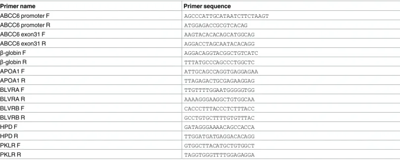
A fraction of the DNA was used as a template for quantitative PCRs. The primers used were designed with BiSearch [57] and their sequences are shown inTable 3. PCR products were between 100 and 200 bp. qPCRs were performed in a total volume of 20μL containing 1x SYBR green mix (Roche), a 1/10 fraction of ChIP-enriched DNA, and 250 nM primers in a 96-well plate. Using sonicated genomic DNA samples at different dilutions, we generated stan- dard curves, and relative amounts of immunoprecipitated DNA were calculated by extrapolat- ing from the dilution curves. All standards and samples were run in duplicate. Plates were read
Table 3. List of qPCR primers.
Primer name Primer sequence
ABCC6 promoter F AGCCCATTGCATAATCTTCTAAGT
ABCC6 promoter R ATGGAGACCGCGTCACAG
ABCC6 exon31 F AAGTACACACAGCATGGCAG
ABCC6 exon31 R AGGACCTAGCAATACACAGG
β-globin F AGGACAGGTACGGCTGTCATC
β-globin R TTTATGCCCAGCCCTGGCTC
APOA1 F ATTGCAGCCAGGTGAGGAGAA
APOA1 R TTAGAGACTGCGAGAAGGAG
BLVRA F TTGTTTTGGAATGGGGGTGG
BLVRA R AAAAGGGAAGGCTGTGGCAA
BLVRB F CACCCTTTACCCTCTTTACC
BLVRB R GCCTGTGCTTTTGTGTTTAC
HPD F GATAGGGAAAACAGCCACCA
HPD R TTGGATGATGAGGACACAGG
PKLR F GTGGCTTACATGCTGTGGCT
PKLR R TAGGTGGGTTTTGGAGAGGA
doi:10.1371/journal.pone.0172020.t003
with a LightCycler 480 real-time PCR machine (Roche). Enrichment of a particular DNA frag- ment was calculated by comparing its relative concentration to control regions where no bind- ing to the protein of interest is expected.
Accession number
Sequencing data were submitted to NCBI’s Sequence Read Archive (SRA) database under accession number SRP096703 at the following URL:
https://www.ncbi.nlm.nih.gov/sra/?term=SRP096703
Supporting information
S1 Fig. Recruitment of HNF4αand H3K27ac at promoters. Histogram shows the average tag density of HNF4αand H3K27ac peaks at those 451 HNF4αbinding sites which overlap with the transcription start site (TSS).
(TIF)
S2 Fig. ChIP-qPCR results showing HNF4αoccupancy on seven genomic regions of hepatic HNF4αtarget genes. Immunoprecipitation of chromatin from HepG2 cells was per- formed with anti- HNF4αor IgG antibody untreated or treated with EGF for 30 mins or 24 hours. Enrichment was compared to negative control region (Beta globin) for TF occupancy (y axis). HNF4αbinding (black columns) is decreased after short EGF treatment (dark grey), which is further diminished after long EGF treatment (light grey). BLVR A: Biliverdin A, BLVR B: Biliverdin B, HPD: 4-hydroxyphenylpyruvate dioxygenase, PKLR: Pyruvate kinase, liver and RBC, ABCC6: ATP-binding cassette subfamily C, member 6 and APOA1: Apolipo- protein A1. S.D. is indicated on the figure.
(TIFF)
S1 Table. HNF4αtarget genes identified in ChIP-seq experiment on vehicle treated HepG2 cells.
(XLSX)
S2 Table. P values of one-sample t-test on individual HNF4αtarget genes investigated by ChIP-qPCR.
(XLSX)
Acknowledgments
The authors are grateful for helpful discussions with Dr. Andra´s Va´radi, furthermore, for Ka´roly Liliom and Anna Lovrics for the help in the statistical analyses. The authors thank Gyo¨rgyi Vermes for technical help. The work was supported by grants from the MedinProt Program of the Hungarian Academy of Sciences (LB and TA). BLB is a Szodoray Fellow of the University of Debrecen, Medical Faculty, his research was funded by an Internal Research Uni- versity Grant entitled „Dissecting the genetic and epigenetic components of gene expression regulation in the context of 1000 genomes project”.
Author Contributions
Conceptualization: TA CB BLB LM.
Data curation: DB BLB TA BV CB JK.
Formal analysis: DB BLB.
Funding acquisition: TA BLB LB.
Investigation: BV CB SS TA.
Methodology: CB BV TA SS LB.
Project administration: TA.
Resources: TA LB LM.
Software: DB BLB.
Supervision: TA BLB LB LM.
Validation: TA BLB.
Visualization: JK DB BV.
Writing – original draft: TA BV DB.
Writing – review & editing: BV DB CB JK SS LM LB BLB TA.
References
1. Costa RH, Grayson DR, Darnell JE Jr. Multiple hepatocyte-enriched nuclear factors function in the regu- lation of transthyretin and alpha 1-antitrypsin genes. Molecular and cellular biology. 1989; 9(4):1415–
25. Epub 1989/04/01. PubMed Central PMCID: PMCPMC362558. PMID:2786140
2. Sladek FM, Zhong WM, Lai E, Darnell JE Jr. Liver-enriched transcription factor HNF-4 is a novel mem- ber of the steroid hormone receptor superfamily. Genes & development. 1990; 4(12b):2353–65. Epub 1990/12/01.
3. Gupta RK, Kaestner KH. HNF-4alpha: from MODY to late-onset type 2 diabetes. Trends Mol Med.
2004; 10(11):521–4. doi:10.1016/j.molmed.2004.09.004PMID:15519277
4. Hayhurst GP, Lee YH, Lambert G, Ward JM, Gonzalez FJ. Hepatocyte nuclear factor 4alpha (nuclear receptor 2A1) is essential for maintenance of hepatic gene expression and lipid homeostasis. Molecular and cellular biology. 2001; 21(4):1393–403. Epub 2001/02/07. PubMed Central PMCID:
PMCPMC99591. doi:10.1128/MCB.21.4.1393-1403.2001PMID:11158324
5. Drewes T, Senkel S, Holewa B, Ryffel GU. Human hepatocyte nuclear factor 4 isoforms are encoded by distinct and differentially expressed genes. Molecular and cellular biology. 1996; 16(3):925–31. Epub 1996/03/01. PubMed Central PMCID: PMCPMC231074. PMID:8622695
6. Odom DT, Zizlsperger N, Gordon DB, Bell GW, Rinaldi NJ, Murray HL, et al. Control of pancreas and liver gene expression by HNF transcription factors. Science (New York, NY). 2004; 303(5662):1378–
81. Epub 2004/02/28. PubMed Central PMCID: PMCPMC3012624.
7. Stoffel M, Duncan SA. The maturity-onset diabetes of the young (MODY1) transcription factor HNF4al- pha regulates expression of genes required for glucose transport and metabolism. Proceedings of the National Academy of Sciences of the United States of America. 1997; 94(24):13209–14. Epub 1997/12/
16. PubMed Central PMCID: PMCPMC24288. PMID:9371825
8. Leclerc I, Lenzner C, Gourdon L, Vaulont S, Kahn A, Viollet B. Hepatocyte nuclear factor-4alpha involved in type 1 maturity-onset diabetes of the young is a novel target of AMP-activated protein kinase. Diabetes. 2001; 50(7):1515–21. Epub 2001/06/26. PMID:11423471
9. Mogilenko DA, Dizhe EB, Shavva VS, Lapikov IA, Orlov SV, Perevozchikov AP. Role of the nuclear receptors HNF4 alpha, PPAR alpha, and LXRs in the TNF alpha-mediated inhibition of human apolipo- protein A-I gene expression in HepG2 cells. Biochemistry. 2009; 48(50):11950–60. doi:10.1021/
bi9015742PMID:19883121
10. Bogan AA, Dallas-Yang Q, Ruse MD Jr., Maeda Y, Jiang G, Nepomuceno L, et al. Analysis of protein dimerization and ligand binding of orphan receptor HNF4alpha. Journal of molecular biology. 2000; 302 (4):831–51. Epub 2000/09/20. doi:10.1006/jmbi.2000.4099PMID:10993727
11. Dhe-Paganon S, Duda K, Iwamoto M, Chi YI, Shoelson SE. Crystal structure of the HNF4 alpha ligand binding domain in complex with endogenous fatty acid ligand. The Journal of biological chemistry.
2002; 277(41):37973–6. Epub 2002/08/24. doi:10.1074/jbc.C200420200PMID:12193589
12. Wisely GB, Miller AB, Davis RG, Thornquest AD Jr., Johnson R, Spitzer T, et al. Hepatocyte nuclear factor 4 is a transcription factor that constitutively binds fatty acids. Structure (London, England: 1993).
2002; 10(9):1225–34. Epub 2002/09/11.
13. Yokoyama A, Katsura S, Ito R, Hashiba W, Sekine H, Fujiki R, et al. Multiple post-translational modifica- tions in hepatocyte nuclear factor 4alpha. Biochem Biophys Res Commun. 2011; 410(4):749–53. doi:
10.1016/j.bbrc.2011.06.033PMID:21708125
14. Wang Z, Salih E, Burke PA. Quantitative analysis of cytokine-induced hepatocyte nuclear factor-4alpha phosphorylation by mass spectrometry. Biochemistry. 2011; 50(23):5292–300. PubMed Central PMCID: PMCPMC3115757. doi:10.1021/bi200540wPMID:21598922
15. Caron S, Huaman Samanez C, Dehondt H, Ploton M, Briand O, Lien F, et al. Farnesoid X receptor inhibits the transcriptional activity of carbohydrate response element binding protein in human hepato- cytes. Molecular and cellular biology. 2013; 33(11):2202–11. Epub 2013/03/27. PubMed Central PMCID: PMCPMC3648076. doi:10.1128/MCB.01004-12PMID:23530060
16. Ding X, Lichti K, Kim I, Gonzalez FJ, Staudinger JL. Regulation of constitutive androstane receptor and its target genes by fasting, cAMP, hepatocyte nuclear factor alpha, and the coactivator peroxisome pro- liferator-activated receptor gamma coactivator-1alpha. The Journal of biological chemistry. 2006; 281 (36):26540–51. Epub 2006/07/11. PubMed Central PMCID: PMCPMC2991045. doi:10.1074/jbc.
M600931200PMID:16825189
17. Sun K, Montana V, Chellappa K, Brelivet Y, Moras D, Maeda Y, et al. Phosphorylation of a conserved serine in the deoxyribonucleic acid binding domain of nuclear receptors alters intracellular localization.
Mol Endocrinol. 2007; 21(6):1297–311. doi:10.1210/me.2006-0300PMID:17389749
18. Viollet B, Kahn A, Raymondjean M. Protein kinase A-dependent phosphorylation modulates DNA-bind- ing activity of hepatocyte nuclear factor 4. Molecular and cellular biology. 1997; 17(8):4208–19. Epub 1997/08/01. PubMed Central PMCID: PMCPMC232274. PMID:9234678
19. Guo H, Gao C, Mi Z, Zhang J, Kuo PC. Characterization of the PC4 binding domain and its interactions with HNF4alpha. Journal of biochemistry. 2007; 141(5):635–40. Epub 2007/02/24. doi:10.1093/jb/
mvm066PMID:17317687
20. Hong YH, Varanasi US, Yang W, Leff T. AMP-activated protein kinase regulates HNF4alpha transcrip- tional activity by inhibiting dimer formation and decreasing protein stability. The Journal of biological chemistry. 2003; 278(30):27495–501. Epub 2003/05/13. doi:10.1074/jbc.M304112200PMID:
12740371
21. Varadi A, Szabo Z, Pomozi V, de Boussac H, Fulop K, Aranyi T. ABCC6 as a target in pseudoxanthoma elasticum. Current drug targets. 2011; 12(5):671–82. Epub 2010/11/03. PubMed Central PMCID:
PMCPMC3324121. PMID:21039331
22. Ratajewski M, de Boussac H, Sachrajda I, Bacquet C, Kovacs T, Varadi A, et al. ABCC6 expression is regulated by CCAAT/enhancer-binding protein activating a primate-specific sequence located in the first intron of the gene. J Invest Dermatol. 2012; 132(12):2709–17. PubMed Central PMCID:
PMCPMC3509247. doi:10.1038/jid.2012.218PMID:22763786
23. Aranyi T, Bacquet C, de Boussac H, Ratajewski M, Pomozi V, Fulop K, et al. Transcriptional regulation of the ABCC6 gene and the background of impaired function of missense disease-causing mutations.
Front Genet. 2013; 4:27. PubMed Central PMCID: PMCPMC3593682. doi:10.3389/fgene.2013.00027 PMID:23483032
24. de Boussac H, Ratajewski M, Sachrajda I, Koblos G, Tordai A, Pulaski L, et al. The ERK1/2-hepatocyte nuclear factor 4alpha axis regulates human ABCC6 gene expression in hepatocytes. The Journal of bio- logical chemistry. 2010; 285(30):22800–8. PubMed Central PMCID: PMCPMC2906271. doi:10.1074/
jbc.M110.105593PMID:20463007
25. Xue TC, Jia QA, Bu Y, Chen RX, Cui JF, Tang ZY, et al. CXCR7 correlates with the differentiation of hepatocellular carcinoma and suppresses HNF4alpha expression through the ERK pathway. Oncology reports. 2014; 32(6):2387–96. Epub 2014/09/23. doi:10.3892/or.2014.3501PMID:25242412
26. Lorenz K, Schmitt JP, Schmitteckert EM, Lohse MJ. A new type of ERK1/2 autophosphorylation causes cardiac hypertrophy. Nat Med. 2009; 15(1):75–83. doi:10.1038/nm.1893PMID:19060905
27. Bolotin E, Liao H, Ta TC, Yang C, Hwang-Verslues W, Evans JR, et al. Integrated approach for the iden- tification of human hepatocyte nuclear factor 4alpha target genes using protein binding microarrays.
Hepatology. 2010; 51(2):642–53. PubMed Central PMCID: PMCPMC3581146. doi:10.1002/hep.
23357PMID:20054869
28. Illes A, Enyedi B, Tamas P, Balazs A, Bogel G, Buday L. Inducible phosphorylation of cortactin is not necessary for cortactin-mediated actin polymerisation. Cellular signalling. 2006; 18(6):830–40. Epub 2005/08/20. doi:10.1016/j.cellsig.2005.07.012PMID:16109479
29. Hatzis P, Kyrmizi I, Talianidis I. Mitogen-activated protein kinase-mediated disruption of enhancer-pro- moter communication inhibits hepatocyte nuclear factor 4alpha expression. Molecular and cellular
biology. 2006; 26(19):7017–29. Epub 2006/09/19. PubMed Central PMCID: PMCPMC1592892. doi:
10.1128/MCB.00297-06PMID:16980607
30. Eeckhoute J, Formstecher P, Laine B. Hepatocyte nuclear factor 4alpha enhances the hepatocyte nuclear factor 1alpha-mediated activation of transcription. Nucleic acids research. 2004; 32(8):2586–
93. Epub 2004/05/14. PubMed Central PMCID: PMCPMC419469. doi:10.1093/nar/gkh581PMID:
15141028
31. Struhl K. Histone acetylation and transcriptional regulatory mechanisms. Genes & development. 1998;
12(5):599–606.
32. Aarenstrup L, Falch AM, Jakobsen KK, Neve S, Henriksen LL, Tommerup N, et al. Expression and post-translational modification of human 4-hydroxy-phenylpyruvate dioxygenase. Cell Biol Int. 2002; 26 (7):615–25. PMID:12127941
33. Malik S. Transcriptional regulation of the apolipoprotein AI gene. Front Biosci. 2003; 8:d360–8. PMID:
12456302
34. Jansen RS, Kucukosmanoglu A, de Haas M, Sapthu S, Otero JA, Hegman IE, et al. ABCC6 prevents ectopic mineralization seen in pseudoxanthoma elasticum by inducing cellular nucleotide release. Pro- ceedings of the National Academy of Sciences of the United States of America. 2013; 110(50):20206–
11. PubMed Central PMCID: PMCPMC3864344. doi:10.1073/pnas.1319582110PMID:24277820 35. Gonzalez FJ. Regulation of hepatocyte nuclear factor 4 alpha-mediated transcription. Drug Metab Phar-
macokinet. 2008; 23(1):2–7. PMID:18305369
36. Song Y, Zheng D, Zhao M, Qin Y, Wang T, Xing W, et al. Thyroid-Stimulating Hormone Increases HNF- 4alpha Phosphorylation via cAMP/PKA Pathway in the Liver. Sci Rep. 2015; 5:13409. PubMed Central PMCID: PMCPMC4548215. doi:10.1038/srep13409PMID:26302721
37. Xu Z, Tavares-Sanchez OL, Li Q, Fernando J, Rodriguez CM, Studer EJ, et al. Activation of bile acid biosynthesis by the p38 mitogen-activated protein kinase (MAPK): hepatocyte nuclear factor-4alpha phosphorylation by the p38 MAPK is required for cholesterol 7alpha-hydroxylase expression. The Jour- nal of biological chemistry. 2007; 282(34):24607–14. PubMed Central PMCID: PMCPMC3291957. doi:
10.1074/jbc.M611481200PMID:17603092
38. Puigserver P, Rhee J, Lin J, Wu Z, Yoon JC, Zhang CY, et al. Cytokine stimulation of energy expendi- ture through p38 MAP kinase activation of PPARgamma coactivator-1. Molecular cell. 2001; 8(5):971–
82. PMID:11741533
39. Strub GM, Maceyka M, Hait NC, Milstien S, Spiegel S. Extracellular and intracellular actions of sphingo- sine-1-phosphate. Adv Exp Med Biol. 2010; 688:141–55. PubMed Central PMCID: PMCPMC2951632.
PMID:20919652
40. Cao R, Cronk ZX, Zha W, Sun L, Wang X, Fang Y, et al. Bile acids regulate hepatic gluconeogenic genes and farnesoid X receptor via G(alpha)i-protein-coupled receptors and the AKT pathway. J Lipid Res. 2010; 51(8):2234–44. PubMed Central PMCID: PMCPMC2903791. doi:10.1194/jlr.M004929 PMID:20305288
41. Qiao L, Studer E, Leach K, McKinstry R, Gupta S, Decker R, et al. Deoxycholic acid (DCA) causes ligand-independent activation of epidermal growth factor receptor (EGFR) and FAS receptor in primary hepatocytes: inhibition of EGFR/mitogen-activated protein kinase-signaling module enhances DCA- induced apoptosis. Molecular biology of the cell. 2001; 12(9):2629–45. Epub 2001/09/13. PubMed Cen- tral PMCID: PMCPMC59700. PMID:11553704
42. Kir S, Zhang Y, Gerard RD, Kliewer SA, Mangelsdorf DJ. Nuclear receptors HNF4alpha and LRH-1 cooperate in regulating Cyp7a1 in vivo. The Journal of biological chemistry. 2012; 287(49):41334–41.
PubMed Central PMCID: PMCPMC3510831. doi:10.1074/jbc.M112.421834PMID:23038264 43. Han S, Li T, Ellis E, Strom S, Chiang JY. A novel bile acid-activated vitamin D receptor signaling in
human hepatocytes. Mol Endocrinol. 2010; 24(6):1151–64. Epub 2010/04/08. PubMed Central PMCID:
PMCPMC2875805. doi:10.1210/me.2009-0482PMID:20371703
44. Matsubara T, Li F, Gonzalez FJ. FXR signaling in the enterohepatic system. Mol Cell Endocrinol. 2013;
368(1–2):17–29. PubMed Central PMCID: PMCPMC3491147. doi:10.1016/j.mce.2012.05.004PMID:
22609541
45. Chiang JY. Regulation of bile acid synthesis: pathways, nuclear receptors, and mechanisms. J Hepatol.
2004; 40(3):539–51. doi:10.1016/j.jhep.2003.11.006PMID:15123373
46. Lee YK, Dell H, Dowhan DH, Hadzopoulou-Cladaras M, Moore DD. The orphan nuclear receptor SHP inhibits hepatocyte nuclear factor 4 and retinoid X receptor transactivation: two mechanisms for repres- sion. Molecular and cellular biology. 2000; 20(1):187–95. Epub 1999/12/14. PubMed Central PMCID:
PMCPMC85074. PMID:10594021
47. De Fabiani E, Mitro N, Gilardi F, Caruso D, Galli G, Crestani M. Coordinated control of cholesterol catabolism to bile acids and of gluconeogenesis via a novel mechanism of transcription regulation linked
to the fasted-to-fed cycle. The Journal of biological chemistry. 2003; 278(40):39124–32. doi:10.1074/
jbc.M305079200PMID:12865425
48. Miao J, Xiao Z, Kanamaluru D, Min G, Yau PM, Veenstra TD, et al. Bile acid signaling pathways increase stability of Small Heterodimer Partner (SHP) by inhibiting ubiquitin-proteasomal degradation.
Genes & development. 2009; 23(8):986–96. PubMed Central PMCID: PMCPMC2675865.
49. Lin CY, Hu CT, Cheng CC, Lee MC, Pan SM, Lin TY, et al. Oxidation of heat shock protein 60 and pro- tein disulfide isomerase activates ERK and migration of human hepatocellular carcinoma HepG2.
Oncotarget. 2016; 7(10):11067–82. Epub 2016/02/04. PubMed Central PMCID: PMCPMC4905458.
doi:10.18632/oncotarget.7093PMID:26840563
50. Li T, Chiang JYL. Bile Acid Signaling in Metabolic Disease and Drug Therapy. Pharmacological Reviews. 2014; 66(4):948. doi:10.1124/pr.113.008201PMID:25073467
51. Barta E. Command line analysis of ChIP-seq results. EMBnetjournal; Vol 17, No 1: Next Generation Sequencing Data Analysis. 2011.
52. Heinz S, Benner C, Spann N, Bertolino E, Lin YC, Laslo P, et al. Simple combinations of lineage-deter- mining transcription factors prime cis-regulatory elements required for macrophage and B cell identities.
Molecular cell. 2010; 38(4):576–89. Epub 2010/06/02. PubMed Central PMCID: PMCPMC2898526.
doi:10.1016/j.molcel.2010.05.004PMID:20513432
53. An integrated encyclopedia of DNA elements in the human genome. Nature. 2012; 489(7414):57–74.
Epub 2012/09/08. PubMed Central PMCID: PMCPMC3439153. doi:10.1038/nature11247PMID:
22955616
54. Quinlan AR, Hall IM. BEDTools: a flexible suite of utilities for comparing genomic features. Bioinformat- ics (Oxford, England). 2010; 26(6):841–2. Epub 2010/01/30. PubMed Central PMCID:
PMCPMC2832824.
55. Kanehisa M, Goto S. KEGG: kyoto encyclopedia of genes and genomes. Nucleic acids research. 2000;
28(1):27–30. Epub 1999/12/11. PubMed Central PMCID: PMCPMC102409. PMID:10592173 56. Saldanha AJ. Java Treeview—extensible visualization of microarray data. Bioinformatics (Oxford,
England). 2004; 20(17):3246–8.
57. Tusnady GE, Simon I, Varadi A, Aranyi T. BiSearch: primer-design and search tool for PCR on bisulfite- treated genomes. Nucleic acids research. 2005; 33(1):e9. Epub 2005/01/18. PubMed Central PMCID:
PMCPMC546182. doi:10.1093/nar/gni012PMID:15653630
![Fig 1. In vitro phosphorylation assay of HNF4α by ERK1 kinase. The phosphorylation assay was carried out with in vitro translated human recombinant HNF4α protein, ERK1 kinase and radioactively labelled [γ- 32 P] ATP](https://thumb-eu.123doks.com/thumbv2/9dokorg/1383366.114255/4.918.181.867.377.1008/phosphorylation-phosphorylation-carried-translated-recombinant-protein-radioactively-labelled.webp)