C A S E R E P O R T Open Access
Preserved retinal sensitivity in spatial correspondence to an intrachoroidal
cavitation area with full thickness retinal defect: a case report
Yoshiyuki Kita1*, Makoto Inoue1, Gábor Hollό2, Ritsuko Kita1, Masahiko Sano1and Akito Hirakata1
Abstract
Background:Intrachoroidal cavitation (ICC) in the temporal peripapillary area is a relatively frequent finding in high myopia. However, ICC associated with a full thickness retinal defect rarely observed. We report an unusual case of ICC combined with a full thickness retinal defect in the papillo-macular bundle, in which the spatially corresponding visual field sensitivity was preserved.
Case presentation:A high myopic and pseudophakic left eye of a 79-year-old Japanese woman was evaluated with swept source optical coherence tomography (SS-OCT) and Humphrey 30-2 visual field testing for moderate glaucoma.
The best-corrected visual acuity was 20/20, the axial length was 28.77 mm, the mean deviation -8.94 dB, and the intraocular pressure was 15 mmHg without medication. The horizontal SS-OCT scans showed a wide ICC with a full thickness retinal defect in the papillo-macular area at the outer border of the myopic peripapillary beta zone atrophy. The retina was herniated into the ICC area. While no sensitivity loss was seen in the central visual field corresponding to the full thickness retinal defect, a glaucomatous visual field deterioration spatially corresponding to the glaucomatous disc damage was present. The preserved retinal sensitivity spatially corresponding to the full thickness retinal defect was confirmed with microperimetry.
Conclusions:Our case suggests that retina herniated in peripapillary ICC temporal to the disc may preserve some function despite the presence of a retinal defect.
Keywords:Glaucoma, High myopia, Intrachoroidal cavitation, Perimetry, Retinal defect, Retinal nerve fiber layer, Visual field
Background
On retinal photography and with clinical evaluation of the retina peripapillary intrachoroidal cavitation (ICC) typically appears as an area surrounded by a yellowish–
orange lesion that is located inferior or temporal to the optic disc in highly myopic eyes [1, 2]. Several case re- ports were published on ICC [1–5], and there have been reports on ICC case associated with a full thickness ret- inal defect [3, 5]. It has been shown that a glaucomatous visual field defect is also typically seen in ICC, although a causal relationship between ICC (with or without a
retinal defect) and the visual field sensitivity loss has not been established [3]. We report on an unusual case of temporal peripapillary ICC associated with retinal her- niation and full thickness retinal defect with preserved retinal sensitivity in the corresponding central visual field.
Case presentation
A high myopic and pseudophakic 78-year-old Japanese woman was referred to us for assessment of her moder- ate glaucoma of the left eye. The right eye previously underwent an unsuccessful pars plana vitrectomy for macular hole. On the left eye best-corrected visual acuity was 20/20, the refractive error -3.00 diopters, the axial length 28.93 mm, and the intraocular pressure 15 mmHg
* Correspondence:kitakita220@gmail.com
1Department of Ophthalmology, Kyorin University School of Medicine, 6-20-2 Shinkawa, Mitaka, Tokyo 181-8611, Japan
Full list of author information is available at the end of the article
© The Author(s). 2016Open AccessThis article is distributed under the terms of the Creative Commons Attribution 4.0 International License (http://creativecommons.org/licenses/by/4.0/), which permits unrestricted use, distribution, and reproduction in any medium, provided you give appropriate credit to the original author(s) and the source, provide a link to the Creative Commons license, and indicate if changes were made. The Creative Commons Public Domain Dedication waiver (http://creativecommons.org/publicdomain/zero/1.0/) applies to the data made available in this article, unless otherwise stated.
Fig. 1Fundus photograph of the left eye. Glaucomatous optic neuropathy, a large temporal beta zone peripapillary atrophy and ayellowish–orange lesion located temporal to the optic disc (blue arrows) are present. Swept-source optical coherence tomography scan directions for Fig. 3a, b and c are shown asblack lines with arrows. The retinal defect is in the peripapillary atrophy (red arrow)
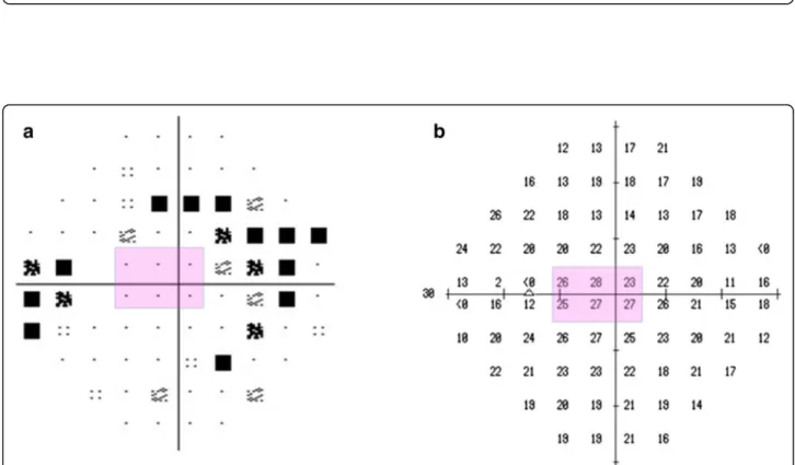
Fig. 2Visual field of the left eye. Pattern deviation plot (a) and threshold plot (b) of the central 30° visual field (Humphrey Field Analyzer, Swedish Interactive Threshold Algorithm) show superior and inferior Bjerrum scotomas corresponding to the superior and inferior neuroretinal rim loss, respectively. The retinal sensitivity is preserved in the papillo-macular bundle area (pink area) which spatially corresponds with the full thickness retinal defect
Kitaet al. BMC Ophthalmology (2016) 16:186 Page 2 of 5
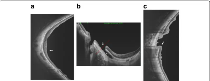
Fig. 3Optical coherence tomography (OCT) image of the posterior pole of the left eye.aVertical swept source (SS) -OCT B scan through the fovea. Localized retinal nerve fiber layer thinning is particularly evident in the inferior area (white arrow).bHorizontal SS-OCT scan shows an intrachoroidal cavitation area (white asterisk) temporal to the optic nerve, and a full thickness retinal defect (white arrow) along the temporal border of the myopic peripapillary atrophy. Thered dotted lineshows the scanned area of (c).cVertical OCT scan shows herniation of the retina toward the intrachoridal cavitation (white arrow)
Fig. 4MP-3 microperimetry results. The sensitivity of the papillomacular bundle was tested with a customised 52 points program using“white”test lights (stimulus size Goldmann III, duration 200 ms) presented on a dim“white”background using a 4-2 threshold strategy. MP-3 microperimetry showed an absolute scotoma spatially corresponding to the optic disc and the peripapillary beta zone area(purple circles, 0 dB). In contrast, no scotoma was found in the papillo-macular bundle, which spatially corresponds to the full thickness retinal defect
with no medication. The optic nerve head showed a moderate superior and a severe inferior glaucomatous neuroretinal rim loss with a laminar dot sign (Fig. 1).
The central 30° visual field evaluated with the Humphrey Field Analyzer, 30-2 Swedish Interactive Threshold Algo- rithm standard program (Carl Zeiss Meditec, Dublin, CA) was reliable and showed a mean deviation value of -8.94 dB and typical glaucomatous deterioration (super- ior and inferior Bjerrum scotomas) spatially correspond- ing to the disc damage (Fig. 2). In addition to the above findings on clinical examination a yellowish–orange le- sion was seen in the horizontal meridian temporal to the optic disc (Fig. 1, asterisk). Swept source optical coher- ence tomography (SS-OCT; DRI OCT-1 Atlantis, Topcon, Tokyo, Japan) was performed. In the suspected location the horizontal OCT scans showed a wide ICC with a full thickness retinal defect at the outer border of the myopic peripapillary beta zone atrophy, and hernia- tion of the retina in the ICC area (Fig. 3). Retinal nerve fiber layer thinning in the superior and inferior hemi- sphere areas were also found with SS-OCT (Fig. 3).
Since no sensitivity loss was seen in the central visual field spatially corresponding to the position of the full thickness retinal defect [6] (Fig. 2), microperimetry with the MP-3 system (Nidek, Aichi, Japan) was performed.
Microperimetry revealed an absolute scotoma spatially corresponding with the peripapillary atrophy but showed no scotoma in the area of the papillo-macular bundle, where the full thickness retinal defect was found (Fig. 4).
Discussion and conclusions
ICC is common in pathological myopia and visual field defects are typical in eyes with ICC. However, associ- ation of ICC and a full thickness retinal defect in the area of the papillo-macular nerve fiber bundle has not yet been published. In one investigation 71 % of eyes with ICC showed a glaucomatous visual field defect [3].
Ohno-Matsui et al. [1] reviewed the medical records of 125 pathological myopia patients whose papillary and peripapillary regions were examined with SS-OCT. Six- teen of the 125 patients (12.8 %) were found to have a temporal ICC. In addition, an inner retinal defect was observed in 2 of the 16 patients. Though, our case is typical in terms of clinical characteristics (high myopia in the background; ICC location temporal to the disc;
and typical clinical appearance of the ICC) it is unusual due to the association of ICC and a full thickness retinal defect in the papillo-macular bundle area; and the lack of association of the full thickness retinal defect with any spatially corresponding functional loss in the central visual field and in microperimetry of the macula. Since preservation of retinal function was confirmed with two different functional tests (perimetry and microperime- try); the visual field was reliable; and the coexisting
glaucomatous visual field deterioration was correctly de- tected, we think that the preservation of visual function corresponding with the papillo-macular bundle is a valid finding. As far as we know this is the first report in which preserved retinal sensitivity, confirmed with two different methods is seen in ICC associated with a full thickness retinal defect. We suppose that the herniated retina maintained some of its function in the papillo- macular area despite the full thickness damage, which may suggest a relatively a recent development of the ICC. Since the time course of the development of func- tional deterioration due to ICC formation is unknown further prospective investigations are proposed to clarify the relationship between ICC formation and correspond- ing perimetric function.
Abbreviations
ICC:Intrachoroidal cavitation; SS-OCT: Swept source optical coherence tomography
Acknowledgement None.
Funding None.
Availability of data and materials
The dataset supporting the conclusions of this article is included within the article.
Authors’contributions
YK drafted this manuscript, collected the data, and reviewed the literature.
MI and GH drafted this manuscript and reviewed the literature. RK, MS and AH critically reviewed the manuscript finally. All authors read and approved the final manuscript.
Competing interests
Gábor Hollόis a consultant of Optovue, Inc. and Carl Zeiss Meditec, Inc.
The authors declare that they have no competing interests.
Consent for publication
The patient has given a written consent for the publication of this report.
Ethics approval and consent to participate
Procedures followed the tenets of Declaration of Helsinki, with the retrospective protocol approved by the Institutional Review Board for Human Research of the Kyorin University.
Author details
1Department of Ophthalmology, Kyorin University School of Medicine, 6-20-2 Shinkawa, Mitaka, Tokyo 181-8611, Japan.2Department of Ophthalmology, Semmelweis University, Budapest, Hungary.
Received: 6 May 2016 Accepted: 13 October 2016
References
1. Ohno-Matsui K, Shimada N, Akiba M, Moriyama M, Ishibashi T, Tokoro T.
Characteristics of intrachoroidal cavitation located temporal to optic disc in highly myopic eyes. Eye. 2013;27:630–8.
2. Wei YH, Yang CM, Chen MS, Shih YF, Ho TC. Peripappillary intrachoroidal cavitation in high myopia: reappraisal. Eye. 2009;23(1):141–4.
3. Shimada N, Ohno-Matsui K, Yoshida T, Yasuzumi K, Kojima A, Kobayashi K, Futagami S, Tokoto T, Mochizuki M. Characteristics of peripapillary detachment in pathologic myopia. Arch Ophthalmol. 2006;124(1):46–52.
Kitaet al. BMC Ophthalmology (2016) 16:186 Page 4 of 5
4. Lee KM, Lee EJ, Lee SH, Kim TW. Disc haemorrhage associated with an enlarged peripapillary intrachoroidal cavitation in a non-glaucomatous myopic eye: a case report. BMC Ophthalmol. 2015;15:145.
5. Spaide RF, Akiba M, Ohno-Matui K. Evaluation of peripapillary intrachoroidal cavitation with swept source and enhance depth imaging optical coherence tomography. Retina. 2012;32(6):1037–44.
6. Garway-Heath D, Poinoosawmy D, Fitzke FW, Hitchings RA. Mapping the visual field to the optic disc in normal tension glaucoma eyes.
Ophthalmology. 2000;107:1809–15.
• We accept pre-submission inquiries
• Our selector tool helps you to find the most relevant journal
• We provide round the clock customer support
• Convenient online submission
• Thorough peer review
• Inclusion in PubMed and all major indexing services
• Maximum visibility for your research Submit your manuscript at
www.biomedcentral.com/submit
Submit your next manuscript to BioMed Central and we will help you at every step: