Chapter 8
Electrospun Nanofibers for Entrapment of Biomolecules
Diána Balogh-Weiser, Csaba Németh, Ferenc Ender, Benjámin Gyarmati, András Szilágyi and
László Poppe
Additional information is available at the end of the chapter http://dx.doi.org/10.5772/intechopen.76068
Provisional chapter
© 2016 The Author(s). Licensee InTech. This chapter is distributed under the terms of the Creative Commons Attribution License (http://creativecommons.org/licenses/by/3.0), which permits unrestricted use, distribution, and reproduction in any medium, provided the original work is properly cited.
Electrospun Nanofibers for Entrapment of Biomolecules
Diána Balogh-Weiser, Csaba Németh, Ferenc Ender, Benjámin Gyarmati, András Szilágyi and
László Poppe
Additional information is available at the end of the chapter
Abstract
This chapter focuses on nanofiber fabrication by electrospinning techniques for the effec- tive immobilization of biomolecules (such as enzymes or active pharmaceutical ingredi- ents—APIs). In this chapter, the development of precursor materials (from commercial polymer systems to systematically designed biopolymers), entrapment protocols, and precursor-nanofiber characterization methods are represented. The entrapment ability of poly(vinyl alcohol) and systematically modified polyaspartamide nanofibers was investi- gated for immobilization of two different lipases (from Candida antarctica and Pseudomonas fluorescens) and for formulation of the antibacterial and antiviral agent, rifampicin. The encapsulated biomolecules in electrospun polymer fibers could be promising nanoma- terials for industrial biocatalysis to produce chiral compound or in the development of smart drug delivery systems.
Keywords: electrospinning, entrapment, enzyme immobilization, drug delivery
1. Introduction
Recently, a considerable effort has been focused on nanofiber fabrication, nanofibrous materi- als, and the application of such materials. The characteristic features of nanofibers are such as small diameter (within the 100 nm–1 μm range), large specific surface area, infinite length, and high aspect ratio [1]. These properties make nanofibers suitable for a wide range of appli- cations including, but not limited to, medical applications, cancer cell engineering, tissue engineering, drug delivery systems, enzyme immobilization, and electronics [2]. Ceramics,
© 2018 The Author(s). Licensee IntechOpen. This chapter is distributed under the terms of the Creative Commons Attribution License (http://creativecommons.org/licenses/by/3.0), which permits unrestricted use, distribution, and reproduction in any medium, provided the original work is properly cited.
metals, and polymers are used to fabricate nanofibers. In fact, the fabrication techniques can limit the utilization of the nanofibers. Therefore, constant attention is paid to the improvement of the existing fabrication techniques and developing novel fabrication methods. Among vari- ous methods, electrospinning is the most widely used process for nanofiber fabrication [1].
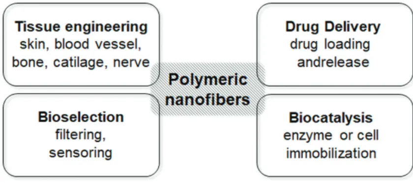
Recently, polymer nanofibers have gained more and more attention in development of “bio- engineered” or “bio-inspired” systems for pharmaceutical, biomedical, or biotechnological applications. Among these widespread issues, tissue engineering for artificial tissue recon- struction and replacement, smart drug formulations for targeted drug delivery, bioselection processes for selective filtering and sensoring materials, as well as biocatalyst production by enzyme or whole-cell immobilization are the most highlighted areas (Figure 1). This chapter focuses on biocatalyst design and drug delivery systems by utilizing the entrapping ability of polymer nanofibers.
Polymer nanofibers can be used to immobilize both small and macromolecules by their physi- cal adsorption or covalent binding on the surface of the fibers or by entrapment within the fiber. The choice between these two different possibilities depends on the application and on the type of small/macromolecules that would be immobilized. The main benefits and disad- vantages of the attachment and entrapment are compared in Table 1. Generally, entrapment of biomolecules is the more beneficial way to immobilize biocatalysts (such as enzymes or whole cells) or encapsulate drugs or vitamins, due to the significant stabilizing and protective effect and the controllable retention and release of the entrapped molecules.
In the process of electrospinning, a polymer solution held by its surface tension at the end of a capillary tube is subjected to an electric field [3, 4]. For entrapment of a biomolecule (small or a macro-sized) by electrospinning, formation of a homogenous precursor mixture from the biomolecule and polymer solution is required, which can be continuously fed by a syringe pump in an electrostatic field with high voltage. Two electrodes are used: one electrode is at the end of a capillary fed by the precursor mixture and the other is attached to a collec- tor. By applying high voltage on one electrode (usually in the range of 10 to 30 kV), while the collector electrode is grounded, strong electrostatic field develops, therefore charge is induced on the liquid surface. Mutual charge repulsion causes a force directly opposite to the force arising from the surface tension. Increasing intensity of electrostatic field elongates
Figure 1. Application fields of nanofibers as functional biomaterials.
the hemispherical surface of the solution at the tip of the capillary tube and forms a conical shape known as the Taylor cone. When the electric field reaches a critical value—at which the repulsive electric force overcomes the force from surface tension—a charged jet of the mixture is ejected from the tip of the Taylor cone [3]. Since this jet is charged, it will move toward the collector, and its trajectory can be controlled by an applied electric field. As the jet travels in air, the solvent evaporates, and the polymer fibers remain on a collector surface.
Thus, completing the electrospinning process, continuous fibers are formed that result in a fibrous material comprising entrapped biomolecules (Figure 2). Experimental results pre- sented in this chapter were carried out by the electrospinning equipment eSpin (Spinsplit Ltd., Budapest, Hungary).
2. Nanobiocatalysts: entrapment of enzymes in electrospun nanofibers
In the simplest sense, biocatalysis can be defined as the use of biological systems to catalyze (speed up) chemical reactions. These substances of natural origin can be one or more enzymes in isolated
Attachment onto polymer
nanofiber Entrapment within polymer fibers
Advantages • The biomolecule and the precur- sor solution can be handled separately
• No solvent or excipient materials limitation during electrospinning
• Easy to prepare
• In situ and rapid immobilization/formulation
• Significant stabilizing effects
• Controllable liberation/retention of the biomolecule
Disadvantages • (Difficult) chemical surface mod-
ification steps are necessary • Chemical compatibility between the precursor system and biomolecule can be a limiting factor
• Immobilization can be achieved step by step
Table 1. Comparison of the attachment and entrapment of biomolecules onto or within polymer nanofibers.
Figure 2. Entrapment of biomolecules by electrospinning technique.
form or within whole cells. An enzyme being a protein catalyst which accelerates the reaction of target molecules under mild conditions can be used in various industries. Major limiting fac- tors of the application of such biocatalysts in various industries are the sensitivity of enzymes to environmental conditions and the high cost of their manufacturing. This is why they ought to be protected from detrimental conditions until their use and—if possible—they should be recycled for subsequent usage in order to reduce the costs. The benefits of nanofibrous structures can be advantageously applied in these processes. Nanofibers can be used as a protective carrier for enzymes and the large surface area of nanofibers allows rapid release of the biomolecules when and where needed. Moreover, enzymes can also be immobilized on or within electrospun nano- fibers for repeated use. Due to the unique properties of nanofibers, such as extraordinarily high surface area and tunable surface morphology, high permeability, low density, ability to retain electrostatic charges, and cost effectiveness, they can be ideally applied for enzyme immobiliza- tion. In our previous study, it was found that electrospun poly(vinyl alcohol) (PVA) and poly (lactic acid) (PLA) nanofibers were applicable for entrapment of lipase from Burkholderia cepacia (lipase PS), Pseudomonas fluorescens (lipase AK) and lipase B from Candida antarctica (CaLB).
The lipase PS and CaLB biocatalysts entrapped in PVA nanofibers were durable biocatalysts retaining significant part of their original biocatalytic activity after ten cycles [5, 6].
2.1. Enzyme immobilization capacity of electrospun polymer nanofibers
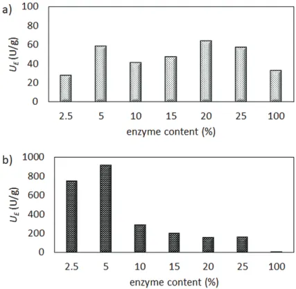
One of the key issues during enzyme entrapment is to reach the optimal enzyme loading with the highest specific activity of the valuable enzyme. To optimize enzyme loading in the course of entrapment, nanofiber entrapment of lipase AK into PVA and of CaLB into a cationic polyaspar- tamide were examined at different enzyme/polymer ratios. The immobilized lipase biocatalysts were tested in kinetic resolution of racemic 1-phenylethanol (rac-1, Figure 3) using vinyl acetate as acylating agents in n-hexane/t-butyl methyl ether (MTBE). The biocatalytic properties of the different electrospun biocatalysts, such as specific enzymatic activity (UE, U/g) and enantiomeric excess (ee, %), were determined from gas chromatographic analysis of the reaction media.
In case of lipase AK-PVA nanofibers, the specific enzyme activity (UE) reached the highest level when fibers were loaded with 5 and 20% enzyme (Figure 4a). In contrast, with CaLB in polyaspartamide, the specific enzyme activity showed a clear tendency with a maximum at moderate enzyme loading. Similar to lipase AK in PVA, 5% enzyme content exhibited the maximal specific activity (Figure 4b).
Figure 3. Kinetic resolution of racemic 1-phenylethanol (rac-1) catalyzed by lipase biocatalysts entrapped within electrospun nanofibers.
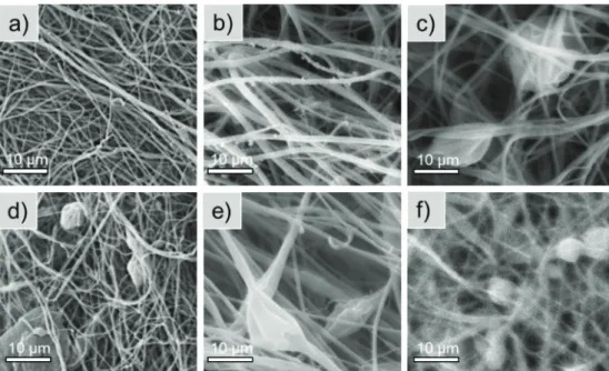
The morphological properties of the nanofibers with different enzyme content were investigated by scanning electron microscopy (SEM). In case of lipase AK in PVA nanofibers, SEM images showed that at 2.5 and 5% enzyme content (Figure 5a and b) the nanofibers were uniform with consistently homogenous surfaces. However, at enzyme content higher than 5%, more and more inhomogeneity could be observed, and nanofibers were not continuous (Figure 5c–f).
Similarly to lipase AK-PVA systems, in case of CaLB-polyaspartamide nanofibers, the mor- phology of fibers depended strongly on the enzyme content. CaLB at higher than 5% loading resulted in significant heterogeneity of the nanofibers (Figure 6c–f).
In summary, enzyme activity tests as well as SEM images of the biocatalyst demonstrated clearly that finding the optimal enzyme content of the nanofibers is a key issue. In case of lipase AK entrapped in PVA and also for CaLB in polyaspartamide nanofibers, the 5%
enzyme content was optimal to reach maximal specific activity of the entrapped enzyme and homogenous nanofiber structure. With both lipases and polymer matrices, enzyme contents higher than 5% resulted in formation of heterogeneous nanotissues with bead-like shapes and forming from aggregates of polymer or enzyme molecules. On the one hand, application of the optimal enzyme loading enables the most economical and cost-effective
Figure 4. Kinetic resolution of 1-phenylethanol (rac-1) with (a) lipase AK entrapped in PVA nanofibers and (b) CaLB entrapped in polyaspartamide nanofibers at different enzyme loading.
Figure 5. Scanning electron microscope images of PVA nanofibers loaded with different amounts of lipase AK: (a) 2.5, (b) 5, (c) 10, (d) 15, (e) 20, (f) 25%.
Figure 6. Scanning electron microscope images of polyaspartamide nanofibers loaded with different amounts of CaLB:
(a) 2.5, (b) 5, (c) 10, (d) 15, (e) 20, (f) 25%.
usage, which is quite important at an industrial scale. In addition, homogenous fiber mor- phology is crucial to produce high-quality nanobiocatalysts in a reproducible manner.
2.2. Bioimprinting for enhanced biocatalytic activity of enzyme entrapped in nanofibers
One of the most promising possibilities for enhancing enzyme activity during immobiliza- tion, especially for entrapment methods, is molecular imprinting which conserves by the aid of added substrates or substrate analogues the proper shape of the enzyme’s active site [7, 8].
This so-called bioimprinting, as can be rationalized by the generally accepted hypothesis of the interfacial activation mechanism, can influence the biocatalytic activity and enantioselectivity of lipases. The positive effect of bioimprinting on lipases was already demonstrated by immo- bilizations using sol-gel entrapment [9, 10]. The active site of many lipases in aqueous solution is covered by a flexible region of the enzyme, often referred to as a lid. Interaction of the lid with hydrophobic molecules can enforce its opening to make the active site accessible [11, 12].
This hypothesis is supported by crystal structures of lipases in their open and closed forms [13, 14]. Because interfacial activation can increase significantly the catalytic activity and selectiv- ity of lipases, it should be considered for all applications of lipases including development of novel immobilization methods. Although bioimprinting proved to be an efficient tool to modu- late the properties of lipases entrapped in sol-gel matrices, this strategy has been extended for entrapment in electrospun nanofibers only recently in one of our previous studies [7].
To demonstrate the efficiency and generality of bioimprinting during entrapment of lipases, lipase AK and CaLB were entrapped in PVA and polyaspartamide nanofibers in the presence of four different additives as bioimprinting agents. Poly(ethylene glycol)s (PEGs) of different molec- ular weights and nonionic detergents such as Tween 80 and Brij 30 were already applied as sub- strate analogues exhibiting bioimprinting effects in sol-gel systems [11] and in PVA nanofibers [6], but their effect in polyaspartamide entrapment has not been investigated yet. To reveal the effect of additives and the difference between fiber-forming polymer matrices, CaLB was chosen as model enzyme for the bioimprinting experiments. Results of testing the immobilized nanofi- brous CaLB biocatalysts by kinetic resolution of rac-1 showed that all tested additives increased the specific enzyme activity (UE) and the enantiomeric excess of the product (eeP) (Table 2). The
Additive CaLB-PVA CaLB-polyaspartamide
UE eeP UE eeP
(U/g) (%) (U/g) (%)
— 208 99.8 1470 99.8
PEG 400 294 99.9 2621 99.9
PEG 1000 204 99.5 2690 99.7
Tween 80 228 99.9 2262 99.9
Brij 30 180 99.8 2563 99.9
Table 2. Kinetic resolution of 1-phenylethanol (rac-1) with CaLB-PVA or CaLB-polyaspartamide nanofibers fabricated in the presence of different additives.
best results were obtained with PEG 400 and Tween 80 as additives in electrospinning of CaLB- polyaspartamide fibers, while, in case of electrospinning of CaLB-polyaspartamide fibers, PEG 400 and PEG 1000 were the most efficient bioimprinting agents. It must be noted, that CaLB in polyaspartamide polymer matrix without additive exhibited much higher activity than in PVA. Consequently, additives could manifest their beneficial effect on the activity of CaLB in polyaspartamide nanofibers better than in PVA nanofibers.
The polymer matrix of nanofibers is able to influence the enzyme activity by the bioimprint- ing effect. In addition, the physicochemical properties of the matrix material may affect also significantly the apparent enzyme activity and the final properties of immobilized biocata- lyst. Interactions between polymer chains can affect significantly the diffusion limitations for substrate or product, which can strongly influence the apparent efficiency of the immo- bilized biocatalyst. Thus, dynamic viscosity as a rheological property of the precursor sys- tems (enzyme-polymer-additive mixtures) and glass transition temperature (Tg) as a thermal property of the resulted enzyme-filled nanofibrous materials were also investigated (Figure 7).
According to Kramers’ theory, the biocatalytic activity of enzymes could strongly depend on the viscosity of solvent because viscosity results in friction of proteins in solution leading to decreased motion and inhibiting catalysis by motile enzymes [15]. Thus, viscosity of the pre- cursor system could affect directly the properties of the immobilized enzyme during its entrap- ment. Moreover, the viscosity of the polymer precursor system can significantly influence fiber formation during electrospinning as well. The dynamic viscosity values of PVA- and polyas- partamide-based precursor systems were determined in the presence of CaLB and the best additives such as PEG 400, PEG 1000, and Tween 80 and were compared to the simple polymer solution without the enzyme or additive. The glass transition temperature of the electrospun products was investigated as well. In case of CaLB-PVA systems, PEG 400 slightly increased the viscosity but Tween 80 resulted in higher increase in the viscosity value. On the other hand, when Tg values of the different CaLB-PVA fibrous biocatalysts were compared, PEG 400 had more pronounced effects on the interaction between the polymer chains, while the Tg of PEG 400-containing CaLB-PVA fibers was significantly lower than that of the CaLB-PVA fibers without additives (Figure 7a). In case of CaLB-polyaspartamide, additives (PEG 400 and PEG 1000) had no significant effect on the viscosity of precursor system, but Tg values became lower due to the presence of PEG molecules, especially in the case of PEG 1000 (Figure 7b).
Figure 7. Dynamic viscosity and glass transition temperature (Tg) of electrospun nanofibers (a) from PVA- and (b) from polyaspartamide-based precursor systems.
3. Entrapment of drugs in electrospun nanofibers
3.1. Encapsulation of rifampicin in polyaspartamide nanofibers for ophthalmic applications
The interest in the development of novel ophthalmic drug formulations has increased consid- erably because of the low bioavailability of drug molecules after their administration on the surface of the eye. The low therapeutic efficiency is caused by the complex structure of the eye, the small absorptive surface, and low transparency of the cornea, lipophilicity of corneal epi- thelium, metabolism, bonding of the drug to proteins in tear liquid, and protective mechanisms such as tear formation, blinking, and the flow of the active pharmaceutical ingredients (APIs) through the nasolacrimal duct [16, 17]. The main challenge in the development of ophthalmic drug formulations is to achieve the required drug concentration at the site of absorption and to improve residence time, which in turn contributes to smaller application frequency [18, 19].
Polymers are often used in ophthalmic formulations to increase viscosity and thus, residence time of the formulation on the corneal surface, which increases the bioavailability of the API. Drug penetration can also be enhanced by various additives such as chelating agents, surfactants, and cyclodextrins, which form inclusion complexes leading to increased solubil- ity, permeability, and bioavailability of the poorly soluble drugs. Controlled release of the drug can also be achieved by novel formulations such as inserts, collagen shields, contact lenses, and in situ gels. These solid or semi-solid formulations provide controlled release and increasing bioavailability of the API due to the extended contact time with cornea. Electrospun nanofibers offer controllable release rate as well as exact and safe dosage of the API.
In our work, a novel solid nanofibrous formulation was designed to encapsulate rifampi- cin (RFP), an important antibiotic and antiviral agent. Rifampicin inhibits bacterial DNA- dependent RNA synthesis; thus, it can be applied for the treatment of many infectious diseases such as tuberculosis and Hansen’s disease [20]. Although rifampicin has great efficiency, the scope of its applications is limited by its poor hydrolytic and thermal stability and by its rather limited solubility [21]. Due to these difficulties and the inaccurate dosing of eye drops, development of a stable solid formulation of rifampicin is still required.
Poly(aspartic acid) (PASP) is a biocompatible and biodegradable synthetic poly(amino acid) having great potential in biomedical applications. For using as a polymer in electrospin- ning, PASP derivatives, including cationic polyaspartamides, can be synthesized under mild reaction conditions [22, 23]. The biological activity of the PASP-based polymers has been investigated carefully and, owing to their protein-like structure, they are expected to be biodegradable. Due to these advantageous properties of polyaspartamide derivatives, rifampicin can be beneficially entrapped in polyaspartamide-based nanofibers fabricated by electrospinning. Poly(aspartic acid) derivatives with butyl and hexyl groups were tested to determine the effect of chemical structure on release properties. For electrospinning of PASP derivatives containing rifampicin (1.6 w/w%), ethanol was used as the solvent to avoid the degradation of the active compound. The morphology of electrospun fibers was investigated by SEM indicating that uniform nanofibers could be produced independently of chemical composition. By using butyl side groups, the fiber diameter was ~800 nm, while
in the presence of hexyl groups, the average diameter of the fibers was ~900 nm. The results of release experiments (Figure 8a) showed that the dissolution rate of rifampicin was larger by using butyl-PASP than hexyl-PASP, but in both cases, the total amount of the encapsu- lated drug was released.
The crystallinity of entrapped API determines its bioavailability; thus, X-ray diffractograms were recorded for the matrices and the native drug. From the comparison of the X-ray spectra of native and encapsulated rifampicin (butyl-PASP + RFP), it can be clearly seen that the origi- nally crystalline rifampicin became amorphous during electrospinning, which can improve the bioavailability even further (Figure 8b).
4. Conclusion
Electrospinning is a promising tool for efficient entrapment of biomolecules, such as expensive enzymes or active pharmaceutical ingredients, within polymer nanofibers which are appli- cable for biotechnology or biomedical purposes. By rational selection of polymer excipients, both small and macromolecules can be immobilized into electrospun matrices and control- lable retention and release can be achieved. Poly(vinyl alcohol) (PVA) and polyaspartamide nanofibers are promising candidates to encapsulate enzymes. In case of industrially relevant lipases, such as lipase from Candida antarctica (CaLB) or Pseudomonas fluorescens (lipase AK), the optimal enzyme loading of nanofibers generated by electrospinning was 5% entrapped in PVA or polyaspartamide matrices. Entrapment of lipases in nanofibers in the presence of different substrate-like additives led to bioimprinting, which could significantly increase the activity and enantioselectivity of the entrapped enzyme. Polyaspartamides could also be used to encapsulate active pharmaceutical ingredients by electrospinning. The encapsula- tion of rifampicin proved that the chemical modification of PASP with different alkyl side groups had a significant effect on the release rate of APIs, opening up new possibilities for the improvement of therapeutic efficiency with solid dosage forms.
Figure 8. (a) Release of rifampicin (RFP) from butyl-PASP and hexyl-PASP electrospun matrices and (b) X-ray diffraction spectra of free rifampicin, butyl-PASP electrospun matrix, and rifampicin entrapped in butyl-PASP.
Acknowledgements
This research was supported by the National Research, Development, and Innovation Office—
NKFIH (FK 125074). Benjámin Gyarmati acknowledges the János Bolyai Research Scholarship of the Hungarian Academy of Sciences for financial support. A. Szilágyi is grateful to the support of the ÚNKP-17-4-III New National Excellence Program of the Ministry of Human Capacities. We thank Anna Szabó for her cooperation in the electrospinning of polyaspar- tamides (Department of Physical Chemistry and Materials Science, Budapest University of Technology and Economics, BME) and Imre Miklós Szilágyi (Department of Inorganic and Analytical Chemistry, BME) for performing the XRD measurements.
Conflict of interest
The authors declare no conflict of interest.
Author details
Diána Balogh-Weiser1,2,3, Csaba Németh2, Ferenc Ender4,5, Benjámin Gyarmati2, András Szilágyi2 and László Poppe1,3,6*
*Address all correspondence to: poppe@mail.bme.hu
1 Department of Organic Chemistry and Technology, Budapest University of Technology and Economics, Budapest, Hungary
2 Department of Physical Chemistry and Materials Science, Budapest University of Technology and Economics, Budapest, Hungary
3 SynBiocat Ltd., Budapest, Hungary
4 Department of Electron Devices, Budapest University of Technology and Economics, Budapest, Hungary
5 Spinsplit Ltd., Budapest, Hungary
6 Faculty of Chemistry and Chemical Engineering, Biocatalysis and Biotransformation Research Centre, Babeș-Bolyai University of Cluj-Napoca, Cluj-Napoca, Romania
References
[1] Xue J, Xie J, Liu W, Xia Y. Electrospun nanofibers: New concepts, materials, and applications. Accounts in Chemical Research. 2017;50:1976-1987. DOI: 10.1021/acs.
accounts.7b00218
[2] Doshi J, Reneker DH. Electrospinning process and applications of electrospun fibers.
Journal of Electrostatics. 1995;35:151-160. DOI: 10.1016/0304-3886(95)00041-8
[3] Yarin AL. Taylor cone and jetting from liquid droplets in electrospinning of nanofibers.
Journal of Applied Physics. 2001;90:4836. DOI: 10.1063/1.1408260
[4] Ramakrishna S, Fujihara K, Teo WE, Lim TC, Ma Z. Electrospinning process. In: An Introduction to Electrospinning and Nanofibers. Singapore: World Scientific Publishing Co.; 2005. pp. 90-154. DOI: 10.1142/9789812567611_0003
[5] Sóti P, Weiser D, Vígh T, Nagy Z, Poppe L, Marosi G. Electrospun polylactic acid and polyvinyl alcohol fibers as efficient and stable nanomaterials for immobilization of lipases. Bioprocess and Biosystems Engineering. 2016;39:449-459. DOI: 10.1007/
s00449-015-1528-y
[6] Weiser D, Sóti P, Bánóczi G, Bódai V, Kiss B, Gellért Á, Nagy ZK, Koczka B, Szilágyi A, Marosi G, Poppe L. Bioimprinted lipases in PVA nanofibers as efficient immobilized biocatalysts. Tetrahedron. 2016;72:7335-7342. DOI: 10.1016/j.tet.2016.06.027
[7] Dickey FH. The preparation of specific adsorbent. Proceedings of the National Academy of Sciences of the United States of America. 1949;35:227-229. http://www.pnas.org/
content/35/5/227
[8] Mingarro I, Abad C, Braco L. Interfacial activation-based molecular bioimprinting of lipolytic enzymes. Proceedings of the National Academy of Sciences of the United States of America. 1995;92:3308-3312. DOI: 10.1073/pnas.92.8.3308
[9] Cao X, Yang J, Shu L, Yu B, Yan Y. Improving esterification activity of Burkholderia cepa- cia lipase encapsulated in silica by bioimprinting with substrate analogues. Process Biochemistry. 2009;44:177-182. DOI: 10.1016/j.procbio. 2008.10.003
[10] Hellner G, Boros Z, Tomin A, Poppe L. Novel sol-gel lipases by designed bioimprint- ing for continuous-flow kinetic resolutions. Advanced Synthesis & Catalysis. 2011;353:
2481-2491. DOI: 10.1002/adsc.201100329
[11] Verger R. Interfacial activation’ of lipases: Facts and artifacts. Trends in Biotechnology.
1997;15:32-38. DOI: 10.1016/S0167-7799 (96)10064-0
[12] Reis P, Holmberg K, Watzke H, Leser ME, Miller R. Lipases at interfaces: A review.
Advances in Colloid Interface. Sciences. 2009;147-148:237-250. DOI: 10.1016/j.cis.2008.06.001 [13] Brady L, Brzozowski AM, Derewenda ZS, Dodson E, Dodson G, Tolley S, Turkenburg JP,
Christiansen L, Hugejensen B, Norskov L, Thim L, Menge U. Nature. 1990;343:767-770.
DOI: 10.1038/343767a0
[14] Schrag JD, Li Y, Wu S, Cygler M. Ser-His-Glu triad forms the catalytic site of the lipase from Geotrichum candidum. Nature. 1991;351:761-764. DOI: 10.1038/351761a0
[15] Uribe S, Sampedro JG. Measuring solution viscosity and its effect on enzyme activity.
Biological Procedures. 2003;5:108-115. DOI: 10.1251/bpo52
[16] O’Connor Davies H, Hopkins GA, Pearson RM. The Actions and Uses of Ophthalmic Drugs. 3rd ed. London: Butterworths; 1989. ISBN: 9781483192048
[17] Pahuja P, Arora S, Pawar P. Ocular drug delivery system: A reference to natural polymers.
Expert Opinion on Drug Delivery. 2012;9:837-861. DOI: 10.1517/17425247.2012.690733 [18] Karthikeyan D, Bhowmick M, Pandeyetal VP. The concept of ocular inserts as drug
delivery systems: An overview. Asian Journal of Pharmaceutics. 2008;2:192-200. DOI:
10.22377/ajp.v2i4.204
[19] Kumari A, Sharma PK, Garg VK, G. Ocular inserts—Advancement in therapy of eye diseases. Journal of Advanced Pharmaceutical Technology and Research. 2010;1:291-296.
DOI: 10.4103/0110-5558.72419
[20] Calvori C, Frontali L, Leoni L, Tecce G. Effect of rifamycin on protein synthesis. Nature.
1965;207:417-418. DOI: 10.1038/207417a0
[21] Shishoo CJ, Shah SA, Rathod IS, Savale SS, Kotecha JS, Shah PB. Stability of rifampicin in dissolution medium in presence of isoniazid. International Journal of Pharmaceutics.
1999;190:109-123. DOI: 10.1016/S0378-5173(99)00286-0
[22] Krisch E, Gyarmati B, Szilágyi A. Preparation of pH-responsive poly(aspartic acid) nano- gels in inverse emulsion. Periodica Polytechnica-Chemical Engineering. 2017;61:19-26.
DOI: 10.3311/PPch.9788
[23] Németh C, Szabó D, Gyarmati B, Gerasimov AV, Varfolomeev M, László K, Szilágyi A.
Effect of side groups on the properties of cationic polyaspartamides. European Polymer Journal. 2017;93:805-814. DOI: 10.1016/j.eurpolymj.2017.02.024