1521-0111/95/6/652–660$35.00 https://doi.org/10.1124/mol.118.115626
MOLECULARPHARMACOLOGY Mol Pharmacol 95:652–660, June 2019
Copyrightª2019 by The American Society for Pharmacology and Experimental Therapeutics
Chemically Modified Derivatives of the Activator Compound Cloxyquin Exert Inhibitory Effect on TRESK (K 2P 18.1)
Background Potassium Channel s
Miklós Lengyel, Ferenc Erdélyi, Enik} o Pergel, Ágnes Bálint-Polonka,
1Alice Dobolyi, Péter Bozsaki, Mária Dux, Kornél Király, Tamás Heged} us, Gábor Czirják, Péter Mátyus,
2and Péter Enyedi
Department of Physiology, Faculty of Medicine (M.L., E.P., A.D., P.B., G.C., P.E.), Department of Organic Chemistry, Faculty of Pharmacy (A.B.-P., P.M.), Department of Pharmacology and Pharmacotherapy (K.K.), and Department of Biophysics and Radiation Biology, Faculty of Medicine (T.H.), Semmelweis University, Budapest, Hungary; Gene Technology Division, Institute of Experimental Medicine–Hungarian Academy of Sciences, Budapest, Hungary (F.E.); and Department of Physiology, Faculty of Medicine, University of Szeged, Szeged, Hungary (M.D.)
Received December 27, 2018; accepted April 9, 2019
ABSTRACT
Cloxyquin has been reported as a specific activator of TRESK [TWIK-related spinal cord K1channel (also known as K2P18.1)]
background potassium channel. In this study, we have synthe- tized chemically modified analogs of cloxyquin and tested their effects on TRESK and other K2Pchannels. The currents of murine K2P channels, expressed heterologously in Xenopus oocytes, were measured by two-electrode voltage clamp, whereas the native background K1conductance of mouse dorsal root gan- glion (DRG) neurons was examined by the whole-cell patch-clamp method. Some of the analogs retained the activator character of the parent compound, but, more interestingly, other derivatives inhibited mouse TRESK current. The inhibitor analogs (A2764 and A2793) exerted state-dependent effects. The degree of inhibition by 100 mM A2764 (77.8%6 3.5%, n 5 6) was larger in the activated state of TRESK (i.e., after calcineurin-dependent stim- ulation) than in the resting state of the channel (42.8%611.5%
inhibition, n 5 7). The selectivity of the inhibitor compounds was tested on several K2P channels. A2793 inhibited TWIK- related acid-sensitive K1channel (TASK)-1 (100mM, 53.4% 6
13, 5%,n 55), while A2764 was more selective for TRESK, it only moderately influenced TREK-1 and TWIK-related alkaline pH-activated K1channel. The effect of A2764 was also examined on the background K1currents of DRG neurons. A subpopulation of DRG neurons, prepared from wild-type animals, expressed background K1currents sensitive to A2764, whereas the inhibitor did not affect the currents in the DRG neurons of TRESK-deficient mice. Accordingly, A2764 may prove to be useful for the identifi- cation of TRESK current in native cells, and for the investigation of the role of the channel in nociception and migraine.
SIGNIFICANCE STATEMENT
TRESK background potassium channel is a potential pharma- cological target in migraine and neuropathic pain. In this study, we have identified a selective inhibitor of TRESK, A2764. This compound can inhibit TRESK in native cells, leading to cell depolarization and increased excitability. This new inhibitor may be of use to probe the role of TRESK channel in migraine and nociception.
Introduction
Background (leak) potassium currents are responsible for the generation of the resting membrane potential and also play an important role in the adjustment of cellular excitability. The molecular correlates of the background potassium currents are the two-pore domain potassium channels (K2P). To date, 15 K2Psubunits have been identi- fied in mammalian cells. These channels are regulated by a large variety of physicochemical factors, intracellular signaling pathways, and drugs (for review, see Enyedi and Czirják (2010).
This work was supported by the Hungarian Research Fund [Grant NKFIH K-127988] and the Higher Education Institutional Excellence Program of the Ministry of Human Capacities in Hungary, within the framework of the Molecular Biology thematic program of the Semmelweis University. M.L. was supported by the New National Excellence Program of the Ministry of Human Capacities [ÚNKP-18-3-III-SE-43] and [EFOP-3.6.3-VEKOP-16-2017-00009, program name:
“Az orvos-, egészségtudományi- és gyógyszerészképzés tudományos muhelyeinek} fejlesztése”]. No potential conflicts of interest relevant to this article are reported.
1Current affiliation: Hungarian Accreditation Committee, Budapest, Hungary.
2Current affiliation: Faculty of Health and Public Services, Semmelweis University, Budapest, Hungary.
https://doi.org/10.1124/mol.118.115626.
s This article has supplemental material available at molpharm.aspetjournals.org.
ABBREVIATIONS: cRNA, complementary RNA; DRG, dorsal root ganglion; KO, knockout; K2P, two-pore domain potassium channel; mTRESK, mouse tandem of pore domains in a weak inward rectifying K1channel–related spinal cord K1channel; RR, ruthenium red; TALEN, transcription activator–like effector nuclease; TALK, TWIK-related alkaline pH-activated K1 channel; TASK, TWIK-related acid-sensitive K1 channel; THIK, tandem pore domain halothane-inhibited K1 channel; TREK, TWIK-related K1 channel; TRAAK, TWIK-related arachidonic acid-stimulated K+ channel; TRESK, TWIK-related spinal cord K1channel; TWIK, tandem of pore domains in a weak inward rectifying K1channel.
652
http://molpharm.aspetjournals.org/content/suppl/2019/04/11/mol.118.115626.DC1
at ASPET Journals on December 10, 2019molpharm.aspetjournals.orgDownloaded from
TRESK [TWIK-related spinal cord K1channel (also known as K2P18.1)] was first cloned from human spinal cord (Sano et al., 2003). TRESK is activated by the cytoplasmic Ca21 signal, via the calcium/calmodulin-dependent phosphatase calcineurin (Czirják et al., 2004). TRESK is abundantly expressed in the primary sensory neurons of the dorsal root (DRG) (Yoo et al., 2009) and trigeminal ganglion (Bautista et al., 2008; Yamamoto et al., 2009). Single-channel studies indicated that TRESK provides a major component of the background potassium current in rat DRG neurons (Kang and Kim, 2006). It is a plausible hypothesis that TRESK modulates the activity of sensory neurons and the absence of TRESK current results in an increased excitability, leading to disorders in nociception. A decrease of TRESK expression has been reported in a model of neuropathic pain (Tulleuda et al., 2011). In further support of this hypothesis, a mutation of TRESK leading to a nonfunctional channel fragment with dominant-negative effect has been recognized in a cohort of patients suffering from migraine (Lafrenière et al., 2010).
To understand the role of TRESK in physiological and pathophysiological nociception, appropriate pharmacological tools are necessary for selective modulation of the channel.
However, pharmacological targeting of TRESK and other K2P channels is a difficult task (for a recent review on the pharmacology of TRESK, see Enyedi and Czirják, 2015).
The antiamoebic drug cloxyquin was identified as an activator of TRESK (Wright et al., 2013). We have recently reported that cloxyquin is a selective activator of TRESK in the K2P family. The stimulatory effect of cloxyquin was independent of the Ca21/calcineurin pathway, suggesting that cloxyquin is a direct activator of the channel. We have also shown that cloxyquin activates TRESK in isolated mouse DRG neurons (Lengyel et al., 2017). In the present study, we have produced 28 chemically modified analogs of cloxy- quin and examined their effects on TRESK channels.
Some of these derivatives activated TRESK with similar efficiency and potency as the parent compound. On the other hand, we obtained several compounds suitable for the selective inhibition of TRESK current in an activation state–dependent manner.
Materials and Methods
Plasmids, Complementary RNA Synthesis. The cloning of mouse TWIK-related acid-sensitive K1channel (TASK)-1/2/3, TWIK- related K1 channel (TREK)-2, TWIK-related alkaline pH-activated K1channel (TALK)-1, tandem pore domain halothane-inhibited K1 channel (THIK)-1, and TRESK has been described previously (Czirják et al., 2004; Czirják and Enyedi, 2006). The plasmids coding mouse TREK-1 and TRAAK channels were provided by Professor M.
Lazdunski and Dr. F. Lesage (Institut de Pharmacologie Moleculaire et Cellulaire -CNRS -UPR 411, 660 route des Lucioles, Sophia Antipolis, 06560 Valbonne, France). The plasmids were linearized and used for in vitro complementary RNA (cRNA) synthesis using the mMESSAGE mMACHINE T7 in vitro transcription kit (Ambion, Austin, TX). The structural integrity of the RNA was checked on denaturing agarose gels.
Animal Husbandry, Preparation, and Microinjection of Xenopus Oocytes. Generation of a TRESK Knockout Mouse Line: Isolation of DRG Neurons. Xenopus laevis oocytes were prepared as previously described (Czirják and Enyedi, 2002). For the expression of the different channels, the oocytes were injected with 57 pg to 4 ng of cRNA (depending on the channel type) 1 day after defolliculation. Injection was performed with a Nanoliter Injector
(World Precision Instruments, Sarasota, FL). X. laevisfrogs were housed in 50-l tanks with continuous filtering and water circulation.
Room temperature was 19°C. Frogs were anesthetized with 0.1%
tricaine solution and killed by decerebration and pithing.
FVB/Ant (FVB.129P2-Pde6b1Tyrc-ch/Ant) mice were obtained from the Institute of Experimental Medicine of the Hungarian Academy of Sciences (Budapest, Hungary). TRESK knockout (KO) animals were generated by the transcription activator–like effector nuclease (TALEN) technique using plasmids ordered from Addgene (TALE Toolbox Kit number 1000000019, deposited by Feng Zhang) (Broad Institute of MIT and Harvard). Mouse TRESK (mTRESK)- specific TALEN recognition sites were designed for the genomic sequence of the first exon (corresponding to the N-terminal intracel- lular domain of the channel) using the 59-TN19 N14220 N19A-39 formula, with the following sequences: left TALEN recognition site, 59tgaggagccacctgaggcca; right TALEN recognition site, 59ccctgg- ggaaggccagggga; and an 18 base pair (59ggagatgctgtcctgagg) FokI nuclease dimerization and cutting sequence in between. mTRESK recognizing TALEN plasmids were assembled according to the pro- tocol of Sanjana et al. (2012). TALEN mRNAs were produced using the Ambion mMESSAGE mMACHINE T7 in vitro transcription kit (Ambion). TALEN mRNAs were microinjected at a concentration of 20–20 ng/ml into the pronuclei of fertilized eggs of FVB/Ant mice. Pups were analyzed with Surveyor assay plus sequencing. In 12 mice, among the 61 born pups the TRESK (KCNK18) gene was changed, and a founder bearing a 33 base pair deletion and also a mutation introducing a stop codon was chosen to establish a colony.
Adult female wild-type and TRESK KO mice (2–3 months of age) were used for the patch-clamp experiments in this study. The animals were maintained on a 12-hour light/dark cycle with free access to food and water in a specific pathogen-free animal facility. Mice were killed humanely by CO2 exposure (CO2 was applied until death of the animals). DRGs were dissected from the thoracic and lumbar levels of the spinal cord and collected in sterile PBS (137 mM NaCl, 2.7 mM KCl, and 10 mM NaH2PO4, pH adjusted to 7.4 with NaOH) at 4°C.
Ganglia were incubated in PBS containing 2 mg/ml collagenase enzyme (type I; Worthington Biochemical Corporation, Lakewood, NJ) for 30 minutes with gentle shaking at 37°C. For further details regarding the isolation and culturing of the cells, see Braun et al.
(2015). All experimental procedures using animals were conducted in accordance with theGuide for the Care and Use of Laboratory Animals as adopted by the National Institutes of Health, local state laws, and institutional regulations. All animal experiments were approved by the Animal Care and Ethics Committee of Semmelweis Univer- sity (approval ID: XIV-I-001/2154-4/2012).
Two-Electrode Voltage-Clamp and Patch-Clamp Measure- ments.Two-electrode voltage-clamp experiments were performed 1–3 days after the microinjection of cRNA intoXenopusoocytes, as previously described (Czirják et al., 2004). For each channel type, the oocytes contributing to thennumber (the exactnnumber is indicated in the text or on the figures) were derived from at least two, but usually three, separate frogs. The holding potential was 0 mV. Background potassium currents were measured at the end of 300-millisecond-long voltage steps to2100 mV applied every 4 seconds. The low-potassium recording solution contained the following (in mM): NaCl 95.4, KCl 2, CaCl21.8, and HEPES 5, at pH 7.5, adjusted by NaOH. The high- potassium solution contained 80 mM K1 (78 mM NaCl in the low- potassium solution was replaced with KCl). For the measurement of TREK-1, TREK-2, and TRAAK currents, the high-potassium solu- tion contained 40 mM K1. Solutions were applied to the oocytes using a gravity-driven perfusion system. Experiments were performed at room temperature (21°C). Data were analyzed by pCLAMP 10 software (Molecular Devices, Sunnyvale, CA).
Whole-cell patch-clamp experiments in the voltage-clamp configu- ration were performed as described previously (Lengyel et al., 2016).
The resting membrane potential was recorded in the current-clamp mode with no current injection (I 5 0 mode). The rheobase was determined by injecting depolarizing current (in 100 pA increments,
at ASPET Journals on December 10, 2019molpharm.aspetjournals.orgDownloaded from
up to 1500 pA) for 1 second every 4 seconds. Isolated DRG neurons were used for experiments 1 to 2 days after isolation. For the current- clamp study of DRG neurons, only cells with a membrane potential between245 and270 mV were accepted, as described in previous studies (Petruska et al., 2000). Because the focus of this study was to examine the effects of our new cloxyquin analogs on the electrophys- iological parameters of isolated DRG neurons, no other selection criteria were used. The cutoff frequency of the eight-pole Bessel filter was adjusted to 200 Hz, and data were acquired at 1 kHz. The pipette solution contained (in mM): 140 KCl, 3 MgCl2, 0.05 EGTA, 1 Na2-ATP, 0.1 Na2-GTP, and 10 HEPES. The low-potassium solution contained (in mM): 140 NaCl, 3.6 KCl, 0.5 MgCl2, 2 CaCl2, 11 glucose, and 10 HEPES. The high-potassium solution contained 30 mM KCl (26.4 mM NaCl of the low-potassium solution was replaced with KCl). The pH of the bath solutions was adjusted to 7.4 with NaOH.
Experiments were performed at room temperature (21°C). Data were analyzed using pCLAMP 10.7 software (Molecular Devices).
Data and Statistical Analysis. Results are expressed as the mean6S.D. Normalized concentration-response curves were fitted with the following modified Hill equation:acn/[cn1K1/2n]11, wherecis the concentration,K1/2is the concentration at which half- maximal stimulation occurs,nis the Hill coefficient, andais the effect of the treatment. The normality of the data was estimated using the Shapiro-Wilk test. If the Shapiro-Wilk test showed a significant difference between the examined groups, statistical significance was determined using the Mann-WhitneyUtest. Otherwise, statistical significance was determined with the Student’s t test or Fisher’s ANOVA followed by Tukey’s post hoc test for multiple groups. Results were considered to be statistically significant at P , 0.05. The difference in sample sizes among different groups is a consequence of the differing number of cells suitable for experimentation; sample sizes were not modified after obtaining initial results. The determined P values are descriptive and not the result of testing prespecified hypotheses. Curve fitting was performed using SigmaPlot10 (Build 10.0.0.54; SyStat Software, San Jose, CA). Statistical calculations were performed using Statistica (version 13.2; Dell, Round Rock, TX).
Materials.Chemicals of analytical grade were purchased from Sigma-Aldrich (St. Louis, MO), Fluka (Milwaukee, WI), and Merck (Whitehouse Station, NJ). Enzymes and kits for molecular biology were purchased from Ambion, Thermo Fisher Scientific (Waltham, MA), New England Biolabs (Beverly, MA), or Stratagene (La Jolla, CA). Cloxyquin and ruthenium red (RR) were purchased from Sigma-Aldrich. Ionomycin (calcium salt) was purchased from Enzo Life Sciences, Inc. (Farmington, NY), dissolved in DMSO as a 5 mM stock solution, and stored at220°C.
Synthesis and chemical analysis of the new compounds described in this study are detailed in the Supplemental Material.
Results
A2764 Is a State-Dependent Inhibitor of Mouse TRESK Channel. We have synthesized 28 compounds struc- turally similar to cloxyquin and tested their effects on
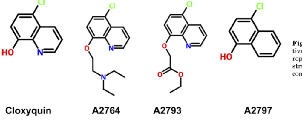
mTRESK channels expressed inX. laevis oocytes using the two-electrode voltage-clamp technique. The background K1 current was estimated at2100 mV in an extracellular solu- tion containing high K1(80 mM) after the subtraction of the small nonspecific leak current measured in 2 mM K1. Seven derivatives had a stimulatory effect on mTRESK activity in a manner similar to that of cloxyquin, 10 compounds did not influence the K1 current, while 11 analogs inhibited the channel. Based on our pilot screening of the compounds, the most promising inhibitors (A2764 and A2793) and an activator (A2797) of mTRESK were chosen for in-depth analysis. For the chemical structure of the novel cloxyquin analogs, see Fig. 1 (for the chemical structures of the new compounds and details of synthesis and chemical analysis for A2764 and A2793, see the chemical structures of compounds and the Supplemental Material).
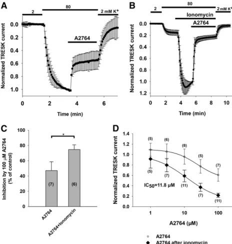
The background K1current was inhibited by 42.8%611.5%
when A2764 (100mM) was applied to the oocytes expressing mTRESK (n57 oocytes) (Fig. 2A). In addition to the inhibition of the basal current, we also examined the effect of A2764 on the current of activated mTRESK. Ionomycin was used to evoke the elevation of the cytoplasmic [Ca21], which has been previously shown to activate the channel by dephosphoryla- tion via the stimulation of the serine phosphatase calcineurin (Czirják et al., 2004). When mTRESK current was increased 7.161.8-fold by the pretreatment with ionomycin (0.5mM), the subsequent application of A2764 (100 mM) strongly inhibited the current (77.8%63.5% inhibition,n56 oocytes) (Fig. 2B). The effect of A2764 on mTRESK activated by dephosphorylation was more pronounced than the inhibition of the basal current (P,0.05, Mann-WhitneyUtest) (Fig. 2C).
The concentration dependence of the channel inhibition was also shifted; while the IC50value evoked by A2764 was 11.8 mM for the activated channel, the inhibitory potency of the drug was an order of magnitude lower for mTRESK in the resting state (Fig. 2D).
A2764 Is the Most Selective Modulator Among the Cloxyquin Analogs, Tested Among a Wide Range of K2P
Channels. To estimate the selectivity of the novel cloxyquin analogs within the K2Psubfamily, we tested them on several mouse K2Pchannels (at least one member from each subfamily being functional under physiologic ionic conditions): TASK-1 (K2P3.1), TASK-2 (K2P5.1), TASK-3 (K2P9.1), TALK-1 (K2P16.1), TREK-1 (K2P2.1), TREK-2 (K2P10.1), TRAAK (K2P4.1), and THIK-1 (K2P13.1). The channels were expressed inXenopus oocytes, and the effect of the compounds was measured by two- electrode voltage clamp (n5 5–7 oocytes per channel type).
Among the three examined cloxyquin analogs, A2764 proved to
Fig. 1.Chemical structures of novel cloxyquin deriva- tives. Chemical structures of the new cloxyquin analogs reported in this study (A2764, A2793, and A2797). The structure of the parent compound is shown for comparison.
at ASPET Journals on December 10, 2019molpharm.aspetjournals.orgDownloaded from
be the most selective agent; it inhibited the current of TRESK under resting conditions and in the activated state by 42.8%6 11.5% (n57) and 77.8%63.5% (n56), respectively (Fig. 3).
TREK-1 and TALK-1 currents were reduced only by about 20%, while the other channels were even less affected. The difference in the degree of inhibition between TRESK (both in the resting
and in the activated state) and all the examined K2Pchannels was statistically significant.
The efficiency of the other tested inhibitory analog (A2793) was higher than that of A2764. At 100 mM concentration, A2793 inhibited the unstimulated channel by 43.0%68.9%
(n 5 5), whereas after ionomycin activation the reduction of the TRESK current was 85.5% 6 2.9% (n 5 5) and was practically reduced to the nonactivated level (Fig. 4, A and B).
The concentration-response relationship was also determined for both the activated and the basal mTRESK currents (Fig.
4D). The activated channel was inhibited with an IC50of 6.8 mM, whereas the inhibitory potency of the compound was more than an order of magnitude smaller in the case of TRESK at basal activity. The specificity of A2793 showed a strikingly different pattern from A2764; it inhibited TASK-1 by 53.4%6 13.5% (n55), while all the other channels were influenced by less than 20% (Fig. 4C). These properties may have advantage under experimental conditions when the presence of TASK-1 can be excluded. This was not the case in our studies on native DRG cells, accordingly, we used A2764 in our subsequent experiments.
The chemical structure of the activator analog (A2797) differed in the aromatic ring from cloxyquin. Its potency was not favorable compared with cloxyquin, and it activated TRESK to a similar degree (437.6%6139.8% of control,n5 9 oocytes) as the parent compound. Unlike cloxyquin, it activated other K2Pchannels (TREK-2, TASK-2, and TRAAK) in addition to TRESK (details of these data are available in Supplemental Fig. 1).
Fig. 2. A2764 is a state-dependent inhibitor of mTRESK. (A) Normalized currents of oocytes expressing mTRESK, measured by the two-electrode voltage-clamp technique. Currents were measured at the end of 300- millisecond voltage steps to2100 mV applied every 4 seconds from a holding potential of 0 mV. The extracellular K+concentration was increased from 2 to 80 mM, as indicated by the bars above the graph. Oocytes were challenged with A2764 (100mM; see the horizontal bar). Currents were normalized to the value measured in 80 mM K+before the application of the drug. The current measured in 80 mM K+before the application of A2764 was 2.961.5mA. Data are plotted as the mean6S.D. (B) The effect of A2764 on the activated mTRESK channels was determined under the same experimental conditions as in (A). The current measured in 80 mM K+ before the application of ionomycin was 1.5 6 0.8 mA. The TRESK current was activated by the application of 0.5 mM ionomycin (by 7.161.8 fold), which was followed by the addition of A2764 (100mM) to the bath solution (the timing of the changes of K+concentration and drug application are marked by horizontal bars above the graph). Currents were normalized to the value measured after ionomycin stimulation, before the application of A2764. (C) The data from (A and B) have been summa- rized as a column graph. Mean inhibitions of the resting and activated TRESK currents are plotted, and the number of oocytes in each group is given in parenthe- ses. The difference in the degree of inhibition was statistically significant (*: P, 0.05, Mann-Whitney U test). (D) Concentration-response relationship of A2764 and mTRESK current. The inhibitory effects of different concentrations of A2764 (in a range from 1 to 100mM) on basal (white circles) and activated (black diamonds) TRESK current are plotted. Each data point represents the average of 5–11 oocytes (the exact number is indicated in parentheses). The data points were fitted with a modified Hill equation (seeMaterials and Methods).
Fig. 3. A2764 is a selective inhibitor of mTRESK. Mouse K2Pchannels were expressed inXenopusoocytes. The effect of A2764 (100mM) on the inward current in 80 mM extracellular K+(or 40 mM in the case of TREK-1, TREK-2, and TRAAK) was determined in 5–7 oocytes per channel type, as in Fig. 2A. As a comparison, the data points corresponding to the inhibition of basal or activated TRESK current from Fig. 2 are also plotted. The mean inhibition for each channel type is plotted as a column graph. The error bars represent the S.D. iono, ionomycin.
at ASPET Journals on December 10, 2019molpharm.aspetjournals.orgDownloaded from
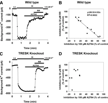
A2764 Inhibits the Background K1Currents in DRG Neurons. To examine the TRESK current in native cells by the application of A2764, we isolated DRG neurons and performed whole-cell patch-clamp recordings. Since the func- tional state of TRESK in cell culture may vary depending on inhibitory kinases and the calcium homeostasis of the neuron, we uniformly induced calcineurin-dependent TRESK dephos- phorylation by adding 0.5 mM ionomycin to our recording solutions before the application of A2764. In addition to the inhibitory effect of A2764 (100 mM), the sensitivity of the currents to RR (30mM) was also examined. It is well established that TREK-2, TRAAK, and TASK-3 K2Pchannels are inhibited by RR, and TREK-2 and TRAAK were previously reported to be expressed in large subpopulations of DRG neurons.
We applied the two inhibitors (A2764 and RR) into the bath solution successively. A recording from a representative, A2764-sensitive DRG neuron is shown in Fig. 5A. The sensitivity of the background current of the examined indi- vidual cells to A2764 and RR is presented on a scatter plot; 6 of the examined 10 cells showed substantial A2764 sensitivity (above 40%), whereas in the other four cells the degree of inhibition was below 20% (Fig. 5B). At the same time, we observed a significant negative correlation (r5 20.9499,P, 1024, Pearson’s correlation coefficient) between the ampli- tudes of A2764-sensitive and RR-sensitive current compo- nents (Fig. 5B). The current remaining after the elimination of the A2764-sensitive component was substantially reduced by RR (the residual current was 16%69.2%), suggesting that the A2764-sensitive (TRESK) and RR-inhibited (most proba- bly TREK-2 and TRAAK) currents were mainly responsible for the background K1 conductance in the majority of DRG neurons of the examined unselected population.
To validate the assumption that the A2764-sensitive cur- rent component of these native cells was indeed TRESK current, we also performed whole-cell patch-clamp experi- ments in the DRG neurons from TRESK KO mice under identical conditions. As shown on a representative recording in Fig. 5C, the application of A2764 (100mM) had no effect, but RR substantially inhibited the background K1current in this neuron. As apparent on the scatter plot in Fig. 5D, A2764 (100mM) did not influence the background K1current of the DRG neurons derived from the TRESK KO animals (the average effect on the K1 current was marginal, 6.5% 6 15.8% inhibition,n58). This was significantly different from the substantial inhibition of the current in the wild-type cells (43.0%629.7%,n510;P,0.05, Mann-WhitneyUtest). This result indicates that the A2764-sensitive component corre- sponds to TRESK current, and A2764 is suitable for the investigation of the channel in native cells.
A2764 Increases the Excitability of DRG Neurons.
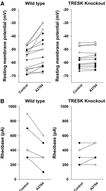
The effect of A2764 on the resting membrane potential and excitability of wild-type and TRESK KO DRG neurons was determined by using the current-clamp technique. In the case of the wild-type DRG neurons, the application of A2764 (100 mM, in low-K1 recording solution) depolarized the membrane potential by 6.665.1 mV, from257.368.5 to 250.7 6 12.5 mV (P , 0.05, paired t test; n 5 15) (for representative recording, see Fig. 6A, middle panel). In TRESK KO DRG neurons, the application of A2764 did not change the resting membrane potential (255.867.3 vs.255.0 67.6 mV, before and after A2764, respectively,n513) (for representative recording, see Fig. 6B, middle panel). The effect of A2764 on the membrane potential was significantly different between the wild-type and the KO animals (P,0.05, Fig. 4.A2793 is an inhibitor of mTRESK and mTASK-1.
(A) mTRESK was expressed inXenopus oocytes. The oocytes were challenged with A2793 (100 mM). The current measured in 80 mM K+ before application of A2793 was 2.6 6 0.6 mA. Inhibition by A2793 was determined and presented as described in Fig. 2A. (B) The effect of A2793 on the activated mTRESK current was determined under the same experimental conditions detailed in Fig. 2B. The current measured in 80 mM K+ before the application of ionomycin was 1.76 0.7mA.
Ionomycin activated the current by 6.762.0 fold. (C) To determine the selectivity of A2793, mouse K2Pchannels were expressed inXenopusoocytes. The effect of A2793 on the inward current in 80 mM extracellular K+(or 40 mM in the case of TREK-1, TREK-2, and TRAAK) was determined in five oocytes per channel type, as in Fig. 3.
As a comparison, the data points corresponding to the inhibition of TRESK current from (A and B) are also plotted.
Data are plotted as the mean + S.D. (D) Concentration- response relationship of A2793 and mTRESK current. The inhibitory effect of different concentrations of A2793 (in a range from 1 to 100 mM) on basal (white circles) and activated (black diamonds) TRESK current are plotted.
Each data point represents the average of five oocytes. The data points were fitted with a modified Hill equation (see Materials and Methods). iono, ionomycin.
at ASPET Journals on December 10, 2019molpharm.aspetjournals.orgDownloaded from
ttest) (Fig. 7A), indicating that the depolarization by A2764 was mediated by the inhibition of TRESK.
We also investigated the effect of A2764 application on the excitability of DRG neurons. The rheobase of DRG neurons before and after the application of A2764 (100 mM) was determined by the injection of depolarizing current impulses of gradually increasing amplitude. The minimum amplitude of current injection necessary for the generation of action potential was reduced after the application of A2764 in most
neurons (for representative recording, see Fig. 6A, left and right panels). The reduction of the rheobase was statistically signifi- cant (control rheobase: 5006223 pA,n56; A2764 rheobase:
3506160 pA,n56;P,0.05, pairedttest) (Fig. 7B, left panel).
In sharp contrast, the rheobase of TRESK-deficient DRG neu- rons was not influenced by A2764 (for representative recording, see Fig. 6B, left and right panels). The rheobase was 3206 130 pA (n55) before and 3606134 pA (n55) after, respectively, the application of the cloxyquin derivative (Fig. 7B, right panel).
Fig. 6. A2764 increases the excitability of DRG neurons by inhibiting TRESK. DRG neurons isolated from either wild-type or TRESK KO mice were used for current-clamp experiments. The rheobase was determined by injecting depolarizing current impulses in 100 pA increments before and after the application of A2764 (100mM). The effect of A2764 (100mM) was determined without any current injection (I= 0 mode). (A) Representative recordings are shown from a wild-type DRG neuron. (Left and right panels) The first traces where current injection resulted in an action potential before and after the application of A2764 are plotted. (Middle panel) The application of A2764 leads to depolarization of the resting membrane potential. Application of the drug is marked by the horizontal line above the recording. (B) Representative recordings are shown from a TRESK KO DRG neuron. (Left and right panels) The first traces where current injection resulted in an action potential before and after the application of A2764 are plotted. (Middle panel) The application of A2764 has no effect on the resting membrane potential. Application of the drug is marked by the horizontal line above the recording.
Fig. 5. A2764 inhibits the background K+current in DRG neurons. DRG neurons isolated from either wild-type or TRESK KO mice were used for whole-cell patch-clamp experiments. Inward currents were measured in 3.6 or 30 mM extracellular K+ at the end of 200-millisecond voltage steps to2100 mV from a holding potential of 280 mV. Changes in extracellular K+concentration and the application of A2764 (100mM) and RR (30mM) are marked above the graphs. To convert TRESK channels into their active state, the bath solution contained 0.5mM ionomycin. (A) A representative recording is shown from a wild-type, A2764-sensitive DRG neuron. (B) The effect of A2764 and RR on the background K+current was mea- sured in 10 DRG neurons. The degrees of inhibition by A2764 and RR were plotted against each other. Linear regression was performed, and the regression line was plotted in addition to the data points (the corresponding equation andRvalue are also shown). (C) A representative recording is shown from a TRESK KO DRG neuron. (D) The effect of A2764 and RR on the background K+current was measured in eight DRG neurons isolated from TRESK KO mice. The degrees of inhibition by A2764 and RR were plotted against each other.
at ASPET Journals on December 10, 2019molpharm.aspetjournals.orgDownloaded from
These data consistently indicate that the enhanced excit- ability induced by A2764 relied on the inhibition of the TRESK channel, which is a major determinant of action potential generation in DRG neurons.
Discussion
K2Pchannels are expressed in a wide variety of cell types, where they play a major role in the determination of the resting membrane potential and in the regulation of cellu- lar excitability (for review, see Enyedi and Czirják, 2010;
Feliciangeli et al., 2015). In comparison with the other K2P channel subunits, the expression pattern of TRESK is more restricted. The channel is abundantly and specifically expressed in the primary sensory neurons of the DRG and trigeminal ganglion. In accordance with this localization,
TRESK has been implicated in the modulation of both physiological and pathophysiological nociception (for recent reviews, see Enyedi and Czirják, 2015; Mathie and Veale, 2015). Important functional data highlighting TRESK as a regulator of cellular excitability in primary sensory neurons were obtained by eliminating TRESK (by transient knock- down or by using KO animal strains), transiently overexpress- ing the channel, or by using pharmacological modulators of channel activity (Dobler et al., 2007; Tulleuda et al., 2011;
Guo and Cao, 2014). An accumulating body of evidence also indicates that TRESK current alterations may contribute to the pathologic mechanism of neuropathic, migraine, and cancer pain (Lafrenière et al., 2010; Tulleuda et al., 2011;
Yang et al., 2018).
The examination of the role of TRESK in primary sensory neurons is impeded by the lack of specific modulators. Several pharmacological agents, including inorganic modulators as zinc or mercuric ion (Czirják and Enyedi, 2006); the natural com- pounds hydroxy-a-sanshool from Szechuan peppers (Bautista et al., 2008); and aristolochic acid fromAristolochiaceaeplants (Veale and Mathie, 2016), approved medicines such as lamotri- gine or verapamil (Kang et al., 2008; Park et al., 2018); and also the insecticide pyrethroids (Castellanos et al., 2018) were demonstrated to inhibit the TRESK current.
Although these agents are known to have other pharmaco- logical targets in addition to TRESK, and in most cases they were also shown to influence the activity of other K2Pchannels, they provided the first options to experimentally manipulate the TRESK current of sensory neurons. As another tool to achieve this goal, cloxyquin, an antiamoebic drug, was identi- fied as a TRESK activator in a high-throughput screening (Wright et al., 2013). We have recently shown that cloxyquin is selective for TRESK in the K2P family and can activate the background K1current in mouse DRG neurons (Lengyel et al., 2017). Nevertheless, a high concentration of cloxyquin (in the 50–100mM range) was required for TRESK activation. There- fore, we have synthesized 28 chemically modified analogs of cloxyquin to see whether we can find more potent activators of TRESK. After a pilot screening on an mTRESK channel expressed in Xenopus oocytes, it became apparent that the potency of these derivatives for TRESK activation was not increased by the chemical modification; however, their phar- macological profile has been changed substantially. Some of the analogs turned out to be inhibitors of TRESK, in sharp contrast to the activator parent compound cloxyquin. We have chosen the most potent analogs (one activator and two inhibitors) for further analysis.
The A2797 compound activated mTRESK current about 4-fold at a 100mM concentration. This degree of activation was similar to the previously reported effect of cloxyquin (Lengyel et al., 2017). However, as opposed to the relative selectivity of cloxyquin for TRESK in the K2Pfamily, A2797 also activated TREK-2, TASK-2, and TRAAK channels. This indicates that the modification of the aromatic ring of cloxyquin may sub- stantially influence the effect of the compound on various members of the background potassium channel family.
Modification of the hydroxyl sidechain in the chlorinated hydroxyquinoline parent molecule resulted in inhibitory deriv- atives. We have recently reported that cloxyquin showed state dependence in its effect; it was only able to activate TRESK in the resting state, but not when the channel was acti- vated by the Ca21/calcineurin pathway (Lengyel et al., 2017).
Fig. 7.Summary of the effect of A2764 on wild-type and TRESK KO DRG neurons. (A) The resting membrane potential of 15 wild-type (left panel) and 13 TRESK KO (right panel) DRG neurons was measured before (Control) and after (100mM) A2764 application. Data points corresponding to one neuron are connected with a straight line. (B) The rheobase of six wild-type (left panel) and five TRESK KO (right panel) DRG neurons was measured before (Control) and after (100mM) A2764 application. Data points corresponding to one neuron are connected with a straight line.
at ASPET Journals on December 10, 2019molpharm.aspetjournals.orgDownloaded from
Intrigued by the possibility that our inhibitors also show state- dependent effects, we determined the degree of inhibition by A2793 for both the resting mTRESK and the channels preactivated by application of the calcium ionophore ionomy- cin. Interestingly, the state dependence of the effect of A2793 proved to be the mirror image of that of cloxyquin. The inhibition by the compound was definitely more pronounced when TRESK current was stimulated with ionomycin before the application of A2793 than under the resting conditions of the channel.
The effect of A2793 was also tested on the other K2P
channels in addition to TRESK. It proved to be an efficient inhibitor of TASK-1, which was also reported to be expressed in some primary sensory neurons (Cooper et al., 2004). The effect on TASK-1 may limit the potential usage of A2793 to selectively inhibit TRESK in the native cells coexpressing these two specific K2P channels. However, A2793 may be considered as a tool to discriminate between the resting and activated channels in heterologous expression systems, and to block TRESK activated by calcineurin in the native cells, which do not express TASK-1.
The other cloxyquin derivative A2764 was also found to exert a state-dependent effect. Similar to A2793, inhibition by this compound was larger in the case of the activated TRESK than at the resting channel. The concentration-response relationship between A2764 and the mTRESK current was also shifted to the left by the calcineurin-dependent dephos- phorylation of the channel induced by the application of the Ca21ionophore ionomycin.
We have also determined the effect of A2764 on the other K2Pchannels expressed inXenopusoocytes. The substantial inhibition of TASK-1 (by A2793) was missing from the profile of A2764; thus, A2764 showed much better overall selectivity for TRESK among the K2Pchannels than the other inhibitor.
The degree of inhibition of the most affected other K2P
channels (TREK-1 and TALK-1) was moderate (21.0% 6 9.7% and 22.4% 6 10.3%, respectively), compared with the robust inhibition of dephosphorylated TRESK (77.8%63.5%), suggesting that A2764 could be used as a selective inhibitor of TRESK in isolated cells in vitro.
To test the hypothesis that A2764 can be applied to inhibit TRESK and thus determine the contribution of the channel to the ensemble background K1current in native cells, we have performed whole-cell patch-clamp experiments in DRG neu- rons from both wild-type and TRESK KO mice. Based on previous single channel and immunocytochemical results, in the DRG neurons primarily the expression of two major K2P
channels, TRESK and TREK-2 was expected (Acosta et al., 2014; Kang and Kim, 2006). To estimate the contribution of these channels to the background K1 current by a further independent method, we determined the degree of inhibi- tion by A2764 and RR. We have previously described that RR inhibits background K1 currents in DRG neurons (Braun et al., 2015). The molecular correlates of the RR- sensitive K1 current may be TREK-2, TASK-3, or TRAAK homodimers, or heterodimers of the TREK subfamily (Braun et al., 2015; Blin et al., 2016; Lengyel et al., 2016). We found that in neurons derived from wild-type animals, A2764 strongly inhibited the background K1current in a subset of DRG neurons. However, this subset of A2764-sensitive neu- rons was completely missing from the DRG neurons prepared from TRESK KO animals. Based on these results from the
wild-type and TRESK KO DRG neurons, we propose that A2764 is a useful selective inhibitor of TRESK.
To determine the contribution of TRESK and the RR- sensitive channels to the background K1current, the sensi- tivity of the current to the two inhibitors was determined in the same DRG neurons. We found an inverse correla- tion between the amplitude of the A2764-sensitive and RR- sensitive currents, indicating that the cells where TRESK is the major determinant of the background K1 current express relatively less RR-sensitive K2P channels and vice versa. In the DRG neurons analyzed, around 80% of the background K1current was sensitive to the combination of A2764 and RR. We have thus shown for the first time using whole-cell patch clamp that TRESK and various RR-sensitive K2Psubunits are the major determinants of the background K1current of isolated mouse DRG neurons. The application of A2764 offers a viable alternative to the previous methods for the investigation of TRESK and its regulation in the different functionally important subpopulations of primary sensory neurons.
In good accordance with the efficient inhibition of TRESK current by A2764, the application of this inhibitor also depolarized DRG neurons and appreciably increased their excitability in vitro, as indicated by the decreased rheobase in current-clamp experiments. Since these effects and also the inhibition of the background K1 current was completely absent in the neurons isolated from the KO animals, we concluded that the current component blocked by A2764 corresponds to TRESK. We propose that cloxyquin and A2764 can be used to modulate the activity of TRESK channels in native cells, and that the TRESK KO mouse line can serve as a reliable negative control in these experiments. We hope that with this combined genetic and pharmacological approach the functional relevance of the enigmatic TRESK channel can be further elucidated.
Acknowledgments
The skillful technical assistance of Irén Veres is acknowledged.
Authorship Contributions
Participated in research design:Lengyel, Bálint-Polonka, Hegedus,} Czirják, Enyedi.
Conducted experiments:Lengyel, Erdélyi, Pergel, Bálint-Polonka, Dobolyi, Bozsaki, Király.
Contributed new reagents or analytic tools:Erdélyi, Mátyus.
Performed data analysis: Lengyel, Pergel, Bozsaki, Király, Heged}us, Czirják, Mátyus.
Wrote or contributed to the writing of the manuscript: Lengyel, Erdélyi, Pergel, Dobolyi, Bozsaki, Dux, Király, Hegedus, Czirják,} Mátyus, Enyedi.
References
Acosta C, Djouhri L, Watkins R, Berry C, Bromage K, and Lawson SN (2014) TREK2 expressed selectively in IB4-binding C-fiber nociceptors hyperpolarizes their membrane potentials and limits spontaneous pain.J Neurosci34:1494–1509.
Bautista DM, Sigal YM, Milstein AD, Garrison JL, Zorn JA, Tsuruda PR, Nicoll RA, and Julius D (2008) Pungent agents from Szechuan peppers excite sensory neurons by inhibiting two-pore potassium channels.Nat Neurosci11:772–779.
Blin S, Ben Soussia I, Kim EJ, Brau F, Kang D, Lesage F, and Bichet D (2016) Mixing and matching TREK/TRAAK subunits generate heterodimeric K2P channels with unique properties.Proc Natl Acad Sci USA113:4200–4205.
Braun G, Lengyel M, Enyedi P, and Czirják G (2015) Differential sensitivity of TREK-1, TREK-2 and TRAAK background potassium channels to the polycationic dye ruthenium red.Br J Pharmacol172:1728–1738.
Castellanos A, Andres A, Bernal L, Callejo G, Comes N, Gual A, Giblin JP, Roza C, and Gasull X (2018) Pyrethroids inhibit K2P channels and activate sensory neu- rons: basis of insecticide-induced paraesthesias.Pain159:92–105.
at ASPET Journals on December 10, 2019molpharm.aspetjournals.orgDownloaded from
Cooper BY, Johnson RD, and Rau KK (2004) Characterization and function of TWIK- related acid sensing K1 channels in a rat nociceptive cell.Neuroscience129:
209–224.
Czirják G and Enyedi P (2002) Formation of functional heterodimers between the TASK-1 and TASK-3 two-pore domain potassium channel subunits.J Biol Chem 277:5426–5432.
Czirják G and Enyedi P (2006) Zinc and mercuric ions distinguish TRESK from the other two-pore-domain K1channels.Mol Pharmacol69:1024–1032.
Czirják G, Tóth ZE, and Enyedi P (2004) The two-pore domain K1channel, TRESK, is activated by the cytoplasmic calcium signal through calcineurin.J Biol Chem 279:18550–18558.
Dobler T, Springauf A, Tovornik S, Weber M, Schmitt A, Sedlmeier R, Wischmeyer E, and Döring F (2007) TRESK two-pore-domain K1channels constitute a significant component of background potassium currents in murine dorsal root ganglion neurones.J Physiol585:867–879.
Enyedi P and Czirják G (2010) Molecular background of leak K1currents: two-pore domain potassium channels.Physiol Rev90:559–605.
Enyedi P and Czirják G (2015) Properties, regulation, pharmacology, and functions of the K2p channel, TRESK.Pflugers Arch467:945–958.
Feliciangeli S, Chatelain FC, Bichet D, and Lesage F (2015) The family of K2P channels: salient structural and functional properties.J Physiol593:2587–2603.
Guo Z and Cao YQ (2014) Over-expression of TRESK K(1) channels reduces the excitability of trigeminal ganglion nociceptors.PLoS One9:e87029.
Kang D and Kim D (2006) TREK-2 (K2P10.1) and TRESK (K2P18.1) are major background K1channels in dorsal root ganglion neurons.Am J Physiol Cell Physiol291:C138–C146.
Kang D, Kim GT, Kim EJ, La JH, Lee JS, Lee ES, Park JY, Hong SG, and Han J (2008) Lamotrigine inhibits TRESK regulated by G-protein coupled receptor ago- nists.Biochem Biophys Res Commun367:609–615.
Lafrenière RG, Cader MZ, Poulin JF, Andres-Enguix I, Simoneau M, Gupta N, Boisvert K, Lafrenière F, McLaughlan S, Dubé MP, et al. (2010) A dominant- negative mutation in the TRESK potassium channel is linked to familial migraine with aura.Nat Med16:1157–1160.
Lengyel M, Czirják G, and Enyedi P (2016) Formation of functional heterodimers by TREK-1 and TREK-2 two-pore domain potassium channel subunits.J Biol Chem 291:13649–13661.
Lengyel M, Dobolyi A, Czirják G, and Enyedi P (2017) Selective and state-dependent activation of TRESK (K2P18.1) background potassium channel by cloxyquin.Br J Pharmacol174:2102–2113.
Mathie A and Veale EL (2015) Two-pore domain potassium channels: potential therapeutic targets for the treatment of pain.Pflugers Arch467:931–943.
Park H, Kim EJ, Ryu JH, Lee DK, Hong SG, Han J, Han J, and Kang D (2018) Verapamil inhibits TRESK (K2P18.1) current in trigeminal ganglion neurons in- dependently of the blockade of Ca21influx.Int J Mol Sci19:E1961.
Petruska JC, Napaporn J, Johnson RD, Gu JG, and Cooper BY (2000) Subclassified acutely dissociated cells of rat DRG: histochemistry and patterns of capsaicin-, proton-, and ATP-activated currents.J Neurophysiol84:2365–2379.
Sanjana NE, Cong L, Zhou Y, Cunniff MM, Feng G, and Zhang F (2012) A tran- scription activator-like effector toolbox for genome engineering. Nat Protoc7:
171–192.
Sano Y, Inamura K, Miyake A, Mochizuki S, Kitada C, Yokoi H, Nozawa K, Okada H, Matsushime H, and Furuichi K (2003) A novel two-pore domain K1channel, TRESK, is localized in the spinal cord.J Biol Chem278:27406–27412.
Tulleuda A, Cokic B, Callejo G, Saiani B, Serra J, and Gasull X (2011) TRESK channel contribution to nociceptive sensory neurons excitability: modulation by nerve injury.Mol Pain7:30.
Veale EL and Mathie A (2016) Aristolochic acid, a plant extract used in the treatment of pain and linked to Balkan endemic nephropathy, is a regulator of K2P channels.
Br J Pharmacol173:1639–1652.
Wright PD, Weir G, Cartland J, Tickle D, Kettleborough C, Cader MZ, and Jerman J (2013) Cloxyquin (5-chloroquinolin-8-ol) is an activator of the two-pore domain potassium channel TRESK.Biochem Biophys Res Commun441:463–468.
Yamamoto Y, Hatakeyama T, and Taniguchi K (2009) Immunohistochemical coloc- alization of TREK-1, TREK-2 and TRAAK with TRP channels in the trigeminal ganglion cells.Neurosci Lett454:129–133.
Yang Y, Li S, Jin ZR, Jing HB, Zhao HY, Liu BH, Liang YJ, Liu LY, Cai J, Wan Y, et al. (2018) Decreased abundance of TRESK two-pore domain potassium channels in sensory neurons underlies the pain associated with bone metastasis.Sci Signal 11:eaao5150.
Yoo S, Liu J, Sabbadini M, Au P, Xie GX, and Yost CS (2009) Regional expression of the anesthetic-activated potassium channel TRESK in the rat nervous system.
Neurosci Lett465:79–84.
Address correspondence to: Péter Enyedi, Department of Physiology, Semmelweis University, P.O. Box 2, H-1428 Budapest, Hungary. E-mail:
enyedi.peter@med.semmelweis-univ.hu
at ASPET Journals on December 10, 2019molpharm.aspetjournals.orgDownloaded from