REGULATED CELL DEATH: APOPTOSIS AND NECROPTOSIS IN U937 CELL LINE
Ph.D thesis
Zsuzsanna A. Dunai
Pathological Sciences Doctoral School Semmelweis University
Supervisor: Dr. Rudolf Mihalik Ph.D.
Official reviewers:
Dr. Gabor Koncz, Ph.D.
Dr. Tibor Vantus, Ph.D.
Head of the Final Examination Committee:
Dr. Gabor Banhegyi, M.D., D.Sc.
Members of the Final Examination Committee:
Dr. Andras Kiss, M.D., Ph.D.
Dr. Gabor Rez, Ph.D.
Budapest, 2012
Table of Contents
Table of Contents ... 2
The list of Abbreviations ... 5
1. Introduction ... 10
1. 1. Types of cell death subroutines and actual questions of nomenclature ... 10
1. 2. Apoptosis, secondary necrosis, necrosis and necroptosis ... 11
1. 2. 1. Apoptosis ... 13
1. 2. 1. 1. Molecular background of apoptosis ... 13
1. 2. 1. 2. Caspases are the central initiators and executioners of the apoptotic process ... 13
1. 2. 2. Secondary necrosis ... 16
1. 2. 3. Necrosis ... 17
1. 2. 4. Necroptosis ... 18
1. 3. Extrinsic cell death pathway and/or survival induction – through TNFR1 ... 19
1. 3. 1. NF-κB activation – induction of the survival pathway ... 19
1. 3. 2. Apoptosis induction – a route to cell death ... 21
1. 3. 3. Necroptosis induction ... 23
1. 4. Extrinsic cell death and survival induction – through Fas/CD95 and TRAIL receptors ... 26
1. 4. 1. Apoptosis and necroptosis induction ... 26
1. 5. Intrinsic apoptotic pathway ... 29
1. 6. Further type of programmed necrosis ... 32
1. 6. 1. PARP-AIF-mediated programmed necrotic pathway ... 32
1. 7. Physiological and pathological aspects of programmed necrotic cell death ... 33
1. 7. 1. Physiological aspects ... 33
1. 7. 2. Pathological aspects ... 36
2. Objectives ... 41
3. Methods ... 42
3. 1. Materials ... 42
3. 2. Cell culture ... 42
3. 3. Detection of the cell death-associated functional changes by flow cytometry ... 43
3. 3. 1. Assay of PI uptake of native cells representing the damage of plasma
membrane ... 43
3. 3. 2. Characterization of PS distribution in the plasma membrane by flow cytometric analysis of Annexin V-FITC and PI double-labeled cells ... 44
3. 3. 3. Changes of mitochondrial transmembrane potential were characterized by the DiOC6(3) uptake method ... 45
3. 3. 4. Functioning lysosomal compartments are characterized by the red fluorescence of acridine orange emitted in the acidic environment of lysosomes . 45 3. 3. 5. Determination of oligonucleosomal DNA fragmentation by the measurement of sub-G1 population of cells ... 46
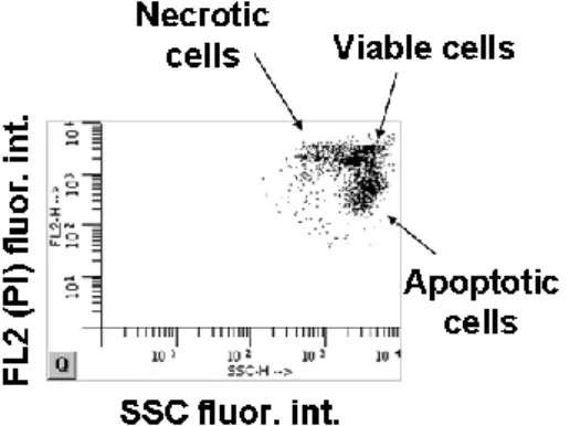
3. 3. 6. Representation of side scatter change and DNA fragmentation ... 46
3. 4. Agarose gel electrophoresis ... 47
3. 5. Light microscopic studies ... 47
3. 6. Fluorescent microscopic studies ... 48
3. 7. Western blot representation of PARP-1 and RIPK1 cleavage ... 48
3. 8. DEVD-ase activity assay ... 49
3. 9. Statistics ... 49
4. Results ... 50
4. 1. TRAIL induces necroptosis in U937 cell line in the presence of a caspase inhibitor... 50
4. 2. STS induces primary necrosis in the presence of a caspase inhibitor ... 52
4. 3. STS and TRAIL induce RIPK1 and MLKL-dependent necroptosis ... 59
4. 4. 3-MA inhibits STS-induced necroptosis ... 63
4. 5. CA inhibits both the TRAIL and STS-induced necroptosis in the presence of a caspase inhibitor ... 64
4. 6. PJ-34 does not arrest either the TRAIL or STS-induced necroptosis in the presence of a caspase inhibitor ... 70
5. Discussion ... 73
6. Conclusions ... 78
7. Summary ... 80
8. Összefoglaló ... 82
9. Bibliography ... 84
10. Publications ... 104
10. 1. Publications related to the thesis ... 104
10. 2. Publications not directly related to the thesis ... 104
11. Acknowledgements ... 105
The list of Abbreviations
A20 dual E3 ligase and hydrolase cleaves off K63-linked polyubiquitin chains
aa amino acid
AA arachidonic acid
ACAT acyl-CoA:cholesterol acyltransferase ADP adenosine diphosphate
AIF apoptosis-inducing factor ALL acute lymphoblastic leukemia AMC 7-amino-4-methylcoumarin AML acute myeloid leukemia
ANT adenine nucleotide translocase, ADP/ATP translocase
AO acridine orange
Apaf1 apoptotic protease activating factor-1 atg1 autophagy related 1 homolog gene ATP adenosine triphosphate
BAD BCL-2 antagonist of cell death BAK BCL-2 homologous antagonist/killer BAX BCL-2-like protein 4
BCL-2 B-cell lymphoma 2 BCL-XL BCL-2-like protein 1 BHA butylated hydroxyanisole
BID BH3 interacting domain death agonist C. elegans Caenorhabditis elegans
CA cathepsin-B inhibitor, CA-074-OMe CARD caspase recruitment domain
CCI controlled cortical impact
cFLIP cellular FLICE-like inhibitory protein, CASP8 and FADD-like apoptosis regulator
CHAPS 3-[(3-cholamidopropyl)dimethylammonio]-1-propanesulfonate CHOP C/EBP homologous protein
CICD caspase-independent cell death CYLD ubiquitin carboxyl-terminal hydrolase
CYPD cyclophilin D
D. melanogaster Drosophila melanogaster
DAMP danger-associated molecular pattern molecule DED death effector domain
DIF-1 differentiation-inducing factor DiOC6(3) 3,3′-Dihexyloxacarbocyanine iodide DISC death-inducing signaling complex DNA deoxyribonucleic acid
DR death receptor
DR4 TRAILR1, tumor necrosis factor-related apoptosis-inducing ligand receptor 1, tumor necrosis factor receptor superfamily member 10A DR5 TRAILR2, tumor necrosis factor-related apoptosis-inducing ligand
receptor 2, tumor necrosis factor receptor superfamily member 10B DRP1 mitochondrial fission factor
DTT DL-Dithiothreitol
EDTA ethylenediamine-tetraacetic acid
EtBr ethidium bromide
FACS fluorescence-activated cell sorting, flow cytometer FADD Fas-associated death domain protein
Fas receptor also known CD95 receptor, tumor necrosis factor receptor superfamily member 6
FasL also known CD95L, tumor necrosis factor ligand superfamily member 6
FSC forward scatter
GA geldanamycin
GLUD1 glutamate dehydrogenase 1
GLUL glutamate–ammonia ligase, glutamine synthetase GM-CSF granulocyte-macrophage colony stimulating factor
GSH glutathione
HEPES 4-(2-Hydroxyethyl)piperazine-1-ethanesulfonic acid sodium salt
HMGB high mobility group box protein
hr- human recombinant
HSP70 heat-shock protein 70 HSP90 heat-shock protein 90
IAP-1 inhibitor of apoptosis protein 1, baculoviral IAP repeat-containing protein 3
IAP-2 inhibitor of apoptosis protein 2, baculoviral IAP repeat-containing protein 2
IKK IκB kinase
IKKα and β alpha and beta kinases of IKK complex
IκB inhibitor of κB
JNK c-Jun N-terminal kinase 1, mitogen-activated protein kinase 8 LDH lactate dehydrogenase
LOX lypoxigenase
MA 3-methyladenine
MDR-ABC multi-drug resistant ATP binding cassette MEF mouse embryonic fibroblast
MLKL mixed lineage kinase domain-like protein
MNNG N-methyl-N’-nitro-N-nitrosoguanidine, DNA alkylating agent MnSOD superoxide dismutase [Mn]
NAD nicotinamide adenine dinucleotide NCCD Nomenclature Committee on Cell Death
Nec necrostatin-1
NEMO NF-kappa-B essential modulator NF-κB nuclear factor kappa-B
NK cell natural killer cell NMDA N-Methyl-D-aspartate
NO nitric oxide
NOX1 plasma membrane NADPH oxidase-1 NSA necrosulfonamide, inhibitor of MLKL
Omi/HtrA2 high temperature requirement protein A2, serine protease, IAP antagonist
PAR poly(ADP-ribose)
PARP-1 poly(ADP-ribose) polymerase-1 PBS phosphate buffered saline
PCD programmed cell death
PGAM5 serine/threonine-protein phosphatase
PI propidium iodide
PJ-34 N-(6-Oxo-5,6-dihydrophenanthridin-2-yl)-(N,N-
dimethylamino)acetamide hydrochloride, PARP inhibitor PRR pathogen recognition receptor
PS phosphatidylserine
PTPC mitochondrial permeability transition pore complex PYGL glycogen phosphorylase
RHIM RIP homotypic interaction motif
RIPK1 also known RIP1, receptor-interacting protein kinase 1, receptor- interacting serine/threonine-protein kinase 1
RIPK3 also known RIP3, receptor-interacting protein kinase 3, receptor- interacting serine/threonine-protein kinase 3
RNA ribonucleic acid
RNase A ribonuclease A
ROS reactive oxygen species SDS sodium dodecyl sulfate
Smac mimetics cIAP inhibitors, facilitate the proteasomal degradation of cIAPs and sensitize various type of tumor cell to death
Smac/Diablo IAP-binding mitochondrial protein
SSC side scatter
STS staurosporine
TBI traumatic brain injury
tBID truncated BID
TNFR1 tumor necrosis factor receptor 1, tumor necrosis factor receptor superfamily member 1A
TNFα tumor necrosis factor-alpha, tumor necrosis factor
TRADD TNFα receptor-associated death domain protein, tumor necrosis factor receptor type 1-associated death domain protein
TRAF2 TNFα receptor-associated factor 2, TNF receptor-associated factor 2, E3 ligase enzyme
TRAIL also known Apo-2L, tumor necrosis factor-related apoptosis-inducing ligand, tumor necrosis factor ligand superfamily member 10
TRAILR1 tumor necrosis factor-related apoptosis-inducing ligand receptor 1, tumor necrosis factor receptor superfamily member 10A, DR4
TRIS-HCl tris(hydroxymethyl)aminomethane hydrochloride VDAC voltage-dependent anion channel
XIAP X-linked inhibitor of apoptosis protein
z-DEVD.amc Benzyloxycarbonyl-Asp(OMe)-Glu(OMe)-Val-DL-Asp(OMe)-7 aminomethylcoumarin
zVAD.fmk N-Benzyloxycarbonyl-Val-Ala-Asp(OMe)-fluoromethyl ketone zVD.fmk z-Val-DL-Asp-fluoromethylketone
1. Introduction
In 1972 Kerr, Wyllie and Currie proposed a controlled cell elimination process, which acts complementary but opposite to cell division, to keep tissue homeostasis. That was suggested to be an active and programmed process which can be initiated or inhibited by various physiological or pathological stimuli [1]. Since then, apoptosis as they termed, became a widely investigated cell physiological process. Later Horvitz et al.
described the molecular genetic pathway responsible for apoptosis that leads to genetically determined cell elimination during the development of the model organism Caenorhabditis elegans [2]. Apoptosis has become a widely used term and is often considered to be synonymous with programmed cell death (PCD), while necrosis remained a cell death type lacking the morphological signs of apoptosis. In the last decades accumulating evidences imply that necrotic cell death can also be a genetically regulated event and can be classified as programmed cell death in line with apoptosis.
However, contrary to the fairly well characterized pathways of apoptosis the molecular constituents of necrotic pathway(s) are hardly known.
1. 1. Types of cell death subroutines and actual questions of nomenclature
As the experimental scope widened various sub-types of basic cell death forms were defined based not only on morphological criteria but also considering other biochemical, functional or immunological aspects too. Besides the morphology based classification of Clarke: type I. cell death - apoptosis, type II. cell death mediated through autophagy, type III. - necrosis [3], new expressions such as caspase- independent cell death (CICD) [4], non-apoptotic PCD [5], tyrosine kinase inhibitor- triggered Clarke III cell death [6], oncosis [7], necrapoptosis [8], necrotic-like cell death [9], paraptosis [10] or programmed necrosis [11] were established to better define the variegated appearance of cell death types. Some of these terms refer to a cell death type characterized by necrotic morphology but reported as a regulated event.
At the same time, this expansion of cell death related terminologies, without precisely defined terms of cell death subtypes caused confusions in interpretation of results. The term of PCD is often used as a synonym of apoptosis although it has been proved that necrosis can also be a programmed process, as a result of the involvement of a regulated signaling cascade [12-14]. Moreover necrosis can be genetically regulated programmed cell death type too [15-18]. In comparison, during CICD the dying cells can display the morphological signs of apoptosis, necrosis or autophagy [19,20]. Additionally in some cases apoptosis may provoke immune response too [21].
Due to the need for a more precise classification of cell death types, the Nomenclature Committee on Cell Death (NCCD) has been set up by the Editors of Cell Death and Differentiation with the following goals: “The Nomenclature Committee on Cell Death (NCCD) proposes unified criteria for the definition of cell death and of different cell death morphologies, while formulating several caveats against the misuse of words and concepts that slow down progress in the area of cell death research. Nomenclature must be open to improvements and amendments to entail new discoveries, and the NCCD will help to update and clarify these points.” [19]. „The NCCD provides a forum in which names describing distinct modalities of cell death are critically evaluated and recommendations on their definition and use are formulated, hoping that a non-rigid, yet uniform, nomenclature will facilitate the communication among scientists and ultimately accelerate the pace of discovery.” [22]
The NCCD published its recommendations in 2005, 2009 and 2012 [19,22,23]. These reviews define dying cell, point of no return and the point of cell death, contain guidelines for functional classification of cell death types, suggest methods for detection of the different cell death types, and present the main biochemical features and examples of inhibitory interventions [19,22,23]. In the last review the NCCD discussed 13 different types of regulated cell death subroutines including necroptosis.
1. 2. Apoptosis, secondary necrosis, necrosis and necroptosis
Conventional knowledge considers apoptosis as a caspase-dependent, programmed, non-immunogenic process, characterized by cellular shrinkage, membrane blebbing, chromatin condensation and DNA degradation. During apoptosis dying cell looses its
contacts to the neighbouring cells and finally is fragmented into compact membrane- enclosed structures, called apoptotic bodies. Under normal physiological circumstances apoptotic bodies are engulfed by macrophages and are removed from the tissue without activating immune response.
It is accepted that the main, but not obligate, hallmarks of apoptosis are the activation of caspases resulting in cleavage of a selective pool of proteins, the loss of phospholipid asymmetry of the plasma membrane, cell shrinkage and the early oligonucleosomal DNA fragmentation (Table 1).
In absence of corpse clearing mechanism the apoptotic process is terminated in an autolytic necrotic outcome, with loss of plasma membrane integrity (Table 1). This phenomena was called as secondary necrosis by Wyllie et al. [24] with the intention to better distinguish this mode of cell elimination from “cellular necrosis occurring ab initio”, which should be called “primary necrosis” [25].
Table 1. Morphological features of apoptosis, secondary necrosis and (primary) necrosis
Apoptosis Secondary necrosis1 (Primary) necrosis Cell shrinkage Karyolysis (dissolution
of the chromatin matter) Cell volume increase Intense chromatin
condensation (pyknosis) Pyknosis Dilatation of ER, mitochondria Nuclear fragmentation
(karyorrhexis) Karyorrhexis Chromatin clamping
Oligonucleosomal DNA
fragmentation Cytoplasmic swelling Karyolysis Intact cytoplasmic
membrane
Rupture of lysosomal membrane
Rupture of cytoplasmic and lysosomal membrane Phosphatidylserine
externalization
Rupture of cytoplasmic membrane
1Apoptotic and necrotic features are in bold and in italic, respectively.
Originally the word necrosis was used as a pathological term which describes the morphology of dead cells observed in many human diseases such as neurodegenerative diseases with necrotic outcome [26] as pancreas adiponecrosis [27], trauma [28], ischemia-reperfusion in myocardial infarction [29] or in cerebral infarction [30],
bacterial infection [31,32] tumor malignancies [33,34] and not as description characterising the way how cells dye. Morphologically, necrosis is marked by oncosis accompanied by early loss of the integrity of plasma membrane and intracellular compartments. (Table 1.) Due to the bias, that this is a passive type of cell death without underlying regulatory mechanisms involved, the general research interest was turned away from this field of cell death.
Nevertheless, accumulating evidences have confirmed that necrotic cell death can also be a regulated event and therefore be classified as programmed cell death in line with apoptosis [9,14,35-38] (see at 1.2.2).
1. 2. 1. Apoptosis
1. 2. 1. 1. Molecular background of apoptosis
The definitive evidences for the genetic control of the apoptotic machinery were gained in the studies using the nematode C. elegans as experimental object [2]. Horvitz and his colleagues identified the crucial genes involved in the process of apoptosis [39]. The cysteine protease CED-3 is the executioner molecule of apoptosis in C. elegans and is proteolytically activated from its pro-form with the help of CED-4 protein [39].
Activation of the CED-3/4 complex is regulated by the product of the apoptosis inhibitor gene, ced-9 and also by the apoptosis inducer egl-1 gene product, during the developmental cell loss of C. elegans [39]. ced-3 shows similarity to the mammalian caspases, ced-4 corresponds to apoptotic protease activating factor-1 (Apaf1), while egl- 1 and ced-9 are members of the bcl-2 family of pro- and antiapoptotic genes respectively [40]. Subsequent studies in Drosophila melanogaster and mammalian cells demonstrated that the core components of the apoptotic cell death machinery are highly conserved through evolution [41].
1. 2. 1. 2. Caspases are the central initiators and executioners of the apoptotic process
Mammalian cysteine proteases show homology to the C. elegans CED-3 protein, play crucial role in the signaling network of apoptosis. The acronym word caspase derives from the cystein-dependent aspartate-specific protease expression. The catalytic activity of these enzymes depends on the –SH group of the cysteine residue located at the active
site of the enzyme in the middle of a characteristic QACXG sequence (where X is R, Q or G) [42,43]. Upon binding, caspases specifically cleave their substrates at carboxyl groups of aspartates present in the recognition sequence within the substrate protein [42]. Caspases are synthesized as inactive zymogens. Procaspases carry a prodomain at their N-terminus site, followed by a polypeptide from where the large and small subunits of the enzyme will be carved out [44-46] (Fig. 1A). The active caspase is a heterotetramer, a homodimer of two heterodimers [46]. The prodomain is also frequently but not necessarily removed from the procaspase upon activation (Fig. 1A).
In the procaspase family 14 member have been identified, that are play essential roles in apoptosis and inflammation (Fig. 1B). Eleven members of this family have been identified in the human genome: caspase-1 to caspase-10, and caspase-14. Based on their homology in amino acid sequences and their function, caspases are divided into three groups (Fig. 1B): Group I - Inflammatory mediators (procaspase-1, -4, -5, -11, - 12, -13, -14). Group II - The initiator, apical or apoptosis activator caspases derives from procaspase-2, -8, -9, -10 and Group III - The effector or executioner caspases (procaspase-3, -6, -7) [47]. While the effector caspases have short prodomains, the initiator caspases possess long prodomains. The death effector domain (DED) is present in the prodomains of procaspase-8 and -10, while the caspase recruitment domain (CARD) can be found in the prodomains of procaspase-1, -2, -4, -5, -9, -11, -12, -13.
Via their prodomains initiator procaspases can be recruited and activated by the close proximity model at death inducing signaling complexes through homotypic interactions [48,49].
Much less is known about how caspases are involved in apoptosis-related events like phosphatidylserine (PS) externalization, cellular shrinkage, chromatin condensation apoptotic body formation, etc. The inflammatory caspases appear to be much more specific proteases than those involved in apoptosis [50].
Figure 1. Domain structure and subfamily members of caspase family [48]
(A) Members of the caspase family share some common properties like: aspartate- specific cysteine protease function; have a conservative pentapeptide active site region
‘QACXG’ (X can be R, Q or D); their precursors are synthesized as zymogens known as pro-caspases. Pro-caspases are capable for autoactivation or also are able to activate other procaspases. The active caspases are heterotetramers, a homodimers of two heterodimers with a large and a small subunit. (B) The caspase family consists of fourteen members and eleven members of this family have been identified in the human genome: caspase-1 to caspase-10, and caspase-14. Based on their homology in amino acid sequences and their function, caspases are divided into three groups: Group I - Inflammatory mediators; Group II - Apoptosis activator caspases; Group III - Executioner caspases. The long prodomain of the activator caspases contains DED is present in the prodomains of procaspase-8 and -10, while the CARD can be found in the prodomains of procaspase -1, -2, -4, -5, -9, -11, -12, -13.
Approximately 400 apoptosis-associated caspase substrates have been predicted and there are likely to be hundreds yet unknown [50]. However, there is still a debate whether caspases are essential for apoptosis or other proteases can replace their function [51-54]. If the caspase activity is experimentally diminished, either through genetic inactivation or by caspase inhibitors, cell death still occurs in response to many proapoptotic triggers and this is taken as evidence that caspases are not required essentially for apoptosis. However, in the majority of these cases the morphological endpoints differ from the common hallmarks of apoptotic cell death [55]. Moreover, caspase inhibition typically converts the phenotype of the dying cell from apoptosis into necrosis [56,57]
1. 2. 2. Secondary necrosis
In multicellular animals, under physiological circumstances apoptosis leads to cell elimination mostly during the embryogenesis and tissue homeostasis. However the apoptotic mode of cell deletion is a “two-cell” process, as it involves other cells that assist the engulfment the apoptotic bodies [58]. Several cell types are able to phagocyte apoptotic cells but mainly macrophages are the prime phagocytes for this task [59].
Engulfment of apoptotic cells is regulated by receptors and bridging molecules on the surface of phagocytic cells that detect molecules, specific for dying cells [60].
Scavengers recognize the dying cells through “eat-me” signals like translocation of PS to the outer leaflet of the lipid bilayer [61]. Besides the “eat-me” signals, “find-me”
signals [62,63], as well as absence of “don’t eat-me” signals contribute to the appropriate corpse clearance [64]. When scavenger cells do not operate, the apoptotic pathway progresses until a terminal disintegration of the cells by secondary necrosis [24].
Progression of apoptosis to secondary necrosis can be observed in (i) some physiological situations where apoptotic cells are shed into ducts or into territories topologically outside the organism (like cell death during involution of lactating breast or during autoimmun diseases) [65,66] ii) in case of mer gene deificent (phagocyte receptor) knockout mice, which animals have macrophage defects resulting in insufficient clearance of apoptotic cells and as a consequence accumulation of secondary necrotic cells [67]. (iii) extensive secondary necrosis can be observed in
multicellular animals during massive apoptosis that overwhelm the available scavenging capacity [34].
The completion of the apoptotic process in multicellular animals might include an autolytic termination by secondary necrosis, which makes that process self-sufficient leading to self-elimination when scavengers are not available [58]. However the main difference between the two outcomes (i.e. apoptosis and secondary necrosis) is that while apoptosis is non-immunogenic process, secondary necrosis serves to elicit immune response (see more detailed in 2.2.4.).
1. 2. 3. Necrosis
Necrosis or necrotic cell death is morphologically characterized by oncosis, gain in cell volume, swelling of organelles, chromatin clumping, karyolysis, early plasma membrane rupture and subsequent loss of intracellular contents. Historically necrosis was defined in negative fashion: a cell death type without the hallmarks of apoptosis and autophagic vacuolization caused by overwhelming stress. It is known as a harmful process that often associated with pathological cell loss and promotes inflammation.
Indeed, during necrosis, due to the early plasma membrane rupture, necrotic cells can release multiple proinflammatory factors, including heat-shock proteins (such as HSP70, HSP90), histone proteins, high mobility group box proteins (HMGBs) and several other factors (RNA, DNA) which act on different pathogen recognition receptor (PRRs) on immune effector cells to activate inflammatory reactions (see review [68]).
These factors function as danger signals, i.e. danger-associated molecular pattern molecules (DAMPs) as they appear in the extracellular space and acts on several immune cells to trigger immune responses.
Necrosis is considered as passive type of cell death which lacks specific biochemical markers, except the presence of early plasma membrane permeabilization. Interestingly, similar inducers can activate the apoptotic program, autophagy or induce necrosis. Just the intensity of the stressors or the duration of the exposure time are different. Physical stressors like irradiation (UV, X-ray, γ), heat or cold, or chemical agents like cytotoxic drugs, lack of nutrients necessary for adenosine triphosphate (ATP) production,
hypoxia, protein accumulation can also trigger different type of cell death included necrosis based on the intensity of the stressor.
Formerly, it was published by others (reviewed in [69]) and also by us [20] that apoptosis can be converted into necrosis. Similarly, the process of autophagy also can be shifted into necrosis via inhibition of the early steps of autophagy [70]. More interestingly we published earlier that necrosis can also be turned into apoptosis [20].
These observations imply that necrosis can also have a programmed mechanism carried out by cascades of activated enzymes. The actual rates of different participating subroutes, the length of induction and the cell type will all influence the final outcome.
See below in chapter 2.2.4. and 2.3.3.
1. 2. 4. Necroptosis
A novel, necrotic-like, caspase-independent cell death form has been recently described and termed as necroptosis [12]. Degterev et al. demonstrated that stimulation of the extrinsic apoptotic pathway by tumor necrosis factor-alpha (TNFα) or Fas ligand (FASL) under caspase-compromised conditions in certain cell types resulted in a necrotic-like cell death process [12]. This pathway can be hampered by a small molecular weight inhibitor called necrostatin-1 (Nec), which acts by inhibiting the kinase activity of receptor-interacting protein kinase 1 (RIPK1) [71] and by necrosulfonamide (NSA), an inhibitor of mixed lineage kinase domain-like protein (MLKL), substrate of receptor-interacting protein kinase 3 (RIPK3) [72]. As it was published recently one of the targets of MLKL is serine/threonine-protein phosphatase PGAM5, which by its enzymatic activity influences the fusion/fission equilibrium of mitochondria through the regulation of mitochondrial fission factor (DRP1) enzyme activity [73]. Here we use the term necroptosis a type of programmed necrosis which requires the kinase activity of RIPK1 and RIPK3 (receptor-interacting protein kinase 1 and 3) and fulfills under caspase-compromised conditions according to the recommendation of the NCCD [23].
1. 3. Extrinsic cell death pathway and/or survival induction – through TNFR1
1. 3. 1. NF-κB activation – induction of the survival pathway
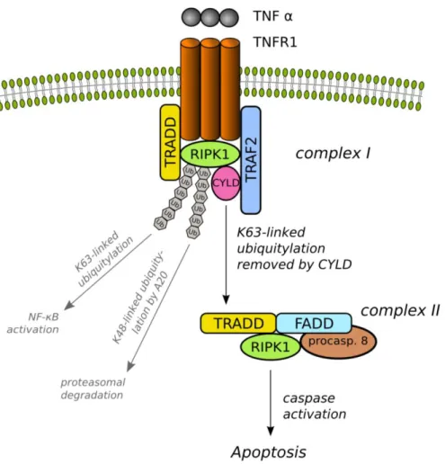
The most widely studied pathway leading to necroptosis is triggered by TNFα (see reviews [74,75]), a classical inducer of the extrinsic apoptotic pathway or activator of nuclear factor kappa-B (NF-κB) survival pathway (Fig. 2). Tumor necrosis factor receptor 1 (TNFR1) upon activation by TNFα undergoes rapid conformational changes.
Rearrangement of the intracellular part of TNFR1 provides docking surface for TNFα receptor-associated death domain protein (TRADD) and RIPK1 through their DD.
TRADD binding in turn recruits E3 ubiquitin ligase enzymes like the TNFR-associated factor 2 (TRAF2) or the inhibitor of apoptosis protein 1/2 (cIAP1/2), and create the multicomponent membrane-associated structure called complex I [76] (Fig.2).
Polyubiquitylation of RIPK1 via lysine 63 (K63) of ubiquitin attached to the K377 residue of RIPK1 in complex I contributes to the liberation of NF-κB from its inhibitory complex formed with the inhibitor of κB (IκB) protein and leading this way to the activation of the pro-survival pathway [14,77]. During this process, the polyubiquitylated RIPK1 directly recruits the inhibitor of κB kinase (IKK) protein via the IKK regulatory subunit of NEMO and activates IKKα and β kinases [78]. IKK phosphorylates the NF-κB inhibitor protein IκB and targets this protein for degradation by the ubiquitin-proteasome pathway [79]. NF-κB then liberated and translocated to the nucleus to activate expression of downstream target genes involved in the immune, inflammatory and survival responses [77] (Fig. 2).
Figure 2. Schematic figure of extrinsic apoptotic signaling pathway induced by TNFα.
TNFR1 upon activation by its ligand (TNFα) trimerises and re-arrange their intracellular part which serves as a platform for the formation of multiprotein signaling complex.
TNFR1 signaling complex I is composed of the adapter proteins TRADD, the E3 ligase TRAF2, the death domain-containing RIPK1 and other associated proteins for instance another E3 ligase cIAP1/2. cIAP1/2 and TRAF2 in the complex I ubiquitylate RIPK1 via K63-linked polyubiquitylation. Modified RIPK1 then recruits NEMO, the regulatory subunit of the IKK complex and subsequently activates the IKKα and β. Active IKK complex phosphorylates the IκB which results in ubiquitylation and dissociation of IκB from NF-κB, and eventual degradation of IκB by the proteosome. Liberated NF-κB then translocates into the nucleus and activate target genes that are contribute to inflammation, proliferation and cell survival.
1. 3. 2. Apoptosis induction – a route to cell death
As we have seen during the formation of complex I, polyubiquitin chains function not only as protein degradation signal but also provide a platform for the assembly of complex I (Fig. 2). After complex I formation, several other E3 ubiquitin ligases and ubiquitin hydrolases compete to activate or shut down the canonical NF-κB pathway and reform the polyubiquitin meshwork paving the way to form other macromolecular complexes with different biological functions (Fig. 3). E.g. A20 a dual E3 ligase and hydrolase cleaves off K63-linked polyubiquitin chain from RIPK1 and subsequently marks it for proteasomal degradation through its K48-linked polyubiquitylation [80]. In addition another ubiquitin hydrolase, the ubiquitin carboxyl-terminal hydrolase (CYLD) protein negatively influences the activation of the NF-κB pathway via removing the K63-linked ubiquitin chain from RIPK1 [13]. The exact details are not known and are targets of intensive research efforts. If the pro-death signal is stronger or lasts longer than the pro-survival signal, the internalized TNFR1 and the deubiquitylated RIPK1 form a new cytoplasmic complex, called complex II (Fig. 3). After dissociation from TNFR1 the DD-s of RIPK1 and TRADD molecules become available to form other complexes with different DD-containing proteins. E.g. Fas-associated death domain protein (FADD) can be adsorbed, leading to subsequent binding of caspase-8 [76]. In the cytosolic complex II the activated caspase-8 then proteolytically cleaves various substrate molecules including RIPK1 and by activating downstream effector caspases the apoptotic cell death is unavoidable (Fig. 3). In vitro, in the absence of corpse clearing, apoptosis turns into secondary necrosis.
Figure 3. Schematic figure of apoptosis induction via the TNFR1-triggered extrinsic apoptotic signaling pathway.
TNFR1 upon activation by its ligand (TNFα) trimerises and re-arrange their intracellular part which serves as a platform for the formation of multiprotein signaling complex.
TNFR1 signaling complex I is composed of the adapter proteins TRADD, the E3 ligase TRAF2, the death domain-containing RIPK1 and other associated proteins for instance another E3 ligase cIAP1/2. cIAP1/2 and TRAF2 in the complex I ubiquitylate RIPK1 via K63-linked polyubiquitylation. In complex I the polyubiquitylated RIPK1 activates the NF-kB pathway, see detailed in Fig.2. Activation of the NF-kB pathway is negatively regulated by cIAP inhibitor Smac mimetics and E3 ubiquitin ligases, such as A20 that target TRAF2 and RIPK1 for proteasomal degradation via K48-linked polyubiquitylation. In the meantime the ubiquitin hydrolase CYLD can also remove the K63-linked ubiquitin chain from the RIPK1 similar to A20. Deubiquitylated RIPK1 then serves as a platform to form the cytosolic complex II. Complex II formed with the participation of the deubiquitylated RIPK1, adapter protein FADD and TRADD, and the procaspase-8. In this complex caspase-8 be activated and then proteolytically cleaves various substrate molecules including RIPK1 and by activating downstream effector caspases leads the fulfillment of the apoptotic cell death program.
1. 3. 3. Necroptosis induction
The mechanism how the enzymatic activities of RIP kinases predispose cells to dye either by apoptosis or necrosis/necroptosis is intensively studied and explored. Recently the spontaneous formation of a high-molecular-weight complex has been reported by the research groups of Tenev and Feoktistova [81,82]. Core components of this complex, called as Ripoptosome are RIPK1, FADD, and procaspase-8 [81-83] (Fig. 4).
Ripoptosome seems to be a death-inducing platform that can direct the cell into apoptosis and necroptosis too. Feoktistova reported that presence of the protease- deficient caspase homolog cellular FLICE-inhibitory protein (cFLIPL) isoform in the Ripoptosome promotes necroptosis instead of apoptosis [81]. This notion is supported by Chang et al. as they published that cFLIPL can either promote or inhibit apoptosis [84]. In the meantime Tenev reported that presence of cFLIPL in the Ripoptosome limits the activity of the complex either for apoptosis either for necroptosis induction [82].
Interestingly Ripoptosome assembly can be independent of death receptor activation and mitochondrial pathways, and it is distinct form cytosolic complex II too [82].
Ripoptosome formation requires the kinase activity of RIPK1 and it is negatively regulated by cIAPs [82]. Tenev et al. showed that cIAPs and X-linked inhibitor of apoptosis protein (XIAP) directly polyubiquitylate RIPK1 which stimulate the recruitment and activation of various kinases necessary for prosurvival NF-κB signaling (see chapter 2. 3. 1.) [82]. Additionally cIAPs can also mark RIPK1 for proteasomal degradation by polyubiquitylation [85] (Fig. 4). Not surprisingly IAPs are highly expressed in various tumor cell types and their protein level may have prognostic significance [86]. Consequently depletion of cIAPs and XIAP contributes to the assembly of Ripoptosome and direct the cell to death. During apoptosis, natural IAP antagonists Omi/HtrA2 and Smac/Diablo translocate from the mitochondria to the cytoplasm and through their IAP-binding motif, inactivate IAPs and this way facilitate caspase activation [87]. Based on this fact a novel class of anti-cancer drugs namely Smac mimetics were developed. Smac mimetics facilitate the proteasomal degradation of cIAPs and sensitize various type of tumor cell to death [88].
Figure 4. Schematic figure of necroptosis induction via the death receptor TNFR1 In complex I the polyubiquitylated RIPK1 activates the NF-κB pathway, see detailed in Fig. 2. Deubiquitylated RIPK1 serves as a platform to form the cytosolic complex II with the participation of the adapter protein FADD and TRADD, and the procaspase-8.
In the complex II caspase-8 can be activated and trigger apoptosis (see detailed in Fig.
3). In the complex II, if the caspase activation is halted by inhibitor, caspase-8 activation and the subsequent cleavage of RIPK1 are suppressed. Due to arrested caspase activation and the kinase activity of RIPK1, another cytosolic complex, the ripoptosome is established. In the ripoptosome RIPK1 and RIPK3 are phosphorylated and elicit the necroptotic pathway.
RIPK3 contains an N-terminal kinase domain and a C-terminal RIP homotypic interaction motif (RHIM), which mediates its interaction with RIPK1 and therefore can attach to the Ripoptosome [89] (Fig. 4). Under caspase-compromised conditions or in the presence of high level of cFLIP in the Ripoptososme the cleavage of RIPK1 and RIPK3 by caspase-8 is postponed and, as a consequence, kinase activities of RIPK1 and RIPK3 remain active [90]. In 2005, Degterev and colleagues identified a small molecule allosteric inhibitor called Nec which blocks the kinase activity of RIPK1, thereby inhibiting necroptosis but leaving RIPK1-mediated activation of NF-κB unaffected [12].
The cross-talk between RIPK1 and RIPK3 is not clear yet. Cho et al. showed that kinase activity of RIPK3 is responsible for the phosphorylation of RIPK1 that stabilizes their association and initiates pro-necrotic kinase cascade [91] (Fig. 4). Kinase activity of RIPK1 and RIPK3 regulates various mechanisms during necroptosis involving reactive oxygen species (ROS) production, calcium overload, mitochondrial permeability, ATP level of cells and glucose metabolism [14,92].
It was recently published that the adenine nucleotide translocase (ANT), an integral protein of the inner mitochondrial membrane protein which, exchanges mitochondrial ATP with cytosolic adenosine diphosphate (ADP), can be inhibited via RIPK1- dependent way [93]. Inhibition of ANT by RIP1 can be expected to reduce intramitochondrial ADP levels resulting in a reduction of ATP level [93]. On the other hand ANT contributes to the pore formation on the mitochondrial permeability
transition pore complex (PTPC), which is a feature of necroptosis, together with the voltage-dependent anion channel (VDAC) presents on the outer mitochondrial membrane and cyclophilin D (CYPD) localizes in the mitochondrial matrix [94]. Smith and colleagues demonstrated that the CYPD, a core component of the PTPC complex, has to be present in order to observe cardyoprotective effect by Nec [95]. This corresponds to results discovering that RIPK1 may alter PTPC formation (see reviews [14,96]).
RIPK3 was published to be involved directly in glucose metabolism related enzyme activation and consequent respiratory burst and ROS generation that characterizes necroptosis [97]. Via allosteric activation of glycogen phosphorylase (PYGL), glutamate–ammonia ligase (GLUL) and glutamate dehydrogenase 1 (GLUD1) RIPK3
contributes to enhance the glycogenolysis, glycolysis and glutaminolysis. Knock down of these enzymes halt the TNFα-induced ROS production and necroptosis under caspase-compromised conditions [97]. In other experimental settings and cell line models non-mitochondrial ROS production by the plasma membrane NADPH oxidase- 1 (NOX1) contributes to TNFα-triggered necroptosis [98]. It is possible, that the RIPK1-recruited NOX1-generates ROS, that triggers the subsequent ROS production by the mitochondrial respiratory chain [99].
ROS can react with polyunsaturated fatty acids in cellular membranes and produce reactive aldehydes, which in turn can attack protein and lipid moieties of various membranes, thereby compromising their integrity. Lipid peroxidation mediated destabilization of cellular membranes (including the plasma, lysosomal and ER membranes) results in leakage of proteases or an elevation of cytosolic Ca2+
concentrations, two phenomena that participate in necrotic cell death [100].
The further downstream events of necroptosis are less known but intensively studied (see reviews [14,83]). Nowadays, extensive research focuses on the molecular background of necroptosis [81-83,101] and on the identification of necroptosis in physiological [102,103] or pathological [104,105] conditions. Recently MLKL was identified as the target of RIPK3 [72]. MLKL is phosphorylated by RIPK3 and this step seems to be critical for the fulfillment of necroptosis [57,72]. Most recently the mitochondrial protein phosphatase PGAM5 was shown to play role in necroptosis execution [73].
1. 4. Extrinsic cell death and survival induction – through Fas/CD95 and TRAIL receptors
1. 4. 1. Apoptosis and necroptosis induction
In sharp contrast to TNFR1-activated cellular signaling, the binding and activation of tumor necrosis factor-related apoptosis-inducing ligand (TRAIL) and Fas/CD95 receptors through their corresponding cytokine ligands rather promote cell death then the pro-survival of cells. Both TRAIL and FasL-induced apoptosis has crucial role in homeostatic regulation and effector function of the immune system [106,107].
The molecular events involved in TRAIL or FasL-induced apoptosis have been well documented [108]. Upon binding their corresponding ligands Fas/CD95 or TRAIL receptors undergo conformational changes which provides docking surface for FADD, RIPK1, the initiator procaspase-8/-10 and their regulator cFLIP protein (Fig. 5). In this complex, namely death-inducing signaling complex (DISC) the close proximity of procaspase-8 molecules allows the autocatalytic cleavage and formation of active caspase-8 [109]. Activated caspase-8 contributes the induction of apoptosis by initiating a cascade type of activation of downstream effector caspases. Upon receptor internalization the complex translocates from Fas/CD95 or TRAIL receptor and the cytosolic complex II is formed. At this level, based on the RIPK1 kinase activity cell death can be realized via necroptosis or by apoptosis [110] (Fig. 5).
Figure 5. Schematic figure of the extrinsic apoptosis induction via the death receptor DR4/5 and CD95.
In contrast to TNFR1 signaling, the default pathway activated by Fas/CD95 or TRAIL receptors is caspase-8 activation in DISC and induction of apoptosis. Ligation of CD95/FAS or DR4/5 receptor by their ligand FASL and TRAIL respectively cause conformational changes of the death receptors. The re-arrangement of the intracellular parts then serves as a platform for the formation of plasma membrane-associated multiprotein signaling complex called DISC. DISC consists of the death receptor, the adaptor molecule FADD, procaspase-8 and procaspase-10. During the receptor internalization cytosolic complex II is formed together with RIPK1, in which the high local concentration of procaspase-8 is believed to lead to its autoproteolytic cleavage and activation. The active caspase-8 then proteolytically cleaves various substrate molecules including RIPK1 and by activating downstream effector caspases which leads the fulfillment of the apoptotic cell death program. If the caspase activation is halted, the caspase-8 activation and the subsequent cleavage of RIPK1 are suppressed.
Due to the kinase activity of RIPK1, another cytosolic complex, the ripoptosome is established. In the ripoptosome RIPK1 and RIPK3 are phosphorylated and elicit the necroptotic pathway.
1. 5. Intrinsic apoptotic pathway
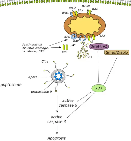
Besides the extrinsic stimuli, initiator procaspases can be activated also by inner death signals like DNA damage, oxidative stress, heat shock, UV radiation, starvation, staurosporine (STS) treatment etc. [111]. The intrinsic apoptotic pathway is characterized by the permeabilization of the mitochondrial outer membrane and as a consequence mitochondrial membrane depolarization, which leads to the cytochrome c and other proteins release from the mitochondrial intermembrane space [112]. In addition to the release of mitochondrial factors the mitochondrial membrane depolarization cause a loss of biochemical homeostasis of the cell: ATP synthesis is inhibited in absence of proton gradient between the intermembrane space and the matrix, and ROS are increasingly generated [113].
This permeabilization is regulated mainly by the B-cell lymphoma 2 (BCL-2) family of proteins which contains both pro- and antiapoptotic proteins [114]. Through their BH3 domains the family members can form hetero- and homodimers. As proposed in their rheostate model, Korsmayer predicted that various stimuli (mentioned above) induce the dimerization of proapoptotic proteins like BCL-2-like protein 4 (BAX), BCL-2 homologous antagonist (BAK) and BCL-2 antagonist of cell death (BAD) which upon their activation are engaged to generate a protein-permeable pore in the mitochondrial outer membrane [115] (Fig. 6). According to the theory the antiapoptotic proteins like BCL-2 and BCL-2-like protein 1 (BCL-XL) can directly interact with the proapoptotic proteins and consequently inhibit the function of their oligomerization and pore formation [116] (Fig. 6). The mechanism of pore formation is up till now an enigma.
As a consequence of BAX and BAK homo-oligomerized, the formed pore on the outer mitochondrial membrane let leaking out cytochrome c and other mitochondrial proteins (like Smac/Diablo, Omi/HtrA2 and apoptosis-inducing factor (AIF) into the cytosol from the mitochondria intermembrane space [117]. The release of cytochrome c induces the formation of apoptosome which consist of Apaf1, procaspase-9, citochrome c and ATP or dATP [118] (Fig. 6). In the aptoptosome procaspase-9 is activated, which in turn promotes the activation of caspase-3 [119]. Active caspase-3 then leads to the fulfillment of the apoptotic program. This process is further inhibited by XIAP protein, which inhibits the activity of caspase-3, -7, -9 [120]. In parallel Smac/Diablo and
Omi/HtrA2 also released from the mitochondria and interacts with and inactivates the XIAP which therefore forces the caspase activation [121] (Fig. 6).
Another released protein during intrinsic apoptotic pathway is AIF, which has both mitochondrial and nuclear signal sequences and normally located in the mitochondrial intermembrane space. In response to apoptotic stimuli it translocates to the nucleus causing caspase-independent DNA fragmentation and peripheral chromatin condensation, but not oligonucleosomal DNA laddering [122].
It has to be mentioned also that there is an interconnecting link between the extrinsically activated caspase signaling and the mitochondrial pathway via activated caspase-8. BH3 interacting domain death agonist (BID) is cleaved by the active caspase-8 and in its truncated form (tBID) translocates to the mitochondria where it acts similar to other proapoptotic proteins and induce formation of mitochondrial permeabilization pore [123]. With this connection between the extrinsic and intrinsic pathway, cells can multiply and reinforce the extrinsic death signal.
STS a poorly selective wide spectrum inhibitor of protein kinases, is a generally accepted model compound for inducing cell death [124] (Fig. 6). Treatment with STS induces the intrinsic, mitochondrial pathway of apoptosis, activates the BAX protein [125] and promotes its translocation to the mitochondria [126]. Consequently, the outer membrane of the mitochondria becomes permeabilized, allowing the release of proapoptotic proteins such as cytochrome c, Omi/ HtrA2 and AIF [127,128].
Cytochrome c initiates apoptosome formation, activation of caspase-9 and the executioner caspases (-3, -6, -7). Caspases process their selective target proteins, thereby evoking the maturation of the apoptotic morphology [129].
Previously we studied the nature of the switch mechanism between apoptosis and necrosis and investigated the intrinsic apoptotic pathway in STS-treated U937 cells [20]. We [20,130] and others [131] found that STS can provoke necrosis in human cancer cells which was independent of caspase activation. Based on these results we were curious to compare STS-induced necrosis under caspase-compromised conditions and the death ligand-triggered necroptosis in spite of the fact that necroptosis is generally defined as a result of a death receptor-triggered cell death pathway [12].
Figure 6. Schematic figure of the intrinsic apoptotic pathway.
The intrinsic apoptotic pathway is initiated by different stimuli mainly represented by like DNA damage, oxidative stress, heat shock, UV radiation etc. The initial events are represented by the activation proapoptotic proteins such as BAX and BAK. These activated cytosolic or mitochondrial membrane-associated proteins are subsequently generate a protein-permeable conduit in the mitochondrial outer membrane. This mitochondrial membrane pore permeable for cytochrome c and other mitochondrial proteins like Smac/Diablo and Omi/HtrA2. The release of cytochrome c contributes to the formation of the so called apoptosome in association with Apaf1 and procaspase-9.
In the apoptosome caspase-9 matures and subsequently proteolytically cleaves various substrate molecules, additionally activates the executioner caspase-3 which leads the fulfillment of the apoptotic program. Caspase-3 and -9 activation can be halted by XIAP, however Smac/Diablo and Omi/HtrA2 released from the mitochondria, interact with and inactivate the XIAP, which contributes the fulfillment of the apoptotic pathway. Antiapoptotic BCL-2 proteins negatively influence the mitochondrial membrane pore formation. BCL-2 and BCL-XL can directly bind and therefore inhibit the function of different pro-apoptotic proteins like BAX and BAD.
1. 6. Further type of programmed necrosis
The cellular events during programmed necrosis are poorly understood. Former studies investigated the role of the lysosomes during necrosis in neurodegenerative diseases.
Investigations in both nematodes and mammals confirm the active participation of calpains (calcium-dependent neutral cysteine proteases) and cathepsins (lysosomal aspartyl proteases) in the execution of necrotic cell death [132,133] .
Tavernakis and his colleagues showed that vacuolar H(+)-ATPase, a pump that acidifies lysosomes and other intracellular organelles, is essential for necrotic cell death in C.
elegans [134]. Cytoplasmic pH drops in dying cells and the intracellular acidification requires the vacuolar H(+)-ATPase, whereas alkalination of endosomal and lysosomal compartments by weak bases protects against necrosis [134].
Calpain proteases are normally requires calcium for activation, whereas cathepsins have a highly acidic pH optimum for full activity [135]. Studies in primates confirmed calpain-mediated disruption of the lysosomal membrane after ischemia [136].
These observations led to the formulation of the “calpain–cathepsin hypothesis,” by Yamashima, which uncover the way of calcium-mediated activation of calpains results in the rupture of lysosomes and leakage of cathepsins that eventually degrade the cell [137].
Formerly we showed that the selective cathepsin-B inhibitor CA-074-OMe (CA) rescued the caspase-independent necrotic and the caspase-dependent apoptotic form of cell death in promyelocytic leukemia (HL60) cells treated by STS [130]. However the applied CA concentration was higher with orders of magnitudes (10 µM) compared to the concentrations (10 nM) required for the inhibition of cathepsin B protease activity [130]. This result point out that that a non-cathepsin B, currently unknown target of CA can be important in necroptotic cell death.
1. 6. 1. PARP-AIF-mediated programmed necrotic pathway
Besides the death receptor-triggered necroptosis, an other intensively investigated type of programmed necrosis is mediated by poly(ADP-ribose) polymerase (PARP) activation called parthanatos. In response to DNA damage, or exposure of DNA
alkylating agent, like MNNG, the DNA repair PARP-1 protein is activated. Following PARP-1 activation the consequent poly(ADP-ribose) (PAR) accumulation and/or the ATP/Nicotinamide adenine dinucleotide (NAD) pool depletion leads to the release of the AIF from the mitochondrial intermembrane space. Cytosolic AIF then enter to the nucleus and perform caspase-independent, large-scale DNA fragmentation. The mechanism requires calpains but not cathepsins or caspases [138].
AIF-deficient harlequin mutant mice are protected against several necrotic stimuli, including ischaemia–reperfusion injury of the brain [139]. Similarly, pharmacological and genetic inhibition of PARP-1 has consistent cytoprotective effects [140].
Further question is the demarcation of different types of programmed necrosis.
Surprisingly, it was published that c-Jun N-terminal kinase (JNK), especially JNK1, but not the other members of mitogen-activated protein kinase family, is required for PARP-1-induced mitochondrial dysfunction, AIF translocation, and subsequent cell death [141]. Moreover this group showed that knockouts of RIPK1 and TRAF2 of mouse embryonic fibroblast (MEF) cells caused a resistance to PARP-1-induced cell death [141].
It means that it is possible that RIPK1 plays role as central molecule of the execution and fulfillment of programmed necrosis, and different types of programmed necrosis are emerged at the level of RIPK1.
1. 7. Physiological and pathological aspects of programmed necrotic cell death
1. 7. 1. Physiological aspects
Apoptosis was described as a regulated cell death form which participates in normal tissue homeostasis during the embryogenesis of different organisms, especially in the genetic control of cell elimination during development of C. elegans. This early description of apoptosis may had influenced researchers to focus on the programmed cell death and at the same time neglect the other modalities of cell death already known [1,2].
At the same time some arguments suggested that apoptosis is not the solely form of cell death responsible for unwanted cell elimination during embryogenesis or physiological tissue renewal. For example interdigital membrane elimination provides a representative evidence for the role of apoptosis in tissue sculpting during embryonic development.
Contrarily to this Yoshida et al. reported that a delayed but apparently normal interdigital membrane removal exists in apaf1-/- mutant mice embryos, where the apoptosome can not be formed [142].
In other cases, necrotic-like cell demise was observed under conditions where the apoptotic machinery was blocked. For instance, in BAK-/- and BAX-/- double mutant mice various defects of apoptotic process were observed: the syndactyly phenotype, excess cells accumulation in the central nervous system and finally most embryos died at or just before birth. Some of the littermates of the dying BAK-/- and BAX-/- mice could survive and lived until adulthood [143,144]. Similarly, in the protist Dictyostelium discoideum stalk cells showed autophagy-like cell death when treated with differentiation-inducing factor (DIF-1) under condition of starvation. Inactivation of autophagy-related 1 homolog gene (atg1) shifted the process to a classical necrotic- like cell death [145]. Based on this results necrosis is a candidate process as a back-up mechanism which can substitute the defective apoptotic pathway. On the other hand, necrosis was also observed during normal mammalian tissue homeostasis as a programmed event. For instance, the human large intestine crypts contain necrotic colonocytes [15], or in human growth plate during late pubertal fusing, chondrocytes showed necrotic phenotype [16]. Moreover, disappearance of linker cells in C. elegans at a well defined time point during development is essential for future fertility. Abraham et al. reported that caspases and other apoptosis-associated gene products do not participate in the loss of linker cells and indeed, the dying linker cells showed necrotic morphology [12,17,18]. For more details see reviews of Kroemer, McCall and Thompson [35,37,146]. However, the above-mentioned reports are based mostly on morphological observations, therefore further investigations would be preferable, to evaluate the type of cell death.
This context raises the question of evolutional appearance and potential hierarchy of different cell death modalities. Golstein and Kroemer discussed the redundancy of cell
death mechanisms and hypothesized that an evolutionary ancestral cell death – resembling necrosis – was overridden by evolutionary younger, more elaborate processes, likely autophagy and apoptosis. Cell death types emerging later in evolution may turn into the major dominant form of cell death while masking the ancestral one, but which can resurface as back-up mechanism when the other cell death pathways are blocked [69].
It is tempting to speculate how necroptosis is related to other cell death modalities and what physiological role it might have. It is conceivable that necroptosis compared to necrosis is a relatively new evolutionary mechanism, an alternative programmed cell death that serves as a back-up process when apoptosis is abrogated. Hitomi et al. in a murine genome-wide siRNA screen for genes involved in zVAD.fmk (zVAD) induced necroptosis in L929 cell line have found that some genes required for necroptosis had increased expression in macrophages, dendritic cells and natural killer (NK) cells, while another gene cluster was upregulated in B and T lymphocytes connecting necroptosis to the immunological processes [13]. Another interaction between the necroptotic process and the immune-response has also been shown [91]. Viruses are known to frequently express antiapoptotic proteins to control the lifespan of their (Vertebrate) host in order to avoid cell death until their replication cycle is finished [147]. As Cho et al. have shown that in vaccinia virus infected T cell, when the apoptotic process was abrogated by caspase inhibitors, the RIP3K directed necroptotic cell death became dominant [91].
Accordingly, necroptosis might be an evolutionary response of host cells to avoid the pathogen-induced apoptosis inhibition. Moreover, further benefit of the necrotic cell demise in a multicellular organism might be that necrosis/necroptosis leads to a proinflammatory response. The release of the intracellular content can provoke the immune response, which serves not only as a warning system, indicating the internal or external hazardous conditions, but also initiating the immune response. This observation raises the clinical benefits of necroptosis induction in antiviral therapy.
To study the phylogenetics of necroptosis, the Inparanoid, a comprehensive database of eukaryotic orthologs was used and results have shown that RIP1 and RIP3 kinases are characteristic to Vertebrates. However, a human RIP1 kinase homolog can be found in an annelid worm (Capitella sp.), and proteins similar to the human RIP3 kinase is
present in the chordate Ciona savignyi, the amoeba Dictyostelium discoideum, and in the smallest free-living unicellular eukaryote, Ostreococcus tauri [148]. The connection of those these proteins to cell death pathways are unknown and their homology might be explained by prior exon shuffling process. Based on the above mentioned facts necroptosis may be an important stage in the arms race where vertebrates are constantly participating, evolving and learning how to cope with viral invasion.
The above mentioned evidences namely that certain cell types can initiate their own death via necrosis, apparently refute the bias that the term necrosis solely refers to accidental, uncontrolled event.
1. 7. 2. Pathological aspects
Necrosis has for a very long time been described as a direct cause or as a simultaneously occurring phenomenon of cell death in many cases of human diseases, such as neurodegenerative diseases with necrotic outcome [26], pancreatic adiponecrosis [27], trauma [28], ischemia-reperfusion in myocardial infarction [29], in cerebral infarction [30], bacterial infection [31,32] or in tumor malignancies [33,34]. However, having the preconception that this type of cell death is random and unregulated, researchers have regarded its harmful effects inevitable. Undoubtedly, overwhelming stress induces necrosis that can not be impeded, it is still worthwhile to examine if there is an activated signaling pathway behind the focal necrotic morphology that at least partially can be hampered. For instance following ischemia in the brain [149] and myocardial tissues [150] an increase in inflammatory cytokines production, including TNFα might occur and resulting in accompanying necroptosis.
Using Nec Degterev et al. investigated the in vivo participation of necroptosis in a murine model of ischemia-reperfusion [12]. As it was reported the area of the infarct and the neurological score due to middle cerebral artery occlusion was significantly reduced by intracerebroventricular administration of 7-Cl-Nec in a wide therapeutic window [12]. On the other hand, Whalen et al. hypothesized that Nec would reduce histopathological appearance of cell death and improve functional outcome in a controlled cortical impact (CCI) model of traumatic brain injury (TBI) in mice [151].
As they reported, short- and long-term Nec treatment attenuated the plasmamembrane
damage and moderated the motor and cognitive deficits compared to untreated littermates after CCI. In similar experiments Nec failed to adjust the outcomes the caspase-mediated apoptosis which also operates in TBI [28]. Moreover, pretreatment with Nec significantly reduced the microglial activation and neutrophil influx 48 hours after CCI. These results underline the possible involvement of necroptosis in acute brain injury and underscore the applicability of Nec in reducing posttraumatic lesion and inflammation [151].
Additional contribution of necroptosis in neurodegenerative diseases was published recently. N-Methyl-D-aspartate (NMDA) exposure through ionotropic glutamate receptor induced excitotoxicity in primary rat cortical cell cultures could be partially reduced by Nec, shown by cell viability assays and lactate dehydrogenase (LDH) leakage. It was also reported by the authors that the elevated intracellular Ca ++ level may contribute to NMDA induced necroptosis [152].
Oxidative glutamate toxicity, also called as oxytosis is characterized by glutathione (GSH) depletion, increased reactive oxygen species production, calcium influx and caspase-independence that could be prevented by antioxidants [153]. Further study showed that Nec could attenuate non-receptor mediated glutamate toxicity in a mouse hippocampal cell line (HT22) and not only blocked the nuclear translocation of AIF but an increase in the GSH level and a decrease of ROS production were also observed [154]. The inhibition of AIF transport to the nucleus by Nec raises the question whether is there a connection between RIP1 kinase activity and AIF transport, thus PAR plays any role in the necroptotic process. These questions need further experimental answers [155].
In another study Kim et al. characterized arachidonic acid- (AA) induced cell death in oligodendrocyte precursors [156]. The authors previously demonstrated that AA- induced cell death was lypoxigenase- (LOX) dependent and mediated by elevated ROS level and JNK activation, but it was independent from caspase activation. Authors confirmed that AA-induced cytotoxicity was markedly prevented by an antioxidant butylated hydroxyanisole (BHA), or a 12-LOX specific inhibitor or by Nec. Moreover, Nec markedly prevented cell death via blocking ROS production and JNK activation.
The authors hypothesized that deubiquitylation of RIP1 kinase is upregulated by

![Figure 1. Domain structure and subfamily members of caspase family [48]](https://thumb-eu.123doks.com/thumbv2/9dokorg/1378890.113532/15.892.132.760.131.740/figure-domain-structure-subfamily-members-caspase-family.webp)