Inflammation leads through PGE/EP 3 signaling to HDAC 5 /MEF 2 -dependent transcription in cardiac myocytes
András D Tóth
1,2,3,†, Richard Schell
1,2,4,†, Magdolna Lévay
2,5, Christiane Vettel
2,5, Philipp Theis
1,2, Clemens Haslinger
1,2, Felix Alban
1,2, Stefanie Werhahn
1,2, Lina Frischbier
1,2, Jutta Krebs-Haupenthal
1,2, Dominique Thomas
6, Hermann-Josef Gröne
7, Metin Avkiran
8, Hugo A Katus
4, Thomas Wieland
2,5,†&
Johannes Backs
1,2,†,*Abstract
The myocyte enhancer factor2 (MEF2) regulates transcription in cardiac myocytes and adverse remodeling of adult hearts. Activa- tors of G protein-coupled receptors (GPCRs) have been reported to activate MEF2, but a comprehensive analysis of GPCR activators that regulate MEF2 has to our knowledge not been performed.
Here, we tested several GPCR agonists regarding their ability to activate a MEF2reporter in neonatal rat ventricular myocytes. The inflammatory mediator prostaglandin E2(PGE2) strongly activated MEF2. Using pharmacological and protein-based inhibitors, we demonstrated that PGE2regulates MEF2via the EP3receptor, the bcsubunit of Gi/oprotein and two concomitantly activated down- stream pathways. The first consists of Tiam1, Rac1, and its effector p21-activated kinase2, the second of protein kinase D. Both path- ways converge on and inactivate histone deacetylase 5 (HDAC5) and thereby de-repress MEF2. In vivo, endotoxemia in MEF2- reporter mice induced upregulation of PGE2and MEF2activation.
Our findings provide an unexpected new link between inflamma- tion and cardiac remodeling by de-repression of MEF2 through HDAC5 inactivation, which has potential implications for new strategies to treat inflammatory cardiomyopathies.
Keywordshistone deacetylase5; myocyte enhancer factor2; p21-activated kinase2; prostaglandin E2; protein kinase D
Subject Categories Cardiovascular System; Immunology
DOI10.15252/emmm.201708536| Received28September2017| Revised17 May2018| Accepted18May2018| Published online15June2018 EMBO Mol Med (2018)10: e8536
Introduction
Evidence has been provided that sustained stimulation of receptors on the plasma membrane of cardiac myocytes by distinct mediators including endothelin-1 ora-adrenergic agonists is able to trigger the development of pathological structural changes along with alter- ations of cardiac gene expression (Backs & Olson, 2006a). A pathog- nomonic change is the reactivation of the dormant embryonic isoforms of genes regulating cardiac growth, myocardial contractil- ity and Ca2+-handling (Backs & Olson, 2006a). In particular, the reactivation of fetal cardiac gene programs is suggested to lead to the progression of cardiac remodeling and heart failure. As those mediators play crucial roles in the activation of fetal gene programs, it seems a promising approach to identify and describe the distinct upstream signaling pathways, which may reveal new targets for therapeutic interventions.
The myocyte enhancer factor 2 (MEF2) transcription factors belong to the family of MADS (MCM1, agamous, deficiens, SRF) proteins (Potthoff & Olson, 2007). In mammals, four different MEF2 proteins exist, named MEF2A, B, C, and D. They bind a common DNA sequence and drive the expression of specific target genes (Potthoff &
Olson, 2007). The MEF2 proteins have a crucial role in the proper cardiac development. Due to severe cardiac abnormalities, MEF2A- and MEF2C-null mice exhibit pre- and perinatal lethality, respectively (Linet al, 1997; Nayaet al, 2002). In addition to their critical role in the embryonic development, the MEF2 isoforms and their splicing variants also govern the stress response in the adult heart by reactiva- tion of fetal cardiac gene programs (van Oortet al, 2006; Kimet al, 2008; Gao et al, 2016). Whereas MEF2 transgenic mice develop
1 Department of Molecular Cardiology and Epigenetics, Heidelberg University, Heidelberg, Germany 2 DZHK (German Centre for Cardiovascular Research), Heidelberg/Mannheim, Germany
3 Department of Physiology, Faculty of Medicine, Semmelweis University, Budapest, Hungary 4 Department of Cardiology, Heidelberg University, Heidelberg, Germany
5 Experimental Pharmacology, European Center of Angioscience, Medical Faculty Mannheim, Heidelberg University, Mannheim, Germany 6 Institute of Clinical Pharmacology, Goethe University Frankfurt, Frankfurt, Germany
7 Department of Cellular and Molecular Pathology, German Cancer Research Center, Heidelberg, Germany
8 Cardiovascular Division, King’s College London British Heart Foundation Centre of Research Excellence, The Rayne Institute, St Thomas’Hospital, London, UK
*Corresponding author. Tel: +49 6221 56 35271; E-mail: Johannes.backs@med.uni-heidelberg.de
†These authors contributed equally to this work
ventricular dilation and contractile dysfunction, mice lacking the MEF2D gene are protected against fetal gene activation, fibrosis, and cardiac hypertrophy (van Oortet al, 2006; Kimet al, 2008). There- fore, the activity of MEF2 is tightly controlled. Whereas phosphoryla- tion by mitogen-activated protein (MAP) kinases and acetylation by the histone acetyltransferase p300 enhance the transcriptional activity of MEF2 (Potthoff & Olson, 2007; Waleset al, 2014; Weiet al, 2017), class IIa histone deacetylases (HDACs; HDACs 4, 5, 7, and 9) physi- cally interact with MEF2 and recruit other repressive epigenetic factors (Backs & Olson, 2006a; Lehmannet al, 2014). A protective role of class IIa HDACs in heart failure has been suggested. For example, HDAC5 and HDAC9 prevent cardiac hypertrophy (Zhanget al, 2002;
Changet al, 2004) and HDAC4 is required for the maintenance of cardiac function during physiological exercise (Lehmannet al, 2018).
Class IIa HDACs can be phosphorylated by several kinases, including the Ca2+/Calmodulin-dependent protein kinase II (CaMKII), protein kinase D (PKD), or G protein-coupled receptor kinase 5 (Backs &
Olson, 2006a; Kreusseret al, 2014; Lehmann et al, 2014; Weeks &
Avkiran, 2015). In turn, phosphorylated HDACs are guided by 14-3-3 chaperones from the nucleus to the cytoplasm and MEF2 is released from the inhibition and can activate downstream genes (Backs &
Olson, 2006a). On the other hand, there are pathways which protect from the harmful hyper-activation of MEF2 (Lehmannet al, 2014).
For instance, activation of protein kinase A (PKA) has been shown to inhibit MEF2 activity and moreover to counteract CaMKII-mediated activation of MEF2 (Backset al, 2011; Weekset al, 2017; Lehmann et al, 2018).
The upstream activation signal of the HDAC-MEF2 axis often originates from G protein-coupled receptors (GPCRs). The divergent pathways resulting from GPCRs could have antagonistic effects on MEF2 activation; therefore, it is difficult to predict the net effect on MEF2 activation by different mediators. The aim of this study was to identify new mediators that signal via GPCRs in cardiac myocytes and which were not known to activate the HDAC-MEF2 axis before.
Furthermore, we sought to explore the detailed resulting down- stream signaling pathway. We found that prostaglandin E2(PGE2), which is one of the main inflammatory mediators, strongly activates MEF2. PGE2binds to the Gi/o-coupled EP3receptor, which activates divergent, parallel operating signaling pathways; one involving Tiam1-, Rac1-, and p21-activated kinases (PAK) and another involv- ing PKD and HDAC5. We found that full MEF2 activation depends on participation of both pathways and that MEF2 is activatedin vivo in inflamed hearts.
Results
PGE2activates MEF2through the EP3receptor
To identify unknown GPCR-dependent signaling pathways that regu- late MEF2 activity, we conducted a screening experiment using neonatal rat ventricular myocytes (NRVMs). NRVMs were infected with an adenovirus harboring a MEF2-reporter (3xMEF2-Luc), which responds to endogenous MEF2. Thereafter, the cells were stimulated with different GPCR agonists for 24 h in serum-free medium. Similar to previous reports, endothelin-1 (100 nM) activated MEF2 to a high extent (Backset al, 2011). On the other hand, theb-adrenergic recep- tor agonist isoproterenol (1lM) significantly decreased the basal
level of MEF2 activity. We identified several mediators, which slightly elevated the activity of MEF2, such as sphingosine-1-phos- phate (1lM) or lysophosphatidic acid (10lM). Strikingly, we observed a 20-fold activation by two related compounds, namely prostaglandin E1(PGE1; 10lM) and prostaglandin E2(PGE2; 10lM;
Fig 1A). In contrast, analogs of other prostaglandins (fluprostenol and treprostinil, agonists of prostaglandin F and prostacyclin recep- tors, respectively) had no significant effect. In the following experi- ments, we focused on PGE2because of its higher abundance in the heartin vivo(Hermanet al, 1987). The PGE2effect on MEF2 activity was concentration-dependent (Fig EV1A). In good agreement with the ability to activate MEF2, PGE2triggered the expression of known specific MEF2 target genes (Potthoff & Olson, 2007; Lehmannet al, 2018), such asNur77,Myomaxin, orAdamts1(Fig 1B). MEF2 activa- tion is commonly associated with the induction of hypertrophic gene programs. Likewise, PGE2increased the mRNA levels of the hypertro- phy marker BNP (Fig 1B) and induced cellular hypertrophy of NRVMs, which correlated with the induction of MEF2 activity (Fig 1C). In addition, we observed a similar extent of protein synthe- sis induction after PGE2stimulation as with the well-known hyper- trophica-adrenoceptor agonist phenylephrine (Fig EV1B). Next, we aimed to determine the receptor involved in PGE2-mediated MEF2 activation. The four different isoforms of PGE2receptors (EP1, EP2, EP3, EP4) show a different degree of G protein coupling and expres- sion patterns (Woodward et al, 2011). Each of the four types is expressed in cardiac myocytes, but EP3and EP4receptors are the most abundant isoforms (Di Benedettoet al, 2008). The EP3receptor antagonist L798106 inhibited PGE2-dependent MEF2 activation (Figs 1D and EV1C), while neither the EP4 receptor antagonist L- 161,982 (2lM) nor the EP1/EP2receptor antagonist AH6809 (10lM) led to an alteration of MEF2 activity, suggesting the EP3receptor as the underlying receptor to activate MEF2 by PGE2in NRVMs.
PGE2activates MEF2through Gi/oproteins
The EP3receptor is generally considered to couple to pertussis toxin (PTX)-sensitive Gi/oproteins, thereby decreasing intracellular cAMP levels. In accordance, EP3receptor inhibition by L798106 increased the isoproterenol-induced phosphorylation of phospholamban, a known cAMP-regulated protein (Cuello et al, 2007; Fig EV1D).
However, the EP3receptor has several splice variants, which differ in the G protein-coupling properties (Woodwardet al, 2011). There- fore, we examined which heterotrimeric G protein types are essential for the PGE2-mediated MEF2 activation by a pharmacological and molecular approach (Fig 2A). Regulator of G protein signaling (RGS) proteins are GTPase activating proteins (GAPs) and specifically block different classes of G proteins. RGS16 is a GAP for Gi/oand Gq/11
proteins, RGS2 suppresses Gq/11, while RGS-LSCII inhibits the G12/13
signaling pathway (Vettelet al, 2012). Like RGS2, p63ΔN, a specific scavenger of activated Gaq/11 proteins did not affect the PGE2- induced activity of MEF2. In contrast, the Gi/oand Gq/11inhibitor RGS16 (Fig 2B), as well as pretreatment with pertussis toxin (PTX, 100 ng/ml) attenuated the PGE2 response (Fig 2C). We did not observe any significant effect of RGS-LSCII overexpression in this setting (Fig 2B). These data suggest that neither Gq/11- nor G12/13-, but the PTX-sensitive Gi/oprotein activation is required for the PGE2
response. To rule out an involvement of Gsproteins, we inhibited adenylyl cyclase, the effector enzyme of Gsproteins. The adenylyl
A
B
C
D
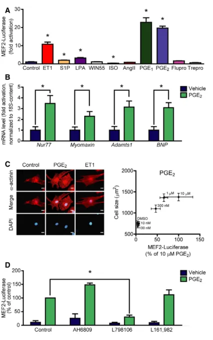
Figure1. Screening the effect of different GPCR agonists on MEF2activity.
A Neonatal rat ventricular myocytes (NRVMs) were infected with the3xMEF2-Luc reporter, serum starved for20h, and stimulated for24h with different agonists:
100nM endothelin-1(ET1),1lM sphingosine-1-phosphate (S1P),10lM lysophosphatidic acid (LPA),1lM WIN55,212-2(WIN55, cannabinoid receptor agonist), 1lM isoproterenol (ISO,b-adrenergic receptor agonist),100nM angiotensin II (AngII),10lM prostaglandin E1(PGE1),10lM prostaglandin E2(PGE2) or100nM prostanoid F receptor agonist fluprostenol (Flupro),100nM treprostinil (Trepro, prostacyclin receptor agonist).
B PGE2induces the expression of MEF2target genes. NRVMs were serum starved for24h, then were stimulated with1lM PGE2for2h, and mRNA levels of the indicated genes were determined.
C Correlation between MEF2activation and NRVM hypertrophy. MEF2activity was measured as in (A) upon increasing concentration of PGE2. Cell size of NRVMs was determined after48-h stimulation with the indicated concentrations of PGE2. Left: Representative images ofa-actinin stained NRVMs stimulated with DMSO,300nM PGE2, or1nM ET1. Scale bar is10lm.
D PGE2activates MEF2via EP3receptor. NRVMs were infected with the3xMEF2-Luc reporter and serum starved for20h. The cells were stimulated with DMSO or1lM PGE2for24h in the presence or absence of different EP receptor antagonists: AH6809(10lM, EP1- and EP2-antagonist) or798106(200nM, EP3-antagonist) or L161,982(2lM, EP4-antagonist).
Data information: Values are means.e.m. In (A), the experiment was performed in triplicates, and similar results were obtained in three different experiments.
Student’s two-tailedt-test, *P<0.05vs. control (ET1,P=0.0013; S1P,P=0.0144; LPA,P=0.026; WIN55,P=0.6205; ISO=0.0399; AngII,P=0.5812; PGE1,P=0.001; PGE2,P<0.0001; Flupro,P=0.0846; Trepro,P=0.1958). In (B),n=5, technical replicates, Student’s two-tailedt-test, *P<0.05vs. control (Nur77,P=0.0148;
Myomaxin,P=0.0417;Adamts1,P=0.0112;BNP,P=0.0065). In (C), NRVM cell size was determined from a minimum of100cells of three technical replicates upon different concentrations of PGE2, and MEF2activity was parallel assessed from six technical replicates. Correlation was statistically analyzed by calculating Pearson correlation coefficient (Pearsonr=0.9626,P=0.0021). In (D),n=3, independent experiments, *represents significant interaction between the two treatments (P<0.05, two-way ANOVA, AH6809,P=0.1092; L798106,P=0.0002; L161,982,P=0.5579).
Source data are available online for this figure.
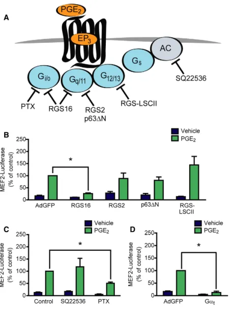
cyclase inhibitor SQ22536 (100lM) did not affect MEF2 activity (Fig 2C). In addition, since the adenylyl cyclase inhibition also mimics the effect of Gai/o, the absence of alteration suggested that not thea, but thebc subunit of Gi/ois the transducer in the PGE2-mediated MEF2 activation. In accordance, overexpression of Gat to scavenge the free Gbc subunits abolished the PGE2 effect (Fig 2D). Similar results were obtained with the overexpression of long isoform of RGS3 (RGS3L), another Gbcscavenger (Vogtet al, 2007; Fig EV2). Taken together, these data indicate that the PGE2-induced MEF2 activation is mediated by thebcsubunit of Gi/o
proteins.
PGE2induces HDAC5hyperphosphorylation and de-repression of MEF2via PKD
Among critical regulators of MEF2 are class IIa HDACs. A well- known mechanism of de-repression of MEF2 activity is HDAC phos- phorylation followed by its concomitant nucleo-cytoplasmic shut- tling. Therefore, we analyzed the phosphorylation status of HDAC5 by Western blot. We overexpressed FLAG-tagged HDAC5 in NRVMs. After stimulation with PGE2, we observed a high extent of hyperphosphorylation of HDAC5 at Ser-498 (Fig 3A), similarly to endothelin-1 (ET1, 100 nM) or phenylephrine (PE, 100lM). Since A
B
C D
Figure2. PGE2-mediated MEF2activation is Gi/o-bcdependent.
A Illustration of the target points of the used pharmacological and protein-based inhibitors.
B–D (B, D) NRVMs were infected with the3xMEF2-Luciferase reporter and with recombinant adenoviruses encoding EGFP (AdGFP), RGS16, RGS2, p63DN, RGS-LSCII, or Gat, as indicated. The cells were serum starved for20h and stimulated with DMSO or1lM PGE2for24h. (C) NRVMs were infected with the3xMEF2-Luciferase reporter and pretreated with the adenylyl cyclase inhibitor SQ22536(100lM) for20min or the Gi/oprotein inhibitor pertussis toxin (PTX,100ng/ml) for20h in serum-starved conditions and treated with DMSO or1lM PGE2for24h.
Data information: In (B–D),n=3, independent experiments, *represents significant interaction between the two treatments (P<0.05, two-way ANOVA, RGS16, P<0.0001; RGS2,P=0.1725; p63DN,P=0.0666; RGS-LSCII,P=0.0818, SQ22536,P=0.385; PTX,P<0.0001; Gat,P<0.0001), values are means.e.m.
Source data are available online for this figure.
Ser-498 is a specific phosphorylation site of protein kinase D (PKD;
Haworthet al, 2011), we examined whether PKD is responsible for the phosphorylation of HDAC5. Indeed, pretreatment with the specific PKD-inhibitor BPKDi (3lM; Meredith et al, 2010;
Monovichet al, 2010) diminished the PGE2-induced HDAC5 phos- phorylation. In accordance, phosphorylation of PKD was increased at Ser-744/Ser-748 and Ser-916 sites upon PGE2 treatment in NRVMs (Fig 3B), which was partly (phosphorylation at Ser-744/
Ser-748) observed in adult mouse ventricular myocytes as well (Fig EV3). Different mechanisms of PKD activation exist. It is known that PKD can be activated in a protein kinase C (PKC)- dependent way, e.g., by PE. In contrast, ET1 stimulates PKD activity in a PKC-independent manner in NRVMs (Vegaet al, 2004; Harrison
et al, 2006; Haworthet al, 2007; Guoet al, 2011). Thus, the involve- ment of PKC in the PGE2-mediated PKD and HDAC5 phosphorylation was examined. Phosphorylation of PKD at Ser-744/Ser-748, a well- known PKC-target site, was diminished upon pretreatment with the PKC-inhibitor bisindolylmaleimide I (BIM, 2lM). However, phos- phorylation of the autophosphorylation site (Ser-916) was only slightly decreased (Fig 3B), and HDAC5 phosphorylation at Ser-498 was not altered (Fig 3A) by BIM, suggesting that the activation of PKD by PGE2occurs both PKC dependently and PKC independently.
Nevertheless, BPKDi prevented MEF2 activation by PGE2(Fig 3C). In contrast, inhibition of another HDAC kinase, CaMKII, by auto- camtide-2-related inhibitory peptide II (AIP, 1lM) did not affect PGE2-mediated MEF2 activity. We therefore conclude that PGE2
A
B
C
Figure3. PGE2induces HDAC5hyperphosphorylation and concomitant MEF2activation via PKD.
A, B NRVMs were infected with recombinant adenovirus encoding Flag-HDAC5(A) or not infected (B) and serum starved for20h. The cells were pretreated with the PKC-inhibitor bisindolylmaleimide I (2lM) or the PKD-inhibitor BPKDi (3lM) for20min and stimulated with100nM endothelin-1(ET1) or100lM phenylephrine (PE) or1lM PGE2for4h, as indicated. HDAC phosphorylation was detected by immunoblotting with the anti-HDAC5phospho-Ser-498antibody, phosphorylation of PKD was assessed using anti-phospho-PKD antibodies (Ser-744/Ser-748and Ser-916), and representative images are shown from three independent experiments.
C NRVMs were infected with the3xMEF2-Luc reporter and serum starved for20h. The cells were pretreated with the PKD-inhibitor BPKDi (3lM) or the CamKII- inhibitor AIP (1lM) for20min and stimulated with DMSO or1lM PGE2for24h.N=3, *represents significant interaction between the two treatments (P<0.05, two-way ANOVA, BPKDi,P<0.0001; AIP,P=0.2197). Values are means.e.m. The exactnandP-values can also be found in the Source Data Excel file for Fig3.
Source data are available online for this figure.
promotes HDAC5 hyperphosphorylation and de-repression of MEF2 activity via PKD.
PGE2triggers Rac1activation via Tiam1
Several effector partners of Gi/o-bc have been described, among others the small G protein Rac1 and RhoA (Vogtet al, 2007). To test whether these small G proteins are activated by PGE2in NRVMs, we
performed effector pull-down assays and we found, that exclusively Rac1, but not RhoA is activated after PGE2stimulation (Fig 4A). To examine whether Rac1 activation is Gi/o-dependent, we used PTX treatment and overexpression of RGS16. Both manipulations suppressed the PGE2-induced Rac1 activation (Fig 4B and C). Earlier studies have shown that the Gi/o-mediated Rac1 activation in NRVM can occur in a Gi/o-bc-, phosphatidyl-inositol-3-kinase (PI3K)-, and the guanine nucleotide exchange factor Tiam1-dependent manner
A
B
C
D
Figure4. PGE2induces Rac1and MEF2activation via Gi/oand the RacGEF Tiam1.
A NRVMs were serum starved for24h and stimulated for2min with10lM PGE2. Rac1and RhoA GTPase activation was determined by effector pull-down assay and analyzed by immunoblot.
B NRVMs were infected with recombinant adenoviruses encoding EGFP (AdGFP) or RGS16 24h prior to stimulation, and then, Rac1activity was measured.
C Cells were pretreated with100ng/ml pertussis toxin (PTX, Gi/o-inhibitor) for24h or with50lM NSC23766(NSC, Tiam1–Rac1interaction inhibitor) for2h.
Thereafter, Rac1activation was determined by effector pull-down assay.
D NRVMs were transduced with the3xMEF2-Luc reporter and were serum starved for20h. After2h of50lM NSC23766(A) pretreatments, the cells were stimulated with DMSO or1lM PGE2for24h, and then, MEF2activity was determined. NSC abolished the MEF2activation by PGE2.
Data information: In (A–C), representative images from at least three independent experiments are shown. In (B–D), values are means.e.m., (B, C)n=4; (D),n=3;
*P<0.05(two-way ANOVA, RGS16,P=0.0415(B); PTX,P=0.039(C), NSC effect on Rac1activation,P=0.0412(C); NSC effect on MEF2induction,P<0.0001. PGE2 effect was significant in Rac1activation,P<0.0001). The exactnandP-values can also be found in the Source Data Excel file for Fig4.
Source data are available online for this figure.
and that the interaction between Tiam1 and Rac1 can be inhibited by NSC23766 (Vettelet al, 2012). Treatment with 50lM NSC23766 (NSC) diminished the PGE2-induced Rac1 activation (Fig 4C), suggesting that PGE2triggers Rac1 activation via Gi/oand Tiam1.
PGE2-induced MEF2but not PKD activation requires Tiam1-dependent Rac1activation
We hypothesized that MEF2 activation by PGE2 may thus occur downstream of Tiam1 and Rac1. For verification, we treated NRVMs with NSC (Fig 4D). The Tiam1–Rac1 interaction inhibitor dimin- ished the MEF2 activation after PGE2stimulation, indicating that the PGE2-induced signaling pathway toward MEF2 includes Tiam1 and Rac1. To investigate the theoretical possibility that Rac1 exerts its effect on MEF2 through PKD, we measured PKD phosphorylation after treatment with NSC (Fig 5A). However, PGE2-induced PKD phosphorylation at Ser-744/Ser-748 and Ser-916 was not affected by NSC treatment. Likewise, NSC could not prevent PGE2-induced HDAC5 phosphorylation at Ser-498 (Fig 5B). Moreover, inhibition of PKD did not block the Rac1 activation (Fig EV4). p21-activated kinases (PAKs) are well-characterized effector proteins of Rac1 (Vettelet al, 2012). In accordance, the phosphorylation of the isoform 2 of PAK (PAK2) was increased upon PGE2at Ser-192/Ser-197 and Thr-402 sites in NRVMs in a partly EP3receptor-dependent manner but not in cardiac fibroblasts, similar to the phosphorylation of PKD (Fig 5C). Thus, we examined their role in MEF2 regulation, as possi- ble participants in the Rac1-induced MEF2 activator pathway.
Indeed, the PAK inhibitor IPA-3 (30lM) significantly attenuated the activation of MEF2 upon stimulation with PGE2(Fig 5D). We then tested whether the Tiam1–Rac1–PAK2 pathway regulates the cellu- lar localization of HDAC5. Like ET1, PGE2induced the nucleo-cyto- plasmic shuttling of HDAC5 (Fig 6). Strikingly, IPA-3 decreased the cytosolic translocation of HDAC5, similar to the PKD-inhibitor BPKDi, which was even stronger through a combined pretreatment with both compounds. These findings suggest that PGE2promotes more likely two parallel pathways regulating nucleo-cytoplasmic shuttling of HDAC5. The first is dependent on PKD activation and HDAC phosphorylation, and the other involves the Tiam1–Rac1- mediated activation of PAK2 affecting nucleo-cytoplasmic shuttling of HDAC5 by a mechanism that remains to be identified. Thus, two pathways converge on HDAC5 and may promote MEF2 activation synergistically.
Cardiac inflammation leads to increased myocardial PGE2levels and MEF2activationin vivo
To confirm thein vivorelevance of these results, we used the LPS- induced endotoxemia model to induce myocardial inflammation.
The inflammatory effect in this model was confirmed by increased mRNA levels of pro-inflammatory cytokines including interleukin 6 (IL6) and tumor necrosis factora(TNFa; Fig 7A). Importantly, the LPS-treated mice displayed more than twofold higher levels of PGE2
in the myocardium (Fig 7B). Histological and “whole organ” stain- ing ofb-galactosidase activity in MEF2-lacZ reporter mice showed a lacZ-positive myocardium and in particular cardiac myocytes as sign for MEF2 activation in cardiac myocytes, which was not observed in sham-treated mice (Fig 7C). Moreover, we found increased activity of the in vitro identified pathways upstream to MEF2 in
inflammation. Phosphorylation of PKD at Ser-744/Ser-748 and of HDAC5 was significantly elevated, and we observed a slight but statistically significant increase of PAK2 phosphorylation at Thr-402 (Fig 7D).
Discussion
Our results identify PGE2as a powerful activator of the MEF2 tran- scription factor in cardiac myocytes. PGE2is one of the main media- tors of inflammation (Woodwardet al, 2011). Several studies linked chronic inflammation with the progression of cardiac remodeling and heart failure but the exact mechanism how signals are trans- duced toward cardiac gene expression are not understood (Hofmann
& Frantz, 2013). Since the role of MEF2 is well established in tran- scriptional activation that drives adverse cardiac remodeling, our findings reveal a yet unrecognized link between myocardial inflammation and adverse remodeling leading to heart failure and unmask potential new drug targets in the setting of inflammatory cardiomyopathies. Accordingly, several groups have shown that PGE2induces cardiomyocyte hypertrophy but could not link their results to a distinct transcriptional pathway (Mendez & LaPointe, 2005; Friaset al, 2007; Heet al, 2010). In these studies, pro-hyper- trophic effects of the Gi/o-coupled EP3and the Gs-coupled EP4recep- tors were reported. Here, we show that the EP3, but not EP4
receptor is responsible for the induction of MEF2 activity, suggesting that EP4signals to another transcriptional pathway that still needs to be identified.
Phosphorylation of class IIa HDACs is a critical signaling event for their nucleo-cytoplasmic shuttling and de-repression of MEF2 activity (Vegaet al, 2004). Here, we show that PGE2leads to activa- tion of PKD and HDAC5 phosphorylation at its 14-3-3 binding site that induces nucleo-cytoplasmic shuttling. HDAC5 phosphorylation was not influenced by CaMKII inhibition, which is consistent with our previous results that CaMKII selectively phosphorylates HDAC4 but not HDAC5 (Backset al, 2006b, 2008). On the other hand, data of our group suggested a role of CaMKII for leukocyte recruitment via a newly identified cardiac myocyte-intrinsic immune response (Weinreuter et al, 2014), suggesting the existence of another CaMKII-HDAC4-dependent pathway in cardiac inflammation that is independent of the here described PGE2-EP3-PKD-HDAC5 axis.
Protein kinase D is considered as a powerful mediator of myocar- dial contraction, cardiac hypertrophy, and remodeling (Cuelloet al, 2007; Avkiran et al, 2008; Fielitz et al, 2008). Interestingly, the properties of PKD activation originating from different GPCRs can show distinct characteristics in cardiac myocytes. For example, the a1-adrenergic receptor agonist phenylephrine only activates PKD in a PKC-dependent manner and the activated form rapidly shuttles from the sarcolemma to the nucleus. In contrast, ET1 can activate PKD via PKC-dependent and PKC-independent signaling pathways and promotes more sustained activation of PKD at the sarcolemma (Bossuyt et al, 2011). We found that PGE2, like ET1, can activate PKD also through a PKC-independent mechanism. Thus, we propose that PGE2stimulates PKD via Gi/o-bc. Gbcwas demonstrated to acti- vate PKD directly by interaction with its pleckstrin homology domain (Jamora et al, 1999). In addition, Gbc was also shown to hinder HDAC5 activity through direct binding, which might add to the effects described in this study (Spiegelberg & Hamm, 2005).
Monomeric GTPases of the Rho family have been associated with regulation of HDAC function. For example, activation of RhoA leads to HDAC5 phosphorylation (Harrisonet al, 2006). Gi/o-coupled receptors are able to activate several RhoGTPases, including RhoA and Rac1 in
cardiac myocytes (Vogtet al, 2007). However, we only could detect increased Rac1 activity after PGE2stimulation. Rac1 is apparently indis- pensable for the hypertrophic response of cardiac myocytes (Pracyk et al, 1998; Vettel et al, 2012). Moreover, Rac1 activation also A
B
C
D
Figure5. PGE2activates MEF2through PAK2in NRVMs.
A After serum starvation, NRVMs were pretreated with or without50lM NSC23766for2h and stimulated with1lM PGE2for1h. Total and phosphorylated PKD levels were analyzed by immunoblot. PKD was activated by PGE2, but it was not affected by NSC.
B After infection with Flag-HDAC5and serum starvation, NRVMs were treated with50lM NSC23766for2h prior to stimulation. The cells were stimulated with1lM PGE2for1h. HDAC5phosphorylation at Ser-498was detected by immunoblot. The phosphorylation of HDAC5at this site was not prevented by NSC.
C NRVMs and neonatal cardiac fibroblasts (CFs) were serum starved, pretreated with vehicle or200nM L798106(EP3receptor antagonist) for20min, and were stimulated with1lM PGE2for10min. PAK2and PKD phosphorylation were assessed by immunoblot. Calsequestrin was used as a myocyte marker.
D MEF2activity was determined in cells infected with the3xMEF2-Luc reporter after serum starvation. Cells were stimulated with vehicle or1lM PGE2for24h after 1-h of30lM IPA-3pretreatment. IPA-3inhibited the MEF2activation by PGE2.
Data information: In (A–C), representative images from three independent experiments are shown. In (D), values are means.e.m.,n=3, biological replicates, *P<0.05 (two-way ANOVA,P<0.0001). The exactnandP-values can also be found in the Source Data Excel file for Fig5.
Source data are available online for this figure.
promotes oxidative stress by activation of NADPH oxidase and contri- butes to cardiac injury and malfunction (Maacket al, 2003; Maet al, 2013). Rac1 signaling was also shown to lower class I HDAC activity in cardiac myocytes (Maet al, 2013). Earlier, we reported that stimulation ofa1A-adrenergic receptors promotes Rac1 activation, which is required for myocyte hypertrophy. This Rac1 activation was mediated by Gi/o- bc/PI3K signaling and by the widely expressed RacGEF Tiam1 (Vettel et al, 2012). Similarly, we found that the PGE2-mediated Rac1 activa- tion depends on Tiam1 and the Tiam1–Rac1–PAK pathway was essen- tial for MEF2 activation. We found that, similar to PKD, PAK2 activation contributes to nucleo-cytoplasmic shuttling of HDAC5 upon PGE2stimulation. These findings suggest that simultaneous activation of at least two different pathways converge on HDAC5 and subse- quently MEF2. Whether and where PAK2 phosphorylates HDAC5 directly or indirectly remains however to be determined. Thus, we propose a model, in which PGE2-dependent MEF2 activation requires nucleo-cytoplasmic shuttling of HDAC5 by PKD and Rac1-PAK2 in parallel (Fig 8). The necessity of multiple signaling pathways for the
complete activation of MEF2 is in good agreement with the results of previous studies. For instance, nuclear export of HDACs can be blocked without altering their phosphorylation, and acetylation of MEF2 was shown to be essential for its complete activation (Weiet al, 2017). In earlier studies, PGE2has also been shown to elevate the intracellular cAMP level in cardiac myocytes through the EP4receptor (Liuet al, 2012). We and others have demonstrated before that increased cAMP levels afterb-adrenergic receptor stimulation counteracts MEF2 activa- tion (Backs et al, 2011; Haworth et al, 2012; Chang et al, 2013;
Lehmann et al, 2014, 2018). Interestingly, PGE2 increases cAMP concentration in different cellular compartments than b-adrenergic receptors (Liuet al, 2012). We speculate that theb-adrenergic receptor- induced cAMP pool may be more efficient to repress MEF2 than the PGE2-induced pool. Since PKA activation was associated with both beneficial and deleterious outcomes in cardiac myocytes (Lehmann et al, 2014), it would be valuable to determine the exact cAMP compartment responsible for MEF2 repression because its pharmaco- logical manipulation may offer advantageous impact.
Figure6. PGE2-induced nuclear export of HDAC5is regulated by both PKD and PAK2.
Top: GFP-HDAC5-infected NRVMs were serum starved for a day, pretreated for2h with the indicated compounds (BPKDi,3lM, IPA-3,30lM) alone or in combination, then were stimulated for4h. Representative images. Scale bar is10lm. Bottom: Statistical analysis of cellular localization of HDAC5. GFP-HDAC5localization (nuclear or cytosolic) was assessed in>100cells of each sample (n=3, technical replicates). *P<0.05(one-way ANOVA with Bonferronipost hoctest, vehicle vs. ET1or vs. PGE2,P<0.0001; PGE2
vs. PGE2+BPKDi,P=0.0004; PGE2vs. PGE2+BPKDi,P=0.0114; PGE2vs. PGE2+BPKDi+IPA-3,P<0.0001). The exactnandP-values can also be found in the Source Data Excel file for Fig6.
Source data are available online for this figure.
A
C
D
B
Figure7. LPS-induced endotoxemia leads to myocardial inflammation, increased myocardial PGE2levels, and MEF2activation.
MEF2-lacZ reporter mice (BALB/c background,6–12weeks old) were treated with7mg/kg lipopolysaccharide fromEscherichia coli(O111:B4) or saline intraperitoneally and were sacrificed after24h.
A mRNA levels of different inflammatory cytokines, as indicated. The graphs show relative mRNA levels, fold increase compared to saline-treated controls, normalized to 18s content.
B PGE2was quantified after mechanical homogenization of deeply frozen hearts using nano-liquid chromatography-tandem mass spectrometry.
C Histologic and macroscopic stainings ofb-galactosidase activity in MEF2-lacZ reporter mice show MEF2activation (blue cells and precipitates, respectively) in the myocardium upon LPS treatment. Saline-treated littermates served as control. Scale bar of histological stainings is100lm. Quantification of whole-heart stainings is shown in the right panel (pixel intensity in blue channel normalized to total intensity).
D Induction of PKD, HDAC5, and PAK2in myocardial inflammations. Immunoblots were performed on extracts of hearts of saline- and LPS-treated mice. Representative blots are shown in the left panel, and the quantification is in the right panel.
Data information: Values are means.e.m. The exactnvalues are shown in the bottom of the bar graphs. *P<0.05, Welch’s two-tailed unpairedt-test was used for statistical analysis, n.s.=not significant. In (C), representative images of myocardial sections are shown from three independent experiments and representative whole hearts of six independent experiments.P-values: IL6,P=0.0007(A); TNFa,P<0.0001(A); PGE2,P=0.0016(B); blue pixel intensity,P=0.0083(C); phospho-PKD-Ser- 744/Ser-748,P=0.0049(D); phospho-PKD-Ser-916,P=0.2684(D); phospho-HDAC5-Ser-498(D),P=0.0019; phospho-HDAC5-Ser-259,P=0.0003(D); phospho-PAK2- Ser-192/Ser-197,P=0.1962(D); phospho-PAK2-Thr-402,P=0.0446(D). The exactnandP-values can also be found in the Source Data Excel file for Fig7.
Source data are available online for this figure.
Therapeutic implications
Besides thein vitrodata, we providein vivoevidence that cardiac inflammation in a sepsis model induces activation of MEF2. Thus, the unmasked upstream signaling pathway consisting of PGE2, EP3, Rac1, and PKD may provide a number of new drug target candi- dates for the treatment of heart failure in the specific setting of inflammatory causes. Cyclooxygenase inhibitors, such as acetylsali- cylic acid, are generally used as anti-inflammatory, anti-nocicep- tive, and anti-thrombotic agents (Woodwardet al, 2011). However, these drugs inhibit the synthesis of all prostaglandin types, and some of them, besides others prostacyclin, were shown to have beneficial effects in cardiovascular homeostasis (Woodward et al, 2011). A more specific approach could be the inhibition of microso- mal prostaglandin E synthases (mPGES), the responsible enzymes for PGE2production. However, mice lacking mPGES show worse cardiac function after angiotensin II infusion and develop adverse
cardiac remodeling in an experimental model of myocardial infarc- tion (Degousee et al, 2008; Harding et al, 2011). Cardiac-specific EP4 /
mice suffer from reduced cardiac function after MI and dilated cardiomyopathy develops with aging in EP4 /
male mice (Qian et al, 2008; Hardinget al, 2010). These results indicate that PGE2may play an important role in maintaining the cardiac home- ostasis partly through EP4 signaling. On the other hand, overex- pression of EP3 receptors induces cardiac hypertrophy in the murine heart (Meyer-Kirchrathet al, 2009). A role of EP3receptor was also demonstrated in the promotion of vascular neointimal hyperplasia and formation of thrombocyte aggregation (Woodward et al, 2011; Zhanget al, 2013). In patients with severe heart failure, PGE2levels are significantly higher, indicating the hyper-activation of the prostaglandin system (Dzauet al, 1984). Thus, our findings imply that EP3 receptors represent a potential target in the treat- ment of inflammatory cardiomyopathy. Moreover, the identified downstream molecules of EP3 such as PKD or Rac1/PAK2 could also represent novel drug targets for inflammatory cardiomy- opathies, for which no specific therapeutic strategies are available yet (Heymans et al, 2009; Suthahar et al, 2017). Further in vivo investigations using, e.g., EP3-, PKD-, or MEF2D-deficient mice in the context of inflammatory cardiomyopathies are warranted to determine a critical role of the above mentioned signaling pathways and may facilitate translational studies toward personalized strate- gies to combat inflammatory cardiomyopathies.
Materials and Methods
Chemical reagents
Phenylephrine (PE), endothelin-1 (ET1), dibutyryl-cAMP, AH6809, L-798106, isoproterenol, and bicinchoninic acid protein assay kit were purchased from Sigma-Aldrich. Prostaglandin E1, prosta- glandin E2, SQ 22536, and treprostinil were purchased from Santa Cruz, autocamtide-2-related inhibitory peptide II, L-161,982, bisin- dolylmaleimide, and pertussis toxin from Calbiochem. Sphingosine- 1-phosphate, lysophosphatidic acid, IPA-3, and NSC23766 were from Tocris. Fluprostenol was from Cayman, and WIN55,212-2 was from Enzo. Angiotensin II was bought from American Peptide, and the Luciferase Assay Kit was purchased from Promega. BPKDi was custom-synthesized (Haworth et al, 2012). [4,5-3H]-leucine was from Perkin-Elmer. Cell culture reagents were from Invitrogen.
Cell culture and adenoviral infection
Neonatal rat ventricular myocytes (NRVMs) were isolated from 1- to 2-day-old Sprague Dawley rats as previously described (Backs et al, 2006b). After isolation, NRVMs were maintained in DME/199 medium (4:1) with 10% FBS, 2 mM L-glutamine, 100 U/ml peni- cillin, and 100lg/ml streptomycin. NRVMs were infected 48 h after plating, grown 24 h in serum-free media. Adenoviruses (Ad) harboring FLAG-HDAC4, FLAG-HDAC5, 3xMEF2-Luc, GFP, RGS2, RGS16, p63DN, RGS-LSCII, RGS3L, and transducinawere described before and used with 10 MOI (Fahimi-Vahidet al, 2002; Vogtet al, 2007; Backset al, 2008, 2011; Zhouet al, 2008; Wuertzet al, 2010;
Carbajo-Lozoya et al, 2012; Vidal et al, 2012). The adenovirus encoding GFP-HDAC5 was a kind gift from Eric N. Olson. Neonatal Figure8. Divergent pathways promoted by PGE2synergistically activate
the transcription factor MEF2.
Schematic representation of the PGE2triggered signal transduction pathways converging on HDAC5and subsequently MEF2. PGE2activates MEF2through EP3 receptor and thebcsubunit of Gi/oproteins. One pathway implicates activation of PKD and concomitant phosphorylation of HDAC5. The phosphorylated HDAC5 is exported from the nucleus, which results in de-repression of MEF2and activation of the embryonic gene programs. In addition, hand in hand activation of Tiam1, Rac1, and PAK2kinase occurs, which pathway also contributes to the nuclear export of HDAC5and is responsible for synergistic activation of MEF2.
cardiac fibroblasts (CFs) were separated from cardiomyocytes by pre-plating. A cell suspension equal to two neonatal rat hearts was seeded per well of a 6-well plate. After 1 h, the supernatant was removed, and the surface was rinsed with culture media to remove cell debris and suspended cells. The attached cells were cultured for 2–3 days to confluence and subsequently deprived of serum 24 h prior to stimulation. Adult mouse ventricular myocytes were isolated as previously described (Kreusser et al, 2014). Briefly, hearts of 6- to 12-week-old wild-type BALB/c mice were excised after isoflurane anesthesia, which were then retrogradely perfused and digested. The cells were plated and incubated on laminin- coated plates for 16 h, then were serum starved for 4 h prior to stimulation.
Pharmacological characteristics of the used inhibitors
Detailed description of the compounds and their use can be found in Appendix Table S1.
MEF2reporter assay
NRVMs were infected with Ad–3xMEF2-Luc to measure endogenous MEF2 activity. 4 h after infection, the cells were serum starved for 20 h. After starvation, cells were stimulated for 24 h. Thereafter, the cells were harvested, and luciferase activity was detected by Promega Luciferase Assay Kit and BMG Labtech FLUOstar OPTIMA luminometer. For standardization, the samples’ protein content was determined by Bicinchoninic Acid Protein Assay Kit according to manufacturer’s instruction.
Immunoblotting
After infection and/or stimulation, NRVMs and adult mouse ventricular myocytes were washed and harvested in a solution containing 150 mM NaCl, 50 mM Tris, pH 7.4, 1 mM EDTA, 1%
Triton X-100, 1 mM PMSF, and protease inhibitors (Complete;
Roche). To determine PAK and phospholamban phosphorylation, NRVMs and neonatal cardiac fibroblasts were harvested in Kranias buffer (30 mM Tris–HCl, pH 8.8, 5 mM EDTA, 30 mM NaF, 3%
SDS, 10% glycerol supplemented with protease inhibitor cocktail and phosphatase inhibitor cocktail). Extracts from cardiac tissue were obtained as previously described (Kreusseret al, 2014). The lysates were denaturated, solved in Laemmli buffer, and elec- trophoresed with SDS–PAGE, then transferred to a PVDF membrane using wet transfer. The membrane was blocked in a 5%
skim milk/PBS solution containing 0.1% Tween-20 (PBST) or Roti- Block (Carl Roth) solution (for detection of phosphorylated proteins) for 30 min. The membrane was then probed with the primary antibody overnight at 4°C. After washing three times with PBST, the membrane was incubated with the secondary antibody for 1 h at room temperature, followed by washing with PBST again. The signal was detected using ECL Advance Western Blot- ting System (GE Healthcare). The antibodies used for immunoblot- ting were as follows: rabbit anti-phospho-HDAC5 (Ser-498;
ab47283, 1:1,000, Abcam), rabbit anti-phospho-HDAC5 (Ser-259;
produced and provided by Timothy A. McKinsey, 1:1,000), rabbit anti-HDAC5 (#20458, 1:1,000, Cell Signaling), mouse anti-Flag M2- peroxidase (HRP, 1:5,000, Sigma), mouse anti-GAPDH (MAB374,
1:5,000, Chemicon, Millipore), mouse anti-RhoA, (26 C4, 1:200, Santa Cruz), mouse anti-Rac1 (610650, 1:1,000, BD Transduction), rabbit anti-PKD/PKCl [#90039, 1:1,000, Cell Signaling (Huynh &
McKinsey, 2006)], rabbit anti-phospho-PKD/PKCl (Ser-744/748;
#2054, 1:1,000, Cell Signaling), rabbit anti-phospho-PKD/PKCl (Ser-916; #2051, 1:1,000, Cell Signaling), rabbit anti-calsequestrin (#PA1-913, 1:2,000, ThermoScientific), rabbit anti-PAK1/2/3 (#2604, 1:1,000, Cell Signaling, used in NRVM and CF experi- ments), rabbit anti-PAK2 (#2615, 1:1,000, Cell Signaling, used for mouse heart lysates), rabbit anti-phosho-PAK1/2 (Ser-199/Ser-204/
Ser-192/Ser-197; #2605, 1:1,000, Cell Signaling), rabbit anti- phospho-PAK1/2 (Thr-423/Thr-402; #2601, 1:1,000, Cell Signaling), rabbit anti-phospholamban (Ser-16; #A010-12, 1:5,000, Badrilla), mouse anti-phospholamban (#A010-14, 1:5,000, Badrilla). HRP- conjugated secondary antibodies (goat anti-rabbit and goat anti- mouse, 1:5,000) were from Bio-Rad.
RhoGTPase activation assay
NRVMs were adenovirally infected or pretreated with the indicated inhibitors before the RhoGTPase activation assay. The cells were lysed in GST-Fish buffer. GTP-bound RhoA was precipitated with the Rho-binding domain of Rhotekin, and the GTP-bound Rac1 was precipitated with the Rac-binding domain of PAK1 coupled to glutathione sepharose, as previously described (Vettelet al, 2012).
To determine the quantity of GTP-bound and total GTPases, immu- noblot analysis was performed.
[3H]-Leucine incorporation assay
NRVMs were serum starved and treated 1lCi/ml [4,5-3H]-leucine containing media. The [3H]-incorporation was determined as previ- ously described (Vettelet al, 2012). Briefly, after thorough washing with ice-cold PBS, the proteins were precipitated with 10% trichlor- oacetic acid for 4 h at 4°C. Following repeated washing with ice- cold PBS, the pellet was dissolved with 0.1 M NaOH and 0.01% SDS for 2 h at 37°C. Thereafter, the samples were mixed scintillation liquid from Carl Roth GmbH. [3H]-scintillation was measured with a Tri-Carb 2100TR Packard scintillation counter.
Immunostaining and analysis of HDAC5cellular localization
NRVMs were grown on collagen-coated coverslips. For cell size measurements, the cells were serum starved for 24 h and were stim- ulated with the indicated stimuli for 48 h; the stimulation was repeated after first 24 h. To determine the cellular localization of HDAC5, NRVMs were transduced with GFP-HDAC5, and the medium was changed the next day to starvation medium for 24 h.
The cells were pretreated for 2 h and stimulated for 4 h with the indicated compounds. After stimulation, the cells were washed, fixed in 4% paraformaldehyde, permeabilized using 0.3% Triton X- 100, and blocked in 5% goat serum-containing PBS solution. The cells were stained with mouse anti-a-actinin (sarcomeric) primary antibody (clone EA-53, 1:400, Sigma) and Alexa Fluor 594-conju- gated goat anti-mouse IgG secondary antibody (#A-11005, 1:1,000, Molecular Probes). DAPI was added in 1:10,000 dilution. Images were taken with an Olympus BX-51 fluorescence microscope using 20×or 63×objectives. Cell size was analyzed with ImageJ software.
Intracellular (i.e., nuclear or cytosolic) localization of GFP-HDAC5 was determined in more than 100 cells in each sample. The investi- gator was blinded.
Induction of endotoxemia
Six- to twelve-week-old male and female BALB/c mice, harboring a MEF2-lacZ reporter, were treated with 7 mg/kg bacterial lipopolysaccharide (LPS) from Escherichia coli (O111:B4, Sigma- Aldrich), as described by others (Rudyk et al, 2013). Littermates received saline and served as a control, and the mice were randomly allocated. Sacrificing and organ harvesting was performed after 24 h. The MEF2-lacZ reporter consists of a lacZ transgene linked to the hsp68 basal promoter and three tandem copies of the MEF2 site from the desmin enhancer and the MEF2-lacZ reporter mice were a kind gift of Eric N. Olson (Naya et al, 1999; Passier et al, 2000).
Animals were kept in a 12-h/12-h light/dark cycle at 21–23°C and fed with a standard chow dietad libitum before experiments. All experimental procedures were reviewed and approved by the Insti- tutional Animal Care and Use Committee at the Regierung- spra¨sidium Karlsruhe, Germany.
Analysis of myocardial PGE2levels
Directly after harvest, murine hearts were frozen in liquid nitrogen and mechanically homogenized without intermittent thawing.
Concentrations of PGE2were determined using liquid chromatogra- phy-tandem mass spectrometry (LC-MS/MS) as described previ- ously (Sisignano et al, 2016). In brief, PGE2 was extracted from homogenized tissue samples (ca.25 mg) using liquid–liquid extrac- tion. PGE2 was analyzed using a Synergi Hydro column (150×2 mm, 4lm, Phenomenex) coupled to a hybrid triple quad- rupole-ion trap mass spectrometer QTRAP 5500 (Sciex, Darmstadt, Germany) equipped with a Turbo-V-source operating in negative ESI mode. The concentrations of the calibration standards, quality controls, and tissue samples were evaluated by Analyst software 1.6 and MultiQuant software 3.0 (Sciex) using the internal standard method (isotope dilution mass spectrometry). The calibration curve was calculated by quadratic regression with 1/x2weighting (calibra- tion range 0.002–25 ng/sample).
RNA analysis
RNA was isolated from mouse ventricular tissue or from NRVMs using Trizol (Invitrogen). Total RNA was digested with DNase, and cDNA synthesis from 1,000 ng of RNA was carried out using a SuperScript first-strand synthesis system for RT–PCR (Invitrogen).
Quantitative real-time PCR (qPCR) was performed with Universal ProbeLibrary (Roche) by using TaqMan Universal PCR Mastermix (Applied Biosystems) and detection on a 7500 Fast Cycler (Applied Biosystems). Primer sequences are shown in Appendix Table S2.
In vivoassay for MEF2activity
In vivotranscriptional activity of MEF2 was monitored using MEF2- LacZ reporter mice, as described previously (Naya et al, 1999;
Passieret al, 2000). Briefly, the MEF2-drivenb-galactosidase expres- sion was determined by measuring the b-galactosidase activity.
After LPS or saline treatment, b-galactosidase staining was performed on cardiac sections and whole organs, respectively, from MEF2-lacZ reporter mice. After fixation,b-galactosidase activity was detected by addition of the chromogenic substrate 5-bromo-4- chloro-3-indolyl-b-D-galactoside (X-gal), which results in creation of bright blue precipitates. Images were acquired with an Olympus SZH zoom stereo dissection scope with an Optronics DEI-750 CCD digital camera (using a 20×objective for histological slides); whole- heart stainings were quantified using ImageJ software by measuring the blue pixel intensity normalized to the total intensity.
Statistics
Results are expressed as means.e.m. No statistical methods were used to predetermine the sample size. Sample size was chosen based on our own experience and other publications studying simi- lar processes. Mice were randomly and blindly allocated to the sham-treated group and the LPS-treated group. Besides that, no randomization was used. Animals, which died in the first 24 h after treatment, were not further examined. Besides that, no animals or samples were excluded. To assess b-galactosidase expression and cell size of NRVMs, the investigator was blinded during both the image acquisition and the analysis. Analysis of HDAC5 cellular localization was blinded as well. For statistical analysis Student’s or Welch’s unpaired two-tailed tests, Pearson’s correlation test, one- way or two-way ANOVA with Bonferroni post hoc tests were performed. D’Agostino-Pearson test was used to assess normal distribution. We consideredP-values<0.05 statistically significant.
The paper explained Problem
The underlying mechanisms responsible for the development of cardiac remodeling and myocardial dysfunction are still poorly under- stood. The aim of the study was to identify and describe new signal- ing pathways, leading to an activation of the transcription factor myocyte enhancer factor 2 (MEF2), a central regulator of adverse cardiac remodeling, and thereby potentially contributing to the patho- genesis of heart failure.
Results
We found that the inflammatory mediator prostaglandin E2(PGE2) is a powerful activator of MEF2 in neonatal rat ventricular cardiomy- ocytes. In accordance with that, the levels of PGE2are increased and MEF2 is strongly activated in a murine model of sepsisin vivo. We provide evidence that the effects of PGE2are transmitted by the EP3 receptor subtype and thebcsubunit of the heterotrimeric Gi/oprotein, which initiate at least two parallel signaling pathways. One consists of the guanine exchange factor Tiam1, the monomeric GTPase Rac1, and the p21-activated kinase2. In addition, the other pathway results in the activation of protein kinase D, which relieves the histone deacetylase 5-mediated repression of MEF2 activity. Inhibition of either of the participating pathway members prevented the MEF2 activation by PGE2.
Impact
Our findings reveal an unexpected new link between inflammation and adverse cardiac remodeling via MEF2 activation. The unmasked upstream signaling pathway consisting of PGE2, EP3, Rac1, and PKD may provide a number of new drug target candidates for the treat- ment of heart failure, in particular of inflammatory cardiomyopathies.