THESIS OF PHD DISSERTATION
CHARACTERISATION OF PEANUT STUNT VIRUS (PSV) ISOLATES FROM THE PANNON ECOREGION
László Kiss
Budapest 2011
PhD School/Program
Name:
PhD School of Horticultural Sciences
Field:
Crop Production and Horticultural Sciences
Head of PhD school: Dr. Magdolna Tóth
university teacher, DSc
Corvinus University of Budapest, Faculty of Horticultural Sciences,
Department of Pomology
Supervisors:
Dr. Katalin Salánki
senior research fellow, PhD
Agricultural Biotechnology Center
Dr. Ervin Balázs
university teacher, MHAS
Corvinus University of Budapest, Faculty of Horticultural Sciences,
Department of Applied Plant Biology and Organic Farming
The applicant met the requirement of the PhD regulations of the Corvinus University of Budapest and the thesis is accepted for the defence process.
...
...
...
..
...
.
Dr. Magdolna Tóth Head of school
Dr. Katalin Salánki Supervisor
Dr. Ervin Balázs
Supervisor
1. ANTECEDENTS OF WORK, TARGETS SET
Economic significance of viruses is determinant from several aspects. Infected plants can not be healed, therefore the primary method of defence is prevention. Symptoms may vary considerably among host plants; plants are even frequently symptomless. At least partial knowledge of the nucleotide sequence is essential for accurate identification of pathogens.
Based on it, much data becomes available regarding the species, relationships and evolutionary connections of the virus.
In my dissertation I carried out the analysis of the Hungarian isolates of Peanut stunt virus (PSV). The extremely heterogeneous pathogen has caused severe damages in North America, Africa and Asia.
Complete nucleotide (nt) sequence of the pathogen has not been published yet from Europe or from black locust (Robinia pseudoacacia). There are relatively few literature records available on its occurrence in Hungary. Nevertheless, we may catch sight of its mosaic leaf malformation on the so-called ’old black locust’ near the Roosevelt square (Budapest), and similarly on the row of acacias bordering arable lands.
During our work we set the following targets for my dissertation:
1. Synthesis of genomic complementer DNA (cDNA) clones of a full length of a PSV isolate deriving from black locust in Hungary;
2. Determination of complete nucleotide sequences of genomic cDNA clones (as the first European PSV isolate);
3. Phylogenetic and recombination analysis of nucleotide sequences. Revealing of its relationships and better understanding of the evolution of pathogen.
As a result of the 3 items above:
4. Synthesis of partial RNA3 cDNA clones of further PSV isolates deriving from the Pannon ecoregion;
5. Determination of the nucleotide sequence of partial RNA3 cDNA clones;
6. Phylogenetic and recombination analysis of new isolates;
7. Analysis of host range and symptomatology of isolates.
2. MATERIALS AND METHODS 2.1 Viruses analysed in the dissertation and their isolation
The PSV-Rp strain was isolated by Pál Salamon from black locust in Gödöllő (2002), and he made it available for our research team. The further nine PSV isolates analyised during our research were collected in the Pannon ecoregion in 2007 and 2008 from black locust plants showing mild mosaic and leaf malformation symptoms (Table 1). After being passaged on C. quinoa plants, isolates were propagated on N. benthamiana test plants in order to carry out further analyses.
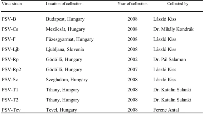
Table 1: Collection data of PSV isolates analysed
Virus strain Location of collection Year of collection Collected by
PSV-B Budapest, Hungary 2008 László Kiss
PSV-Cs Mezőcsát, Hungary 2008 Dr. Mihály Kondrák
PSV-F Füzesgyarmat, Hungary 2008 László Kiss
PSV-Ljb Ljubljana, Slovenia 2008 László Kiss
PSV-Rp Gödöllő, Hungary 2002 Dr. Pál Salamon
PSV-Rp2 Gödöllő, Hungary 2007 László Kiss
PSV-Sz Szeghalom, Hungary 2008 László Kiss
PSV-T1 Tihany, Hungary 2008 Dr. Katalin Salánki
PSV-T2 Tihany, Hungary 2008 Dr. Katalin Salánki
PSV-Tev Tevel, Hungary 2008 Ferenc Antal
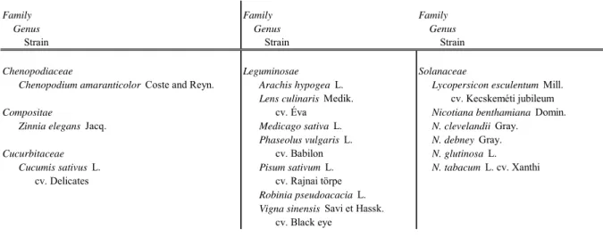
2.2 Examination of host range
Among natural and experimental host plants, 16 species of 5 plant families were infected with PSV isolates analysed (Table 2). In each case, three to eight plants were infected in a status having 2–8 leaves. Symptomatologically, plants were examined visually for up to 1 month after infection. In the case of symptomless plants, local and / or systemic spread of the pathogen was tested by Northern blot analysis (Sambrook et al. 1989). For the Northern blot analysis, samples involving complete nucleic acid extracted by using the method described by White and Kaper (1989) were denaturated in a buffer containing formaldehyde and formamide and were fractionated with electrophoresis on a 1% agarose gel containing
formaldehyde. Nucleic acids were transported to Hybond-N membrane and fixed to the membrane by UV light.
Table 2: Plants used for the analysis of host range
Chenopodiaceae Leguminosae Solanaceae
Chenopodium amaranticolor Coste and Reyn. Arachis hypogea L. Lycopersicon esculentum Mill.
Lens culinaris Medik. cv. Kecskeméti jubileum
Compositae cv. Éva Nicotiana benthamiana Domin.
Zinnia elegans Jacq. Medicago sativa L. N. clevelandii Gray.
Phaseolus vulgaris L. N. debney Gray.
Cucurbitaceae cv. Babilon N. glutinosa L.
Cucumis sativus L. Pisum sativum L. N. tabacum L. cv. Xanthi
cv. Delicates cv. Rajnai törpe
Robinia pseudoacacia L.
Vigna sinensis Savi et Hassk.
cv. Black eye
Family Genus Strain Family
Genus Strain
Family Genus Strain
To determine the nucleotide sequence, virus was purified from isolates propagated on N. benthamiana test plants in the case of PSV-Rp by using the method described by Lot et al.
(1972). Regarding plants infected by further isolates, complete nucleic acid extraction was carried out using the method of White and Kaper (1989) aforementioned. Phenol-chloroform extraction and precipitation with ethanole was used for extracting RNA.
2.3 Molecular and bioinformatic analyses
2.3.1. Synthesis of cDNA clones of PSV-Rp strain
Three genomic and one subgenonic virus RNAs characterised of cucumoviruses were extracted with phenol/SDS from purified virus particles. RNAs were detected by gel electrophoresis. During the synthesis of cDNA, RNAs were polyadenylated for determining the 3’ ends of RNAs. In accordance with that, the first-strand cDNA synthesis was primed with oligo dT primer, having an opposite (complementary) nucleotide sequence. The second strand cDNA was synthesized in the presence of ribonuclease H and DNA polymerase I, as described by Gubler and Hoffman (1983). The cDNA segments obtained were ligated into pBluescriptII SK(+) phagemid cloning vectors. The cDNA clones were propagated on Escherichia coli DH5-α and GM2163 strains. The cDNA clones of virus origin were selected by colony hybridization, indicating the purified virus RNA (Sambrook et al. 1989). The virus origin of nucleotide sequence of cDNA clones selected was controlled with automated fluorescence-based stopnucleotide method (Applied Biosystems Gene Analyzer 3100). On the basis of the sequences partially known, the 5’ terminal sequence of the three genomic RNAs was determined using the 5’ Rapid Amplification of cDNA Ends kit. Virus-specific antisense
primers including RNS1-(486–513 nt), RNS2-(257–276nt), and RNS3-(269–288nt) designed by us were used for the determination.
With knowledge of 5’ and 3’ real terminal sequences, the synthesis of full-strand cDNA was primed with oligonucleotide PSV123–3’-PstI for all three virus RNAs. The synthezis was performed with M-MuLV reverse transciptase.
The two-strand molecules synthesized were multiplied by PCR (Mullis and Faloona, 1987). The homology of 5’ terminal sequences of RNA1 and RNA2 enabled to multiply those in a joint reaction using the PSV12-T7-BamHI primary sequence. In the case of RNA3, PSV3-T7-BamHI primary sequence was used for multiplying the molecule. During the PCR, cDNA molecules were propagated in the presence of Taq DNA polymerase enzyme, using the 5’ and 3’ primers, the latter used uniformly for cDNA synthesis. In the case of RNA1 and RNA2, the reaction occurred in two phases. During the synthesis of RNA3, the reaction took place in one phase, within 30 cycles. The size-specific PCR products obtained were detected on a 1% agarose gel and purified afterwards (High Pure Purification Kit). The cDNA molecules were digested with restriction endonucleases, and ligated into pBluescriptII SK+
vector digested with the same enzymes. The PSV-Rp RNA1, RNA2, RNA3 clones completed were preserved in the DH5α and GM2163 strains of Escherichia coli. The nucleotide sequence of clones was determined with the aid of subclones and internal primers, using the automated fluorescence-based stopnucleotide method. Sequence data of the partial nucleotide sequences were joined by using the Clone manager 7 (SciEd Central) and Emboss-Align (Rice et al. 2000) programs, and deposited in the GeneBank international databank.
2.3.2 Synthesis of partial cDNA clones of PSV isolates
In the case of the further 9 PSV isolates tested, complete nucleic acid extraction and reverse transcription was performed from the infected N. benthamiana plants. Following that, a particular sequence of RNA3 was propagated by PCR technique, including a certain segment of movement protein gene, the full intergenic region, the coat protein gene and the 3’
untranslated region. Degenerated uniPSV5’ and PSV123-PstI primer pairs designed for the PSV strains found in the GeneBank and for the PSV-Rp RNS3 conservative region defined by us were used for the tests. PCR technique was used for the multiplication of heteroduplex- specific sequences. The PCR products obtained were detected on a 1% agarose gel and were purified (High Pure Purification Kit, Roche). The DNA samples were ligated into a pGEM-T- Easy cloning vector, thus synthesizing the cDNA clones of the partial RNA3 molecule of
isolates. The nt sequence of cDNAs was determined with automated fluorescense-based stopnucleotide method using the plasmide-specific M13 reverse and M13 forward primers.
2.4 Bioinformatic analyses
2.4.1 Nucleotide sequence comparison and phylogenetic analysis
The nucleotide and amino acid sequences obtained were compared using the Emboss- Align and Clustal X (Thomson et al. 1997) program kits with PSV sequences available in the EMBL/NCBI international databank. Following that, the Neighbor-Joining procedure was used for carrying out a phylogenetic tree from the data on the surface of TreeWiew 1.6.6.
(Page, 1996) using the Clustal X program.
2.4.2 Recombination analyses
For the recombination analyses built on PSV sequences available in international databanks, the PDM (Probabilistic Divergence Measures) analysis of TOPALi v2 (Milne et al.
2004) program kit generally used and approved was used.
3. RESULTS
3.1 Molecular characterisation of the cDNA clones of PSV-Rp
During our work the cDNA clones of RNA1, RNA2 and RNA3 of Peanut stunt virus collected from the domestic black locust were synthesized and named as ’PSV-Rp isolate’.
After that the complete primary structure of all three of clones was determined. The cDNA sequences obtained were deposited in the GeneBank international database (AccNo:
AM905353, AM905354, AM905355). The length of RNA1, RNA2 and RNA3 was found to be 3,325 nt, 2,942 nt, and 2,208 nt, respectively. These data are approximately identical with the length of other PSV sequences found in the databank. After determining the nt sequence, the five open reading frames (ORF) characteristic of cucumoviruses were identified.
The 1a ORF transcripted from RNA1 ranges from 85 nt to 3,078 nt, coding 998 amino acids. The H…DxxR…Y motive referring to the activity of methyltransferase can be identified at the N-terminal sequence of the molecule. In the C-terminal sequence there are helicase motives and a region rich in proline can be observed between amino acids 540 to 555.
The 2a ORF transcripted from RNA2 ranges from 80 nt to 2,581 nt, coding 834 amino acids. Amino acids 601–603 of ORF code a GDD motive, which is significant regarding the Mg2+ bond of RNA dependent RNA polymerases (RdRp). Also RNA2 determines a 2b ORF overlapping with 2a, located between 2,406 nt to 2,684 nt, from which a protein coding 93 amino acids is transcripted.
The RNA3 codes the 3a and 3b Open Reading Frames with an intermediate IR of a length of 257 nt. The sequence between 1,143–1,152 nt of IR includes the 5’- GGTTCAATTC-3’ conservated motive. The MP gene ranges from 142 nt to 1,011 nt, coding 290 amino acids. The amino acid sequence deduced from the nt sequence includes the MSVPQVLCAITRTVMANAEGSIRIYLADLGDDE conservated region of a length of 33 aa characteristic of the 30K superfamily. In the case of PSV-Rp (underlined aa-s), the above can differ from the other PSV isolates by 8 amino acids.
Regarding the RNA3 molecule, the ORF3b ranges from 1,269 nt to 1,919 nt, coding 217 amino acids. The N-terminal part of CP (10–17 aa) is rich in arginine.
The complete nucleotide sequence of PSV-Rp, as well as the nt and coded amino acid sequence of the individual ORFs were compared with the sequence data of the same regions of some cucumovirus isolates available in the international database. Complete nt sequence comparison of PSV-Rp RNAs showed 74.1–84.6% identity with PSV isolates deriving from
the different subgroups, whereas the identity is the following for the entire genome: 79.5% for PSV-J, 83.1% for PSV-W, and 78.2% for PSV-Mi. Identity with other cucumovirus isolates varied between 56.8–68.1% at this level.
With regard to ORF1a, ORF2a, ORF2b and ORF3a, PSV-Rp RNS1 and RNS2 showed the highest identity with PSV-W belonging to subclass II, whereas approximately the same level of identity was observed for RNA3 for all three subgroups (80.9–83.9%). In terms of ORF3b coding the CP, the PSV-J showed 83.3% identity, PSV-Mi showed 83.2% identity, whereas the PSV-W showed 78.8% identity with PSV-Rp at the nucleotide level. At the amino acid level, the highest degree of identity was observed in the case of PSV-Mi belonging to subgroup III. In every case, nt values obtained were considerably far from the 90% limit indicating the belonging to the same subgroup. Based on the results, our isolate remarkably differed from the other PSV isolates and could not be classified into any of the subgroups known. Low levels of identity verify the establishment of subgroup IV.
3.2 Phylogenetic and recombination analysis of PSV-Rp
Using the nucleotide sequence of genes of cucumovirus isolates, phylogenetic trees were made. With regard to genes coding the 1a, 2a and 3a proteins, PSV-Rp showed the closest phylogenetic relationship to PSV-W. Yet, it significantly separated from PSV-W at the subgroup level. In the case of the 3b gene coding the CP, however, the closest relationship was observed to PSV-Mi.
The divergent phylogenetic results, as well as the low sequence identity values defined for RNAs clearly referred to the presence of a recombination event taking place at the RNA3 level during the evolution of PSV.
To clarify the above, recombination tests were carried out on the complete PSV RNA3 sequences available in the GeneBank. During the analysis, 2 recombination points (hot spots) presumed were defined at the 95% significance level in the region around 1,199 nt and 1,873 nt. One of the recombination points is located in the IR, 70 nt ahead of the ORF3a, while the other point is situated 47 nt ahead of the stop codone of 3b gene coding the CP. Taking the sequences determined by recombination points dividing the RNA3 molecule into 3 sections a phylogenetic analysis was carried out again. Regarding the region of a length of approx. 1,200 nt ahead of the first hot spot, and the region of a length of 335 nt after the second hot spot, the PSV-Rp showed the closest relationship to PSV-W. Whereas the nucleotide section of a length of 674 nt between the two hot spots showed the closest relationship to PSV-Mi. The results obtained prove the different origin of the central region, and are in line with the results
of nt sequence identities. Results of the phylogenetic trees confirm that a recombination occurred between the isolates during the evolution of PSV.
3.3 Molecular characterisation of PSV isolates deriving from the Pannon ecoregion Following the complete molecular analysis of PSV-Rp, the characterisation of 9 additional PSV isolates deriving from the Pannon ecoregion and isolated from black locust was performed. During the molecular work, the RNA3 molecule of PSV isolates propagated on N. benthamiana test plants were partially cloned, and its nt sequence was determined.
Sequence data determined were deposited in the GeneBank international database.
The size of nucleotide sequence of the 9 gene coding partial MP, the complete IR, the gene coding the CP, and the 3’ NCR region varied between 1,342 to 1,351 nt, except the PSV- Cs isolate of a length of 1,542 nt. In the 3’ NCR region of the latter, a 196 nt duplication was observed. The RNA3 segments analysed completely included the region that implied the two recombination points and the CP gene in the case of PSV-Rp (Figure 1).
CP gen 3a
1201 1873
5’ oligo 3’ oligo
Figure 1. The PSV RNA3 molecule
The nucleic acid sequence determined for isolates deriving from the Pannon ecoregion is indicated with green colour. Red arrows and the numbers above indicate the recombination points determined for PSV-Rp.
The nucleotide and coded amino acid sequence of CP regions of the 9 isolates analysed were compared to each other and to the nucleotide and amino acid sequence of the same region of some cucumovirus isolates available in the international database. Nucleotide sequence of isolates deriving from the Pannon ecoregion showed 96.3–98.0% identity, whereas the amino acid sequence of those showed 98.1–100% identity with that of PSV-Rp isolate. Regarding the other cucumovirus isolates, the following identities were established:
PSV-J (nt: 82.1–83.2%, aa: 81.1–82.9%), PSV-W (nt: 77.6–79.3%, aa: 70.5–72.8%), PSV-Mi (nt: 82.6–84.3%, aa: 85.3–86.6%), CMV-Trk7 (nt: 55.3–58.6%, aa: 49.3–50.2%), CMV-Fny (nt: 53.4–54.8%, aa: 47.5–49.3%), TAV-KC (nt: 65.2–66.7%, aa: 70.2–72.4%), GMMV (nt:
63.5–65.4%, aa: 70.5–72.4%). The high degree of identity among isolates deriving from black locust clearly indicates that these isolates belong to the same subgroup, whereas close relationship can be excluded if compared with other PSV isolates.
3.4 Phylogenetic analysis of PSV isolates deriving from the Pannon ecoregion
Taking several cucumoviruses as a basis, phylogenetic analyses were carried out for the segment between the 2 recombination points (Figure 2a) and the complete cloned region including the recombination points (Figure 2b) determined in the case of PSV-Rp. In the first case, the isolates showed the closest relationship to subgroup III, whereas the relationship proved to be the closest to subgroup II as a result of analysing the entire region. Both results were identical with former results gained for PSV-Rp, i.e., a recombination occurred between subgroups II and III. It can be observed on the evolutionary tree that isolates deriving from the Pannon ecoregion are clearly separated from the other samples tested, and form a new, clearly confinable subgroup.
0.1
BMV-F CMV-Fny
CMV-Trk7 1000
TAV-KC PSV-J
PSV-P PSV-ER 821
1000
PSV-W PSV-Mi PSV-S PSV-Rp2
PSV-T2 PSV-Rp PSV-Tev 883 426
PSV-B PSV-Cs
PSV-Sz PSV-T1 PSV-F PSV-Ljb 707 428 890 538 656 365 1000
958 I
II III
IV
a
0.1
BMV-F
CMV-Trk7
CMV-Fny 1000
TAV-KC
PSV-MI
PSV-P
PSV-J
PSV-Er 534 1000
PSV-W
PSV-Cs
PSV-T1 PSV-Ljb
PSV-F
PSV-Sz 545 622 863
PSV-B
PSV-T2
PSV-Rp2
PSV-Tev
PSV-Rp 916 446 343 421 747 988 1000 1000 b
I
II III
IV
Figure 2: Phylogenetic analysis of the different regions of cucumovirus RNA3 molecules
A: analysing the nt sequence between the recombination points, b: analysing the nt sequence coding the partial MP, the complete IR, the complete CP, and the segment including the 3’ NCR regions. The latter also includes the recombinant region. Numbers indicated at the branches show the result of bootstrap analysis, while BMV-F is a control isolate beyond the group. Squares and Roman numbers contribute to the visual separation and identification of PSV groups.
3.5 Host range examination and sympthomic characterisation of PSV isolates deriving from the Pannon ecoregion
Following the molecular and phylogenetic analyses, infection and symptomic characteristic features of all the 10 PSV isolates were analysed on 16 species of 5 plant families. Regarding symptomless plants, presence of the virus was checked with Northern blot both at the inoculated and upper leaves to decide whether the plant concerned has latent infection.
As a result of infection induced by the PSV isolates analysed, the plants started to stunt and upper leaves became mosaic. As regards C. amaranticolor, each PSV isolate induced chlorotic local lesions on the inoculated leaves. The virus systemized in the plant, as well as mosaic patches and leaf malformation occurred on peak leaves. Following the infection, no symptoms were observed on lentils and peas. The Northern hybridisation, however, showed the presence of pathogen; the virus was latently present on the upper leaves of plants in every case. During artificial infections, mild mosaic was observed on seedling black locust plants, with additional ringspot in the case of certain isolates (PSV-Rp, PSV-Rp2, PSV-T2). By the end of the test period taking one month, the completemasking of symptoms was observed on the plants.
Among the Nicotiana species tested, significant differences were observed only on N.
glutinosa test plants from a symptomic perspective. Pathogens were present symptomless in the inoculated leaves in each case, which was verified by the Northern analysis made from samples taken the first week after infection. Systemic movement of pathogens, however, could be seen with the unaded eye only in the case of PSV-Rp2, PSV-F, PSV-Sz and PSV- Ljb. In those cases, mosaic and ringspot could be observed on the upper leaves, whereas even radioactive indication did not show the systemic spread of pathogens in the other cases.
Regarding the plants of Cucumis sativus cv. Delicates, Lycopersicon esculentum cv.
Kecskeméti jubileum, N. debney, N. tabacum cv. Xanthi, none of the isolates could be detected from the inoculated or systemically infected leaves.
3.5 New scientific results
1. During our work, the cDNA clones of all three of genomic RNAs of PSV-Rp isolate deriving from black locust in Hungary were synthesized.
2. The complete primary structure of genomic RNAs of the pathogen was determined and deposited in the international database.
3. The sequence of PSV-Rp isolate is the first complete PSV sequence known both from Europe and black locust host.
4. The detailed nucleotide sequence of RNA molecule of the other 9 PSV isolates collected from black locust deriving from the Pannon ecoregion, as well as the amino acid sequence of derived CP were determined. Based on the nucleotide sequence, all the PSV isolates collected have shown high degree of identity, and considerably differed from isolates of other known subgroups. Based on the above the establishment of subgroup IV is proposed.
5. Extended characterisation of the host range and symptomatology of isolates of subgroup IV was carried out, during which the narrowing of host range and the occurrence of latent infections was experienced.
6. Based on the complete nucleotice sequence and the phylogenetic and recombination analyses we found that a recombination connected to 2 hot spots was carried out during the evolution of PSV isolates examined. Recombination points took place in the IR and in the region coding the CP, and most probably took place between isolates of subgroups II and III. The recombination experienced during the analysis of PSV-Rp is the first recombination event precisely characterised for cucumoviruses deriving from natural ecosystems.
4. DISCUSSION OF RESULTS, CONCLUSION
In the light of results, the following findings were made after analysing the nucleotide sequence of PSV-Rp isolated by us:
Following the determination of the complete nucleotide sequence we found that the length of the three genomic RNAs was identical with the genome dimensions generally known of cucumoviruses. All the five ORFs could be found in the nucleic acid sequence of RNA, the location thereof is also identical with that in cucumoviruses.
The complete nucleotide sequence of RNAs of PSV-Rp showed 74.1–84.6% identity with PSV isolates deriving from different subgroups. Based on the above and on criteria provided for ICTV cucumoviruses (Roosinck et al. 2006), PSV-Rp is an isolate clearly belonging to the Peanut stunt virus strain. Hajimorad et al. (1999) proposed that isolates classified within the same subgroup should have at least 90% nt sequence identity, while the nt sequence identity between the different subgroups should be between 70 to 80%. PSV-Rp is clearly far from all three of PSV subgroups, since at the nucleotide level the degree of identity slightly higher than 80% for all three of genomic RNAs could be observed only in the case of PSV-W belonging to subgroup II. These values, however, are far below the degree of identity (90%) determining the belonging to the same subgroup. On the basis of the above, it is justified to establish a new, fourth subgroup, in which the PSV-Rp deriving from black locust can be regarded as a type isolate characteristic of the subgroup.
4.1 The role of recombination in the evolution of PSV
By using the TOPALi v2 (Milne et al. 2004) program kit, two recombination points were identified that intercept the great majority of the CP gene of PSV-Rp. In the recombination region it showed the closest relationship to PSV-Mi of subgroup III. The results gained during identification, phylogenetic and recombination alalytical tests of nucleotide sequence all prove that recombination occurred during the evolution of PSV.
The rate of recombination occurrence is fairly frequent in genus Cucumovirus (Codoñer and Elena, 2008). In most cases, these recombinations take place in RNA3, which figure is identical with our own research results. Occurrence of recombinations were observed between CMV and TAV isolates, as well as between CMV isolates as a result of mixed infection under greenhouse conditions. Recombinations occurred at the RNA3 level in the regions of NCR and IR, as well as in the gene coding CP and MP. The recombinant isolates, however, were less viable than the parent isolates, therefore were selected within a short
period of time (de Wispelaere et al. 2005; Pierrugues et al. 2007). Analyses of isolates collected in the open also proved the poorer viability and the contraselection of isolates (Bonnet et al. 2005; Escriu et al. 2007). Remaining of the recombination in the case of PSV- Rp is probably due to the fact that almost the entire gene had been replaced. In this case, there is higher chance for survival of the recombinant pathogen (Bonnet et al. 2005; Escriu et al.
2007). In the light of results we can establish that PSV-Rp is the isolate of subgroup IV, separated from the isolates known so far. The above also proves that a recombination event played a significant role in the evolution of PSV. Although, we do not know its evolutionary advantage which contributed to the remaining and spread of the recombinant virus.
4.2 Molecular characterisation of PSV isolates deriving from the Pannon ecoregion
Following the molecular characterisation of PSV-Rp, several questions arose, including whether the isolate is unique, or there are other isolates with similar genetic stock in the Carpathian Basin.
With regard to domestic data, the isolate collected near Putnok from red clover proved that the pathogen can not be classified into any subgroups. The molecular technology of that time did not enable to determine the nucleotide sequence; the isolate was lost, therefore it cannot be characterised. We can make consequences to the level of relationship between PSV-Rp and PSV-Tp from serological and host range results only.
During our research, the molecular and pathological characterisation of 9 further PSV isolates collected from black locust was carried out. The partial RNA3 cDNA clones were synthesized, which included the region surrounded by the recombination points determined for PSV-Rp. The size of sections examined was approximately the same, except the PSV-Cs isolate collected in Mezőcsát, where a duplication of RNA3 of a length of 196 nt was identified in the NCR region. This phenomenon has not been known so far, its occurrence is fairly typical for TAV isolates within cucumoviruses (Palukaitis and Garcia-Arenal; 2003).
Since the PSV isolates showed an identity above 90% (described by Hajimorad et al. 1999) with the PSV-Rp isolate in the regions tested, the classification of isolates is unequivocal. On the grounds of the above, the 9 isolates deriving from the Pannon ecoregion are in close relationship to the PSV-Rp and may be regarded as isolates belonging to subgroup IV.
Phylogenetic analyses verified that the other isolates tested also include the recombination observed in the case of PSV-Rp. Therefore, it is not only characteristic of PSV-Rp isolate but also the occurrence of recombinant isolates is common in the area of Pannon ecoregion for samples collected from black locust.
4.3 Host range analysis of PSV isolates deriving from the Pannon ecoregion, characterisation of symptoms caused
According to information available, PSV is provided with much diverse genetic background as compared to CMV. Still, CMV has more than 1,000 hosts, while PSV infects 73 species only.
During our research, also symptomatology analyses confirmed that the 10 PSV isolates analysed significantly differ from the three PSV subgroups currently known. Isolates induce similar symptoms on the 16 hosts tested. At the same time, it can be observed that the isolates often induce no or mild symptoms on the plants analysed. On the basis of results we assume that the adaptation of pathogen to black locust causes symptomic changes. Yet, a more certain picture on this issue can be gained only after analysing the isolates collected from other hosts.
According to literature records available, the C. amaranticolor species is infected systematically only by isolates of subgroup III. In the other cases [e.g. PSV-Tp isolate in Hungary (Beczner and Devergne, 1979)], isolates induced chlorotic (neurotic) local lesions on the infected leaves. The current research proved that isolates of the subgroup IV also systemize in the infected plants of C. amaranticolor. In addition, the heavy malformation of upper leaves can be observed. Since the phylogenetic and recombination analyses showed the closest relationship between PSV isolates and subgroup III in the region coding the CP, we assume that also the CP gene played a role in the occurrence of systemic spread in C.
amaranticolor. This result has been verified by several former researches (Taliansky and Garcia-Arenal, 1995; Salánki et al. 1997). On the basis of systemization and leave malformation, C. amaranticolor is proposed for separating the fourth subgroup.
Symptoms caused by different PSV isolates tested deviated significantly only in the case of N. glutinosa test plant. While the majority of isolates could be detected only from inoculated leaves during the Northern blot analysis, the pathogen caused mosaic and ringspot on upper leaves. Du et al. (2008) mentioned the role of CP and 2a protein played in the long- distance movement of the virus or in the inhibition thereof. The aa sequence of CP of PSV-Rp and PSV-Rp2 producing different symptoms is identical to a degree of 100%, therefore the role of 2a protein may be determinant in the long-distance movement of PSV isolates in the case of N. glutinosa. The analysis of aa sequence of 2a proteins is ongoing in our research team.
5. LITERATURE
1. Beczner, L. and Devergne, J.C. (1979): Characterization of a New Peanut Stunt Virus Strain Isolated from Trifolium pratense L. in Hungary. Acta Phytopatologica Academiae Scientiarum Hungaricae, 14: 247–267.
2. Bonnet, J., Fraile, A., Sacristan, S., Malpica, J.M. and Garcia-Arenal, F. (2005): Role of recombination in the evolution of natural populations of Cucumber mosaic virus, a tripartite RNA plant virus. Virology, 332: 359–368.
3. Codoñer, F.M and Elena, S.F. (2008): The promiscuous evolutionary history of the family Bromoviridae. Journal of General Virology, 89: 1739–1747.
4. de Wispelaere, M., Gaubert, S., Trouilloud, S., Belin, C. and Tepfer, M. (2005): A map of the diversity of RNA3 recombinants appearing in plants infected with Cucumber mosaic virus and Tomato aspermy virus. Virology, 331: 117–127.
5. Du, Z., Chen, F., Zhao, Z., Liao, Q., Palukaitis, P., and Chen, J. (2008): The 2b protein and the C-terminus of the 2a protein of Cucumber mosaic virus subgroup I strains both play a role in viral RNA accumulation and induction of symptoms. Virology, 380:
363–370.
6. Escriu, F., Fraile, A. and García-Arenal, F. (2007): Constraints to genetic exchange support gene coadaptation in a tripartite RNA virus. PLoS Pathogenes, 3 e8.
doi:10.1371/s0030008.
7. Gubler, U., Hoffman, B.J. (1983): A simple and very efficient method for generating cDNA libraries. Gene, 25: 263–269.
8. Hajimorad, M.R., Hu, C.C. and Ghabrial, S.A. (1999): Molecular characterization of an atypical old world strain of Peanut stunt virus. Archives of Virology, 144: 1587–
1600.
9. Lot, H.W., Marrou J., Quiot J.B. and Esvan C. (1972): Contribution à l’étude du virus de la mosaique du concombre (CMV). I. Méthode de purification rapide du virus.
Annual Review of Phytopathology, 4: 25–38.
10. Milne, I., Wright, F., Rowe, G., Marshall, D.F., Hushmeier, D. and McGuire, G.
(2004): TOPALi: software for automatic identification of recombinant sequences within DNA multiple alignments. Bioinformatics, 20: 1806–1807.
11. Mullis, K. B. and Faloona, F. A. (1987): Specific synthesis of DNA in vitro via a polymerase-catalyzed chain reaction. Methods in Enzymology, 155: 335–350.
12. Page, R. D. M. (1996): TreeView: An application to display phylogenetic trees on personal computers. Computer Applications in the Biosciences, 12: 357–358.
13. Palukaitis, P. and García-Arenal, F. (2003): Cucumoviruses. Advances in Virus Research, 62: 241-323.
14. Pierrugues, O., Guilbaud, L., Fernandez-Delmond, I., Fabre, F., Tepfer, M. and Jacquemond, M. (2007): Biological properties and relative fitness of inter-subgroup cucumber mosaic virus RNA 3 recombinants produced in vitro. Journal of General Virology, 88: 2852–2861.
15. Rice, P., Longden, I. and Bleasby, A. (2000): EMBOSS: the European molecular open software suite. Trends in Genetics, 16: 276–277.
16. Roosinck, M.J., Bujarski, J., Ding, S.W., Hajimorad, R., Hanada, K., Scott, S. and Tousgnant, M. (2006): Index of Viruses – Bromoviridae In: ICTVdB – The Universal Virus Database, version 4. Büchen-Osmond, C (Ed), Columbia University, New York, USA http://www.ncbi.nlm.nih.gov/ICTVdb/Ictv/fs_ index.htm
17. Salánki, K., Carrére, I., Jacquemond, M., Balázs, E. and Tepfer, M. (1997): Biological properties of pseudorecombinant and recombinant strains created with cucumber mosaic virus and tomato aspermy virus. Journal of Virology, 71: 3597–3602.
18. Sambrook, J., Fritsch, E. F. and Maniatis, T. (1989): Molecular cloning: A laboratory manual (2nd edition). Cold Spring Harbor, N.Y: Cold Spring Harbor Laboratory.
19. Taliansky, M.E. and García-Arenal, F. (1995): Role of cucumovirus capsid protein in long-distance movement within the infected plant. Journal of Virology, 69: 916–922.
20. Thompson, J.D., Gibson, T.J., Plewniak, F., Jeanmougin, F. and Higgins, D.G. (1997):
The Clustal X windows interface: flexible strategies for multiple sequence alignment aided by quality analysis tools. Nucleic Acids Research, 24: 4876–4882.
21. White, J.L. and Kaper, J.M. (1989): A simple method for detection of viral satellite RNAs in small tissue samples. Journal of Virological Methods, 23: 83–94.
6. PUBLICATIONS PUBLISHED ON THE TOPIC OF DISSERTATION Publications in journals with IF:
• Kiss, L., Sebestyén, E., László, E., Salamon, P., Balázs, E. and Salánki, K. (2008):
Nucleotide sequence analysis of Peanut stunt virus Rp strain suggests the role of homologous recombination in cucumovirus evolution. Archives of Virology, 153:
1373–1377. p. (IF: 2,02)
• Kiss, L., Balázs, E. and Salánki, K. (2009): Characterisation of black locust isolates of Peanut stunt virus (PSV) from Pannon ecoregion show the frequent occurrence of the fourth taxonomic PSV subgroup. European Journal of Plant Pathology, 125: 671–
677. (IF: 2,054)
Publications in journals without IF:
• Kiss L. Salánki K. és Balázs E. (2008): Magyarországi, fehér akácról (Robinia pseudoacacia L.) származó földimogyoró satnyulás vírus (Peanut stunt virus, PSV) izolátumok jellemzése. Növényvédelem, 44: 573–578.
Conference publications (abstract)
• Kiss, L., Sebestyén, E., László, E., Salamon, P., Balázs, E. and Salánki, K. (2007):
Nucleotide sequence analysis of Peanut stunt virus Rp isolate, prove the role of recombination in Cucumovirus evolution. 15th International Congress of the Hungarian Society for Microbiology, (July 18-20, Budapest) 61. p.
• Kiss, L., Sebestyén, E., László, E., Salamon, P., Balázs, E. and Salánki, K. (2008):
Molecular characterization of a black locust strain of Peanut stunt virus. The 3rd Conference of the International Working Group on Legume and Vegetable Viruses, (August 20-23, Ljubljana, Slovenia) 61. p.
• Kiss L., Sebestyén E., László E., Salamon P., Balázs E., Salánki K. (2008): In vivo rekombináns földimogyoró satnyulás virus (Peanut stunt virus, PSV) izolátum molekuláris jellemzése. 54. Növényvédelmi Tudományos Napok, Növénykórtani szekció, (február 27-28, Budapest) 29. p.
• Kiss L., Salánki K., Balázs E. (2009): Pannon ökorégióból származó földimogyoró satnyulás vírus (Peanut stunt virus, PSV) izolátumok jellemzése. 55. Növényvédelmi Tudományos Napok, Növénykórtani szekció, (február 23, Budapest)