Lower Limb following Ischemia-Reperfusion Using mPTP Inhibitor NIM-811
David Garbaisz1*, Zsolt Turoczi1, Peter Aranyi1, Andras Fulop1, Oliver Rosero1, Edit Hermesz2, Agnes Ferencz2, Gabor Lotz3, Laszlo Harsanyi1, Attila Szijarto1
1Semmelweis University, 1stDepartment of Surgery, Budapest, Hungary,2University of Szeged, Department of Biochemistry and Molecular Biology, Szeged, Hungary, 3Semmelweis University, 2ndDepartment of Pathology, Budapest, Hungary
Abstract
Introduction:Operation on the infrarenal aorta and large arteries of the lower extremities may cause rhabdomyolysis of the skeletal muscle, which in turn may induce remote kidney injury. NIM-811 (N-metyl-4-isoleucine-cyclosporine) is a mitochondria specific drug, which can prevent ischemic-reperfusion (IR) injury, by inhibiting mitochondrial permeability transition pores (mPTP).
Objectives:Our aim was to reduce damages in the skeletal muscle and the kidney after IR of the lower limb with NIM-811.
Materials and methods: Wistar rats underwent 180 minutes of bilateral lower limb ischemia and 240 minutes of reperfusion. Four animal groups were formed called Sham (receiving vehicle and sham surgery), NIM-Sham (receiving NIM- 811 and sham surgery), IR (receiving vehicle and surgery), and NIM-IR (receiving NIM-811 and surgery). Serum, urine and histological samples were taken at the end of reperfusion. NADH-tetrazolium staining, muscle Wet/Dry (W/D) ratio calculations, laser Doppler-flowmetry (LDF) and mean arterial pressure (MAP) monitoring were performed. Renal peroxynitrite concentration, serum TNF-aand IL-6 levels were measured.
Results:Less significant histopathological changes were observable in the NIM-IR group as compared with the IR group.
Serum K+and necroenzyme levels were significantly lower in the NIM-IR group than in the IR group (LDH: p,0.001; CK: p, 0.001; K+: p = 0.017). Muscle mitochondrial viability proved to be significantly higher (p = 0.001) and renal function parameters were significantly better (creatinine: p = 0.016; FENa: p,0.001) in the NIM-IR group in comparison to the IR group. Serum TNF-a and IL-6 levels were significantly lower (TNF-a: p = 0.003, IL-6: p = 0.040) as well as W/D ratio and peroxynitrite concentration were significantly lower (p = 0.014; p,0.001) in the NIM-IR group than in the IR group.
Conclusion:NIM-811 could have the potential of reducing rhabdomyolysis and impairment of the kidney after lower limb IR injury.
Citation:Garbaisz D, Turoczi Z, Aranyi P, Fulop A, Rosero O, et al. (2014) Attenuation of Skeletal Muscle and Renal Injury to the Lower Limb following Ischemia- Reperfusion Using mPTP Inhibitor NIM-811. PLoS ONE 9(6): e101067. doi:10.1371/journal.pone.0101067
Editor:Raghavan Raju, Georgia Regents University, United States of America ReceivedFebruary 19, 2014;AcceptedJune 2, 2014;PublishedJune 26, 2014
Copyright:ß2014 Garbaisz et al. This is an open-access article distributed under the terms of the Creative Commons Attribution License, which permits unrestricted use, distribution, and reproduction in any medium, provided the original author and source are credited.
Funding:The authors have no support or funding to report.
Competing Interests:In the present experimental study, NIM-811 was provided as a gift from Novartis International AG, Basel, Switzerland. Authors declare that there are no competing interests with Novartis International AG related to either employment, consultancy, patents, products in development, marketed products, etc. This does not alter the authors’ adherence to PLOS ONE policies on sharing data and materials.
* Email: garbaiszdavid@t-online.hu
Introduction
Arterial occlusive diseases are the most frequent causes of acute limb ischemia (ALI). Operative treatments for these diseases, as well as other reconstructive surgeries on abdominal aorta or other major arteries of the lower limb may induce sudden onset ischemia, in which event lower limb tissues suffer ischemic- reperfusion (IR) injuries.
There are two sides to IR injury since, in a paradox manner, reestablishment of the blood supply is associated with further damages. Beyond local muscle cell necrosis and consequent rhabdomyolysis, serious remote organ injuries may also develop.
The metabolites and proinflammatory cytokines released from the
damaged muscles [1–3] may induce systemic inflammatory response syndrome (SIRS), which could possibly affect the gastrointestinal system, lungs and kidneys, thereby inducing multiple organ dysfunction syndrome (MODS).
Mitochondria play a leading role in IR injury [4]. Damage caused by mitochondrial membrane depolarization can signifi- cantly jeopardize energy production, which can lead to cell damage, then further to cell death. The mechanism of mitochon- drial membrane depolarization, called mitochondrial permeability transition (MPT), is induced by the opening of the mitochondrial permeability transition pores (mPTP) in the mitochondrial membrane [5]. Several proteins participate in the structure of the pore, but the most important during mPTP opening is
cyclophilin-D in the mitochondrial matrix [6,7]. After opening of the pore, the mitochondrial electron transport chain is interrupted, which disarrays mitochondrial energy production and induces production of reactive oxygen species (ROS) [6]. Decline of the mitochondrial membrane potential causes further opening of the mPTPs, thus release of different molecules from the dysfunctional mitochondria may provoke cell death [6].
Cyclosporine-A (CsA) – besides its known immunosuppressive effects – is a potential inhibitor of the opening of mPTP. It can bind to both the cyclophilin-D component of mPTPs and the cytosolic cyclophilin-A molecules [8–11]. N-methyl-4-isoleucine cyclosporine (NIM-811) is a derivative of CsA, which does not bind to cyclophilin-A, therefore it has no immunosuppressive effects; it does however inhibit the opening of mPTP by binding to cyclophilin-D, thus preventing the development of MPT [12].
NIM-811 has an enormous advantage over CsA in that it has no known systemic side effects.
Based on our earlier investigations, attenuation of local IR damage can reduce the risk of remote organ injury (i. e. in the kidneys) [13–15]. During IR, opening of the mPTP plays a pivotal role in cell injury, thus inhibition of the pore opening may mitigate the damages. In the current study therefore, our aim was to examine the potentially favourable effects of NIM-811 as an inhibitor of mPTP opening in a skeletal muscle model of IR injury, with respect to local damages, consequential remote organ complications, as well as renal dysfunction.
Materials and Methods
Male Wistar rats (n = 40) weighing 220–250 g were used (Charles Rivers Hungary Ltd., Budapest, Hungary). Animals were kept under specific pathogen-free conditions at 22–24uC, on standard rat chow and water ‘ad libitum’. The experimental design was carried out in strict accordance with the recommen- dations in the Guide for the Care and Use of Laboratory Animals of the National Institutes of Health (The National Academies Press, Washington, D.C., USA, 8th edition). The protocol was approved by the Committee on Animal Experimentation of Semmelweis University (Permit Number: 22.1/2409/3/2011). All surgeries were performed under general anesthesia with efforts to minimize suffering.
Preoperative procedures
Animals were anesthetized by intraperitoneal injection of 75 mg/bwkg ketamine and 7.5 mg/bwkg xylazine. Anesthesia was maintained by intravenous (through right jugular vein) administration of 25 mg/bwkg/h ketamine and 2.5 mg/bwkg/h xylazine. Heating blankets were used to keep body temperature at 37uC 60.5uC, monitored with rectal probe (Homeothermic Blanket Control Unit, Harvard Apparatus, Holliston, MA, USA).
Mean arterial pressure (MAP) was registered by invasive blood pressure monitor (Kent Scientific Corporation, Torrington, CT, USA) through the internal carotid artery.
Surgical procedures and experimental design
Before induction of ischemia, a 20-minute baseline period was allowed. Tourniquet was applied around both lower limbs in the femoral region [16], and rats underwent a 180-minute long total bilateral lower limb ischemia. After median laparotomy, a laser- Doppler flowmeter (LDF) probe was placed over the front surface of the left kidney in order to monitor circulation of the renal parenchyma [17]. Following a 1 cm longitudinal skin incision along the lateral side of the thigh, another LDF probe was placed
on the femoral biceps muscle to monitor the microcirculation of the left lower limb [14].
The animals were randomized to the following four groups according to treatment: Sham (receiving vehicle and sham surgery, n = 10), NIM-Sham (receiving NIM-811 and sham surgery, n = 10), IR (receiving vehicle and surgery, n = 10) and NIM-IR (receiving NIM-811 and surgery, n = 10). Five minutes before reperfusion, NIM-811 (10 mg/bwkg; Novartis International AG, Basel, Switzerland) was administered intravenously to the NIM- Sham and NIM-IR groups. After removal of the tourniquet, reperfusion was allowed for 240 minutes. Sham animals received all procedures including anesthesia and laparotomy except for the 180-minute-long ischemia.
During the experiment, urine output was determined by gathering the spontaneously excreted urine in two fractions: (1) urine samples collected for 20 minutes before the ischemia served as control; (2) the amount of urine gathered during the last 3 hours of reperfusion was completed with bladder puncture at termina- tion of the experiment.
Determining skeletal muscle injury
Serum laboratory parameters. Tests consisted of measur- ing serum creatine kinase (CK), lactate dehydrogenase (LDH) and K+ levels. After centrifuging (2610 min, 1050 g) the blood samples, serum was snap-frozen in liquid nitrogen and stored at 280uC until analysed with a clinical chemistry analyzer automate (Beckman Coulter AU480/2011, Beckman Coulter Inc, Brea, CA, USA).
Histological evaluation of skeletal muscle. Samples were collected from the anterior tibial muscle and fixed in 4% neutral buffered formalin solution for one day. Thereafter samples were embedded in paraffin, 3mm thin cross- and longitudinal sections were cut and stained for hematoxillin and eosin. Histological examinations were carried out with Olympus BX50 microscope equipped with Olympus DP70 camera (Olympus Corporation, Tokyo, Japan). The examining pathologist received no informed about the applied pretreatment.
Assessment of muscle fiber viability. Samples from the left anterior tibial muscle were snap frozen in liquid nitrogen and stored at280uC until further processing. Cross sections of 3mm thickness were cut in cryostat. Slides were incubated for 30 minutes at 37uC in a solution consisting of nitroblue tetrazolium (1.8 mg/dL), NADH (15 mg/dL) and 0.05 M TRIS buffer (pH 7.6) (Sigma-Aldrich Inc, St. Louis, MO, USA). Unused tetrazolium reagent was removed using ascending and descending concentrations of acetone. Ten different fields were photographed randomly in each slide with 6006 magnification. Viability assessment was performed by detection of NADH-tetrazolium reductase staining of muscles using Leica QWin Pro (Leica Microsystems Ltd, Wetzlar, Germany) morphometric software.
Muscle fiber viability was calculated as a proportion of the total stained area and the total muscle fiber area of the slide. Average of the 10 slides was calculated for each animal. The final result is expressed as a percentage of the values of untreated control muscles (obtained in a previous experiment) [18].
Skeletal muscle Wet/Dry (W/D) ratio. Tissue edema was quantified with the wet/dry ratio utilizing the remaining tibial anterior muscle. Following careful excision, muscles were weighed immediately after the end of reperfusion (wet weight) and placed in a drying oven set at a temperature of +80uC until reaching constant weight. Muscles were then reweighed (dry weight) [19]
and wet/dry ratio calculated using the following equation: (wet weight – dry weight)/wet weight * 100.
Measurement of tissue microcirculation. Registration of microcirculation of the lower limb skeletal muscle was performed by laser Doppler flowmeter (MOOR Instruments Ltd, London, UK; DRT4). The probe of the device was placed on the surface of the left femoral biceps muscle. To characterize the microcircula- tion, reperfusion area (RA) was used, based on the mathematical calculations of our research group. The mathematical transfor- mations required for correct interpretation of the circulation data were described previously [20].
Systemic inflammatory parameters
Measurement of TNF-aand IL-6 levels. Proinflammatory cytokine concentrations were measured by sandwich ELISA kits according to manufacturer’s instructions (R&D Systems, Minne- apolis, MN, USA). Absorbance was measured at 450nm.
Determination of kidney dysfunction
Histopathology. Tissue sampling was performed from the same anatomical location (left kidney) in case of every animal, regardless of group. As in case of skeletal muscle samples, kidney samples were fixed in 4% neutral buffered formalin solution (24 h), followed by embedment in paraffin, then 3mm thin sections were cut and stained for hematoxillin and eosin. During evaluation of the sections (Olympus DP70 camera, Olympus Corporation, Tokyo, Japan), features of tubular and interstitial injuries were considered.
Evaluation of histological damage was performed in keeping with a score described previously [21]. The degree of tubular damage to the kidney tissue was scored by determining the percentage of tubules in the kidney cortex, which showed tubular damage, tubular cell necrosis and cast formation as follows: 0, none; 1#10%; 2, 10–25%; 3, 25–45%; 4, 45–75%; 5.75%. Ten randomly chosen, non-overlapping fields per section were examined. Scoring was carried out by an independent pathologist in a blinded manner.
Laboratory examinations. Blood samples were investigated according to the same method as serum necroenzymes. Levels of Na+-, creatinine- and BUN (blood urea nitrogen) were deter- mined, from which renal injury parameters (BUN/creatinine ratio, fractional Na+-excretion (FENa = UNa+6Pkreat. x100/Uk- reat.6PNa+) were calculated.
Registration of renal microcirculation. During the exper- iment, microcirculation of the left kidney cortex was registered with LDF, placing the laser probe on the anterior surface of the left kidney.
Measurement of peroxynitrite concentrations in the kidneys. Homogenized samples of kidney tissue were analyzed by spectrophotometric method after dilution in 1.0 M NaOH (60:1) solution; absorbance was measured at 302 nm. As control, 100 mM potassium-phosphate (pH 7.4) was added to the sample (60:1). The rate of decline in absorbance was measured on neutral pH [22].
Biochemicals
NIM-811 was the generous gift of Novartis International AG (Basel, Switzerland). The agent (10 mg/bwkg) was dissolved in the vehicle, which contained 1.3 mL cremophor oil (Cremophor EL, polyethoxylated castor oil), 0.7 mL ethanol and 8 mL 0.9% saline solution [23]. The injection volume range was standard (0.44–
0.5 mL) in each animal according to their weights (220–250 g).
Other reagents were purchased from Sigma-Aldrich Corporation (St Louis, MO, USA).
Statistical analysis
Data are shown as means 6SEM. Statistical analysis of data was performed using IBM SPSS Statistics 20.0 software (IBM Corporation, Armonk, NY, USA). Two-way analysis of variance (ANOVA) was used for comparison of all groups with LSD post- hoc tests. A 95% confidence interval was considered as statistically significant (p,0.05).
Results
Determining skeletal muscle injury
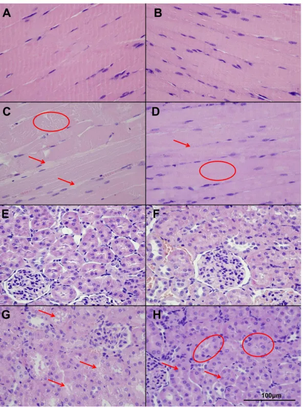
Histopathology. As compared with the nearly normal histological picture detectable in case of the Sham and NIM- Sham groups, segmental necroses and disintegrated filaments were observable in the muscle fibers in case of the IR group. The muscle fibers were separated by significant edema. By contrast, in the NIM-IR group, intact conditions were observable (Figure 1 A–
D).
Laboratory measurements. Both CK and LDH levels were significantly increased in the IR group compared with the Sham and NIM-Sham groups (values were almost similar in the latter two groups). Both examined parameters were significantly lower in the NIM-IR group than in the IR group (CK: p,0.001; LDH: p, 0.001). The serum K+level was higher in the IR group compared with the Sham and NIM-Sham groups, and the elevation was significantly lower in the NIM-IR group (p = 0.017) (Figure 2 A–B, Table 1).
Muscle fiber viability. In the IR group, muscle fiber viability was significantly decreased as compared with the Sham and NIM-Sham groups (where values were nearly 100 percent).
Viability in the NIM-IR group was significantly less reduced in comparison with the IR group (p = 0.001) (Figure 2C, Table 1).
Wet/Dry ratio of skeletal muscle tissues. Calculations of wet content of muscles revealed a significantly higher value in the IR group as compared with the Sham and NIM-Sham groups. In the NIM-IR group, the amount of tissue edema was significantly lower than in the IR group (p = 0.014) (Figure 2D, Table 1).
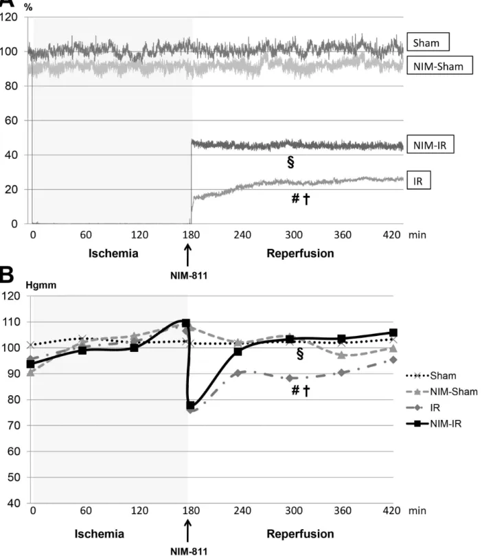
Microcirculation of skeletal muscle of the lower limb. Calculations of the reperfusion areas displayed signifi- cantly compromised microcirculation observable in the IR group when compared with the Sham and NIM-Sham groups.
Microcirculation became stabilized at a significantly higher level in the NIM-IR group than in the IR group (RA: p,0.001) (Figure 3A, Table 1).
Systemic parameters
Systemic hemodynamics. MAP values in case of the Sham and NIM-Sham groups remained at the same level during the entire experimental period. In case of both the IR and NIM-IR group, the MAP values showed a decline by 30 and 32 mmHg, respectively at the beginning of the reperfusion. After the beginning of the reperfusion the MAP values were found to be significantly higher in the NIM-IR group compared with the IR group (p = 0.044) (Figure 3B, Table 1).
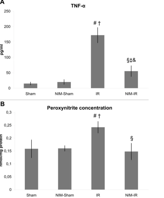
Measurement of TNF-aand IL-6 levels. Both TNF-aand IL-6 levels were markedly increased in the IR group in comparison with the Sham and NIM-Sham groups. The TNF-a level was considerably lower in the NIM-IR group than in the IR group (p = 0.003) (Figure 4A, Table 1). Regarding IL-6 measurements, significantly lower levels were noticeable in the NIM-IR group than in the IR group (p = 0.040). (Table 1).
Determination of kidney dysfunction
Histopathology. In the IR group, swollen tubular cells, intracellular vacuolization, disintegrated necrotic cells and blurred cell borders were observable, as compared with the Sham and
NIM-Sham groups. The morphological picture of the kidney was more favourable in the NIM-IR group, cell necrosis could rarely be seen, intact cell borders and nearly normal cell morphology were present. (Figure 1 E–H).
Figure 1. Muscle and kidney histopathology (hematoxillin-eosin (HE) stain, light microscopy). (A–B): An almost physiologically histological picture is observable in the Sham (A) and NIM-Sham (B) groups, with intact muscle fibers and normal wide interstitial spaces. (C):In the IR group segmental necrosis (circle), disintegrated myofilaments and thicker interstitial spaces (arrow) are detectable. (D):NIM-IR group shows intact muscle fibers (circle) with no expanded interstitial spaces (arrow), similar to the normal structure. (E–F):Sham and NIM-Sham groups show the normal structure of kidney cortical tissue. Tubules have normal appearance. (G):In the IR group massive injury can be detected with loss of cell integrity and intracellular vacuolization (arrows). (H):In the NIM-IR group cell necrosis is less severe (arrow) and the tubular integrity remained intact (circle), an almost normal picture can be seen.
doi:10.1371/journal.pone.0101067.g001
The applied histopathological score was significantly lower in the NIM-IR group, as compared with the IR group (p,0.001) (Table 1).
Urine output and laboratory findings. Based on the urine volume measured during the reperfusion, diuresis was significantly lower in the IR group than in the Sham and NIM-Sham groups.
In the NIM-IR group, urine output was significantly higher when compared with the IR group (p = 0.022) (Figure 5A, Table 1).
The serum creatinine level observable in the IR group was significantly higher than the levels found in case of the Sham and NIM-Sham groups. The level manifest in the NIM-IR group was significantly less elevated than in case of the IR group (p = 0.001) (Figure 5B, Table 1).
Calculated renal functional parameters. The fractional Na+-excretion was significantly higher in the IR group compared with the Sham and NIM-Sham groups. A significantly lower value
could be calculated in the NIM-IR group than in the IR group (p,0.001) (Figure 5D, Table 1).
The serum BUN/creatinine ratio was lower in the IR and NIM- IR groups compared with the Sham and NIM-Sham groups. No statistically significant differences could be seen between the IR and NIM-IR groups, though less decreased values were measured in the latter group (Figure 5C, Table 1).
Renal microcirculation. After onset of reperfusion, the microcirculatory flow rates of the IR and NIM-IR groups showed a declining tendency in comparison with the Sham and NIM- Sham groups; however, calculations of the reperfusion area (RA) values did not point to any significant differences measurable between the groups (Table 1).
Measurement of peroxynitrite concentrations in the kidney. Significantly higher concentration of peroxynitrite could be observed in the IR group when compared with the Sham and NIM-Sham groups. In case of the NIM-IR group, such Figure 2. Parameters of muscle injury, muscle fiber viability and muscle wet content.(A–B):Serum creatine-kinase (CK) and lactate- dehydrogenase (LDH) concentrations were significantly elevated in the IR group compared with the Sham and NIM-Sham groups. Significantly lower value was detectable in the NIM-IR group compared with the IR group, leading to the conclusion that there may be a lower extent of muscle necrosis (CK:#p,0.01 vs. Sham;{p,0.01 vs. NIM-Sham;1p,0.01 vs. IR;`p,0.01 vs. Sham; & p,0.01 vs. NIM-Sham; LDH:#p,0.01 vs. Sham;{p, 0.01 vs. NIM-Sham;1p,0.01 vs. IR;`p,0.01 vs. Sham; & p,0.01 vs. NIM-Sham; U/l – Unit/liter). (C):Muscle fiber viability was assessed by NADH- tetrazolium staining and was expressed as percentage of viability measured in untreated controls (%). In the IR group there was a significant decline in viability compared with the Sham and NIM-Sham groups. Significantly higher value was found in the NIM-IR group, compared with the IR group (#
p,0.01 vs. Sham;{p,0.01 vs. NIM-Sham;1p,0.01 vs. IR;`p,0.01 vs. Sham; & p,0.05 vs. NIM-Sham). (D):Wet/Dry ratio is suitable to determine the amount of interstitial edema. The wet content of the skeletal muscle tissue was significantly lower in the NIM-IR group compared with the IR group (#p,0.01 vs. Sham;{p,0.01 vs. NIM-Sham;1p,0.05 vs. IR;`p,0.05 vs. Sham; & p,0.05 vs. NIM-Sham).
doi:10.1371/journal.pone.0101067.g002
elevation was preventable by administering NIM-811 (p,0.001) (Figure 4B, Table 1).
Discussion
Acute limb ischemia is a sudden onset decline in the perfusion of the limb, which is a serious concern regarding limb function and even patient survival [24]. The aim of the present study was to set up an experimental model and search for a therapeutic option suitable for treatment of the postoperative complications of this vascular disease. Several pharmacological interventions [25,26]
and surgical conditioning techniques [14,27] may be suitable approaches to mitigate the IR injuries of the skeletal muscle [28].
CsA was widely investigated in previous studies during ischemia-reperfusion [12]. In addition we can find papers in the literature pertaining to the human usage of this drug in clinical trials [29], but not in the clinical practice. The main problem with this drug is its wide range adverse effects, as immunosuppression, nephrotoxicity and hepatotoxicity. For this reason it is important to find a medication which can prevent ischemic-reperfusion injury without any severe adverse effects and additionally its usage is simple, quick, effective and specific. Therein lies one of the main novelties of our experiment.
The present study investigated the effects of the mPTP inhibitor, NIM-811 after three hours of lower limb ischemia and four hours of reperfusion in a rat model. MPTP opening has a pivotal role in the development of IR injuries, triggering cell death [30,31]. NIM-811 is a non-immunosuppressant derivative of CsA, which is able to inhibit the opening of mPTP at the onset of reperfusion, similarly to the effects of CsA [12]. Previous studies [25,32–36] have shown that NIM-811 can reduce infarct size after
myocardial IR. Argaud et al. found that during 30 minutes of ligation and 4 hours of reperfusion of the circumflex coronary artery, CsA and NIM-811 pretreatment could significantly decrease the extension of the necrotic area [25]. Lim et al.
investigated the effects of CsA in mice myocardial ischemic- reperfusion model. They found that i.v. CsA treatment was capable of reducing infarct size compared with placebo treated controls following IR [37]. The favourable effects of CsA have been successfully demonstrated in previous IR models [32–35] as well as following lower limb IR [36]. Further investigations have confirmed that both CsA and NIM-811 are able to attenuate injuries of the skeletal muscle [26,38]. NIM-811 has not yet been investigated in regard to its effects on the systemic and distant organ impacts of these vascular diseases. Upon reviewing the literature, no studies were found which investigate the effects of this drug during lower limb skeletal muscle ischemia-reperfusion, however rhabdomyolysis and its remote organ complications are very common following vascular surgery of the lower limb arteries.
The present study is therefore quite novel in this field of vascular surgical research. Moreover, mortality within 1 year following revascularisation of acute limb ischemia may be 15–20 per cent [39], which emphasizes the importance of our experiment.
There are only two publications in the literature so far reporting that NIM-811 is effective during skeletal muscle IR after autogen transplantation and replantation [26,38]. Our results support this finding concerning the skeletal muscle. In the histological sections of the IR group, apparent necrosis, muscle fiber disintegration and edema were detectable in the muscles upon comparison with the nearly normal muscle morphology of the NIM-IR group. Our histological pictures are well supported by quantitatively with our Table 1.Laboratory measurements, haemodynamics data and wet/dry ratios.
Sham NIM-Sham IR NIM-IR
P-value (IR vs. NIM-IR) Parameters
of muscle injury
LDH (U/l) 153.40657.60 164.40615.19 1464.06143.82 832.56153.78 P,0.001
CK (U/l) 90.0636.24 95.60622.05 3632.226256.66 1530.336500.0 P,0.001
K+(mmol/l) 4.060.56 4.2060.65 7.7960.96 6.5361.05 P = 0.017
Muscle fiber viability (%) 94.10615.0 90.0612.0 7.9465.71 36.97611.06 P = 0.001
W/D (%) 77.3560.36 77.4160.91 81.6460.89 79.1761.05 P = 0.014
LDF RA (%) 101.2362.63 91.9862.47 22.81614.86 45.43623.74 P,0.001
Systemic parameters
TNF-a(pg/ml) 15.065.0 20.068.0 172.142625.0 55.34617.69 P = 0.003
IL-6 (pg/ml) 110.0625.0 125.0632.0 563.296120.0 224.736107.13 P = 0.040
MAP (during reperfusion; Hgmm) 102.1760.66 102.3263.67 88.0467.22 97.83611.47 P = 0.044 Parameters
of remote kidney injury
Histopathological score 1.0860.20 1.3360.25 4.060.81 2.6460.37 P,0.001
Urine output (during reperfusion; ml/h) 1.060.8 0.9560.10 0.0960.04 0.2760.07 P = 0.022
Creatinine (mmol/l) 79.14647.23 83.94619.37 150.17642.91 96.95632.21 P = 0.001
BUN/creatinine 0.0960.01 0.0960.01 0.0760.01 0.0960.01 N.S.
FENa (%) 0.2160.10 0.2260.12 1.2760.32 0.3260.15 P,0.001
LDF RA (%) 97.9061.76 90.8564.73 86.67615.63 83.01616.93 N.S.
Peroxynitrite (nmol/mg protein) 0.1560.03 0.1660.01 0.2460.02 0.1460.03 P,0.001
LDH: lactate dehydrogenase; CK: creatine kinase; W/D: wet/dry ratio; LDF RA: Laser Doppler flowmeter, reperfusion area; TNF-a: tumor necrosis factor alpha; IL-6:
interleukin 6; MAP: mean arterial pressure; BUN: blood urea nitrogen; FENa: fractional Na+excretion.
doi:10.1371/journal.pone.0101067.t001
Figure 3. Systemic hemodinamycs and microcirculation of skeletal muscle of lower limb.(A):Microcirculation of the lower limb skeletal muscle was monitored by laser Doppler flowmeter (LDF). Data are shown as percentage of baseline flow before ischemia (%). In the IR group a significant decline can be observed compared with the Sham and NIM-Sham groups after the onset of reperfusion. Microcirculation became stabilized at a significantly higher level in the NIM-IR group than in the IR group (#p,0.05 vs. Sham;{p,0.05 vs. NIM-Sham;1p,0.05 vs. IR). (B):
Mean arterial pressure (MAP) was registered during blood pressure monitoring. MAP of the Sham and NIM-Sham groups remained constant during the entire experimental period, whereas values of both the IR and NIM-IR groups decreased at the beginning of reperfusion. MAP of the NIM-IR group was significantly higher after the onset of reperfusion as compared with the IR group (#p,0.05 vs. Sham;{p,0.05 vs. NIM-Sham;1p,0.05 vs. IR).
doi:10.1371/journal.pone.0101067.g003
muscle fiber viability results. Nevertheless, Cheng et al. examined the effect of NIM-811 in a rat small intestine IR model and in agreement with our skeletal muscle findings, they found NIM-811 effective in attenuating intestinal IR injury. [40] During a three hour long period of exclusion, rhabdomyolysis may develop, with the release of different metabolites, necroenzymes and K+into the systemic circulation. Our study demonstrated that NIM-811 was capable of reducing the serum levels of necroenzymes and ions, which could be attributed to the lower extent of fiber disintegra- tion possibly as the result of the inhibitory effect of NIM-811 on the opening of mPTP. Our observations are consistent with previous data, namely that both LDH level and rhabdomyolysis could be significantly attenuated by different conditioning techniques (preconditioning, postconditioning), as well as by
pretreatment with CsA and NIM-811 in an experimental model of skeletal muscle IR [26].
During IR injury of the skeletal muscle, there is an increase in vascular permeability, with the development of escalating intersti- tial edema as a consequence of the extravascular presence of plasma proteins [41]. Upon the effect of NIM-811 treatment, significantly lower tissue wet content (edema) was measurable as compared with the IR group. Our observations are in good concordance with several other reports on the effects of CsA on interstitial edema content of the skeletal muscle. Troitzsch et al.
studied the effects of CsA in an ischemia-reperfusion model of rabbits. Ischemic period was 4 hours, followed by 2 hours of reperfusion. The results of skeletal muscle W/D ratio supported the decreased edema formation in the CsA treated group. [42].
Figure 4. Serum TNF-aand peroxynitrite concentration in the kidney.(A):Serum TNF-aconcentration was significantly elevated in the IR group, compared with the Sham and NIM-Sham groups. A significantly lower level was detected in the NIM-IR group compared with the IR group, leading to the conclusion that there may be a lower extent of systemic inflammation (#p,0.01 vs. Sham;{p,0.01 vs. NIM-Sham;1p,0.01 vs. IR;` p.0.05 vs. Sham; & p.0.05 vs. NIM-Sham; pg/ml – pictogram/mililitre). (B):A significantly elevated peroxynitrite concentration can be observed in homogenized kidney samples in the IR group, compared with the Sham and NIM-Sham groups. In the NIM-IR group, a significantly lower value was measured compared with the IR group (#p,0.01 vs. Sham;{p,0.05 vs. NIM-Sham;1p,0.01 vs. IR).
doi:10.1371/journal.pone.0101067.g004
Mitochondria play a central role in the pathophysiology of IR injury, therefore NADH-tetrazolium enzyme histochemistry was performed in our study for the more accurate detection of subcellular muscle injury. This reaction is suitable for analysis of the intactness of mitochondria and the viability of muscle fibers [43]. With the use of NADH-tetrazolium enzyme histochemistry, Mowlavi et al. demonstrated that CsA can induce substantially less decrease in muscle fiber viability in a rat IR model compared with a nontreated control group [44]. This result is also consistent with our findings, namely, the NADH-tetrazolium reductase reaction of active mitochondria was significantly higher in the NIM-IR group.
As NIM-811 is a specific drug for inhibition of mitochondrial mPTPs, it can prevent the potentially fateful pore opening at onset of reperfusion [25]. Inhibition of the pores can prevent the changes in high-energy phosphate metabolism and mitochondrial dysfunction [45], thereby the number of viable mitochondria remains high. A previous study by Troitzsch et al. also indicated that CsA treatment was capable of preserving the tissue viability
following skeletal muscle ischemia-reperfusion according to the performed mitochondrial viability index [42].
In our experiment, we also studied the changes in microcircu- lation of the rectus femoris muscle. During reperfusion, a considerably higher flow was manifest in the NIM-811 treated group than in the IR group. The conservation of cell structure integrity may be in the background of this finding. Maintained integrity of the cell structure results in lower degree of muscle cell necrosis and tissue inflammation. As a consequence, less interstitial wet content is present, which contributes to the mitigation of microvascular external compression, thereby the appearance of the ‘‘no-reflow’’ phenomenon is less likely [46]. The integrity of microcirculation is of great importance as regards survival of the limb during lower limb IR. Microcirculation is completely stopped as the effect of total ischemia. It has been reported that after 4 hours of reperfusion, the capillary flow of skeletal muscle tissues is decreased by 50 percent [47]. Rieber et al. demonstrated the positive effect of CsA on the microcirculation of human ischemic Figure 5. Renal function and calculated renal parameters.(A):Based on the measured urine output during reperfusion, diuresis is significantly lower in the IR group compared to the Sham and NIM-Sham groups. In the NIM-IR group, urine output is significantly higher compared with the IR group (#p,0.01 vs. Sham;{p,0.05 vs. NIM-Sham;1p,0.05 vs. IR;`p,0.01 vs. Sham; & p.0.05 vs. NIM-Sham). (B):Serum creatinine level of the IR group is significantly elevated compared with the Sham and NIM-Sham groups. The serum creatinine level of the NIM-IR group is significantly lower compared with the IR group (#p,0.01 vs. Sham;{p,0.01 vs. NIM-Sham;1p,0.05 vs. IR). (C):Serum BUN/creatinine ratio showed a lower value compared with the Sham and NIM-Sham groups. In the NIM-IR group, a significantly increased value was detectable than in the IR group, however the difference was not significant. (D):The fractional Na+-excretion is significantly increased in the IR group compared with the Sham and NIM-Sham groups. There is a significantly lower value in the NIM-IR group than in the IR group (#p,0.01 vs. Sham;{p,0.01 vs. NIM-Sham;1p, 0.01 vs. IR).
doi:10.1371/journal.pone.0101067.g005
heart following transplantation. This drug was capable of preserving of microvasculature response which has impact on long-term graft function after heart transplantation. [48].
In the background of the evolving systemic complications following lower limb IR, generalization of the local inflammatory process (SIRS) may play an important role [49]. The significant changes taking place in the systemic circulation after a 3 hour long period of exclusion underline the development of SIRS both in the IR and NIM-IR groups. Different inflammatory mediators and proinflammatory cytokines are released during the process. In our experiment, TNF-aand IL-6 showed significantly lower values in the NIM-IR group. The data of Squadrito et al. are in good concordance with our results. These authors investigated the effect of CsA after myocardial ischemia with the finding that it was able to decrease the systemic TNF-alevel and thereby the progression of generalized inflammatory reaction [50].
Patients suffering from rhabdomyolysis are threatened by acute kidney injury [51]. Miller et al. also demonstrated the linkage between lower limb ischemia following vascular operation and postoperative kidney injury. They supported their findings with epidemiological evidences that ischemic rhabdomyolysis of the skeletal muscle may be a major determining factor of renal failure after thoracoabdominal aortic surgery. [52] In the current experimental study, NIM-811 had primary effect on the mPTP opening of lower limb skeletal muscle fibers and thereby an indirect effect on remote kidney injury. Based on a similar principle, Cour et al. investigated the effect of NIM-811 on the kidneys in rabbits presenting post-cardiac arrest syndrome following cardiac arrest and they demonstrated that the treatment improved the short-term survival rates and kidney function [53].
Cast formation and tubular cell injury play central role in the renal complications of rhabdomyolysis after lower limb IR. In the present study, histological assessment of the kidneys demonstrated acute tubular necrosis and intracellular vacuolization in the IR group. By comparison, the NIM-IR group showed a more favourable histological picture, also supported by the results of the performed histological scoring of the kidney tissue.
Based on the results of the skeletal muscle viability and fiber necrosis assessment, a lower degree of muscle damage developed in the NIM-IR group, thus the release of metabolites and inflammatory factors into the circulation was reduced. This could be an explanation for the observed favourable histological picture of the kidney.
The question arises regarding the direct effect of NIM-811 on the mitochondria of the kidney, since this drug is used systemically, however the main part of this pathophysiological process is the skeletal muscle damage and consequently the releasing factors and molecules which have damaging effect on the kidney. Therefore the skeletal muscle should be considered as the therapeutic target.
Nevertheless our results on kidney microcirculation might support that NIM-811 has no direct effect on the kidney.
Registration of the kidney microcirculation revealed diminished blood flow in both the IR and NIM-IR groups after the onset of reperfusion, which phenomenon has also been reported in the literature after rhabdomyolysis [54]. In the current experimental model, NIM-811 treatment had no effect on the microcirculatory changes in the kidney, blood flow did not differ significantly between the IR and the NIM-IR groups. The conclusion could be drawn that NIM-811 pretreatment is able to reduce kidney injury after lower limb exclusion, but the renal protective effect is not exerted directly on the kidney.
Oxidative stress and the released free-radicals have important roles in renal dysfunction following lower limb IR [54,55]. There are two main pathways of forming free-radicals: by way of
oxidative and nitrosative reactions. Peroxynitrite (ONOO-) has several deleterious effects on the cell [56–60]. In the current experiment, accumulation of ONOO- in the kidney suggests intensive production of nitrogen monoxide (NO) and development of nitrosative stress. Besides measuring the formation of ONOO- in our preliminary screening studies, we also investigated the level of oxidative stress by means of measuring superoxide-dismutase (SOD) and catalase (CAT) activities, as well as accumulation of hydrogen peroxide (H2O2). Under the experimental conditions of the current study, there were no significant changes in any of the above parameters after IR (these data of preliminary studies are unpublished). The unaltered SOD and CAT activities could be the consequence of the fast depletion of O2˙-through collision with NO and the formation of an increased amount of ONOO-. This implies that the damage caused by O2˙-is mostly due to the rapid accumulation of ONOO- and not to the formation of H2O2. Formation of ONOO-was significantly reduced by administration of NIM-811 in the NIM-IR group. This finding correlates with the results of Packer et al., who showed that peroxynitrite induces the opening of mPTP in isolated mitochondria and can lead to the disturbance of cellular Ca2+homeostasis, which contributes to cell injury. CsA has been shown to prevent this process as well [61].
In the current experimental model, kidney injury after rhabdomyolysis was determined predominantly by renal causes, namely the appearance of acute tubular necrosis. Our histological results and scoring showed that NIM-811 is capable of mitigating the tubular damages. Based on these observations, we assessed the indirect efficiency of NIM-811 treatment on the renal functions.
Urine output was significantly higher in the NIM-IR group and this finding is independent from the fluid intake. Standard 5 ml/
bwkg/h continuous fluid infusion was given via jugular vein cannula for each animal during the whole experiment. To take a closer look at the renal function, we used calculated renal parameters. The higher BUN/creatinine ratio observed in the NIM-IR group could be explained by the lower levels of creatinine and higher values of BUN. Urea is reabsorbed by tubular cells after glomerular excretion, which does not occur when the tubular cells are injured. We could therefore assume from the higher BUN/creatinine ratio that less tubular cells were affected in the NIM-IR group.
The fractional Na+-excretion is another easily calcualted parameter for renal function. The majority of filtrated Na+ is reabsorbed by the proximal tubular cells. A higher excretion of Na+in the urine represents impaired reabsorption, thus a more severe damage of the tubular cells. NIM-811 has a favourable effect on the tubular function, which is supported by the significantly lower value of fractional Na+-excretion detected in the NIM-IR group.
Conclusion
Our results demonstrated the favourable effect of NIM-811 on skeletal muscle histology and necroenzyme release. Based on our observations, NIM-811 can improve the viability of mitochondria and the microcirculation and mitigates the interstitial wet content in the injured muscle tissues of the lower limb. Systemic inflammatory parameters proved the beneficial effects of this drug on the development of systemic inflammatory reactions. Our histological and laboratory evaluations of the renal function have demonstrated the positive effect of NIM-811 on kidney injuries.
NIM-811 treatment was able to prevent and reduce rhabdo- myolysis of the skeletal muscle and the remote kidney complica- tions in our experimental model of lower limb arterial occlusive diseases and IR injuries during vascular operations.
Acknowledgments
Authors express their special thanks for the generous gift from Novartis International AG (Basel, Switzerland), providing NIM-811 for the present experimental study.
Author Contributions
Conceived and designed the experiments: DG ZT AS. Performed the experiments: DG ZT OR A. Fulop PA. Analyzed the data: DG ZT AS EH. Contributed reagents/materials/analysis tools: EH A. Ferencz LH GL. Wrote the paper: DG.
References
1. Groeneveld AB, Raijmakers PG, Rauwerda JA, Hack CE (1997) The inflammatory response to vascular surgery-associated ischaemia and reperfusion in man: effect on postoperative pulmonary function. Eur J Vasc Endovasc Surg 14: 351–359.
2. Paterson IS, Klausner JM, Pugatch R, Allen P, Mannick JA, et al. (1989) Noncardiogenic pulmonary edema after abdominal aortic aneurysm surgery.
Ann Surg 209: 231–236.
3. Roumen RM, Hendriks T, van der Ven-Jongekrijg J, Nieuwenhuijzen GA, Sauerwein RW, et al. (1993) Cytokine patterns in patients after major vascular surgery, hemorrhagic shock, and severe blunt trauma. Relation with subsequent adult respiratory distress syndrome and multiple organ failure. Ann Surg 218:
769–776.
4. Javadov S, Kuznetsov A (2013) Mitochondrial permeability transition and cell death: the role of cyclophilin d. Front Physiol 4: 76.
5. Zhong Z, Theruvath TP, Currin RT, Waldmeier PC, Lemasters JJ (2007) NIM811, a mitochondrial permeability transition inhibitor, prevents mitochon- drial depolarization in small-for-size rat liver grafts. Am J Transplant 7: 1103–
1111.
6. Ravikumar R, McEwen ML, Springer JE (2007) Post-treatment with the cyclosporin derivative, NIM811, reduced indices of cell death and increased the volume of spared tissue in the acute period following spinal cord contusion.
J Neurotrauma 24: 1618–1630.
7. Duina AA, Chang HC, Marsh JA, Lindquist S, Gaber RF (1996) A cyclophilin function in Hsp90-dependent signal transduction. Science 274: 1713–1715.
8. Halestrap AP, Davidson AM (1990) Inhibition of Ca2(+)-induced large- amplitude swelling of liver and heart mitochondria by cyclosporin is probably caused by the inhibitor binding to mitochondrial-matrix peptidyl-prolyl cis-trans isomerase and preventing it interacting with the adenine nucleotide translocase.
Biochem J 268: 153–160.
9. Fournier N, Ducet G, Crevat A (1987) Action of cyclosporine on mitochondrial calcium fluxes. J Bioenerg Biomembr 19: 297–303.
10. Halloran PF (1996) Molecular mechanisms of new immunosuppressants. Clin Transplant 10: 118–123.
11. Zoratti M, Szabo I (1995) The mitochondrial permeability transition. Biochim Biophys Acta 1241: 139–176.
12. Gill RS, Bigam DL, Cheung PY (2012) The role of cyclosporine in the treatment of myocardial reperfusion injury. Shock 37: 341–347.
13. Garbaisz D, Turoczi Z, Fulop A, Rosero O, Aranyi P, et al. (2013) Therapeutic option for managing lung injury induced by infrarenal aortic cross-clamping.
J Surg Res 185: 469–476.
14. Gyurkovics E, Aranyi P, Stangl R, Onody P, Ferreira G, et al. (2011) Postconditioning of the lower limb–protection against the reperfusion syndrome.
J Surg Res 169: 139–147.
15. Gyurkovics E, Ara´nyi P, Turo´czi Z, Garbaisz D, Varga M, et al. (2010) Postconditioning attenuates remote organ injury after lower limb arterial occlusion. Interventional Medicine and Applied Science 2: 169–177.
16. Rosenthal SM, Tabor H (1945) Electrolyte changes and chemotherapy in experimental burn and traumatic shock and hemorrhage. Arch Surg 51: 244–
252.
17. Ara´nyi P TZ, Garbaisz D, Varga M, Lotz G, Kupcsulik P, Szija´rto´ A. (2011) Postconditiona´la´s: e´rsebe´szeti technika posztoperatı´v veseele´gtelense´g megelo˝ze´- se´re – kı´se´rletes modell. Hypertonia e´s Nephrologia 15: 117–124.
18. Turoczi Z, Aranyi P, Lukats A, Garbaisz D, Lotz G, et al. (2014) Muscle fiber viability, a novel method for the fast detection of ischemic muscle injury in rats.
PLoS One 9: e84783.
19. Homer-Vanniasinkam S, Rowlands TE, Hardy SC, Gough MJ (2001) Skeletal muscle ischaemia-reperfusion injury: further characterisation of a rodent model.
Eur J Vasc Endovasc Surg 22: 523–527.
20. Szijarto A, Hahn O, Lotz G, Schaff Z, Madarasz E, et al. (2006) Effect of ischemic preconditioning on rat liver microcirculation monitored with laser Doppler flowmetry. J Surg Res 131: 150–157.
21. Li W, Zhang Q, Wang M, Wu H, Mao F, et al. (2013) Macrophages are involved in the protective role of human umbilical cord-derived stromal cells in renal ischemia-reperfusion injury. Stem Cell Res 10: 405–416.
22. Huie RE, Padmaja S (1993) The reaction of no with superoxide. Free Radic Res Commun 18: 195–199.
23. Lin HC, Lee TK, Tsai CC, Lai IR, Lu KS (2012) Ischemic postconditioning protects liver from ischemia-reperfusion injury by modulating mitochondrial permeability transition. Transplantation 93: 265–271.
24. Rajan DK, Patel NH, Valji K, Cardella JF, Brown DB, et al. (2009) Quality improvement guidelines for percutaneous management of acute limb ischemia.
J Vasc Interv Radiol 20: S208–218.
25. Argaud L, Gateau-Roesch O, Muntean D, Chalabreysse L, Loufouat J, et al.
(2005) Specific inhibition of the mitochondrial permeability transition prevents lethal reperfusion injury. J Mol Cell Cardiol 38: 367–374.
26. Naparus A, Ashrafpour H, Hofer SO, Zhong T, Huang N, et al. (2012) Efficacy and mechanism of hypoxic postconditioning in salvage of ex vivo human rectus abdominis muscle from hypoxia/reoxygenation injury. Eur J Pharmacol 686:
90–96.
27. Carroll CM, Carroll SM, Overgoor ML, Tobin G, Barker JH (1997) Acute ischemic preconditioning of skeletal muscle prior to flap elevation augments muscle-flap survival. Plast Reconstr Surg 100: 58–65.
28. Wang WZ, Baynosa RC, Zamboni WA (2011) Therapeutic interventions against reperfusion injury in skeletal muscle. J Surg Res 171: 175–182.
29. Gomez L, Li B, Mewton N, Sanchez I, Piot C, et al. (2009) Inhibition of mitochondrial permeability transition pore opening: translation to patients.
Cardiovasc Res 83: 226–233.
30. Lemasters JJ, Nieminen AL, Qian T, Trost LC, Elmore SP, et al. (1998) The mitochondrial permeability transition in cell death: a common mechanism in necrosis, apoptosis and autophagy. Biochim Biophys Acta 1366: 177–196.
31. Griffiths EJ, Halestrap AP (1995) Mitochondrial non-specific pores remain closed during cardiac ischaemia, but open upon reperfusion. Biochem J 307 (Pt 1): 93–98.
32. Matsumoto S, Friberg H, Ferrand-Drake M, Wieloch T (1999) Blockade of the mitochondrial permeability transition pore diminishes infarct size in the rat after transient middle cerebral artery occlusion. J Cereb Blood Flow Metab 19: 736–
741.
33. Argaud L, Gateau-Roesch O, Raisky O, Loufouat J, Robert D, et al. (2005) Postconditioning inhibits mitochondrial permeability transition. Circulation 111:
194–197.
34. Piot C, Croisille P, Staat P, Thibault H, Rioufol G, et al. (2008) Effect of cyclosporine on reperfusion injury in acute myocardial infarction. N Engl J Med 359: 473–481.
35. Puglisi RN, Strande L, Santos M, Schulte G, Hewitt CW, et al. (1996) Beneficial effects of cyclosporine and rapamycin in small bowel ischemic injury. J Surg Res 65: 115–118.
36. Pottecher J, Guillot M, Belaidi E, Charles AL, Lejay A, et al. (2013) Cyclosporine A normalizes mitochondrial coupling, reactive oxygen species production, and inflammation and partially restores skeletal muscle maximal oxidative capacity in experimental aortic cross-clamping. J Vasc Surg 57: 1100–
1108.e1102.
37. Lim SY, Davidson SM, Hausenloy DJ, Yellon DM (2007) Preconditioning and postconditioning: the essential role of the mitochondrial permeability transition pore. Cardiovasc Res 75: 530–535.
38. McAllister SE, Ashrafpour H, Cahoon N, Huang N, Moses MA, et al. (2008) Postconditioning for salvage of ischemic skeletal muscle from reperfusion injury:
efficacy and mechanism. Am J Physiol Regul Integr Comp Physiol 295: R681–
689.
39. Abdulhannan P, Russell DA, Homer-Vanniasinkam S (2012) Peripheral arterial disease: a literature review. Br Med Bull 104: 21–39.
40. Cheng CH, Lin HC, Lai IR, Lai HS (2013) Ischemic postconditioning attenuate reperfusion injury of small intestine: impact of mitochondrial permeability transition. Transplantation 95: 559–565.
41. Odeh M (1991) The role of reperfusion-induced injury in the pathogenesis of the crush syndrome. N Engl J Med 324: 1417–1422.
42. Troitzsch D, Moosdorf R, Hasenkam JM, Nygaard H, Vogt S (2013) Effects of cyclosporine pretreatment on tissue oxygen levels and cytochrome oxidase in skeletal muscle ischemia and reperfusion. Shock 39: 220–226.
43. Freeman R, King B (1972) Technique for the performance of the nitro-blue tetrazolium (NBT) test. J Clin Pathol 25: 912–914.
44. Mowlavi A, Ghavami A, Song YH, Neumeister M (2001) Limited use of cyclosporin A in skeletal muscle ischemia–reperfusion injury. Ann Plast Surg 46:
426–430.
45. Zorov DB, Juhaszova M, Yaniv Y, Nuss HB, Wang S, et al. (2009) Regulation and pharmacology of the mitochondrial permeability transition pore. Cardio- vasc Res 83: 213–225.
46. Olivas TP, Saylor TF, Wong HP, Stephenson LL, Zamboni WA (2001) Timing of microcirculatory injury from ischemia reperfusion. Plast Reconstr Surg 107:
785–788.
47. Menger MD, Sack FU, Barker JH, Feifel G, Messmer K (1988) Quantitative analysis of microcirculatory disorders after prolonged ischemia in skeletal muscle. Therapeutic effects of prophylactic isovolemic hemodilution. Res Exp Med (Berl) 188: 151–165.
48. Rieber J, Klauss V, Konig A, Henneke KH, Spes C, et al. (1998) Effects of tacrolimus and cyclosporine on the coronary microcirculation after heart
transplantation: a prospective study with serial intracoronary flow measure- ments. Transplant Proc 30: 1098–1099.
49. Norwood MG, Bown MJ, Sayers RD (2004) Ischaemia-reperfusion injury and regional inflammatory responses in abdominal aortic aneurysm repair. Eur J Vasc Endovasc Surg 28: 234–245.
50. Squadrito F, Altavilla D, Squadrito G, Saitta A, Campo GM, et al. (1999) Cyclosporin-A reduces leukocyte accumulation and protects against myocardial ischaemia reperfusion injury in rats. Eur J Pharmacol 364: 159–168.
51. Holt SG, Moore KP (2001) Pathogenesis and treatment of renal dysfunction in rhabdomyolysis. Intensive Care Med 27: 803–811.
52. Miller CC, 3rd, Villa MA, Achouh P, Estrera AL, Azizzadeh A, et al. (2008) Intraoperative skeletal muscle ischemia contributes to risk of renal dysfunction following thoracoabdominal aortic repair. Eur J Cardiothorac Surg 33: 691–
694.
53. Cour M, Loufouat J, Paillard M, Augeul L, Goudable J, et al. (2011) Inhibition of mitochondrial permeability transition to prevent the post-cardiac arrest syndrome: a pre-clinical study. Eur Heart J 32: 226–235.
54. Lucas CE (1976) The renal response to acute injury and sepsis. Surg Clin North Am 56: 953–975.
55. Goksin I, Adali F, Enli Y, Akbulut M, Teke Z, et al. (2011) The effect of phlebotomy and mannitol on acute renal injury induced by ischemia/
reperfusion of lower limbs in rats. Ann Vasc Surg 25: 1118–1128.
56. Klebl BM, Ayoub AT, Pette D (1998) Protein oxidation, tyrosine nitration, and inactivation of sarcoplasmic reticulum Ca2+-ATPase in low-frequency stimu- lated rabbit muscle. FEBS Lett 422: 381–384.
57. Muriel P, Sandoval G (2000) Nitric oxide and peroxynitrite anion modulate liver plasma membrane fluidity and Na(+)/K(+)-ATPase activity. Nitric Oxide 4:
333–342.
58. Rubbo H, Radi R, Trujillo M, Telleri R, Kalyanaraman B, et al. (1994) Nitric oxide regulation of superoxide and peroxynitrite-dependent lipid peroxidation.
Formation of novel nitrogen-containing oxidized lipid derivatives. J Biol Chem 269: 26066–26075.
59. Szabo C, Ohshima H (1997) DNA damage induced by peroxynitrite: subsequent biological effects. Nitric Oxide 1: 373–385.
60. Szabo C (2003) Multiple pathways of peroxynitrite cytotoxicity. Toxicol Lett 140–141: 105–112.
61. Packer MA, Murphy MP (1994) Peroxynitrite causes calcium efflux from mitochondria which is prevented by Cyclosporin A. FEBS Lett 345: 237–240.