SIGMA-1 RECEPTOR AGONISM:
THE NOVEL TREATMENT OPTION IN RENAL DISEASE
Ph.D. Thesis
Ádám Hosszú
Doctoral School of Clinical Medicine Semmelweis University
Supervisor: Andrea Fekete, M.D., Ph.D.
Official reviewers:
Attila Szijártó, M.D., D.Sc.
Szilveszter Dolgos, M.D., Ph.D.
Head of the Final Examination Commitee:
Prof. Zoltán Benyó, M.D., Ph.D.
Members of the Final Examination Commitee:
Gábor Kökény, M.D., Ph.D.
Kristóf Dede, M.D., Ph.D.
Budapest, 2016
2 Table of Contents
1. List of Abbreviations ... 4
1.2 List of Figures and Tables ... 7
2. Introduction ... 9
2.1 End-stage renal disease ... 9
2.2 Kidney transplantation ... 12
2.3 Acute kidney injury ... 14
2.4 Sigma-1 receptor ... 18
3. Objectives ... 21
4. Methods ... 22
5. Results ... 35
5.1 S1R in the kidney ... 35
5.2 Dehydroepiandrosterone improves post-ischemic kidney function and ameliorates structural injury ... 37
5.3 High affinity S1R agonist fluvoxamine is renoprotective following kidney IRI ... 40
5.3.1 FLU pretreatment improves survival after sub-lethal IRI ... 41
5.3.2 FLU is protective against renal IRI-induced AKI ... 42
5.3.3 FLU ameliorates IRI-induced inflammation ... 45
5.3.4 FLU ameliorates renal structural damage ... 45
5.4 S1R in the ischemic kidney ... 48
5.5 The role of S1R in proximal tubular cells ... 49
5.5.1 FLU induces S1R-mediated NO production in HK2 cells ... 51
5.6 FLU induces S1R-mediated vasodilatative NO production in the rat kidney ... 53
5.6.1 S1R-mediated renal vasoregulation in SHAM-operated rats ... 53
5.6.2 S1R-mediated vasodilatation in the post-ischemic kidney ... 53
5.6.3 The S1R - Akt - NOS signaling pathway in the kidney ... 54
5.7 The effect of FLU-treatment on the transplanted kidney ... 55
5.7.1 FLU improves kidney function after transplantation ... 56
5.7.2 FLU improves kidney structure after transplantation ... 57
5.8 Chronic FLU-treatment is protective in DNP ... 58
5.8.1 FLU improves renal function in DNP ... 58
5.8.2 FLU improves histological parameters in DNP ... 60
3
5.8.3 FLU rescues depressed peNOS production in DNP ... 63
6. Discussion ... 64
7. Conclusions ... 77
8. Summary ... 78
9. Bibliography ... 80
10. Bibliography of the candidate’s publications ... 92
11. Acknowledgment ... 93
4 1. List of Abbreviations
ADP - adenosine diphosphate AKI - acute kidney injury Akt - protein kinase B
AMP - adenosine monophosphate AST- aspartate transaminase
ATF6 - activating transcription factor 6 ATP - adenosine triphosphate
BDNF - brain-derived neurotrophic factor BUN - blood urea nitrogen
CKD - chronic kidney disease CNS - central nervous system DAB - 3,3’ diaminobenzidine DHEA - dehydroepiandrosterone DM - diabetes mellitus
DM1 – type 1 diabetes mellitus DM2 - type 2 diabetes mellitus DNA – deoxyribonucleid acid DNP - diabetic nephropathy
eNOS - endothelial nitric oxide synthase ER - endoplasmic reticulum
ESRD - end stage renal disease FLU - fluvoxamine
GFR - glomerular filtration rate
HIF-1α - hypoxia inducible factor 1 alpha HK2 - human kidney-2 cell line
HLA - human leukocyte antigene H2O2 - hydrogen peroxide
HSP - heat shock protein IL-1 - interleukin 1 IL-4 - interleukin 4
5 IL-6 - interleukin 6
IL-10 - interleukin 10
IOD – integrated optical density IRE1 - inositol requiring enzyme 1 IRI - ischemia/reperfusion injury
KDIGO - Kidney Disease Improving Global Outcomes KIM-1 - kidney injury molecule 1
KTx - kidney transplantation
MCP-1 - monocyte chemoattractant protein 1 mRNA - messenger ribonucleic acid
miR21- micro ribonucleic acid 21
miR17-5p - micro ribonucleic acid 17-5p mTOR - mammalian target of rapamycin MTT - methyl-thiazoletetrazolium assay
NE100 - N,N-dipropyl-2-[4-methoxy-3-(2-phenylethoxy)-phenyl]- thylaminemonohydrochloride
NGAL - neutrophil gelatinase-associated lipocalin NO - nitric oxide
nNOS - neuronal nitric oxide synthase pAkt - phosphorylated protein kinase B PAS - periodic acid-Schiff
peNOS - phsophorylated endothelial nitric oxide synthase PERK - protein kinase RNA-like endoplasmic reticulum kinase PI3K - phosphoinositide 3-kinase
RAAS - renin angiotensin aldosterone system
RIFLE - Risk, Injury, Failure, Loss, End stage renal disease ROS - reactive oxygen species
RT – room temperature
RT-PCR - reverse transcriptase polymerase chain reaction RRT - renal replacement therapy
S1R - Sigma-1 receptor SCr - serum creatinine
6 siRNA - short interfering ribonucleic acid SSRI – selective serotonin reuptake inhibitors Tx - transplantation
TNF- α - tumor necrosis factor alpha US – United States
XBP1 - X-box binding protein 1 WHO - World Health Organization
7 1.2 List of Figures and Tables
Figure 1. Incidence of treated end-stage renal disease per million population Figure 2. The number of patients on renal replacement therapy in Hungary by year Figure 3. Number of kidney transplants in Hungary
Figure 4. RIFLE criteria of acute kidney injury
Figure 5. Representative periodic acid-Schiff stained sections of healthy control and ischemic rat kidneys
Figure 6. Secondary structure of the Sigma-1 receptor Figure 7. Experimental design of renal ischemia
Figure 8. Experimental design of renal autotransplantation Figure 9. Experimental design of diabetes
Figure 10. Cell viability assay to determine fluvoxamine, NE100 and H2O2 dosages Figure 11. Sigma-1 receptor expression of Sigma-1 receptor siRNA-treated HK2 cells Figure 12. Sigma-1 receptor is predominantly expressed in the renal cortex
Figure 13. Localization of Sigma-1 receptor in different nephron segments
Figure 14. Dehydroepiandrosterone pretreatment is protective against renal ischemia/reperfusion injury
Figure 15. Dehydroepiandrosterone improves renal structure after ischemia/reperfusion injury
Figure 16. The effect of different dosages of fluvoxamine on tubular damage, serum creatinine and Blood urea nitrogen levels after ischemia/reperfusion injury
Figure 17. Fluvoxamine pretreatment improves post-ischemic survival
Figure 18. Fluvoxamine pretreatment is protective against renal ischemia/reperfusion injury
Figure 19. Fluvoxamine ameliorates renal structural damage
Figure 20. Fluvoxamine ameliorates post-ischemic renal cortical damage
Figure 21. Sigma-1 receptor translocation after renal ischemia/reperfusion and fluvoxamine treatment
Figure 22. Sigma-1 receptor translocation to the cytoplasm and nucleus after ligand stimulation and oxidative stress
8
Figure 23. Sigma-1 receptor induces protein kinase B (Akt)-mediated phsopho endothelial nitric oxide synthase and nitrite production in HK2 cells
Figure 24. Fluvoxamine induces sigma-1 receptor–mediated nitric oxide synthase production and vasodilation in the rat kidney
Figure 25. Sigma-1 receptor signaling pathway in the kideny
Figure 26. Fluvoxamine ameliorates post-transplantational kidney damage
Figure 27. Fluvoxamine ameliorates structural kidney damage after transplantation Figure 28. Fluvoxamine decreases diabetes-induced mesangial matrix expansion Figure 29. Fluvoxamine decreases diabetes-induced mesangial matrix expansion Figure 30. Fluvoxamine decreases diabetes-induced collagen accumulation Figure 31. The Sigma-1 receptor signaling pathway in the diabetic kidney
Table 1. Stages of Chronic kidney disease
Table 2. Sequence of forward and reverse primers for RT-PCR Table 3. Renal cytokine expression
Table 4. Metabolic parameters of fluvoxamine-treated type 1 diabetes mellitus rats Table 5. Laboratory parameters of fluvoxamine-treated type 1 diabetes mellitus rats
9 2. Introduction
2.1 End-stage renal disease
Prevalence: End-stage renal disease (ESRD) is the final stage of chronic kidney disease (CKD) characterized by complete loss of kidney function. It is a leading cause of morbidity and mortality worldwide, its global prevalence is estimated to be 8-16%
(Figure 1) and the overall years of life lost due to premature death is third behind AIDS and diabetes mellitus (DM).1 Similarly to global trends the prevalence of CKD has been increasing rapidly in the past decades in Hungary (ca. 8%) as well.
Figure 1. Incidence of treated end-stage renal disease (ESRD) per million population (US Renal Data System ESRD Database 2015).
Etiology: The leading causes of CKD are mostly lifestyle-related and show geographic differences. In the Euro-Atlantic population DM and hypertension are responsible for the majority of adult cases (45% and 28%, respectively). More than 400
10
million people suffer from DM worldwide and by 2040 this number will exceed 640 million (International Diabetes Federation Diabetes Atlas, 7th Edition). Diabetic nephropathy (DNP) develops in around one-third of the cases (30-40% of Type 1 DM (DM1) and 10-20% of Type 2 DM (DM2))2 and significantly amplifies the risk of cardiovascular disease and death.3 Increasing global trends suggest that DNP will continue to drive the prevalence of CKD and ESRD in the foreseeable future.
In low-income countries different types of glomerulonephritis, polycystic kidney disease and infectious diseases are mainly responsible for CKD, while in children anatomical abnormalities, hereditary disorders, glomerular diseases and secondary causes of glomerulonephritides are the leading causes (US Renal Data System Annual Report 2015).
Acute kidney injury (AKI) is also a relevant risk factor for the development of CKD that accounts for 2 to 3% of ESRD cases annually. However AKI represents a much larger portion of renal disease burden on the long run, due to the significantly increased long-term risk of CKD and ESRD following AKI, even if renal function recovers initially.4 This relation seems to be bi-directional as CKD patients are more vulnerable to AKI as well.5 AKI will be discussed in detail in the following part of the dissertation.
Diagnosis: The severity of CKD can be defined based on the level of kidney function (Table 1). CKD exists if glomerular filtration rate (GFR) is below 60 mL/min/1.73m2 for at least 3 months, irrespective of the cause. In many diseases kidney damage can also be confirmed by the presence of albuminuria, defined as albumin-to- creatinine ratio >30 mg/g in at least two urine spot samples.6
Table 1. Stages of chronic kidney disease (CKD). GFR: glomerular filtration rate (Kidney Disease Outcomes Quality Initiative Guidelines).
Stage 1 Kidney damage with normal or ↑ GFR GFR ≥90 mL/min/1.73m2 Stage 2 Kidney damage with mild ↓ GFR GFR 60-89 mL/min/1.73m2
Stage 3 Moderate ↓ GFR GFR 30-59 mL/min/1.73m2
Stage 4 Severe ↓ GFR GFR 15-29 mL/min/1.73m2
Stage 5 Kidney failure GFR <15 mL/min/1.73m2
11
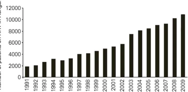
Treatment: If a patient’s GFR falls below 20 mL/min/1.73m2 renal replacement therapy (RRT) (dialysis or kidney transplantation (KTx)) should be considered. Today more than 2.6 million people receive RRT worldwide, but even according to a conservative estimation only less than half of the patients in need get treatment.7 Parallel to international trends in Hungary the number of patients on RRT has also doubled in the last 15 years and now is over 10,000 (Figure 2).8
Figure 2. The number of patients on renal replacement therapy (RRT) in Hungary by year (modified image of Kulcsar et al. 2010).8
Dialysis (peritoneal or hemodialysis) can be a good option in patients not suitable for KTx or awaiting a kidney transplant. However it has several disadvantages;
dialysis opportunities are not available in a lot of areas, transportation of patients is difficult, several side effects can occur and a stable access to the bloodstream is required. Dialysis is not only a health problem, but also imposes a massive economic burden both on affected individuals and health care systems. In the US expenditures are over $40 billion, while in the European Union dialysis alone costs more than €15 billion each year (US Renal Data System Annual Report 2015;
http://www.niddk.nih.gov/health-information/health-statistics). KTx is considerably less expensive. The yearly cost for transplant patients in the US is about $29,000, while the cost for dialysis is over $80,000 per patient (US Renal Data System Annual Report 2015).
12 2.2 Kidney transplantation
The primary RRT treatment option for ESRD is KTx; which is associated with improved survival and quality of life. In a recent review of 110 studies including almost 2 million participants with kidney failure, KTx was associated with reduced risk of mortality and cardiovascular events as well as better quality of life than treatment with chronic dialysis.9
While the number of kidney transplants has not changed in the past decade, the total number of patients living with a functioning kidney transplant continues to grow (US Renal Data System Annual Report 2015). One-year graft survival is 97% for living donor and 92% for deceased donor transplant recipients.
Living donor transplants have superior outcomes as these donors are usually younger and healthier. Cold ischemia time can be markedly reduced also as these Txs can be planned in advance. Graft survival can be further improved by performing preemptive Tx, transplanting when the recipient is in the best medical and social condition. Beside the obvious advantages of living donor Tx, the possible harm of a healthy person should always be considered as well.10
Roughly one third of kidney transplants are from living donors in the US, while this number is around only 12% in Hungary (National Organ Donation Registry/Nemzeti Szervdonációs Regiszter) (Figure 3). Hungary joined the Eurotransplant Foundation in 2012. This is a network of 8 countries with an aim to mediate and improve the allocation and distribution of donor organs for Tx.
13
Figure 3. Number of kidney transplants in Hungary (modified image of Szederkenyi E et al. 2013).11
Although short-term outcomes of KTx have improved substantially due to advances in surgical technique and immunosuppression, long-term outcomes have remained largely unchanged over the past decades.12 The factors affecting long-term outcome may be either alloantigen-dependent (e.g. HLA matching, HLA immunization etc.) or alloantigen-independent (e.g. donor type and age of both the donor and recipient), disease recurrence, comorbidities or time on dialysis).13 Among alloantigen- independent factors ischemia/reperfusion injury (IRI) is a major complication that has special influence on long-term survival after KTx. IRI is unavoidable and the duration of storage and cold ischemia time correlate with delayed graft function.
Treatment: Although effective immunosuppressive regimen is the key to successful Tx, immunosuppressants also have several undesirable effects on the kidney.
They may provoke or reactivate infections (e.g. severe polyoma BK virus, cytomegalovirus and herpes viruses resulting in interstitial nephritis, or urinary tract infections, etc.). Calcineurin-inhibitors (tacrolimus and cyclosporin A) are nephrotoxic by causing persistent vasoconstriction, interstitial fibrosis and tubular atrophy that can eventually lead to chronic graft dysfunction.10 Mainly due to steroids tacrolimus, and mTOR inhibitors about a quarter of KTx patients develop ‘de novo’ post-transplant DM, which can lead to DNP and graft dysfunction.14 For all these reasons continuous
14
and tight control of the immunsuppressive protocol is of special interest during post- transplant nephrological care.
2.3 Acute kidney injury
Etiology: AKI is the abrupt loss of kidney function, resulting in a failure to maintain fluid, acid-base and electrolyte homeostasis. It is a wide-ranging clinical syndrome embodying distinct etiologies, including kidney diseases such as acute glomerular, interstitial or vascular problems. Non - kidney-specific, systemic conditions including global ischemia, hypovolemia or toxic injury along with extrarenal pathology also result in AKI.15
Diagnosis: Traditionally AKI was characterized by severe reduction in kidney function, with severe azotemia and often oliguria or even anuria. According to the most recent Kidney Disease Improving Global Outcomes (KDIGO) guidelines AKI is actually defined as any of the following (KDIGO AKI Guideline 2013):
• Increase in serum creatinine (SCr) by ≥0.3 mg/dL (≥26.5 µmol/L) within 48 hours
• Increase in SCr to ≥1.5 times baseline, which is known or presumed to have occurred within the prior 7 days
• Urine volume <0.5 ml/kg/h for 6 hours
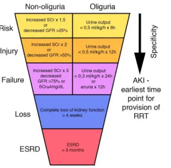
In the past decades to further classify acute impairment of kidney function the RIFLE criteria was developed through a broad consensus of experts. RIFLE stands for the increasing severity classes: Risk, Injury, Failure; and two outcome classes: Loss and End-stage Renal Disease. Risk, Injury and Failure are defined by the changes SCr or urine output (Figure 4). In the past few years however, even moderate decreases of kidney function have been shown to be important. Therefore the Acute Kidney Injury Network added a minor modification to the RIFLE criteria with the inclusion of small changes in SCr (≥26.5 µmol/L) when they occur within 48 hours.16
15
Figure 4. RIFLE criteria of acute kidney injury (AKI); ESRD: end stage renal disease; GFR:
glomerular filtration rate; RRT: renal replacement therapy; SCr: serum creatinine (modified image of Bellomo et al. 2004).17
The most frequently used indicators of renal disease are SCr, blood urea nitrogen (BUN) and creatinine clearance, however these are insensitive, non-specific and do not allow early detection. Therefore ongoing efforts are made to identify new, accurate, real-time indicators of AKI. Kidney Injury Molecule 1 (KIM-1) is upregulated mainly in proximal tubules as soon as a few hours after the ischemic insult, where it may play a role in regeneration as well.18 Neutrophil gelatinase-associated lipocalin (NGAL) is one of the most highly induced genes after ischemia. NGAL protein is upregulated very early in the postischemic kidney and is a marker of distal tubular injury. It is quickly excreted to the urine making it a sensitive and early biomarker.
Several experimental studies have shown that NGAL is remarkably protective by inducing proliferation and inhibiting apoptosis in tubular epithelial cells.19 However these markers are not introduced to the routine clinical use yet, in everyday diagnosis SCr with GFR and BUN still serve as the basic values.
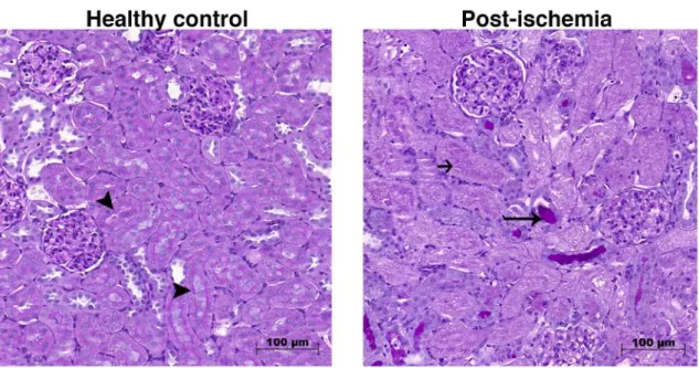
Renal histological changes are also specific and informative for AKI. Notable morphologic features of ischemic AKI include the loss of proximal tubule brush border, depolarization and patchy loss of tubular cells and tubular cast formation20 as well as peritubular capillary congestion, endothelial damage and leukocyte accumulation
16
(Figure 5). Since the main focus of this dissertation is ischemic AKI, this pathophysiology will be discussed in detail.
Figure 5. Representative periodic acid-Schiff stained sections of control and post-ischemic rat kidneys. Black quadrilateral arrow points to intact brush border; long, thin arrow shows hyalin accumulation; short black arrow shows necrotic tubule. 200x magnification, scale bar =100 µm.
Pathophysiology of IRI-induced kidney injury: The endothelium of microcirculation plays a pivotal role in the pathophysiology of IRI. Even under normal conditions the kidney regions housing nephron segments with very high energy requirements such as the S3 segment of the proximal tubule or the medullary thick ascending limb, sustain relative hypoxia due to lower blood flow and exchange of oxygen.21 Relative hypoxia worsens after ischemia, which leads to prolonged cellular injury and cell death. Renal blood flow is reduced by up to 50%, which persists in the outer medulla even during reperfusion. Release of vasoconstrictors such as endothelin is enhanced22 and abundance of vasodilators such as endothelium-derived NO is decreased23 causing vasoconstriction in small arterioles and peritubular capillaries.
In parallel, as we previously also showed Na+K+-ATP-ase is relocated from the basolateral membrane to the cytoplasm making it dysfunctional.24 Due to inadequate sodium reabsorption in injured proximal tubules, the tubuloglomerular feedback mechanism contributes to pre-glomerular arterial vasoconstriction. Additionally endothelial damage causes the disruption of cytoskeleton, loss of glycocalyx and
17
breakdown of the perivascular matrix, which result in increased microvascular permeability and fluid loss to the interstitium.25 Our group previously described an important role of heat shock proteins (HSPs), especially HSP72, in recovering cellular polarity and structure. HSP72 belongs to the chaperone family and acts by refolding denatured proteins, restoring their function and limiting detrimental peptide interactions.26
Oxygen deprivation leads to the degradation of ATP to ADP and AMP, which are further metabolized to adenine nucleotides and hypoxanthine. Hypoxanthine accumulation contributes to the generation of reactive oxygen species (ROS), which in turn cause tubular injury by oxidation of proteins, peroxidation of lipids, DNA damage and apoptosis. ATP depletion also leads to a rise in intracellular free calcium, which activates proteases, phospholipases and leads to cytoskeletal degradation.27
In DNP hyperglycemia activates the renin-angiontensin-aldosterone system (RAAS)28 that leads to ischemia via chronic vasoconstriction as well. Renal blood flow is decreased due to vasoconstriction, which further activates RAAS and increases the production of ROS. Moreover activated RAAS and atherosclerosis increase systemic blood pressure, which in turn not only damages renal perfusion and blood vessels but escalates RAAS activation and atherosclerosis.29 These processes lead to functional and structural damage of the kidneys.
Inflammation is also an important contributor both of acute and chronic ischemic injury. Innate immunity is responsible for early response and comprises neutrophil granulocytes, macrophages, dendritic cells and natural killer cells.
Neutrophils accumulate and attach to the endothelium where they release ROS, proteases and inflammatory cytokines such as IL-1, IL-6, TNF-α, MCP-1, all of which aggravate kidney injury in acute renal injury models30 and DNP.31
Very recently a new molecule, the Sigma-1 receptor (S1R) has become the center of attention in brain ischemia and stroke as a possible mediator of key protective mechanisms, however there is no information about its role in the kidney.
18 2.4 Sigma-1 receptor
S1R is a unique transmembrane chaperone protein, which consists of 223 amino acids and shows no homology with any other mammalian protein. The receptor consists of two subtypes: the S1R and the S2R, which can be distinguished by molecular weight, tissue distribution and ligand binding profile.32 The topic of this thesis is S1R, therefore only this isoform will be discussed further on.
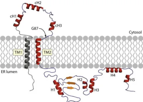
Figure 6. Secondary structure of the Sigma-1 receptor. cH: cytosolic helic; ER: endoplasmic reticulum; TM1: transmembrane helix 1; TM2: transmembrane helix 2, H: helix; G87: the first residue in the GGWMG sequence. (Ortega-Roldan JL et al. 2015).33
S1R is vastly expressed in the central nervous system (CNS)34, but has also been detected in various peripheral tissues including the heart, liver and kidney.35, 36 In the brain S1R is localized in the endoplasmatic reticulum (ER) (Figure 6), but upon ligand stimulation it translocates to the cytosol or the cell periphery using the ER-associated reticular network.37
S1R has many different ligands and thus has been implicated in a large number of CNS diseases and conditions. The involvement of the receptor in drug and alcohol addiction has been extensively studied, especially in the context of drug self- administration.38, 39 The effect of S1R agonists on learning and memory has been
19
studied as well. These compounds seem to improve learning in animal models of amnesia. 40
Several lines of evidence suggest that S1R agonists also have effective antidepressant properties. Several selective serotonin reuptake inhibitors (SSRI) such as fluvoxamine (FLU) or sertraline bind to S1Rs with very high affinity41, suggesting a role of S1R in the pharmacological action of these drugs. Other S1R agonists also had effective antidepressant actions in forced swimming, tail suspension or conditioned fear stress tests and these effects were blocked by S1R.42, 43
The role of S1R agonists has been demonstrated in models of brain ischemia as well. Most of these studies used the middle cerebral artery occlusion ischemic stroke model to examine the neuroprotective characteristics of S1R agonists. S1R agonist treatment reduced infarct areas, presumably by promoting cell survival and reducing the inflammatory response.44 However not very much is known about the regulation and function of S1R outside the CNS.
In a recent study S1R stimulation was proven to be effective in minimizing infarct size in a pressure overload induced cardiac hypertrophy model. This protection seemed to be mediated by the upregulation of protein kinase B (Akt)-endothelial nitric oxide synthase (eNOS) signaling.45 Similarly S1R agonist fluvoxamine attenuated cardiac hypertrophy in another ischemic heart model of transverse aortic constriction.46
On the molecular level S1Rs take part in many different processes. The modulation of voltage-dependent ion channels, especially Ca2+ channels has been the main area of S1R research. Recent studies demonstrated that S1Rs regulate Ca2+
signaling between the ER and mitochondria by modulating inositol triphosphate (IP3) receptors in the ER membrane.47 A possible protective mechanism of S1R agonists under pathological conditions is rescuing ATP production by restoring mitochondrial Ca2+ transport.48 S1Rs have been shown to modulate potassium, sodium and chloride channels as well.49, 50
S1Rs promote cell survival possibly by stabilizing anti-apoptotic protein Bcl-251 and suppressing pro-apoptotic proteins Bax and caspase-352, however the exact mechanism by which S1Rs are related to caspases and hypoxia-inducible factor alpha (HIF-1α) is yet unknown.
20
Summarizing the literary data one can conclude that renal IRI plays a central role in the patomechanism of acute kidney injury and KTx, as well as in the development of CKD such as DNP. Via decreased NOS activity and NO production renal vasoconstriction and hypoperfusion occurs leading to the progression of kidney injury both acutely and on the long run. Since renal IRI has high morbidity and mortality and treatment options are still very limited better understanding of the underlying molecular mechanisms are essential for developing novel renoprotective therapies.
Preliminary results in the brain and heart suggest that S1R activates vasodilatative and protective pathways in brain and cardiac ischemia. Therefore it is rather presumable that S1R agonists could trigger similar mechanisms in renal IRI. In our preclinical experiments - during my PhD work - we aimed to investigate this promising option.
21 3. Objectives
The purpose of our experiments was to investigate the pathomechanisms of IRI- induced AKI in order to identify new therapeutic targets that can be used in KTx as well as in DNP. Based on the current literature demonstrating that S1R is protective in brain and heart ischemia the main aim was to investigate the effect of S1R and its modulation in renal diseases.
Objectives:
The following objectives have been set to fulfill the aims:
1. To investigate the intrarenal and subcellular localization of S1R in the normal and ischemic kidney
2. To analyze the molecular mechanism of S1R - mediated effects
3. To determine the possible renoprotective effect of S1R agonism in acute (renal IRI) and chronic (DNP) models of kidney disease
4. To evaluate the protective effect of S1R agonist pretreatment in KTx
22 4. Methods
Animals
Adult, male Wistar rats weighing 200±15g (Toxi-Coop Toxicological Research Center, Dunakeszi, Hungary) were used in all experiments (n=8-10/each group). Rats were housed in standard laboratory cages and were allowed free access to food and water. Animal procedures were approved by the Committee on the Care of Laboratory Animals at Semmelweis University, Budapest, Hungary (PEI/001/1731-9/2015).
Renal ischemia/reperfusion injury model and treatment groups
General anesthesia was performed by inhalation of isoflurane (3% vol/vol) mixed with synthetic air (1 L/min) in an isoflurane vaporizer (Eickemeyer Veterinary Equipment Ltd., Twickenham, UK). Renal ischemia was accomplished by cross- clamping the left renal pedicles for 50 min with an atraumatic vascular clamp, ischemia was visually confirmed. Before the end of the ischemic period the contralateral kidney was taken out, the clips were removed and the left kidneys were observed for 5 min to ensure reperfusion. Sham animals underwent laparotomy of the same duration without clamping. At pre-determined times of reperfusion, blood samples were collected from the abdominal aorta, the remnant kidneys were harvested, instantly snap-frozen in liquid nitrogen, and stored at −80 °C or fixed in buffered 4% formalin for further processing.
To test the effect of DHEA in the first set of experiments rats were pretreated first 25 hours, then 1 hour before the surgical procedure with (i) isotonic saline as vehicle; (ii) 4 mg/bwkgDHEA (Sigma Aldrich, Budapest, Hungary).
To investigate the effect of FLU in the second set of experiments 30 min prior to the ischemic insult animals were treated as follows: (i) isotonic saline as vehicle; (ii) 20 mg/bwkg fluvoxamine maleate (FLU; Sigma Aldrich, Budapest, Hungary); (iii) 20 mg/bwkg FLU and 1 mg/bwkg N,N-dipropyl-2-[4-methoxy-3-(2-phenylethoxy)- phenyl]-ethylamine monohydrochloride (NE100, selective S1R antagonist) (Tocris Bioscience, Bristol, UK).
In the third set of experiments to test the NO-mediated effect the following
23
pretreatment was applied 30 min prior to the ischemic insult: 20 mg/bwkg FLU and 10 mg/bwkg N-omega-Nitro-L-arginine methyl ester (L-NAME, non-selective NOS inhibitor) (Sigma Aldrich, Budapest, Hungary) or 20 mg/bwkgN5-(1-Iminoethyl)-L- ornithine dihydrochloride (L-NIO, selective eNOS inhibitor; Sigma Aldrich, Budapest, Hungary) or 25 mg/bwkg 7-Nitroindazole (7-NI, selective nNOS inhibitor) (Sigma Aldrich, Budapest, Hungary). DHEA was administered subcutaneously; all other substances were administered intraperitoneally.
Experimental protocol is summarized in Figure 7.
In one series postischemic survival was followed for 7 days.
In another series animals were randomly divided into the following groups:
(i) Sham-operated, vehicle treated group as controls; (ii) T30’ group that was not subjected to ischemic insult and was sacrificed or underwent intravital two-photon microscopic analysis 30 min after drug pretreatment; (iii) T2 group was subjected to ischemia 30 min after drug pretreatment and was sacrificed after 2 hours of reperfusion;
(iv) T24 group was subjected to ischemia 30 min after drug pretreatment and was sacrificed or underwent two-photon microscopic analysis after 24 hours of reperfusion.
Figure 7. Experimental design of renal IRI
24
Rat model of kidney isograft autotransplantation
Male Wistar rats (n=8/group) were anesthetized with isoflurane (3% vol/vol) mixed with synthetic air (1 L/min) before surgery and placed on a temperature- controlled table to maintain core body temperature. Left kidneys were perfused with cold Custodiol perfusion solution (Na+: 15 mmol/L; K+: 9 mmol/L; Mg2+: 4 mmol/L;
Ca2+: 0.015 mmol/L; histidine: 198 mmol/L; tryptophan: 2 mmol/L; ketoglutarate: 1 mmol/L; mannitol: 30 mmol/L) (Franz Kohler Chemie GMBH, Bensheim, Germany), then removed from the animal: kidneys were placed into a container for 2 hours filled with either (i) cold Custodiol perfusion solution (Tx) or (ii) cold Custodiol perfusion solution containing 0.003 mg/mL FLU (Tx FLU). After 2 hours kidneys were placed back into the rats and end-to-end anastomoses of the renal artery, vein and ureter were performed. Contralateral kidneys were removed and the autotransplanted kidneys were observed to ensure reperfusion. Total warm ischemia time was 35 min in all animals.
Sham operated animals served as controls (Figure 8).
After 24 hours of reperfusion, blood samples were collected from the abdominal aorta, the remnant kidneys were harvested, instantly snap-frozen in liquid nitrogen, and stored at −80 °C or fixed in buffered 4% formalin for further processing.
Figure 8. Experimental design of renal autotransplantation
Rat model of type 1 diabetes mellitus (DM1) and experimental groups
DM1 was induced in male Wistar rats (n=6-8/group) by a single intraperitoneal injection of 65 mg/bwkgstreptozotocin (STZ, Sigma Aldrich, Budapest, Hungary) in freshly prepared 0.1 M citrate buffer (pH 4.5). Rats were considered diabetic if the peripheral blood glucose concentration in three random samples was higher than 15 mmol/L at 72 hours after the injection of STZ. Age-matched control rats received an equivalent volume of citrate buffer and were used along with diabetic animals.
25
DM1 rats were randomly divided into four groups and were treated by oral gavage daily at 10:00 AM as follows: (i) DM1 rats treated with isotonic saline as vehicle for 7 weeks (D); (ii) 20 mg/bwkg FLU for 7 weeks (D + 7 wk FLU (20 mg/bwkg)); (iii): 20 mg/bwkgFLU for 2 weeks after 5 weeks of DM1 (D + 2 wk FLU (20 mg/bwkg)); (iv) 2 mg/bwkgFLU for 2 weeks after 5 weeks of DM1 (D + 2 wk FLU (2 mg/bwkg)). Age-matched, non-diabetic control rats (Control) were treated with saline daily for two weeks at the same time as the diabetic animals.
At the end of the experimental protocol all rats were anesthetized with a mixture of 60 mg/bwkgketamine and 5 mg/bwkgxylazine (rats did not receive drug treatment on this day). Blood samples were collected from the abdominal aorta. Kidney samples were collected and immediately snap-frozen stored at −80 °C or fixed in buffered 4%
formalin for further processing.
Figure 9. Experimental design of type 1 diabetes mellitus.
Plasma chemistry and metabolic parameters
The development of DM1 was followed by measurement of serum glucose and fructosamine levels. Serum electrolytes (sodium, potassium, chloride), urea, creatinine, albumin, total protein, triglycerides, total cholesterol, aspartate transaminase (AST) and glutamate-pyruvate transaminase (GPT) were evaluated using commercially available kits on a Hitachi 912 photometric chemistry analyzer by our hospital’s research services.
26 In vitro models
Cell culture and treatment
Human proximal tubular epithelial cell line (HK2; American Type Culture Collection; Rockville, Maryland, USA) were grown in Dulbecco’s Modified Eagle Medium (DMEM) (Life Sciences, Budapest, Hungary) supplemented with 10% fetal calf serum (FCS) 1% L-glutamine and 1% antibiotic, antimycotic solution (100x) (Sigma Aldrich, Budapest, Hungary) containing 10000 IU/mL penicillin, 10 mg/mL streptomycin and 25 µg/mL Amphotericin B. The cells were incubated at 37°C in 5%
CO2 and 95% air. In all experiments, there was a “growth arrest” period of 24 hours in serum-free medium before treatment.
Cell viability and proliferation assay
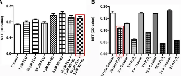
Prior to the experiments the non-toxic dosage of FLU and NE100 was confirmed by methyl-thiazoletetrazolium assay (MTT) (Roche Diagnostics, Mannheim, Germany).
Cell viability was determined by MTT assay according to the manufacturer’s instructions (Figure 10/A).
Cell viability was also assessed by trypan blue exclusion. Cells were detached with trypsin-EDTA and re-suspended in medium diluted 1:1 with trypan blue solution (Sigma Aldrich, Budapest, Hungary). Live cells from triplicate wells were counted in a Burker chamber.
In vitro model of oxidative stress
Oxidative stress was induced by 400 µM hydrogen-peroxide (H2O2) treatment for 30 min. Pilot experiments were performed to determine the effective dose and duration of H H2O2 treatment (Figure 10/B).
Cells were treated as follows: (i) 10 µM FLU (30 min prior to harvest); (ii) FLU + 3 µM NE100 (iii) FLU + 10 µM AktVIII inhibitor (AktVIII; 1 hour prior to harvest;
Santa Cruz Biotechnology, Budapest, Hungary); (iv) FLU + 2 µM AktIV inhibitor
27
(AktIV; 1 hour prior to harvest; Santa Cruz Biotechnology, Budapest, Hungary). After treatment the cells were processed for nitrite measurement.
Figure 10. Cell viability assay to confirm the non-toxic dosage of (A) Fluvoxamine (FLU) and Sigma-1 receptor antagonist NE100. (B) Cell viability assay to determine the protocol for hydrogen peroxide (H2O2)-induced oxidative stress. The dose and duration for final treatment protocols are marked with red.
RNA interference
All reagents for siRNA were purchased from Invitrogen, Budapest, Hungary.
HK2s were transfected with 10 nM S1R specific siRNA or negative control siRNA using Lipofectamine 2000. Successful transfection with 10nM fluorescently labeled siRNA was visualized with an Olympus IX81 (Olympus America, Center Valley, PA) fluorescent microscope. The efficacy of knockdown was determined by Western blot (Figure 11).
28
Figure 11. Validation of Sigma-1 receptor (S1R) knockdown. S1R protein level of S1R siRNA-treated HK2 cells compared to scrambled siRNA-treated Negative Control cells. +P<0.05 versus Negative Control (n=6/group). Bars indicate means ± SEMs, and data were analyzed by one-way ANOVA with Bonferroni multiple comparison test.
Subcellular protein fractionation
HK2 cells were treated either with 10 µM FLU or 10 µM FLU and 400 µM H2O2. After treatment the cells were processed for further analysis by Subcellular Protein Fractionation Kit for Cultured Cells (Thermo Scientific, Budapest, Hungary).
The adherent cells were harvested with trypsin - EDTA and separated into cytoplasmic, membrane, soluble nuclear, chromatin-bound nuclear and cytoskeletal extract. After fractionating the different cell extracts S1R protein levels were determined by Western blot.
Imaging techniques
Conventional histology
Paraffin-embedded, 3 µm kidney sections were stained with periodic acid-Schiff (PAS) and haematoxylin eosin or Masson’s trichrome or Sirius red to assess injury.
Images were taken with a Zeiss AxioImager A1 light microscope (Zeiss, Jena, Germany). Tubular injury was evaluated based on a semi-quantitative scale.53 Each cortical tubule showing epithelial cell necrosis and brush border loss was assigned a score of 0: normal; 1: loss of brush border; cell necrosis in less than 25% of tubular
29
cells 2: cell necrosis in 25% - 50% of tubular cells 3: cell necrosis in 50% - 75% of tubular cells; 4: cell necrosis in >75% of tubular cells. Two fields of x200 magnification/animal were examined and averaged in a double - blinded fashion by two different pathologists.
In the case of transplanted kidneys sections were stained with PAS, then tubular lumen areas were measured on 6 fields of x20/animal using Adobe Photoshop (Adobe Systems, San José, California) software.
To evaluate mesangial matrix expansion sections were stained with PAS. 15 fields of x40 magnification containing glomeruli were randomly selected per animal and the ratio of mesangial area per glomerular taft area was measured in each glomerulus using Adobe Photoshop and Image J (US National Institute of Health, Bethesda, USA, http://rsb.info.nih.gov/ij/) softwares.
To evaluate tubulointerstitial fibrosis sections were stained with Masson’s trichrome. 10 - 10 fields of x20 magnification were randomly selected from cortical and cortico-medullary regions respectively per animal and the ratio of Masson-stanied fibrotic area per total area was measured in each field using Adobe Photoshop and Image J softwares.
To evaluate collagen accumulation sections were stained with Sirius red. 10-10 fields of x20 magnification were randomly selected from cortical and cortico-medullary regions respectively per animal and the ratio of Sirius red-stanied extracellular matrix area per total area was measured in each field using Adobe Photoshop and Image J softwares.
Two-photon microscopy
Femto 2D high sensitivity galvanoscanner-based two-photon microscope system (Femtonics Inc., Budapest, Hungary) was used for intravital two-photon microscopy.
Fluorescence excitation is provided by a Mai Tai mode-locked titanium-sapphire laser (Spectra-Physics Inc., Irvine, CA) and collected in separate photomultiplier tube detectors to a maximal depth of 100 µm. To inject the mixture of dyes the rat was anesthetized as described in the surgical procedure and a cannula was placed in the carotid artery. 70 kDa Rhodamine dextran (Life Technologies, Budapest, Hungary) was
30
used to label the vasculature, Texas Red (Life Technologies, Budapest, Hungary) to evaluate the reabsorption capacity and the preservation of the brush border and Hoechst 33342 (Life Technologies, Budapest, Hungary) to visualize nuclei.
The changes in capillary diameters were measured continuously in every minute for a 30-min period and the difference in diameter between the first, 10th, 20th and 30th minute was calculated. ~150 capillaries were measured per animal.
Images and data volumes were processed using Matlab (Femtonix Inc., Budapest, Hungary) and Image J softwares.
DAB (3,3'-diaminobenzidine) immunohistochemical staining
Slides were deparaffinized in xylene, rehydrated in graded ethanol series and washed in dH2O. Heat-induced epitope retrieval was performed by boiling the tissue sections in citrate buffer (HISTOLS®-Citrate Buffer) followed by cooling at room temperature (RT) for 20 min. Nonspecific sites were blocked (HISTOLS® Background Blocking Protein Solution) for 10 min at RT. Without washing, the slides were incubated with the primary antibody (Rabbit anti-S1R, Thermo Scientific, Budapest, Hungary) in 1:50 dilution for 1 hour at RT and repeatedly washed in TBS. Secondary antibody (HISTOLS® MR anti mouse and rabbit Detection Systems Histopathology Ltd., Pecs, Hungary) was applied for 30 min at RT followed by repeated washing in TBS. Sections were incubated with 3-amino-9-ethylcarbazol (HISTOLS® -Resistant AEC Chromogen/Substrate System, Histopathology Ltd., Pecs, Hungary) and washed in dH2O, counter stained with haematoxylin or PAS followed by washing in tap water.
Fluorescent immunohistochemistry
Frozen kidney sections were embedded in Shandon cryomatrix (Thermo Scientific, Budapest, Hungary) and cut to 5 µm slides. Samples were incubated with the specific rabbit S1R (Invitrogen, Budapest, Hungary); goat Akt (Santa Cruz Biotechnology, Budapest, Hungary) and mouse eNOS (BD Biosciences, Budapest, Hungary) primary antibodies. After repeated washing slides were incubated with the specific secondary anti-rabbit Alexa Fluor 568 (Invitrogen, Budapest, Hungary), anti-
31
goat Alexa Fluor 647 (Invitrogen, Budapest, Hungary) or anti-mouse Alexa Fluor 488 (Invitrogen, Budapest, Hungary) conjugates and counterstained with Hoechst 33342 (Life Technologies, Budapest, Hungary).
HK2 cells were cultured in tissue culture chambers (Sarstedt Kft., Budapest, Hungary). After repeated washing the cells were fixed in 4% paraformaldehyde, washed again and permeabilized with Triton X-100 (Sigma Aldrich, Budapest, Hungary). Cells were incubated with the specific mouse S1R antibody (Santa Cruz Biotechnology, Budapest, Hungary). After repeated washing the chambers were incubated with anti- mouse Alexa Fluor 488 conjugate and counterstained with Hoechst 33342. Appropriate controls were performed omitting the primary antibody to assure the specificity and to avoid autofluorescence. Sections were analyzed with a Zeiss LSM 510 Meta confocal laser-scanning microscope (Zeiss, Jena, Germany) with objectives of 20x and 63x magnification.
Other molecular biology techniques
Quantitative real-time PCR
Total RNA from kidney, hippocampus and prefrontal area samples was extracted using the RNeasy RNA isolation Kit (Quiagen GmbH, Hilden, Germany).
Hif1a, Ngal, Kim1, Sigmar1, glyceraldehyde-3-phosphate dehydrogenase (Gapdh), Mcp1 and 18S ribosomal RNA (Rn18s) mRNA expression were determined in duplicates by real-time (RT-PCR) using SYBR Green I Master enzyme mix (Invitrogen, Budapest, Hungary) and specific primers (Table 2). Results were analyzed by LightCycler 480 SYBR Green I Light Cycler system (Roche Diagnostics, Mannheim, Germany). The mRNA expression of Hif1a, Ngal, and Kim1 was normalized against Gapdh as housekeeping gene.
32
Table 2. Sequence of forward and reverse primers for RT-qPCR
Forward primer 5’-3’ Reverse primer 5’-3’
Ngal CAA GTG GCC GAC ACT GAC TA GGT GGG AAC AGA GAA AAC GA Hif1a AAG AAA CCG CTT ATG ACG TG CCA CCT CTT TTT GCA AGC AT Kim1 CGC AGA GAA ACC CGA CTA AG CAA AGC TCA GAG AGC CCA TC Gapdh CAC CAC CAT GGA GAA GGC TG GTC ATG GCA TGG ACT GTG
Mcp1 ATG CAG TTA ATG CCC CAC TC TTC CTT ATT GGG GTC ACC AC
Measurement of Nitric Oxide Levels
The total stable oxidation products of NO metabolism (NO2/NO3) of serum and HK2 cell homogenates were assessed using Griess reagent (Promega, Budapest, Hungary). Following manufacturer's directions 50 µL of the samples were incubated with 50-50 µL Griess reagent (part I: 1% sulphanilamide; part II: 0.1%
naphthylethylene diamide dihydrochloride and 2% phosphoric acid) at RT. Ten minutes later the absorbance was measured at 540 nm using a Plate Chameleon V Fluorometer- Luminometer-Photometer reader (Hidex, Turku, Finland). Relative concentration was calculated on the basis of a sodium nitrite reference curve and was expressed as µM.
Detection of microRNA (miRNA) expression
Total RNA was extracted from the kidney using TRIzol Reagent (Invitrogen, Budapest, Hungary) according to the protocol provided by the manufacturer. Micro- RNA expression was evaluated and quantified using TaqMan probes (Applied Biosystems, Budapest, Hungary). First, complementary DNA (cDNA) was reverse- transcribed from 5-ng RNA samples using miRNA-specific primers (for miR-21, miR- 17-5p, and U6 snRNA) as described in the manufacturer's protocol. Next, PCR products were amplified from the cDNA samples using the TaqMan Small RNA Assay together with the TaqMan Universal PCR Master Mix 2. All measurements were done in duplicates, and the miRNA expressions were normalized to U6 small nuclear RNA (snRNA).
33 Western Blot analysis
All reagents for PAGE and Western blot were purchased from Bio-Rad Hungary. Kidney samples were sonicated and re-suspended in lysis buffer. Protein concentration measurement was performed with a detergent-compatible protein assay kit. Samples were electrophoretically resolved on 7.5 %, 10 % or 12% polyacrylamide gels and transferred to nitrocellulose membranes. Membranes were stained with Ponceau S, then washed and blocked with 5 % non-fat dry milk. The membranes were incubated with antibodies specific for rat or human S1R (rat: #423300, Invitrogen, Budapest, Hungary; human: #sc-166392, Santa Cruz Biotechnology, Budapest, Hungary); peNOS (Ser1177) (#9571, Cell Signaling Technology, Budapest, Hungary), pAkt (Ser473) (#9271, Cell Signaling Technology, Budapest, Hungary) and nNOS (#sc- 5302, Santa Cruz Biotechnology, Budapest, Hungary) respectively. After repeated washing the blots were incubated with the corresponding horseradish-peroxidase- conjugated secondary antibodies (#7074, goat anti-rabbit, Cell Signaling Technology, Budapest, Hungary; #sc2005, goat anti-mouse, Santa Cruz Biotechnology, Budapest, Hungary).
Bands of interest were detected using enhanced chemiluminescence detection (GE Healthcare Life Sciences, Budapest, Hungary) and quantified by densitometry (VersaDoc, Quantity One Analysis software; Bio-Rad Hungary) as integrated optical density (IOD) after subtraction of background. IOD was factored for Ponceau red staining to correct for any variations in total protein loading and for internal control.
Protein abundance is represented as IOD/Ponceau S/Internal control.54
Cytometric bead array
All reagents and equipment for CBA were purchased from BD Biosciences (Budapest, Hungary). Saline-perfused kidney homogenates were measured for TNFα, IL-1α, IFN-γ, IL-4, and IL-10 peptide levels using appropriate rat CBA Flex Sets according to the manufacturer’s protocol. Measurements have been performed using a FACSVerse flow cytometer and data analyzed using FCAP Array software.
34 Statistical analysis
Parametrical data are expressed as means ± SEM, while non-parametrical data as median ± range. Statistical analyzes were performed using Prism software (version 5.00; GraphPad Prism Software). Survival studies were assessed by Logrank test.
Multiple comparisons and possible interactions were evaluated by one-way ANOVA followed by Bonferroni post-hoc test. For non-parametrical data the Kruskal–Wallis ANOVA on ranks followed by Fischer exacts test was used. P values of <0.05 were considered significant.
35 5. Results
5.1 S1R in the kidney
The peripheral expression, localization and function of S1R are under-discussed in the literature. S1R was first detected in the kidney in 1994 with radioimmunbinding assay (Hellewell 1994), but since then only one group investigated renal S1R (Bhuiyan 2010). However they only measured protein levels from whole kidney lysates, exact localization and subcellular distribution has not been discussed at all. According to the Protein Atlas of human kidney biopsies S1R shows no staining in glomeruli and
moderate staining in tubules (online source:
http://www.proteinatlas.org/ENSG00000147955-SIGMAR1/tissue/kidney).
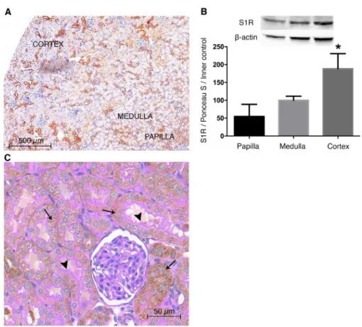
Our group is the first to show evidence of S1R expression in different kidney regions. To gain an overview of the renal distribution of the receptor S1R-specific DAB staining was applied on rat kidney sections. S1R expression was most prominent in the renal cortex, but was also present in the medulla and papilla (Figure 12/A). Western blot measurement of S1R protein levels from homogenates of renal cortex, medulla and papilla confirmed these results (Figure 12/B). Next, co-staining with proximal tubular brush border-specific PAS and anti-S1R DAB revealed that S1R is definitely expressed in proximal tubules but not in glomeruli (Figure 12/C).
36
Figure 12. Sigma-1 receptor (S1R) is predominantly expressed in the renal cortex. (A) Representative rat kidney section stained with anti-S1R (brown; magnification: 6x DAB). Scale bar=500 µm. (B) Immunoblot for renal S1R. S1R is expressed in the papilla, medulla, and most prominently the cortex. Representative blots are shown. Bars indicate means ± SEMs; data were analyzed by one-way ANOVA with Bonferroni multiple comparison test. *P<0.05 versus papilla and medulla (n=6/group). (C) Representative kidney section developed with peroxidase anti-S1R (brown) and counterstained with PAS (pink). Thin black arrows show S1R in proximal tubules but not in glomerulus. Black arrowheads point to intact, PAS–stained brush borders of proximal tubules. Scale bar=50 µm.
To further clarify the subcellular localization of S1R fluorescent immunohistochemistry double staining with S1R and antibodies specific to different nephron segments was performed as well.
S1R was co-localized with proximal tubule-specific gamma-glutamyltransferase (GGT), but not with Na+/K+-ATP-ase in distal tubules, eNOS in glomeruli or neuronal NOS (nNOS) in the macula densa supporting the finding that S1R is mainly present in proximal tubules (Figure 13).
37
Figure 13. Localization of Sigma-1 receptor in different nephron segments. Representative fluorescent immunohistochemistry images stained for nuclei (blue), S1R (red) in all images. Green represents gamma-glutamyltransferase (GGT) in the proximal tubule, Na+/K+-ATPase in the distal tubule, endothelial nitric oxide synthase (eNOS) in the glomerulus or neuronal nitric oxide synthase(nNOS) in the macula densa. Scale bar=25 µm.
5.2 Dehydroepiandrosterone improves post-ischemic kidney function and ameliorates structural injury
After confirming the presence of S1R in the kidney our goal was to investigate the effect of S1R agonist treatment on renal IRI. Dehydroepiandrosterone (DHEA) is a highly abundant steroid hormone, which is also an endogenous agonist of S1R.
To determine the effect of S1R activation rats received only vehicle (ischemia/reperfusion (I/R)) or DHEA (I/R DHEA) pretreatment. In the first series of experiments post-ischemic survival was followed and compared to sham-operated controls (SHAM) for seven days. DHEA-pretreated rats survived longer than vehicle-
38
treated ones (median survival: 72 versus 36 hours, P<0.001). One third of DHEA- treated rats survived the seven-day period and recovered completely, while all vehicle- treated ones died within 75 hours (Figure 14/A).
Another series of animals were sacrificed after 24 hours of reperfusion to investigate the acute effects of IRI (T24 I/R and T24 I/R DHEA groups; resp.).
Increased SCr and BUN in all treatment groups confirmed post-ischemic AKI (Figure 14/B-C). Intravital two-photon microscopy allowed us to measure changes of peritubular capillary diameters in live rats. Post-ischemic vasoconstriction was apparent (Figure 14/D) suggesting that the subsequent decline in renal blood flow could be a causative factor of renal functional and structural damage. DHEA pretreatment (T24 I/R DHEA) improved kidney function and prevented peritubular vasoconstriction (Figure 14/B-D).
Figure 14. Dehydroepiandrosterone (DHEA) pretreatment is protective against renal ischemia/reperfusion injury. (A) Post-ischemic survival was followed for 7 days in rats pretreated with vehicle (I/R) or DHEA (I/R DHEA) 25 and 1 hour before the 50-minute ischemia. Log rank test (n=8/group). *P<0.001 versus ischemia/reperfusion(I/R). (B) Serum creatinine levels. *P<0.05 versus T24 I/R; +P<0.05 versus SHAM-operated (SHAM) (n=6/group). (C) Blood urea nitrogen levels of vehicle (T24 I/R) and DHEA (T24 I/R DHEA) -pretreated rats after 24 hours of reperfusion.
39
*P<0.05 versus T24 I/R (n=6/group); +P<0.05 versus SHAM (n=6/group). (D) Renal capillary diameters measured using intravital two–photon microscopy. Approximately 150 capillaries per animal. *P<0.05 versus T24 I/R (n=3/group); +P<0.05 versus SHAM (n=3/group). Bars indicate means ± SEMs, and data were analyzed by one-way ANOVA with Bonferroni multiple comparison test.
Histologic changes were consistent with functional decline: brush borders disappeared, the majority of tubules showed cell necrosis and extensive cast formation (Figure 15/A-C). DHEA pretreatment (T24 I/R DHEA) considerably reduced tubular necrosis and partly preserved brush borders (Figure 15/A-D).
Figure 15. Dehydroepiandrosterone (DHEA) improves renal structure after ischemia/reperfusion injury (IRI). (A–C) Representative images of structural damage after IRI on PAS–stained kidney sections of (A) SHAM-operated (SHAM), (B) T24 ischemia/reperfusion (I/R), or (C) T24 I/R DHEA–
pretreated rats. Black arrowheads points to intact brush border, long thin black arrow shows hyaline accumulation, and short black arrow shows necrotic tubule. Original magnification: 200x (D) Semi- quantitative evaluation of tubular injury on a zero to four scale. *P<0.05 versus T24 I/R (n=6/group);
+P<0.05 versus SHAM (n=6/group). Bars indicate medians ± ranges, and data were analyzed by Kruskal–
Wallis test with Dunn correction.
40
5.3 High affinity S1R agonist fluvoxamine is renoprotective following kidney IRI
On the basis of the first set of experiments our hypothesis was that S1R activation by DHEA is renoprotective in AKI by improving NO-mediated kidney perfusion. To substantiate the crucial role of S1R in IRI, in a second set of experiments rats were pretreated with fluvoxamine (FLU) that has much higher affinity to S1R than DHEA.55
Since FLU-treatment has not been studied in the kidney yet we performed pilot experiments (with 2 or 20 mg/bwkg) to determine the minimal effective dose, which is not toxic, but still exerts renoprotection. 20 mg/bwkg is comparable to the regular daily dosage of FLU used chronically in everyday clinical treatment (Morishita S 2003), while 2 mg/bwkg is ten-times less than the dosage used in humans.
To determine optimal timing in one series 20 mg/bwkg was given first 30 minutes prior the ischemic insult and then the dose was repeated right after the clip was removed, just at the beginning of reperfusion. All protocols successfully improved renal function; however 20 mg/bwkg and 40 mg/bwkg dosages were more effective in abating structural damage as well. The highest dosage did not cause additional improvement, therefore 20 mg/bwkg dosage was used further on (Figure 16).
A group of rats received selective S1R antagonistN, N-dipropyl-2-[4-methoxy- 3-(2-phenylethoxy)-phenyl]-ethylaminemonohydrochloride (NE100) together with FLU to verify the role of S1R in investigated processes. NE100 dosage of 1 mg/bwkg was based on literary data.56
41
Figure 16. The effect of different dosages offluvoxamine (FLU) on (A) tubular damage, (B) serum creatinine and (C) Blood urea nitrogen (BUN) levels after ischemia/reperfusion injury. +P<0.05 versus ischemia/reperfusion (I/R), *P<0.05 versus 2 mg/bwkg FLU (n=6/group). SHAM-operated (SHAM).
Bars indicate means ± SEMs, and data were analyzed by one-way ANOVA with Bonferroni multiple comparison test. The final dose is marked with red.
5.3.1 FLU pretreatment improves survival after sub-lethal IRI
After determining the 20 mg/bwkg as the minimal effective dose, in the first series of experiments the effect of FLU was tested on post-ischemic survival as the most relevant primary end-point.
FLU-treated rats (I/R F) survived longer than vehicle (I/R) or FLU+NE100 (I/R FN)-treated ones (median survival: 67 versus 36 and 49 hours respectively, P<0.001).
One third of I/R F rats completely recovered from the ischemic insult, while all I/R and I/R FN rats died within 70 hours (Figure 17).
42
Figure 17. Fluvoxamine (FLU; F) pretreatment improves post-ischemic survival. Rats were pretreated with isotonic saline ischemia/reperfusion(I/R), FLU (I/R F), or FLU and NE100 (I/R FN) 30 minutes before the 50-minute ischemia. *P<0.001 versus I/R; §P<0.001 versus I/R F. Post-ischemic survival was followed for 7 days. Log rank test (n=8/group).
5.3.2 FLU is protective against renal IRI-induced AKI
Beside SCr and BUN, serum AST is also acknowledged recently as a marker of ischemia induced renal tubular damage. All these three parameters were less elevated in FLU-treated rats after 24 hours of reperfusion (T24 I/R F) and the improvement was neutralized by NE100 (T24 I/R FN) suggesting that S1R agonism successfully diminishes acute renal injury. All functional parameters returned to normal level by 168 hours in FLU-treated survivors (Figure 18/A-C).
Since SCr, BUN and AST are widely used, but not specific and higly sensitive markers of AKI, we also measured other biormarkers to further prove the protective effect of FLU. HIF-1α is a known mediator of cell protection and epithelium recovery after IRI (Conde E 2012). Hif-1α mRNA expression increased as soon as 2 hours after ischemia (T2 I/R), especially in FLU-treated rats (T2 I/R F) (Figure 18/D). By T24 the difference was disappeared suggesting the HIF-1 alfa shows a quick response after IRI.
NGAL is an indicator of distal tubular injury that correlates with the severity of renal impairment (Bolignano D 2008). However Ngal mRNA expression was robustly increased after 24 hours of reperfusion (T24 I/R) in all groups, but the increase was milder in FLU-treated rats (Figure 18/E).
43
KIM-1 is a specific marker of tubular injury.18 In our study Kim-1 was upregulated in the post-ischemic kidney (T24 I/R). Increased Kim-1 mRNA expression was ameliorated by FLU (T24 I/R F) (Figure 18/F) also suggesting milder proximal tubular damage.
Antagonizing FLU with NE100 diminished functional improvement and suppressed all parameters reflecting milder tubular damage.
MicroRNAs (miRNAs) are short, double-stranded RNAs that can regulate post- transcriptional gene-expression through multiple mechanisms.57 MicroRNA-21 (miR21) and microRNA-17-5p (miR17-5p) are part of the regulatory network that can determine the outcome of IRI.58 Others demonstrated that mir21 is involved in the renoprotective effect of ischemic preconditioning.59 In our experiments both miR21 and miR17-5p expression was elevated after IRI, but no difference was detected between treatment groups (Figure 18/G-H), therefore miRNAs have not been tested further on.