Journal of Reproductive Immunology 148 (2021) 103380
Available online 9 September 2021
0165-0378/© 2021 The Author(s). Published by Elsevier B.V. This is an open access article under the CC BY-NC-ND license
(http://creativecommons.org/licenses/by-nc-nd/4.0/).
Platelet-derived extracellular vesicles may contribute to the hypercoagulable state in preeclampsia
B ´ alint Alasztics
a,*, Arp ´ ´ ad Ferenc Kov ´ acs
b,c, Attila Molvarec
a, Akos Koller ´
d,e,f, G ´ abor Szab ´ o
a, N ´ ora Fekete
b, Edit Ir ´ en Buz ´ as
b, Eva P ´ ´ allinger
b, J ´ anos Rig ´ o Jr.
a,gaDepartment of Obstetrics and Gynecology, Faculty of Medicine, Semmelweis University, Budapest, Hungary
bDepartment of Genetics, Cell- and Immunobiology, Faculty of Medicine, Semmelweis University, Budapest, Hungary
c2nd Department of Pediatrics, Faculty of Medicine, Semmelweis University, Budapest, Hungary
dDepartment of Translational Medicine, Faculty of Medicine, Semmelweis University, Budapest, Hungary
eDepartment of Morphology and Physiology, Faculty of Health Sciences, Semmelweis University, Budapest, Hungary
fDepartment of Physiology, New York Medical College, Valhalla, NY, 10595, USA
gDepartment of Clinical Studies in Obstetrics and Gynecology, Faculty of Health Sciences, Semmelweis University, Budapest, Hungary
A R T I C L E I N F O Keywords:
Platelet-derived extracellular vesicles Activated platelets
Tissue factor Annexin-V
A B S T R A C T
It has previously been shown that preeclampsia is associated with disturbed hemostasis and that extracellular vesicles (EVs) play important role in the regulation of hemostatic homeostasis. Thus, we hypothesized that the altered procoagulant characteristics of circulating platelet-derived EVs may contribute to the disturbed hemo- stasis in preeclampsia.
Using multicolor flow cytometry, we have analyzed both tissue factor expressing procoagulant EVs and platelet-derived EV subpopulations derived from resting and activated thrombocytes by examining them in plasma samples of preeclamptic patients and pregnancy-matched healthy individuals.
Compared to pregnancy-matched healthy individuals in preeclamptic patients a significantly (p <0.05) higher ratio of Annexin-V positive activated platelets and a higher number of CD142+tissue factor bearing procoagulant EVs were found, whereas the absolute amount of circulating CD41a+platelet-derived EVs and CD62P+/CD41a+ EVs produced by activated thrombocytes was significantly lower in the plasma of preeclamptic women. In the plasma samples, there was no significant difference in the amount of CD63+platelet-derived EVs.
We propose that increased platelet activation and tissue factor expression of platelet derived extracellular vesicles may contribute to the hypercoagulable state observed in preeclampsia.
1. Introduction
Hemostasis is a complex process that relies on the balance between procoagulant and anticoagulant factors. Human pregnancy is associated with increased procoagulant activity, which consists of the increased production of procoagulant proteins and the lower levels of natural anticoagulant and fibrinolytic agents (Brenner, 2004; McLean et al., 2012). Preeclampsia occurs in 2–8 % of human pregnancies and is diagnosed by the new onset of hypertension after the 20th gestational week. It is accompanied by proteinuria, maternal renal dysfunction, liver damage, neurological symptoms, hemolysis, thrombocytopenia or fetal growth restriction. In addition, hemolysis, elevated liver enzymes, low platelets (HELLP) syndrome is also part of the preeclampsia
spectrum (Brown et al., 2018).
In preeclampsia, the abnormal early development of placental vasculature renders the placenta chronically hypoxic-ischemic and the defective placenta later produces anti-angiogenic substances, which impair the endothelium of the maternal circulation. As a consequence, clinically manifested maternal hypertension, proteinuria and various end-organ damage can develop (Staff, 2019). In addition to placental hypoperfusion, hemostatic imbalance plays an important role in the pathogenesis of preeclampsia. It is characterized by increased coagula- tion, altered fibrinolysis and extensive platelet activation, fibrin depo- sition, consumptive thrombocytopenia, and coagulopathy. The hypercoagulable state of preeclamptic pregnant women is associated with higher ratio of circulating activated platelets and
* Corresponding author at: 1082 Budapest, Baross u. 27, Hungary.
E-mail address: alasztics.balint@med.semmelweis-univ.hu (B. Alasztics).
Contents lists available at ScienceDirect
Journal of Reproductive Immunology
journal homepage: www.elsevier.com/locate/jri
https://doi.org/10.1016/j.jri.2021.103380
Received 19 May 2021; Received in revised form 3 September 2021; Accepted 8 September 2021
platelet-monocyte aggregates (Macey et al., 2010). On the other hand, the anti-thrombin activity in the early onset of preeclampsia has been proposed to be a predictor of the time of delivery (Morikawa et al., 2019).
The “platelet-like activity of serum” has already been described in 1955 (O’Brien, 1955), although its subcellular character was first shown by electron microscopy in 1967 by Peter Wolf (Wolf, 1967). The pro- coagulant activity of the “platelet dust” was related to membrane phospholipids, and was designated as platelet factor 3 (PF3) (Hardisty and Hutton, 1966). In the recent years, it has become well-known, that vesiculation is a general cell biological process and extracellular vesicles (EV) are constitutively produced by different resting and activated cells (Redman and Sargent, 2008). Platelet-derived EVs (P-EV) have an important role both in hemostasis and in immune response via the regulation of inflammation and endothelial function (Lopez et al., 2019).
The translational aspect of the above and clinical assertion of the disturbed hemostasis is the finding that aspirin - a long established an- tiplatelet agent – is increasingly used in high-risk pregnancies in the last decade. Indeed, preeclampsia may be prevented by taking low dose aspirin starting from the first trimester until near term (Poon et al., 2019). This underlines the importance to reveal the potential patho- physiological links between platelet function and the development of preeclampsia. Based on the above, we hypothesized that preeclampsia alters the procoagulant characteristics of circulating platelet derived extracellular vesicles. Thus, we aimed to analyze circulating procoagu- lant EV patterns in the plasma of preeclamptic patients and pregnancy-matched healthy women.
2. Materials and methods 2.1. Patients
Fifteen preeclamptic patients (PE) and fifteen third trimester healthy pregnant women (HP) were enrolled into this pilot study. Preeclampsia was defined as a new onset high blood pressure (more than 140 mmHg systolic or 90 mmHg diastolic) in the presence of significant proteinuria according to the ISSHP guideline published in 2014 (Tranquilli et al., 2014). Prenatal care (routine obstetrical examination) was provided at the Department of Obstetrics and Gynecology, Semmelweis University in Budapest, Hungary. None of the patients took aspirin nor applied heparin. The study was approved by the Ethical Community of the Semmelweis University according to the Helsinki Declaration and was also authorized by the Ethic Committee of Medical Research Council of Hungary (ETT-TUKEB: 10147-4/2015/EKU (93/2014).
The clinical data of the patients participating in this study show significant (p <0.05) difference between the preeclamptic and control groups in terms of maternal age, gestational age at birth and birth weight of the neonate, whereas the gestational age at the time of sample collection was not different (Table 1). All these data indicate a pre- eclamptic condition in this group of pregnant women.
2.2. Sample collection
Peripheral venous blood samples were collected from the median cubital vein during the pregnancy, but always before the onset of labor.
Vacutainer® Brand Plus Acid Citrate Dextrose (ACD) Tubes of Greiner Bio One International GmbH (Germany) were used for preventing the in vitro platelet activation and EV production (Gyorgy et al., 2014). Sam- ples were transferred at room temperature into the flow cytometric laboratory immediately after blood collection. Platelet count was measured by using an automated blood cell counter at the Department of Laboratory Medicine, Semmelweis University.
2.3. Separation of platelet free plasma for EV measurements
Platelet-free plasma (PFP) was prepared using a 3-step centrifugation procedure: 1) 2000 rpm centrifugation for 5 min at room temperature for depletion of peripheral blood cells; 2) repeated centrifugation at 2500 g for 5 min at 20 ◦C for preparation of PFP. PFP samples were stored at − 80 ◦C for future experiments.
2.4. Flow cytometric analysis of platelets and platelet derived circulating EVs
Unseparated plasma samples (1:500 dilution in sterile filtered phosphate buffered saline - PBS) were stained by phycoerythrin conju- gated anti-CD42b (von Willebrand factor receptor) and fluorescein iso- thiocyanate (FITC) labelled Annexin V for the determination of resting and activated thrombocytes (Supplementary Fig. 1: gating strategy).
PBS-diluted PFP samples (1:500 dilution in sterile filtered PBS) were used for the identification of platelet derived EVs. Anti-CD41a FITC (gpIIb/IIIa complex), anti-CD42b Pe (von Willebrand factor receptor) were used for the determination of platelet derived EVs. Activation state of circulating platelet derived EVs were analyzed by anti-CD62 P APC (P- selectin), anti-CD142 Pe (tissue factor), anti-CD63 Pe (tetraspanin-30) and Annexin V FITC staining. Anti-CD41a, anti-CD42b, anti-CD62 P and anti-CD63 antibodies were manufactured by BD Biosciences Pharmin- gen (San Jose, CA, USA). Annexin V and anti-CD142 antibody were produced by Sony Biotechnology Inc. (Tokyo, Japan). The „Direct Immunofluorescence Staining of Cells Using a Lyse/No-Wash Proced- ure” protocol of BD Biosciences was adapted for the staining of exofacial molecules of EVs. The presence of EVs was confirmed by differential detergent lysis (Gyorgy et al., 2011). Those events that did not disappear in the presence of 0.1 % Triton-X 100 were rejected from analysis. Count Check Beads (Sysmex Partec GmbH) were used as an internal standard for the calculation of absolute number of circulating platelet-derived EVs.
Measurements were carried out by using a FACSCalibur flow cy- tometer (BD, San Jose, CA, USA) on the day of the staining. Forward Table 1
Patient clinical data.
Preeclamptic third trimester pregnant women (n =15)
Healthy third trimester pregnant women (n =15)
Significance
Maternal age (years, mean ± SD)
30.4 ±5.3 34.4 ±3.7 p <0.05
Primiparity (%) 53 % 27 % p <0.05
Systolic blood pressure (mmHg, mean ±SD)
158 ±20.5 114.3 ±8.6 p <0.05
Diastolic blood pressure (mmHg, mean ±SD)
94.7 ±14.6 71.1 ±5.9 p <0.05
Urine protein
(mean ±SD) 3490 ±1886 mg/24 h not detectable –
Early onset PE (%) 533% (8/15) n/a –
HELLP syndrome
(%) 20 % (3/15) n/a –
Gestational age at sampling (weeks, mean ±SD)
33.0 ±4.4 34.1 ±3.4 ns
Gestational age at birth (weeks, mean ±SD)
33.0 ±4.2 38.5 ±1.2 p <0.05
Birth weight (grams, mean ± SD)
1750 ±891 3584 ±309 p <0.05
Pre-pregnancy BMI (kg/m2, mean ± SD)
25.3 ±7.8 22.9 ±3.2 ns
n/a =not applicable, ns =not significant.
(FSC) and side scatter parameters were set in log scale, and threshold was set at the SSC parameter. EV gating was accomplished by pre- liminary standardization experiments using Megamix-Plus SSC beads (Biocytex, France) and was optimized with 1 μm Silica Beads Fluo-Green Green (Kisker Biotech GmbH & Co; Steinfurt, Germany). CellQuestPro software (BD, San Jose, CA, USA) was used for both the acquisition and analysis.
Circulating platelets were defined on the basis of their CD42b expression, while activated and resting thrombocytes were distin- guished by the exofacial presence or absence of phosphatidylserine (Annexin-V positivity).
Circulating platelet derived EVs were demonstrated by exofacial labelling of gpIIb/IIIa complex (CD41a) and von Willebrand factor re- ceptor (CD42b). Exofacial CD62 P and CD63 were used for the isolation of platelet derived EVs produced by activated thrombocytes.
The procoagulant activity of platelet derived EVs was examined in the presence of tissue factor (TF) on their surface.
2.5. Statistical analysis
Microsoft Excel (2010) software was used for data processing. Sta- tistical differences between the studied groups were tested using
“GraphPad Prism 6′′software (San Diego, CA). If datasets have passed the test of normality, statistical significances were analyzed by two- tailed Student’s t-test, and descriptive statistics have been shown as means ±standard error of mean (SEM). If datasets have not passed the test of normality, statistical significances were analyzed by the non- parametric Mann-Whitney test, and descriptive statistics have been shown as medians and interquartile range (IQR). Statistical significance was defined as p <0.05.
3. Results 3.1. Platelet count
The mean value of platelet count was significantly lower in the preeclamptic group (168.9 G/l ± 19.08, mean ±SEM) compared to healthy pregnant group (242.1 G/l ±9.90) (p <0.05, n =15 in both groups) (Fig. 1).
3.2. Flow cytometric analysis of circulating platelets 3.2.1. Activation state of circulating platelets
Significantly higher ratio of activated (CD42b+/AnnexinV+) plate- lets was detected in the plasma of preeclamptic patients (median: 15.23
%, IQR: 2.50–21.19 %, n = 15) compared to third trimester healthy pregnant women (median: 4.05 %, IQR: 1.72–6.48 %, n =15)(p <0.05) (Fig. 2).
3.3. Flow cytometric analysis of platelet-derived extracellular vesicles (P- EVs)
3.3.1. Tissue factor (TF, CD142)
Significantly higher amount of procoagulant TF bearing (CD142+) circulating P-EVs was detected in the plasma of preeclamptic patients (20,638 events/μL ± 7143, mean ± SEM, n = 10) than in healthy pregnancy (5283 events/μL ±788, mean ±SEM, n =13) (p <0.03) (Fig. 3).
3.3.2. GpIIb/IIIa complex (CD41a)
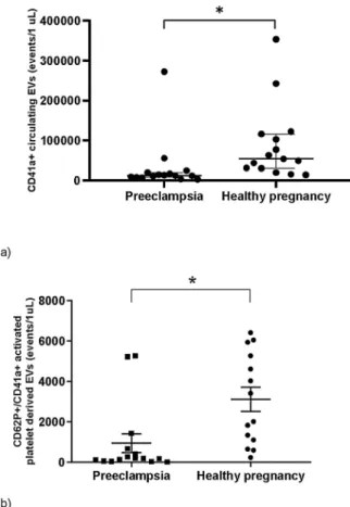
The median value of circulating CD41a +PEVs produced by acti- vated thrombocytes was significantly lower in the plasma of pre- eclamptic patients (median: 11,903 events/μL IQR: 8041–19533, n = 15) compared to third trimester healthy women (median: 54,297 events/μL, IQR: 30395–116438, n =15) (p <0.05) (Fig. 4).
3.3.3. P-Selectin (CD62)
The absolute amount of circulating CD62 P/CD41a double positive P-EVs produced by activated thrombocytes was significantly lower in the plasma of preeclamptic patients (median: 191 events/μL IQR:
62–677, n =15) compared to third trimester healthy women (median:
2717 events/μL, IQR: 997–5445, n =15) (p <0.05) (Fig. 4).
3.3.4. Tetraspanin (CD63)
We observed a higher amount of circulating tetraspanin positive (CD63+) P-EVs in preeclamptic patients (median =4107 events/μL IQR:
2095–19247, n =13) compared to third trimester healthy pregnant women (median: 3352 events/μL, IQR: 1110–5716, n =13), but the difference was not statistically significant p =0.15) (Fig. 5),
4. Discussion
The salient novel finding of the present study is that compared to
Fig. 1. Platelet count in preeclamptic patients and healthy controls (n =15, mean ±SEM) * p <0.05.
Fig. 2.Activation state of circulating platelets in preeclamptic patients and healthy controls (medians and IQRs, n =15) * p <0.05.
Fig. 3. Tissue factor bearing (CD142+) circulating procoagulant EVs in pre- eclamptic patients (n =10) and healthy controls (n =13). * p <0,05.
healthy pregnant women a significantly higher amount of tissue factor positive, thrombogenic, platelet-derived extracellular vesicles were present in the plasma of pregnant women with preeclampsia, a finding, which can have significant translational clinical ramification.
The present study was undertaken on pregnant women in pre- eclamptic conditions as indicated by their parameters compared to those of pregnancy-matched healthy women included in Table 1. Their plasma samples were collected and analyzed for platelets, and various platelet- derived extracellular vesicles.
The maintenance of hemostatic equilibrium is of utmost importance to maintain healthy blood circulation, which can be a major challenge during pregnancy. Even healthy pregnant women can develop throm- boembolic complications four to five times more often during the pregnancy compared to non-pregnant women (Eichinger et al., 1999).
About eighty percent of these thromboembolic events are located on the venous side of circulation and about twenty percent occur in the arteries.
The procoagulant state of pregnancy can be considered as a protective mechanism to prevent major blood loss during and after the childbirth (James, 2009).
Unfortunately, the occurrence of preeclampsia is steadily rising in the developed countries, thereby increasing the risk for maternal and fetal morbidity and mortality (Li et al., 2018). Preeclampsia may pose women to a fivefold increase of risk for pregnancy associated venous thromboembolism compared to the general pregnant population, mainly in the postpartum period (Egan et al., 2015).
Generalized activation of the coagulation system can complicate severe preeclampsia or HELLP syndrome (Bossung et al., 2020).
Disseminated intravascular coagulation (DIC) is a secondary disorder caused by the over-activation of coagulation system that causes the consumption of coagulation factors, platelets and fibrinolytic proteins leading to uncontrollable, multifocal bleeding. DIC is mostly associated with adverse maternal outcome, including massive transfusions, hys- terectomy, and even death (Erez et al., 2015).
More than forty years ago Redman et al. have described the increased platelet consumption as an early feature of preeclampsia (Redman et al., 1978). In line with this early finding, we also observed significantly decreased platelet count in the preeclamptic group. Platelet lifespan is significantly shorter in preeclampsia than in uncomplicated pregnancy (R´ak´oczi et al., 1979). Some of the platelets’ properties, such as mean platelet volume (MPV) and platelet distribution width (PDW) are routinely measured by blood count analyzers and are significantly increased in preeclampsia compared to normal pregnancy (Freitas et al., 2013). Platelet age correlates inversely with the MPV, thus the younger the platelets are, the higher the MPV is (Giles, 1981). MPV is known to increase due to the rapid turnover and activation of platelets thus larger platelets are more prone to aggregation (Park et al., 2002). Out of all platelets, younger platelets have the strongest adhesive and thrombo- genic function (Thattaliyath et al., 2005). According to a recent meta-analysis there is strong evidence, that increased platelet activation can be detected in preeclampsia and MPV is significantly increased in preeclamptic women compared to healthy pregnant women (Jakobsen et al., 2019).
Platelet-derived extracellular vesicles are generally considered as a marker of platelet activation (Antwi-Baffour et al., 2015). This process leads to several changes on the cell surface markers. Phosphatidylserine (PS) is normally located in the cytoplasmic surface of the platelet membrane and platelet activation causes externalization of PS. The exposed PS then activates blood coagulation by enhancing factor Xa-factor Va binding (Lentz, 2003). The significantly higher ratio of PS expressing platelets and tissue factor expressing platelet derived EVs in the plasma of preeclamptic women vs. healthy controls found in our study may explain the hypercoagulable state observed in preeclampsia.
Nevertheless, the exact role of EVs has not been yet entirely elucidated.
Yi et al. have studied the expression levels of CD41a, CD62 P and CD63 of platelets and found that they were higher in preeclampsia than in healthy pregnant women (Yi et al., 2003). We have also found higher albeit not significantly elevated CD63 expression of PEVs’, but the amount of CD41a and CD62 P positive PEVs’ turned out to be markedly lower in preeclamptic patients. This may highlight the difference be- tween the expression patterns of these glycoproteins between platelets and platelet-derived extracellular vesicles.
Tissue factor is an integral membrane receptor for coagulation Factor VII. It is constitutively expressed by cells surrounding blood vessels but it can be also detected on the surface of circulating extracellular vesicles (Bao et al., 2018). Both tissue factor bearing cells and extracellular vesicles are strong activators of extrinsic coagulation cascade (Kleinjan et al., 2012). Elevated levels of TF expressing EVs have already been detected in hypertensive disorders of pregnancy previously (Mackman, 2009; Del Conde et al., 2005), including higher amount of TF-positive syncytiotrophoblast micro-vesicles, which enhance Fig. 4. Circulating platelet derived EVs: Significantly lower amount of CD41a
+platelet derived EVs (a) and CD62P+/CD41a +activated platelet derived EVs (b) could be detected in the plasma of preeclamptic patients compared to third trimester pregnant-matched group. (medians and IQRs; * p <0.05).
Fig. 5. The median amount of CD63 (tetraspanin) positive activated platelet derived extracellular vesicles in preeclampsia (n =13) and healthy controls (n
=13). (medians and IQRs, p =0.15). ns =not significant.
thrombin-formation in preeclampsia. Extracellular vesicles are hypoth- esized to exhibit their procoagulant effect by their tissue factor content (Gardiner et al., 2011). One of the novel findings our study is that we could detect significantly higher amount of TF-positive platelet derived EVs in preeclampsia for the first time. According to our observations, the absolute amount of circulating CD41a + platelet derived EVs and CD41a+/CD62P + EVs produced by activated thrombocytes were significantly lower in the plasma of preeclamptic women than in healthy pregnant women.
CD62 P is a cell adhesion molecule (P-selectin, GMP-140, PADGEM), which plays an essential role in the initial recruitment of leukocytes, in platelet aggregation, in platelet-leukocyte interactions and in the adhesion of those to the endothelium as it is translocated from the cytoplasmic granules to the external cell membrane (Cambien and Wagner, 2004). The majority of previous studies have reported increased circulating platelet-monocyte aggregates during preeclampsia (Macey et al., 2010; Holthe et al., 2005). As platelets bind to monocytes predominantly via P-selectin–P-selectin glycoprotein ligand-1 (CD62P-PSGL-1) pathway (Bournazos et al., 2008), it may explain the contradiction between the elevated ratio of circulating activated plate- lets and the decreased CD62P +P-EV count. The lower count of acti- vated platelet derived EVs may probably be due to the P-selectin mediated aggregate formation in the preeclamptic group.
CD63 is a lysosomal membrane glycoprotein, which belongs to tet- raspanin family. CD63 has been identified as a platelet activation molecule which regulates gpIIb/IIIa function in platelets to aid the stability of the newly formed clots (Goschnick et al., 2006). We have found a trend of increased amount of CD63-positive platelet derived EVs in the preeclamptic samples, which further confirms the presence of increased platelet activation.
All these findings are in line with the idea that preeclampsia alters the procoagulant characteristics of blood and the number of circulating platelet derived extracellular vesicles by increased platelet activation and tissue factor expression. These changes may contribute to the he- mostatic disturbances in preeclampsia, such as thrombotic events in the venous and arterial circulation. Moreover, blocking cerebral micro- vessels could explain seizures observed in eclampsia. Previous studies proposed another mechanism by which platelet derived microparticles may contribute to the development of hypertension, suggesting the presence of a positive feedback mechanism in preeclampsia (Bao et al., 2018). In addition, maternal extracellular vesicles and platelets may promote preeclampsia via inflammasome activation in trophoblasts (Kohli et al., 2016).
Recent evidence suggests that extracellular vesicles can transfer in- formation to adjacent and remote areas by eliciting surface receptor activation and unloading the vesicular cargo into the recipient cell by internalization. In pathological conditions associated with tissue hyp- oxia, acidosis and oxidative stress, such as cardiac infarction, stroke, and abnormal cell proliferation (tumor formation) the degree of vesiculation has been shown to rise in a HIF-1α dependent manner (Belting and Christianson, 2015).
The increased release of thrombogenic extracellular vesicles in pre- eclampsia can lead to a hypercoagulable state as shown in the present study. Indeed, the main features of preeclampsia are uteroplacental hypoxia and oxidative stress both of which are present in tumor devel- opment pointing to the common mechanism of vesiculation. These findings may promote further research in this field and will support clinical decision making regarding diagnosis and treatment.
Finally, it is tempting to entertain the idea that similar disturbed hemostatic conditions can develop in the novel coronavirus disease Covid-19 (SARS-CoV-2), such as coagulopathy, and disseminated intravascular coagulation (DIC)-like massive intravascular clot forma- tion suggesting common mechanism of actions and perhaps similar therapeutic potentials (Ogawa et al., 2021; Taherifard et al., 2021).
Limitation of the study: The occurrence of preeclampsia was mod- erate at our clinic, which limited the sample size, thus we consider this
investigation as a pilot study generating hypothesis.
In conclusion, to the best of our knowledge, this is the first study analyzing the markers of platelet-derived extracellular vesicles in pre- eclampsia, and we have detected a significantly higher amount of tissue factor-positive thrombogenic platelet derived extracellular vesicles in the plasma of preeclamptic women compared to pregnancy-matched healthy women. Thus, we propose that increased platelet activation and tissue factor expression of platelet-derived extracellular vesicles may contribute to the hypercoagulable state observed in preeclampsia.
This idea corresponds to the prothrombotic state and consumptive coagulopathy observed previously in preeclampsia. Further investiga- tion of thrombocyte function might help us to better understanding the underlying mechanisms responsible for the development of the disease as well as to find new diagnostic biomarkers and potential therapeutic strategies for preeclampsia.
Declaration of Competing Interest
The authors report no declarations of interest.
Acknowledgement
This study was supported by the National Research, Development and Innovation Office Fund, OTKA K 132596 and OTKA K 113023.
Appendix A. Supplementary data
Supplementary material related to this article can be found, in the online version, at doi:https://doi.org/10.1016/j.jri.2021.103380.
References
Antwi-Baffour, S., Adjei, J., Aryeh, C., Kyeremeh, R., Kyei, F., Seidu, M.A., 2015.
Understanding the biosynthesis of platelets-derived extracellular vesicles. Immun.
Inflamm. Dis. 3 (3), 133–140.
Bao, H., Chen, Y.X., Huang, K., Zhuang, F., Bao, M., Han, Y., Chen, X.H., Shi, Q., Yao, Q.
P., Qi, Y.X., 2018. Platelet-derived microparticles promote endothelial cell proliferation in hypertension via miR-142-3p. FASEB J. 32 (7), 3912–3923.
Belting, M., Christianson, H.C., 2015. Role of exosomes and microvesicles in hypoxia- associated tumour development and cardiovascular disease. J. Intern. Med. 278 (3), 251–263.
Bossung, V., Fortmann, M.I., Fusch, C., Rausch, T., Herting, E., Swoboda, I., Rody, A., H¨artel, C., Gopel, W., Humberg, A., 2020. Neonatal outcome after preeclampsia and ¨ HELLP syndrome: a population-based cohort study in Germany. Front. Pediatr. 8, 579293.
Bournazos, S., Rennie, J., Hart, S.P., Fox, K.A.A., Dransfield, I., 2008. Monocyte Functional Responsiveness after PSGL-1–Mediated Platelet Adhesion is Dependent on Platelet Activation Status.
Brenner, B., 2004. Haemostatic changes in pregnancy. Thromb. Res. 114 (5–6), 409–414.
Brown, M.A., Magee, L.A., Kenny, L.C., Karumanchi, S.A., McCarthy, F.P., Saito, S., Hall, D.R., Warren, C.E., Adoyi, G., Ishaku, S., (ISSHP), I. S. f. t. S. o. H. i. P, 2018.
Hypertensive disorders of pregnancy: ISSHP classification, diagnosis, and management recommendations for international practice. Hypertension 72 (1), 24–43.
Cambien, B., Wagner, D.D., 2004. A new role in hemostasis for the adhesion receptor P- selectin. Trends Mol. Med. 10 (4), 179–186.
Del Conde, I., Shrimpton, C.N., Thiagarajan, P., Lopez, J.A., 2005. Tissue-factor-bearing microvesicles arise from lipid rafts and fuse with activated platelets to initiate coagulation. Blood 106 (5), 1604–1611.
Egan, K., Kevane, B., Ní Ainle, F., 2015. Elevated venous thromboembolism risk in ´ preeclampsia: molecular mechanisms and clinical impact. Biochem. Soc. Trans. 43 (4), 696–701.
Eichinger, S., Weltermann, A., Philipp, K., Hafner, E., Kaider, A., Kittl, E.M., Brenner, B., Mannhalter, C., Lechner, K., Kyrle, P.A., 1999. Prospective evaluation of hemostatic system activation and thrombin potential in healthy pregnant women with and without factor V Leiden. Thromb. Haemost. 82 (4), 1232–1236.
Erez, O., Mastrolia, S.A., Thachil, J., 2015. Disseminated intravascular coagulation in pregnancy: insights in pathophysiology, diagnosis and management. Am. J. Obstet.
Gynecol. 213 (4), 452–463.
Freitas, L.G., Alpoim, P.N., Komatsuzaki, F., Carvalho, M., Dusse, L.M., 2013.
Preeclampsia: are platelet count and indices useful for its prognostic? Hematology 18 (6), 360–364.
Gardiner, C., Tannetta, D.S., Simms, C.A., Harrison, P., Redman, C.W., Sargent, I.L., 2011. Syncytiotrophoblast microvesicles released from pre-eclampsia placentae exhibit increased tissue factor activity. PLoS One 6 (10), e26313.
Giles, C., 1981. The platelet count and mean platelet volume. Br. J. Haematol. 48 (1), 31–37.
Goschnick, M.W., Lau, L.M., Wee, J.L., Liu, Y.S., Hogarth, P.M., Robb, L.M., Hickey, M.J., Wright, M.D., Jackson, D.E., 2006. Impaired "outside-in" integrin alphaIIbbeta3 signaling and thrombus stability in TSSC6-deficient mice. Blood 108 (6), 1911–1918.
Gyorgy, B., Modos, K., Pallinger, E., Paloczi, K., Pasztoi, M., Misjak, P., Deli, M.A., Sipos, A., Szalai, A., Voszka, I., Polgar, A., Toth, K., Csete, M., Nagy, G., Gay, S., Falus, A., Kittel, A., Buzas, E.I., 2011. Detection and isolation of cell-derived microparticles are compromised by protein complexes resulting from shared biophysical parameters. Blood 117 (4), e39–48.
Gyorgy, B., Paloczi, K., Kovacs, A., Barabas, E., Beko, G., Varnai, K., Pallinger, E., Szabo- Taylor, K., Szabo, T.G., Kiss, A.A., Falus, A., Buzas, E.I., 2014. Improved circulating microparticle analysis in acid-citrate dextrose (ACD) anticoagulant tube. Thromb.
Res. 133 (2), 285–292.
Hardisty, R.M., Hutton, R.A., 1966. Platelet aggregation and the availability of platelet factor 3. Br. J. Haematol. 12 (6), 764–776.
Holthe, M.R., Lyberg, T., Staff, A.C., Berge, L.N., 2005. Leukocyte-platelet interaction in pregnancies complicated with preeclampsia. Platelets 16 (2), 91–97.
Jakobsen, C., Larsen, J.B., Fuglsang, J., Hvas, A.M., 2019. Platelet function in preeclampsia - a systematic review and meta-analysis. Platelets 30 (5), 549–562.
James, A.H., 2009. Pregnancy-associated thrombosis. Hematology Am. Soc. Hematol.
Educ. Program 277–285.
Kleinjan, A., Boing, A.N., Sturk, A., Nieuwland, R., 2012. Microparticles in vascular disorders: how tissue factor-exposing vesicles contribute to pathology and physiology. Thromb. Res. 130 (Suppl 1), S71–3.
Kohli, S., Ranjan, S., Hoffmann, J., Kashif, M., Daniel, E.A., Al-Dabet, M.M., Bock, F., Nazir, S., Huebner, H., Mertens, P.R., Fischer, K.D., Zenclussen, A.C., Offermanns, S., Aharon, A., Brenner, B., Shahzad, K., Ruebner, M., Isermann, B., 2016. Maternal extracellular vesicles and platelets promote preeclampsia via inflammasome activation in trophoblasts. Blood 128 (17), 2153–2164.
Lentz, B.R., 2003. Exposure of platelet membrane phosphatidylserine regulates blood coagulation. Prog. Lipid Res. 42 (5), 423–438.
Li, X., Zhang, W., Lin, J., Liu, H., Yang, Z., Teng, Y., Duan, S., Li, Y., Xie, Y., Lin, X., Xie, L., Peng, Q., Huang, J., Chen, J., Duan, W., Luo, J., Zhang, J., 2018. Preterm birth, low birthweight, and small for gestational age among women with preeclampsia: does maternal age matter? Pregnancy Hypertens. 13, 260–266.
Lopez, E., Srivastava, A.K., Burchfield, J., Wang, Y.W., Cardenas, J.C., Togarrati, P.P., Miyazawa, B., Gonzalez, E., Holcomb, J.B., Pati, S., Wade, C.E., 2019. Platelet- derived- extracellular vesicles promote hemostasis and prevent the development of hemorrhagic shock. Sci. Rep. 9 (1), 17676.
Macey, M.G., Bevan, S., Alam, S., Verghese, L., Agrawal, S., Beski, S., Thuraisingham, R., MacCallum, P.K., 2010. Platelet activation and endogenous thrombin potential in pre-eclampsia. Thromb. Res. 125 (3), e76–81.
Mackman, N., 2009. The role of tissue factor and factor VIIa in hemostasis. Anesth.
Analg. 108 (5), 1447–1452.
McLean, K.C., Bernstein, I.M., Brummel-Ziedins, K.E., 2012. Tissue factor-dependent thrombin generation across pregnancy. Am. J. Obstet. Gynecol. 207 (2), pp. 135.e1- Morikawa, M., Umazume, T., Hosokawa-Miyanishi, A., Watari, H., Kobayashi, T., 6.
Seki, H., Saito, S., 2019. Relationship between antithrombin activity and interval from diagnosis to delivery among pregnant women with early-onset pre-eclampsia.
Int. J. Gynaecol. Obstet. 145 (1), 62–69.
O’Brien, J.R., 1955. The platelet-like activity of serum. Br. J. Haematol. 1 (2), 223–228.
Ogawa, F., Oi, Y., Nakajima, K., Matsumura, R., Nakagawa, T., Miyagawa, T., Abe, T., Takeuchi, I., 2021. An evaluation of venous thromboembolism by whole-body enhanced CT scan for critical COVID-19 pneumonia with markedly rises of coagulopathy related factors: a case series study. Thromb. J. 19 (1), 26.
Park, Y., Schoene, N., Harris, W., 2002. Mean platelet volume as an indicator of platelet activation: methodological issues. Platelets 13 (5–6), 301–306.
Poon, L.C., Shennan, A., Hyett, J.A., Kapur, A., Hadar, E., Divakar, H., McAuliffe, F., da Silva Costa, F., von Dadelszen, P., McIntyre, H.D., Kihara, A.B., Di Renzo, G.C., Romero, R., D’Alton, M., Berghella, V., Nicolaides, K.H., Hod, M., 2019. The International Federation of Gynecology and Obstetrics (FIGO) initiative on pre- eclampsia: a pragmatic guide for first-trimester screening and prevention. Int. J.
Gynaecol. Obstet. 145 (Suppl 1), 1–33.
R´ak´oczi, I., Talli´an, F., Bagd´any, S., G´ati, I., 1979. Platelet life-span in normal pregnancy and pre-eclampsia as determined by a non-radioisotope technique. Thromb. Res. 15 (3–4), 553–556.
Redman, C.W., Sargent, I.L., 2008. Circulating microparticles in normal pregnancy and pre-eclampsia. Placenta 29 (Suppl A), S73–7.
Redman, C.W., Bonnar, J., Beilin, L., 1978. Early platelet consumption in pre-eclampsia.
Br. Med. J. 1 (6111), 467–469.
Staff, A.C., 2019. The two-stage placental model of preeclampsia: An update. J. Reprod.
Immunol. 134–135, 1–10.
Taherifard, E., Movahed, H., Mousavi, M.R., 2021. Hematologic autoimmune disorders in the course of COVID-19: a systematic review of reported cases. Hematology 26 (1), 225–239.
Thattaliyath, B., Cykowski, M., Jagadeeswaran, P., 2005. Young thrombocytes initiate the formation of arterial thrombi in zebrafish. Blood 106 (1), 118–124.
Tranquilli, A., Dekker, G., Magee, L., Roberts, J., Sibai, B., Steyn, W., Zeeman, G., Brown, M., 2014. The classification, diagnosis and management of the hypertensive disorders of pregnancy: a revised statement from the ISSHP. Pregnancy Hyperten.- Int. J. Womens Cardiovasc. Health 4 (2), 97–104.
Wolf, P., 1967. The nature and significance of platelet products in human plasma. Br. J.
Haematol. 13 (3), 269–288.
Yi, Z.S., Zhou, S.Y., Meng, F.Y., Feng, R., Liu, Q.F., Lin, R., 2003. Detection of platelet activation by flow cytometry in patients with pre-eclampsia. Di Yi Jun Yi Da Xue Xue Bao 23 (10), 1095–1096.