GENERAL AND DISEASE-SPECIFIC MECHANISTIC THERAPY APPROACHES FOR OPTIMIZATION OF
LIVER TRANSPLANTATION
PhD thesis
Christian Dominik Fingas
Doctoral School of Pathological Sciences Semmelweis University
Consultant: Zoltán Máthé, MD, Ph.D.
Official reviewers: Péter Igaz, MD, Ph.D.
Róbert Gáspár, Ph.D.
Head of the Final Examination Committee: Tibor Tihanyi, MD, Ph.D.
Members of the Final Examination Committee:
Klára Werling Marisné, MD, Ph.D.
Katalin Monostory, Ph.D.
Budapest
2016
TABLE OF CONTENTS
1 LIST OF ABBREVIATIONS IN ALPHABETICAL ORDER ... 3
2 INTRODUCTION ... 5
2.1 General Optimization Approaches For Liver Transplantation ... 5
2.1.1 Preservation solutions and the effect of chloride ... 5
2.1.2 Erythropoietin and liver regeneration/apoptosis ... 7
2.2 Optimization Of Liver Transplantation For Cholangiocarcinoma ... 8
2.2.1 The roles of myofibroblast derived growth factors and hedgehog signaling ... 8
2.2.2 Interactions between hedgehog signaling and polo-like kinase 2 ... 10
3 OBJECTIVES ... 11
4 METHODS ... 12
4.1 Materials ... 12
4.2 Cell lines/Culture/Co-Culture And Human Samples ... 12
4.3 In Vivo Microscopy ... 13
4.4 Light Microscopy ... 14
4.4.1 Histological evaluation after rat liver transplantation ... 14
4.4.2 Morphometric analysis of vessel area/perimeter ... 14
4.4.3 Immunohistochemistry for Ki-67 ... 15
4.4.4 Immunohistochemistry for α-SMA, PDGFR-β, and PDGF-BB ... 15
4.4.5 Immunohistochemistry for PLK1, PLK2, and PLK3 ... 15
4.5 Immunofluorescence Microscopy ... 16
4.5.1 Staining for cytokeratin 7/TUNEL assay ... 16
4.5.2 Staining for PLK2 and Mcl-1 ... 16
4.6 Microscopy For Smoothened (SMO) Trafficking ... 17
4.7 Quantitation Of Apoptosis ... 18
4.8 Real-Time Polymerase Chain Reaction (RT-PCR) ... 18
4.9 Immunoblot Analysis ... 20
4.10 Chromatin Immunoprecipitation (ChIP Assay) ... 21
4.11 Assessment Of Laboratory Parameters ... 21
4.12 Enzyme-Linked Immunosorbent Assay (ELISA) for PDGF-BB... 21
4.13 Assessment Of Bile Production ... 22
4.14 cDNA Array ... 22
4.15 Genome-Wide mRNA Expression Analysis ... 22
4.16 Generation Of A Transfectant Expressing SMO Short Hairpin RNA ... 23
4.17 Generation Of A Transfectant Expressing PLK1, 2, or 3 Short Hairpin RNA. ... 23
4.18 Generation Of AN Enhanced Green Fuorescent Protein (GFP)–Tagged SMO ... 24
4.19 GLI Reporter Construct And Promoter-Reporter Assay ... 24
4.20.1 Orthotopic full size rat liver transplantation ... 25
4.20.2 Orthotopic partial (30%) rat liver transplantation ... 26
4.20.3 Syngeneic, orthotopic rat modell of cholangiocarcinoma ... 26
4.21 Statistical Analysis ... 27
4.21.1 Preservation solution/erythropoietin studies ... 27
4.21.2 Cholangiocarcinoma studies ... 27
5 RESULTS ... 29
5.1 General Optimization Approaches For Liver Transplantation ... 29
5.1.1 Chloride improves survival due to beneficial effects on microcirculation ... 29
5.1.2 Erythropoietin increases liver growth and inhibits apoptosis ... 33
5.2 Optimization Of Liver Transplantation For Cholangiocarcinoma ... 36
5.2.1 Myofibroblast-derived PDGF-BB promotes hedgehog survival signaling ... 36
5.2.2 Polo-like kinase 2 is a mediator of hedgehog survival signaling ... 48
6 DISCUSSION ... 61
6.1 General Optimization Approaches For Liver Transplantation ... 61
6.1.1 Preservation solution/chloride study ... 61
6.1.2 Erythropoietin study ... 64
6.2 Optimization Of Liver Transplantation For Cholangiocarcinoma ... 67
6.2.1 Myofibroblast-derived PDGF-BB/hedgehog signaling study ... 67
7 CONCLUSIONS ... 73
8 SUMMARY ... 74
9 ÖSSZEFOGLALÓ... ...76
10 BIBLIOGRAPHY ... 76
11 BIBLIOGRAPHY OF THE CANDIDATE’S PUBLICATIONS ... 91
11.1 Publications Related To The PhD Thesis ... 91
11.2 Publications Not Related To The PhD Thesis ... 93
12 ACKNOWLEDGEMENTS ... 95
1 LIST OF ABBREVIATIONS IN ALPHABETICAL ORDER
α-SMA α-smooth muscle actin ALT alanine transaminase AP alkaline phosphatase AST aspartate transaminase Bcl-2 B-cell lymphoma
cAMP cyclic adenosine monophosphate CCA cholangiocellular carcinoma ChIP chromatin immunoprecipitation CIT cold ischemic time
CK7 cytokeratin 7
DHH desert hedgehog
DMEM Dulbecco's modified Eagle Medium ELISA enzyme-linked immunosorbent assay
EPO erythropoietin
ErbB-2 erythroblastic leukemia viral oncogene homolog GFP green fluorescent protein
GLDH glutamate dehydrogenase GLI glioma-associated oncogene HCC hepatocellular carcinoma
Hh hedgehog
HIP hedgehog-interacting protein HSC hepatic stellate cells
HTK histidine-tryptophan-ketoglutarate
IHH indian hedgehog
INR international normalized ratio LBWR liver body weight ratio LDH lactate dehydrogenase
LDLT living donor liver transplantation LTx liver transplantation
MAPK mitogen-activated protein kinase
MBF myofibroblast
Mcl-1 myeloid cell leukemia-1 PDGF platelet-derived growth factor
PDGFR platelet-derived growth factor receptor
PH partial hepatectomy
PKA cAMP-dependent protein kinase PLK polo-like kinases
pLTx 30% partial liver transplantation
PT prothrombin time
PTCH1 patched1
PTT prothrombin time
RBC red blood cells
RFU relative fluorescence unit
RT room temperature
RT-PCR real time polymerase chain reaction
SHH sonic hedgehog
shRNA short hairpin RNA sLTx split liver transplantation
SMO smoothened
TIRF total internal reflection fluorescence
TRAIL tumor necrosis factor-related apoptosis-inducing ligand
TUNEL terminal deoxynucleotidyl transferase-mediated dUTP nick end labeling VEGF vascular endothelial growth factor
WIT warm ischemic time
2 INTRODUCTION
Liver transplantation (LTx) is a viable treatment option for acute liver failure and various end- stage liver diseases including malignancies like hepatocellular carcinoma (HCC) and cholangiocellular carcinoma (CCA). The present work will focus on the development of accompanying mechanistic therapies that may be eligible for optimization of this lifesaving surgical procedure. The studies are divided into general optimization approaches of LTx and disease-specific mechanistic experiments aiming to identify suitable targets in order to improve LTx for unresectable CCA patients according to the multimodal therapy concept of the Mayo protocol.1
2.1 General Optimization Approaches For Liver Transplantation
In these experiments we sought to minimize liver preservation injury and improve microcirculation in implanted liver grafts by modification of a histidine-tryptophan- ketoglutarate (HTK)-based preservation solution. In addition, we tested the effect of the pleiotropic substance erythropoietin (EPO) on liver regeneration/donor liver growth and hepatocyte apoptosis (programmed cell death) in the setting of partial liver transplantation (pLTx).
2.1.1 Preservation solutions and the effect of chloride
Preservation injury is still a major concern in liver transplantation, especially for grafts obtained from “extended criteria donors”.2-4 Preservation injury can be regarded as a consequence of hypoxia,5-8 injury triggered by hypothermia,7, 9, 10 a certain toxicity of the preservation solutions,11, 12 and at a later stage, inflammatory processes.7, 13, 14 Taking into account new findings on the mechanisms of the initial processes already occurring during cold
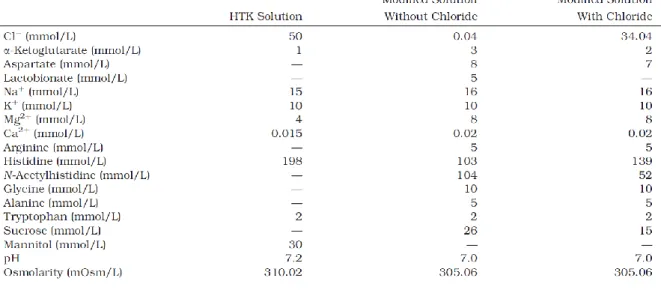
preservation, we developed a new preservation solution on the basis of HTK solution (for composition see Table 1), which, in a preliminary (chloride-poor) version, was already shown to reduce preservation injury to isolated perfused rat livers in comparison with HTK solution.15
Table 1. Compositions of HTK solution and the modified HTK solutions. To obtain a chloride-containing variant of the new solution, further but slight modifications became necessary for reasons of charge and osmolarity;
these modifications mainly affected the concentrations of N-acetylhistidine (partly anionic), histidine (partly cationic), and sucrose.
In the course of the mechanistic studies that formed the basis for developing the new preservation solution, we have described the entity of cold-induced apoptosis, an injury that affects numerous mammalian cell types such as rat hepatocytes, rat liver endothelial cells, rat renal tubular cells, rat coronary endothelial cells, porcine aortic endothelial cells, porcine corneal endothelial cells, and human umbilical vein endothelial cells.7, 9, 16-19 In all these cell types chelatable, ‘‘redox-active’’ iron plays the major role in the development of cold-induced apoptosis.7, 9, 16-21
However, hepatocytes appear to be slightly different since in these cells iron chelators only provide partial protection from cold-induced injury during rewarming after cold incubation in cell culture medium or Krebs-Henseleit buffer.18, 19, 21 We further characterized this iron- independent weaker cold-induced injury to rat hepatocytes and found it to be dependent on extracellular chloride.22
In contrast to that, a another study on cold-induced injury to the endothelium of intact porcine aortic segments exhibited beneficial effects of chloride-containing preservation solutions on endothelial cell survival.23 This is in line with other experiments in cultured porcine aortic endothelial cells revealing strong adverse effects of chloride-poor preservation solutions.24
As it was impossible to judge from these contradictory in vitro experiments which of the chloride-dependent effects has a higher biological relevance regarding the intact liver, we here tested a chloride-poor versus a chloride-containing variant of the new preservation solution in
an orthotopic rat liver transplantation model. Three different post-LTx survival series were performed since survival represents the study criterion with the highest medical relevance and since the clarification of the conflicting in vitro data is crucial for the further development of the preservation solution. The three series were designed to cover different balances of cold ischemic and warm ischemic injury as well as surgical trauma, thereby reflecting the broad range of potential clinical settings most closely. In addition, one LTx series (with intermediate cold and warm ischemic times [CIT/WIT]) was performed for assessment of intrahepatic microcirculation after reperfusion, laboratory data, bile production, and liver histology.
2.1.2 Erythropoietin and liver regeneration/apoptosis
After pLTx (or extended liver resection), efficient regeneration of the liver is essential for the clinical outcome. Especially after living donor liver transplantations (LDLT) and split liver transplantations (sLTx), which have been an important developments to overcome the growing problem of organ shortage,25 an immediate regeneration is most desirable because of the graft’s smaller size and its reduced functional liver mass. A small-for-size graft may not only be functionally insufficient for the recipient, but will also sustain injury characterized by rejection and ischemic insult, which results in an inadequate regeneration and leads to hepatic failure.26 For the LDLT donor, an adequate regenerative response is of comparable importance.
Unfortunately, more than 16% of all LDLT can not be performed, since the suggested graft/recipient- and remnant liver/donor-ratio of at least 0.8% can not be achieved.27
Therefore, improvement of the regenerative capacity is of fundamental importance and novel therapeutic approaches are needed to optimize liver regeneration in the setting of LDLT/sLTx.
Previously, we were able to demonstrate the positive effects of vascular endothelial growth factor (VEGF) as well as tri-iodothyronine as stimulators of liver regeneration after partial hepatectomy (PH). However, none of the above mentioned treatment strategies have proven definitive.28, 29
EPO is a low molecular weight glycoprotein hormone stimulator of erythropoiesis produced in the fetal liver and subsequently in the adult kidney.30, 31 Stimulation of erythropoiesis was considered to be the sole physiological action of EPO, but there is increasing evidence suggesting a wider biological role including angiogenesis and liver regeneration.32, 33 Regarding the latter aspect it is known that not only the fetal but also the adult liver can be an extrarenal source of EPO.34 Indeed, in liver regeneration an increased synthesis of EPO has been
described, whereas enhanced EPO serum levels correlate with the peak of liver regeneration after PH.35 Here, the synthesis of EPO is mediated by erythropoietic hepatic factors 36, 37 and occurs in Kupffer cells 38-40 as well as erythroblastic islets within the liver lobules.37, 41 EPO was also reported as a stimulator of liver regeneration after PH in rats and pigs.32, 33
2.2 Optimization Of Liver Transplantation For Cholangiocarcinoma
Sole LTx for unresectable CCA is often associated with early disease relapse and limited overall survival.1 However, a small percentage of patients have achieved prolonged survival after LTx, suggesting that adjuvant approaches might improve the clinical outcome.1 Thus, a multimodal therapy protocol was developed at the Mayo Clinic, Rochester, Minnesota, USA employing pre-LTx external-beam irradiation, chemotherapy, and iridium brachytherapy for patients with unresectable CCA above the cystic duct and without extrahepatic metastases.1 After pretreatment and before LTx, patients undergo an exploratory laparotomy to exclude metastatic disease. The Mayo protocol has been proven to be quite successful for the treatment of patients with unresectable early-stage CCA.1
However, employing the conventional chemotherapeutic agents fluorouracil and capecitabine, this protocol does not consider new mechanistic findings on CCA tumor biology and, thus, might be improvable by the implementation of „targeted chemotherapy”. The present CCA- specific LTx optimization experiments aim to identify mechanistic processes underlying the pronounced resistance to apoptotic cell death characteristic for CCA cells. Based on these findings, new mechanistic therapy approaches were tested.
2.2.1 The roles of myofibroblast derived growth factors and hedgehog signaling
CCA is a highly lethal malignancy with limited treatment options.42-44 It is the most common biliary cancer and epidemiologic studies suggest that its incidence is increasing in several Western Countries.45 Human CCA in vivo paradoxically expresses the death ligand tumor necrosis factor-related apoptosis-inducing ligand (TRAIL) and its cognate death receptors 46 suggesting that these cancers are reliant on potent survival signals for tumor maintenance and to circumvent apoptotic cell death by TRAIL. However, the mechanisms by which CCA evades apoptosis by TRAIL and other pro-apoptotic stimuli are incompletely understood.
CCAs are highly desmoplastic cancers suggesting cancer-associated fibroblasts within the tumor microenvironment contribute to their development and progression as has been proposed for other
cancers (e.g. breast cancer, prostate cancer, etc.).47, 48 Cancer-associated fibroblasts are perpetually “activated” and express α-smooth muscle actin (α-SMA); cells exhibiting this activated phenotype are often referred to as myofibroblasts (MFBs).49 In the liver, MFBs are derived from periportal fibroblasts, hepatic stellate cells (HSCs), and perhaps an epithelial-to- mesenchymal transition of cholangiocytes, hepatocytes, and/or the tumor itself.50, 51 A role for MFBs in carcinogenesis and tumor biology receives increasing attention.49, 52-54 Cross-talk between the cancer and MFBs appears to be exploited by cancer cells as a tumor promoting mechanism. For example, in CCA the number of MFBs correlates with tumor size and patient survival.55, 56 MFBs also appear capable of providing survival signals as they reduce apoptosis of non-malignant cholangiocytes in co-culture experiments.57 However, information regarding the nature of this cross-talk, and in particular the identity of the potential survival signals, remains obscure.
Growth factor and especially platelet-derived growth factor (PDGF) paracrine signaling between MFBs and cholangiocytes occurs in rodent models of biliary tract inflammation and fibrogenesis.57, 58 Five different ligands of PDGF exist including PDGF-AA, -BB, -AB, -C and –D. However, PDGF-BB appears to be the predominant isoform secreted by liver MFBs.59 Of the two cognate receptors, platelet-derived growth factor receptor (PDGFR)-α and –β, PDGFR- β is the cognate receptor for PDGF-BB. PDGFR-β is a receptor tyrosine kinase that is also known to alter plasma membrane dynamics associated with cell migration by a cyclic adenosine monophosphate (cAMP)-dependent kinase (PKA)-dependent process.60 Thus, PDGF-BB effects on intracellular signaling cascades are pleiotropic. Given an emerging role for PDGF- BB in MFB-to-cholangiocyte cross-talk, a role for PDGF-BB as a survival factor for CCA warrants further investigation.
The Hedgehog (Hh) signaling pathway has been strongly implicated in gastrointestinal tumor biology including CCA.61, 62 Hh signaling is initiated by any of the three ligands Sonic (SHH), Indian (IHH), and Desert (DHH) hedgehog. These ligands bind to the Hh receptor Patched1 (PTCH1) resulting in activation of Smoothened (SMO) and subsequently the transcription factors glioma-associated oncogene (GLI) 1, 2, and 3.63 How PTCH1 modulates SMO was long enigmatic, as the two proteins do not physically associate. SMO trafficking from an intracellular compartment to the plasma membrane apparently results in its activation.64 Hh ligand binding to PTCH1 increases the concentration of intracellular messengers (lipid phosphates), which in turn promote SMO trafficking to the plasma membrane.65, 66 PKA affects SMO trafficking and activation, raising the unexplored possibility that cues from other ligand-receptor systems such
as PDGF-BB may also augment SMO activation by facilitating its trafficking to the plasma membrane.64
Interestingly, SHH mRNA expression is increased by PDGF-BB in immature cholangiocytes58 providing an additional link between Hh signaling and PDGF. Hh signaling also is a master switch mediating resistance of CCA cells to TRAIL cytotoxicity. 67, 68 Taken together, these observations suggest MFB-derived PDGF-BB may modulate Hh survival signaling in CCA cells.
2.2.2 Interactions between hedgehog signaling and polo-like kinase 2
As mentioned above, Hh signaling was reported to be an important survival pathway in CCA.67,
69, 70 Hh ligand SHH is abundantly expressed in CCA cells, 69, 71 and in a recent mRNA expression analysis employing CCA cells, Hh signaling was suggested to positively regulate the cell division modulating enzyme kinase polo-like kinase 2 (PLK2).69
PLK2 (or SNK) is one out of five mammalian PLK family members that orchestrate a wide range of critical cell cycle events.72-74 Besides PLK2, PLK1 (or STPK13), PLK3 (or CNK, FNK and PRK), PLK4 (or SAK and STK18) and PLK5 have been identified.73, 74 All PLK proteins share a similar structure with a canonical serine/threonine kinase domain at the N-terminus and a regulatory polo-box domain at the C-terminus 72; however, PLK4 has a notably divergent structure as compared to other PLK proteins and PLK5 as it lacks kinase activity. 73, 74
About 80% of human cancers express high levels of PLK transcripts in tumor cells (these PLK transcripts are mostly absent in surrounding healthy tissues) and PLK overexpression is often associated with poor prognosis and lower overall survival.75 While PLK1 has been extensively studied and has become an attractive candidate for anti-cancer drug development, the roles of the other PLK proteins including PLK2 are less well understood.74
PLK inhibition in esophageal squamous cell carcinoma and osteosarcoma was reported to decrease protein levels of myeloid cell leukemia-1 (Mcl-1).76, 77 This is of particular interest as Mcl-1, a potent anti-apoptotic member of the B-cell lymphoma (Bcl-2) protein family, has been identified as a survival factor in CCA.78-80 Given the pivotal role of Mcl-1 in mediating CCA resistance to TRAIL-induced apoptosis, 78-80 PLK inhibition is another potential strategy for the targeted treatment of this devastating disease.
.
3 OBJECTIVES
The aims of the present studies were:
1. Optimization of a modified HTK-based preservation solution focusing on chloride- dependent effects on liver preservation injury and microcirculation after LTx.
2. Investigation of the impact of adjuvant administered EPO on liver regeneration/donor liver growth and hepatocyte apoptosis in the setting of pLTx.
3. Examination of the role of MFB-to-CCA cell paracrine signaling for CCA apoptosis resistance in the context of PDGF-BB/Hh co-activation networks
4. Exploration of anti-apoptotic effects mediated by Hh/PLK signaling crosstalk.
5. Based on the observations of 3) and 4), the objectives of subsequent studies were to test whether targeting PDGFR-β, Hh, or PLK signaling would be therapeutic in CCA and, thus, might be a suitable adjuvant therapy to optimize the Mayo LTx protocol for CCA patients.
4 METHODS
4.1 Materials
The chloride-poor (0.04 mmol/l) and chloride-containing (34.04 mmol/l, which is the highest possible chloride concentration within the confines given by all other compounds) preservation solutions were provided by Dr. Franz Köhler Chemie GmbH (Bensheim, Germany). EPO (EPREX®) was purchased from Ortho Biotech, Neuss, Germany.
rhTRAIL, rhPDGF-BB, rhSHH, anti-human PDGF-BB antiserum AB-220-NA (R&D Systems, Minneapolis, MN), PKA inhibitor H-89 (Cayman Chemical, Ann Arbor, MI), MG-132 (Merck, Rockland, MA), GDC-0449 (Selleck, Houston, TX), and cyclopamine (LC Laboratories, Woburn, MA) were prepared according to the suppliers protocols. Imatinib mesylate/STI-571, an inhibitor of the kinase activity of PDGFR(-β), was a generous gift from E. B. Leof (Div. of Pulmonary and Critical Care Medicine, Mayo Clinic, Rochester, MN). Imatinib was dissolved in sterile water (10 mmol/l stock solution) and subsequently diluted in cell culture medium.
BI 6727/volasertib, a potent selective PLK inhibitor 81 was purchased from Active Biochem (Maplewood, NJ), dissolved in dimethyl sulfoxide (DMSO; Sigma, St. Louis, MO; 1 mmol/L stock solution) and subsequently diluted in cell culture medium for use in in vitro experiments.
The SHH-neutralizing antibody 5E1 was obtained from the Developmental Studies Hybridoma Bank (DSHB, University of Iowa, IA). The construct encoding for S peptide-tagged human Mcl-1 mutant resistant to proteasomal degradation due to sequential mutagenesis of the established Mcl-1 ubiquitination sites (amino acids 5, 40, 136, 194, and 197) from lysine to arginine was generated as previously described. 82
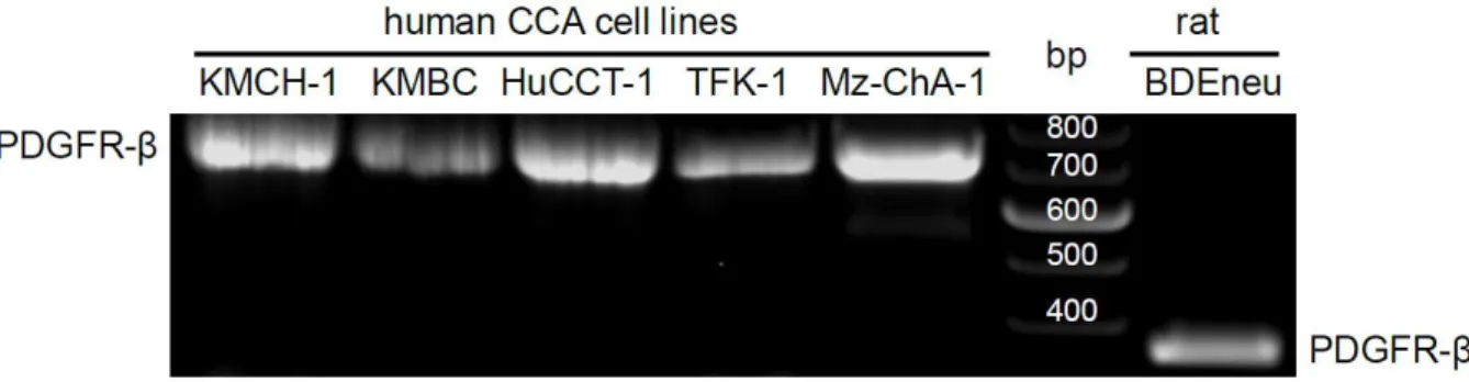
4.2 Cell lines/Culture/Co-Culture And Human Samples
The human CCA cell lines KMCH-1, KMBC, HuCCT-1, TFK-1, and Mz-ChA-1 and as well as the erythroblastic leukemia viral oncogene homolog (ErbB-2)/neu transformed malignant rat cholangiocyte cell line BDEneu (CCA in vivo experiments) and the LX-2 cells, an immortalized myofibroblast cell line derived from human HSCs, were cultured as previously described.46, 83-
86 The human primary myofibroblastic HSCs were kindly provided by V.H. Shah (Division of Gastroenterology and Hepatology, Mayo Clinic, Rochester, MN) and cultured in Dulbecco's modified Eagle Medium (DMEM) supplemented with 10% fetal bovine serum, penicillin G (100 U/mL), and streptomycin (100 μg/mL) under standard conditions.
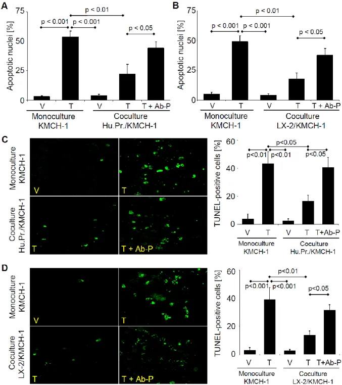
CCA co-culture cell experiments were performed using a transwell insert co-culture system (24 wells) equipped with 0.4 µm pore size polyester (PET) inserts (Corning Coster, Acton, MA) for 6 days according to the manufacturer’s recommendations. Briefly, KMCH-1 or KMBC cells were plated alone or together with myofibroblastic human primary HSCs or LX-2 cells in the transwell insert co-culture system (KMCH-1 or KMBC cells in the bottom and human primary HSCs or LX-2 cells in the top wells; 1:1 ratio). First, all cells were plated alone at a density of 2 x 10³ cells/well overnight. The co-culture insert chambers with the human primary HSCs or LX-2 cells then were transferred the next day. Cells were treated as indicated whereas rhTRAIL was added at the end of the experiment (day 6) for 6 hrs and the anti-human PDGF-BB antiserum was added on day 5 for 24 hrs (anti-human PDGF-BB antiserum was added not longer than 24 hrs to minimize confounding effects on apoptosis measurement due to decreased cell proliferation). After rhTRAIL treatment, the KMCH-1 or KMBC cells in the bottom wells were analyzed for apoptosis by DAPI-staining and TUNEL assay as described in the
“Quantitation of apoptosis” section (for the TUNEL assay, cells were plated on sterilized trimmed coverslips that were placed in the bottom wells prior to cell seeding).
Human samples (from patients with intrahepatic and extrahepatic CCA treated at Mayo Clinic, Rochester, MN, USA) for analysis by immunohistochemistry were collected with Institutional Review Board approval according to the principles embodied in the declaration of Helsinki.
4.3 In Vivo Microscopy
For assessment of microvascular liver perfusion and leukocyte-endothelial interaction (preservation solution study) in vivo microscopy was performed 30 min after reperfusion using a Leica DMLM epifluorescence microscope (Leica Microsystems Wetzlar GmbH, Wetzlar, Germany). The left lateral liver lobe was exteriorized on a specially designed stage. The abdominal cavity was kept moist and body temperature was maintained constant using a heated operation table. Sodium fluorescein (2.0 mmol/kg; Sigma, Deisenhofen, Germany) and rhodamin 6G (0.1 mmol/kg; Sigma) were injected intravenously for fluorescent staining of hepatocytes and leukocytes, respectively.87 During the measurement, hemodynamic parameters (arterial blood pressure and heart rate) were monitored continuously via a polyethylene catheter placed in the right common iliac artery (Pressure measurement set IT2, Smiths Medical Int., Lancashire, UK; Dräger Infinity Delta XL monitor, Dräger Medical GmbH, Lübeck, Germany).
Microcirculation was only assessed if the mean arterial pressure (MAP) was above 60 mmHg (in one case [chloride-poor preservation solution] the measurement was aborted after 40 min
due to systemic hypotension [technical reasons]). The following parameters were determined in 10 randomly selected acinar areas and postsinusoidal venules: i) diameters of sinusoids and postsinusoidal venules [µm]. ii) sinusoidal perfusion rate: ratio of perfused sinusoids to all sinusoids visible in a defined acinar area [%]. iii) red blood cell (RBC) velocity in sinusoids and postsinusoidal venules [µm/s]. iv) temporary leukocyte adherence in postsinusoidal venules (rollers): leukocytes moving along the wall of postsinusoidal venules with a velocity of less than 30 % of the central stream velocity (percentage of rollers of all free-moving leukocytes during the observation period of 20 sec [%]). v) permanent leukocyte adherence (sticker) in sinusoids and postsinusoidal venules: number of leukocytes adhering for at least 20 sec in sinusoids [n/lobule]; number of leukocytes attached for at least 20 sec to the venular surface of postsinusoidal venules ([n/mm2]; calculation of the visible part of the vessel surface [approximately 50 %, since only the back wall or the front wall of the postsinusoidal venules could be observed]: 0.5 · π · d · l [d = mean vessel diameter, l = vessel length]). Video tapes were analyzed by an examiner blinded to the experimental groups using the CapImage 7.3 analysis software (Image Analysis System, Dr. Zeintl, Heidelberg, Germany).
4.4 Light Microscopy
4.4.1 Histological evaluation after rat liver transplantation
Following in vivo microscopy (preservation solution study), specimens of the transplanted livers were taken. Histological evaluation was performed after formalin fixation, paraffin embedding, and hematoxylin/eosin as well as ASDCL (naphthol-AS-D-chloroacetate esterase;
assessment of granulocyte invasion) staining by a pathologist, who also was blinded to the experimental groups. The severity of morphological/pathological changes was graded according to a numeric semiquantitative score (grade 1 = severe changes, grade 2 = moderate changes and grade 3 = minimal changes) evaluating the width of intrahepatic vessels, vacuolization in the cytoplasm of hepatocytes, prominence of Kupffer cells, necrosis/apoptosis as well as cholestasis.
4.4.2 Morphometric analysis of vessel area/perimeter
To assess dilatation of intrahepatic vessels (EPO study), hematoxylin-eosin stained slides were investigated by computed morphometry. Using an image analysis program (Zeiss KS 300, Oberkochen, Germany) the vessel area and perimeter was measured in central veins of the
hepatic parenchyma (10 randomly chosen visual fields). Results are given in µm² (area) and µm (perimeter), respectively.
4.4.3 Immunohistochemistry for Ki-67
Immunostaining for Ki-67, a marker for cell proliferation, was performed to evaluate the proliferation ofhepatocytes (EPO study). The primary antibody was a rabbit monoclonal anti- rat/mouse/human Ki-67 antigen (DCS Diagnostics, Hamburg, Germany 1:1200 dilution).
Immunohistochemistry was performed using a biotin-free enhanced polymer one-step staining technique (EPOS-method) with a peroxidase-conjugated polymer backbone coupled with a goat anti-rabbit secondary antibody (DAKO, Hamburg, Germany). “Proliferation index” was defined as the percentage of Ki-67 positive cells counted in 5 high-power-fields (x40) of a specimen.29
4.4.4 Immunohistochemistry for α-SMA, PDGFR-β, and PDGF-BB
Immunohistochemistry for CCA studies was performed using formalin-fixed, paraffin- embedded human and rat CCA samples (slides were also stained conventionally with hematoxylin/eosin). Slides were deparaffinized in xylene and rehydrated through sequential graded ethanol steps. For α-SMA-, PDGFR-β- and PDGF-BB-staining, the antigen retrieval was performed by permeabilizing the slides in 0.1% Triton X 100 for 2 min (α-SMA-staining) and incubation in sodium citrate (α-SMA- and PDGFR-β-staining; 0.01 M sodium citrate, 0.05% Tween 20; pH 6.0) or Tris-EDTA buffer (PDGF-BB-staining; 0.01M Tris base, 1 mM EDTA solution, 0.05% Tween 20, pH 9.0) using a vegetable steamer (30 min for α-SMA- and 60 min for PDGFR-β-/PDGF-BB-staining). After cooling, further steps were carried out according to the protocols of the EnVision+ System-HRP [DAB] detection kits (α-SMA: K4006 [anti-mouse]; PDGFR-β and PDGF-BB: K4010 [anti-rabbit]; Dako, Carpinteria, CA). The primary antiserum against α-SMA 1A4 (MS-113-R7, ready-to-use dilution; NeoMarkers, Fremont, CA) was applied for 60 min at RT (PDGFR-β: P-20, 1:25, applied overnight at 4°C, Santa Cruz, Santa Cruz, CA; PDGF-BB: ab21234, 1:10, applied overnight at 4°C, Abcam, Cambridge, MA). Finally, the slides were counterstained with Mayer’s Hematoxylin Solution (Sigma, St. Louis, MO), mounted and examined by light microscopy.
4.4.5 Immunohistochemistry for PLK1, PLK2, and PLK3
Immunohistochemistry (CCA studies) was performed using formalin-fixed, paraffin-embedded human CCA samples. Slides were deparaffinized in xylene and rehydrated through sequential graded ethanol steps. The antigen retrieval was performed by permeabilizing the slides in 0.1%
Triton X 100 for 2 min and incubation in sodium citrate (0.01 M sodium citrate, 0.05% Tween 20; pH 6.0) for 30 min using a vegetable steamer. After cooling, further steps were carried out according to the protocols of the EnVision+ System-HRP [DAB] detection kits (K4010 [anti- rabbit for PLK2 and PLK3], K4007 [anti-mouse for PLK1]; Dako, Carpinteria, CA). The primary antiserum against PLK1 (1:200; Merck Millipore, Darmstadt, Germany; CN: 05-844), PLK2 (1:100; Abcam, Cambridge, MA; ab34811) and PLK3 (1:200; Proteintech, Manchester, UK; CN: 10977-1-AP) was applied overnight at 4°C. The slides were counterstained with Mayer’s Hematoxylin Solution (Sigma, St. Louis, MO), mounted and examined by light microscopy. PLK1/2/3 protein expression quantitation of intrahapatic and extrahepatic CCA samples was performed by histological grading according to the number of PLK1/2/3-positive cells and the intensity of PLK1/2/3 immunoreactivity (grade 0 = no protein expression, grade 4
= high protein expression).
4.5 Immunofluorescence Microscopy
4.5.1 Staining for cytokeratin 7/TUNEL assay
For cytokeratin 7 (CK7)-labeling (CCA studies), the antigen retrieval was performed incubating the slides in deionized water containing 5% urea using a vegetable steamer for 20 min (since some slides also were labeled for TUNEL-positive cells, an additional antigen retrieval step was performed with sodium citrate followed directly by cooling and application of the TUNEL reaction mix; the TUNEL assay is described in the “Quantitation of apoptosis” section). The primary antibody against CK7 (1:10; Abcam; ab9021) was applied for 30 min at RT. After being washed, the slides were incubated with Texas Red®-X goat anti-mouse IgG (1:1000;
Invitrogen, Camarillo, CA; T6390) for 1 hr in the dark. The slides were then washed three times in PBS, one time in water and mounted using Prolong Antifade (also Invitrogen). The slides were analyzed by fluorescent confocal microscopy(LSM 510; Zeiss, Jena, Germany).
4.5.2 Staining for PLK2 and Mcl-1
Immunohistochemistry was performed using formalin-fixed, paraffin-embedded (cyclopamine CCA study) or frozen (BI 627 CCA study) rat CCA samples. Paraffin slides were deparaffinized
in xylene and rehydrated through sequential graded ethanol steps Antigen retrieval for the paraffin slides was performed by permeabilizing the slides in 0.1% Triton X 100 for 2 min and incubation in deionized water containing 5% urea using a vegetable steamer for 20 min with subsequent cooling for 20 min. Frozen slides were fixed with 4 % paraformaldehyde in PBS for 10 min at RT and tissue permeabilization was performed with 0.1% Triton X 100 for 15 min at RT. After a blocking step with 5% BSA in PBS for 1 hr at RT the primary antisera/antibodies against PLK2 (1:50; Abcam, Cambridge, MA; ab34811) and Mcl-1 (1:100;
Santa Cruz, Santa Cruz, CA; sc-819) were applied overnight at 4 °C. After washing, the slides were incubated with Alexa Fluor® 488 chicken anti-rabbit IgG (for PLK2 and Mcl-1; 1:1000;
Invitrogen, Camarillo, CA; A21441) for 1 hr in the dark at RT. The slides were then washed three times in PBS, one time in water and mounted using Prolong Antifade with DAPI (Invitrogen). The slides were analyzed by fluorescent confocal microscopy (LSM 510; Zeiss, Jena, Germany) and PLK2 as well as Mcl-1 immunoreactivity was quantitated using the software ImageJ 1.44o (Wayne Rasband, NIH, Bethesda, MD).
4.6 Microscopy For Smoothened (SMO) Trafficking
HuCCT-1 CCA cells were cultured on coverslips, treated as indicated, and fixed with PBS containing 4% paraformaldehyde for 20 min at 37°C. After being washed with PBS, cells were incubated with 0.5% Triton X-100 in PBS for 15 min at RT and then blocked with PBS containing 5% BSA for 60 min at 37°C. Cells were subsequently incubated with anti-SMO antiserum (1:250; Santa Cruz, Santa Cruz, CA; H-300) at 4°C overnight. After being washed, coverslips were incubated with Texas Red®-X goat anti-rabbit IgG (1:1000; Invitrogen, Camarillo, CA; T6391) for 1 hr in the dark. Cells were then washed three times in PBS, one time in water and mounted using Prolong Antifade (Invitrogen). The slides were analyzed by fluorescent confocal microscopy (LSM 510; Zeiss, Jena, Germany). In additional experiments, SMO trafficking was examined by total internal reflection microscopy (TIRF).88 KMCH-1 cells cultured on coverslips were transfected with GFP-SMO plasmid 48 hours prior to study. Cells were treated as indicated, and fixed with ddH2O containing 2.5% formaldehyde, 0.1 M PIPES, 1.0mM EGTA, and 3.0 mM MgSO4 for 20 min at 37°C. Cells were then washed three times in PBS, one time in water and mounted using Prolong Antifade (Invitrogen). The slides were analyzed with a TIRF microscop (Zeiss AxioObserver.Z1, Munich, Germany). GFP-SMO localized to the plasma membrane was quantified using image analysis software (Carl Zeiss
AxioVision 4.8.2.0, Munich, Germany). Data were expressed as the average fluorescence intensity in the cell multiplied by the number of pixels above the background.
4.7 Quantitation Of Apoptosis
Apoptosis in CCA cells was quantified by assessing the characteristic nuclear changes of apoptosis after staining with 4’,6-diamidino-2-phenylindole dihydrochloride (DAPI; Sigma, St.
Louis, MO) using fluorescence microscopy.89 Terminal deoxynucleotidyl transferase-mediated dUTP nick end labeling (TUNEL) assays (cell co-culture and rat liver samples in the EPO and CCA studies) were carried out using the In situ Cell Death Detection kit (Roche, Indianapolis, IN) according to the supplier's protocol and as previously described.84 Caspase 3/7-activity in the CCA studies was quantitated using the ApoONE Homogenous Caspase-3/7 Assay (Promega, Madison, WI) according to manufacturer’s recommendations.89
4.8 Real-Time Polymerase Chain Reaction (RT-PCR)
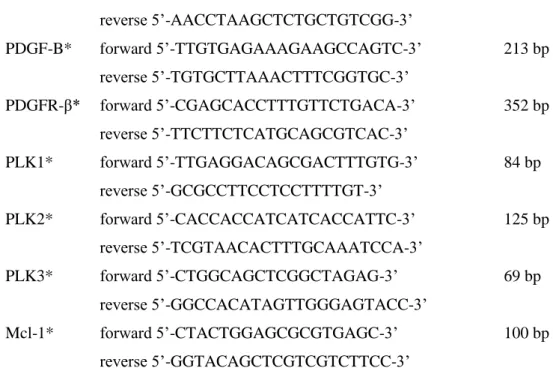
Total RNA was extracted from cells and liver tissue (EPO and CCA studies) using the RNeasy Plus Mini Kit (Qiagen, Hilden, Germany), and was reverse-transcribed with Moloney leukemia virus reverse transcriptase and random primers (Invitrogen, Camarillo, CA). Quantitation of the complementary DNA template was performed with real-time polymerase chain reaction (PCR;
LightCycler, Roche, Indianapolis, IN) using SYBR green (Roche) as a fluorophore.78 Oligonucleotide sequences and expected product sizes for primer pairs used for quantitative RT-PCR analysis are shown in Table 2. Primer pairs for c-jun, Bcl-XL, and the EPO-receptor (EPO study) were purchased from Invitrogen, Mississauga, Ontario, Canada. As an internal control, primers for 18S rRNA (Ambion, Austin, TX) were employed. Using gel purified amplicons, a standard curve was generated to calculate the copy number/µL. The target mRNA expression level of each sample was calculated as the copy ratio of target mRNA to 18S rRNA and then normalized to the target mRNA expression of controls.
Gene Primer sequence Product length
SHH forward 5’-GATGTCTGCTGCTAGTCCTCG-3’
reverse 5’-CACCTCTGAGTCATCAGCCTG-3’
300 bp
IHH forward 5’-TGGCATGCATTGGTACTCTC-3’
reverse 5’-GCTTGCAGCTCTATGACTAC-3’
350 bp
DHH forward 5’-GAGACTCTTTCACAGCTTGG-3’ 250 bp
reverse 5’-TATCACCTCCTCTCAGTACG-3’
PTCH1 forward 5’-CCACCAGACGCTGTTTAGTCA-3’
reverse 5’-CGATGGAGTCCTTGCCTACAA-3’
72 bp
SMO forward 5’-GTTCTCCATCAAGAGCAACCAC-3’
reverse 5’-CGATTCTTGATCTCACAGTCAGG-3’
250 bp
Gli1 forward 5’-TGCAGTAAAGCCTTCAGCAATG-3’
reverse 5’-TTTTCGCAGCGAGCTAGGAT-3’
132 bp
Gli2 forward 5’-TGGCCGCTTCAGATGACAGATGTTG-3’
reverse 5’-CGTTAGCCGAATGTCAGCCGTGAAG-3’
200 bp
Gli3 forward 5’-AAACCCCAATCATGGACTCAAC-3’
reverse 5’-TACGTGCTCCATCCATTTGGT-3’
98 bp
PDGFR-β forward 5’-AATGTCTCCAGCACCTTCGT-3’
reverse 5’-AGCGGATGTGGTAAGGCATA-3’
688 bp
PLK1 forward 5’-CACAGTGTCAATGCCTCCAA-3’
reverse 5’-TTGCTGACCCAGAAGATGG-3’
95 bp
PLK2 forward 5’-TCAGCAACCCAGCAAACACAGG-3’
reverse 5’-TTTCCAGACATCCCCGAAGAACC-3’
230 bp
PLK3 forward 5’-GAAGGTGGGGGATTTTGG-3’
reverse 5’-GGGTGCCACAGATGGTCT-3’
74 bp
Mcl-1 forward 5’-AAGCCAATGGGCAGGTCT-3’
reverse 5’-TGTCCAGTTTCCGAAGCAT-3’
121 bp
Shh* forward 5’-CTGGCCAGATGTTTTCTGGT-3’
reverse 5’-TAAAGGGGTCAGCTTTTTGG-3’
117 bp
Ihh* forward 5’-ACCCCACCTTCAGCGATGT-3’
reverse 5’-GAGTCTCGATGACCTGGAAAGC-3’
78 bp
Dhh* forward 5’-CGTTACGTGCGCAAGCAA-3’
reverse 5’-GGTCCGCTCGGGCATACT-3’
69 bp
Ptch1* forward 5’-GCAGAGGACTTACGTGGAGG-3’
reverse 5’-CTGACAGTGCAACCAACAGG-3’
245 bp
Smo* forward 5’-GGGAGGCTACTTCCTCATCC-3’
reverse 5’-TAGCACATAGTCCCGGAAGC-3’
226 bp
Gli1* forward 5’-TGGAAGGGGACATGTCTAGC-3’
reverse 5’-GCTCACTGTTGATGTGGTGC-3’
195 bp
Gli2* forward 5’-CCATCCATAAGCGGAGCAAG-3’
reverse 5’-CCAGATCTTCCTTGAGATCAG-3’
105 bp
Gli3* forward 5’-CATAGCTTCGACCTTCAGACC-3’ 211 bp
reverse 5’-AACCTAAGCTCTGCTGTCGG-3’
PDGF-B* forward 5’-TTGTGAGAAAGAAGCCAGTC-3’
reverse 5’-TGTGCTTAAACTTTCGGTGC-3’
213 bp
PDGFR-β* forward 5’-CGAGCACCTTTGTTCTGACA-3’
reverse 5’-TTCTTCTCATGCAGCGTCAC-3’
352 bp
PLK1* forward 5’-TTGAGGACAGCGACTTTGTG-3’
reverse 5’-GCGCCTTCCTCCTTTTGT-3’
84 bp
PLK2* forward 5’-CACCACCATCATCACCATTC-3’
reverse 5’-TCGTAACACTTTGCAAATCCA-3’
125 bp
PLK3* forward 5’-CTGGCAGCTCGGCTAGAG-3’
reverse 5’-GGCCACATAGTTGGGAGTACC-3’
69 bp
Mcl-1* forward 5’-CTACTGGAGCGCGTGAGC-3’
reverse 5’-GGTACAGCTCGTCGTCTTCC-3’
100 bp
Table 2. Primer sequences and expected product sizes of human and rat primer pairs used for quantitative RT-PCR analysis. All primers were designed to have an optimum annealing temperature between 50 and 60 °C.
* = rat primer pairs (all others are complementary to human targets).
4.9 Immunoblot Analysis
For CCA studies, whole cell lysates were obtained as previously described.90 For the examination of GLI2 activation, nuclear protein extracts were obtained using the NE-PER Nuclear and Cytoplasmatic Extraction Kit (Thermo Scientific , Barrington , IL; Product no.:
78833). Primary antisera/antibodies used were: Actin (1:2000; Santa Cruz, Santa Cruz, CA; C- 11), Lamin B (1:1000; Santa Cruz, Santa Cruz, CA; M-20), PDGFR-β (1:1000; Santa Cruz, Santa Cruz, CA; P-20), phospho-PDGFR-β (Tyr857; 1:1000; Cell Signaling, Danvers, MA;
#3170), GLI2 (R&D Systems, Minneapolis, MN; Antibody Part 965887 from the GLI2 ExactaChIP Kit Catalog no.: ECP3526), PLK1 (1μg/ml; Merck Millipore, Darmstadt, Germany; CN: 05-844), PLK2 (1 μg/ml; Abcam, Cambridge, MA; ab34811), PLK3 (1:1000;
Cell Signaling Danvers, MA; CN: D14F12), Mcl-1 (1:1000; Santa Cruz, Santa Cruz, CA; sc- 819), and Bcl-2 (1:1000; Santa Cruz, Santa Cruz, CA; sc-492). The mouse anti-S peptide antibody was a generous gift from S. H. Kaufmann (Oncology Research, Mayo Clinic, Rochester, MN). Horseradish peroxidase-conjugated secondary antibodies for rabbit (Santa Cruz; sc-2004), goat (Santa Cruz; sc-2020), mouse (Santa Cruz; sc-2031), and sheep (Santa Cruz; sc-2770) were incubated at a dilution of 1:2000 for 1 hr at RT. Proteins were visualized using enhanced chemiluminescence reagents (ECL, Amersham Biosciences, Buckinghamshire, UK) and Kodak X-OMAT films.
4.10 Chromatin Immunoprecipitation (ChIP Assay)
ChIP was performed from KMCH-1 CCA cells treated with rhSHH (500 ng/ml, 5 hrs) plusminus cyclopamine (10 µM, 5 hrs) using total cellular DNA sheared to ≈ 500 bp fragments employing an automated cooled sonication device (Bioruptor XL; Diagenode, Denville, NJ; 20 cycles with 30 seconds sonication/30 second intervals). ExactaCHIP chromatin immunoprecipitation kits for GL1, GLI2 and GL3 (R&D Systems, Minneapolis, MN; GLI1:
ECP3324, GLI2: ECP3526, GLI3: ECP3690) and streptavidin agarose beads (Merck, Rockland, MA; #69203), were used following the manufacturer’s instructions. Samples were pre-cleared using agarose beads plus salmon sperm slurry (Upstate, Lake Placid, NY) prior to immunoprecipitation (also performed with agarose beads). Primers for RT-PCR were forward 5’-TCA TGT CTC CCC GTT CCA ACT-3’, reverse 5’-TGC AAA GCC ACC CTG AAA GGA-3’ (PLK pomotor site I, 277 bp) and forward 5’-CAT TTG GGT CAG CTC CAA GT-3’, reverse 5’-TCT CAC GCC AGT TAA AAT GGC G-3’ (PLK pomotor site II, 297bp). Positive control primers supplied by the manufacturer were for the Bcl-2 promoter (bound by GLI1 and GLI2, 147 bp) and the GLI1 promoter (bound by GLI3, 211 bp).
4.11 A
ssessment Of Laboratory Parameters
Subsequent to in vivo microscopy (approximately 90 min after reperfusion; preservation solution study) blood samples were drawn (vena cava puncture) for analysis of liver enzymes (aspartate transaminase [AST], alanine transaminase [ALT], lactate dehydrogenase [LDH] and alkaline phosphatase [AP]) and prothrombin time. The samples were processed using standard blood analysis tests. In addition, blood samples from n=8 untreated Lewis rats were drawn as reference. In the EPO experiments, glutamate dehydrogenase (GLDH), total bilirubin, prothrombin time (PTT), international normalized ratio (INR), and hematocrit were additionally assessed as described before.29 Furthermore, EPO serum concentrations were measured by means of an immunoluminometric assay (Limbach, Heidelberg, Germany)
4.12 Enzyme-Linked Immunosorbent Assay (ELISA) for PDGF-BB
Levels of secreted human PDGF-BB in CCA and MFB cell experiments were determined by an enzyme-linked immunosorbent assay using a commercially available kit (RayBiotech, Norcross, GA) according to the suppliers protocol.
4.13 Assessment Of Bile Production
As an indicator of liver function, postoperative bile production was measured (preservation solution study). The amount of bile draining from the bile duct via a polyethylene tube (prepared from a 22G IV Catheter, Medex Medical GmbH, Germany) over a period of 90 min was collected, weighed and given in mg per g of liver wet weight.
4.14 cDNA Array
A customized cDNA array consisting of 183 rat genes was established as previously described (EPO study).28 As a control, 13 GAPDH and ß-actin gene probes were added. Subsequently, the gene products were spotted on Hybond N+ nylon membranes (Amersham Pharmacia, Freiburg, Germany) and hybridized overnight at 65°C with P32 labeled cDNA prepared from 10μg total RNA of each rat liver sample. The membranes were stored in a Phosphoimager Cassette (Amersham Biosciences, Freiburg, Germany), exposed for 2-3 days and scanned on the STORM Phosphoimager (Amersham Biosciences, Freiburg, Germany). The data were analyzed using Image Quant software (Amersham Biosciences, Freiburg, Germany) as described bevore.91
4.15 Genome-Wide mRNA Expression Analysis
For the CCA studies, KMCH-1 cells were treated with vehicle, PDGF-BB (200 ng/ml, 8hrs), or SHH (500 ng/ml, 8hrs) in the presence or absence of cyclopamine (10 μM, 8hrs). After total mRNA extraction (see section real time polymerase chain reaction) and confirmation of the sample quality by Agilent bioanalysis, 150-500 ng of total RNA per sample were analyzed for 33617 target genes (after MAS5 noise filtering) employing an Affymetrix GeneChip Platform with the Affymetrix Human U133 Plus 2.0 labeling method. Specifically, biotin-labeled cRNA, produced by in vitro transcription, was hybridized to the Affymetrix Human Genome U133 Plus 2.0 GeneChips. These experiments were conducted in collaboration with the Advanced Genomics Technology Center Core Mayo Clinic, Rochester, MN.
4.16 Generation Of A Transfectant Expressing SMO Short Hairpin RNA
Short hairpin RNA (shRNA) lentiviral plasmid for SMO was from Thermo Fisher Scientific (Hunsville, AL; Oligo ID: V2LHS_56569; GenBank accession no.: NM_005631). KMCH-1 CCA cells were transfected using OptiMEM I (Gibco-Invitrogen, Carlsbad, CA) containing 6 μL/mL Lipofectamine (Invitrogen),1 μg/mL plasmid DNA, and 6 μL/mL Plus reagent (Invitrogen). Forty-eight hours after transfection, fresh DMEM containing 0.5 μg/mL puromycin was added. Surviving clones were separated using cloning rings and individually cultured. A clone with a scrambled shRNA was employed as a control (stable scrambled KMCH-1 cells). The expression/knockdown of SMO in the clones was assessed by immunoblot analysis.4.17 Generation Of A Transfectant Expressing PLK1, 2, or 3 Short Hairpin RNA.
Short hairpin RNA (shRNA) lentiviral plasmids for PLK1 and PLK3 were obtained from Thermo Fisher Scientific/Open Biosystems (Hunsville, AL; Oligo ID: V2LHS_241437, Gen Bank accession no.: NM_005030 and Oligo ID: V2LHS_172853, Gen Bank accession no.:
NM_004073, resp.). PLK2 shRNA lentiviral plasmids were obtained from Sigma-Aldrich (St.
Louis, MO; Gen Bank accession no.: NM_006622.2). KMCH-1 cells were transfected using OptiMEM I (Gibco-Invitrogen, Carlsbad, CA) containing 6 µl/ml Lipofectamine (Invitrogen), 1 µg/ml plasmid DNA and 6 µl/ml Plus reagent (Invitrogen). Forty-eight hours after transfection, fresh DMEM containing 0.5 µg/ml puromycin was added. Surviving clones were separated using cloning rings and individually cultured. A clone with a scrambled shRNA was employed as a control (stable scrambled KMCH-1 cells). The expression/knockdown of PLK1, PLK2 or PLK3 in the clones was assessed by immunoblot analysis.
4.18 Generation Of AN Enhanced Green Fuorescent Protein (GFP)–Tagged SMO
A pRK7 plasmid containing the human SMO sequence (GenBank accession no.: NM_005631) for the CCA studies was a generous gift from M. Fernandez-Zapico (Division of Oncology Research, Mayo Clinic, Rochester, MN). The pRK7-SMO plasmid was modified to accept the green fluorescent protein (GFP) tag first by inserting recognition sites for EcoRI and NotI at the C-terminus of SMO, replacing the stop codon. For this, a PCR-generated EcoRI/NotI modified SMO C-terminal coding sequence was inserted into pRK7-SMO. Next, GFP from the pEGFP-N1 protein fusion vector (Clontech Laboratories, Inc., Mountain View, CA; Catalog no.: 6085-1; GenBank accession no.: U55762) was digested and inserted into the modified pRK7-SMO plasmid to generate a SMO construct fused to GFP at the C-terminal cytoplasmic domain. The GFP-SMO plasmid was sequenced to confirm that the construct was in frame and no polymerase chain reaction artifacts were introduced.
4.19 GLI Reporter Construct And Promoter-Reporter Assay
To determine GLI activity in the CCA studies, a reporter containing eight directly repeated copies of a consensus GLI-binding site (8x-GLI) downstream of the luciferase gene was employed (pδ51LucII plasmid; δ-crystalline promoter). 92 The 8x-GLI reporter was kindly provided by M. Fernandez-Zapico (Division of Oncology Research, Mayo Clinic, Rochester, MN). The plasmid was transfected into normal, stable scrambled, or shSMO KMCH-1 cells (0.5 μg/well) using FuGene HD (Roche Diagnosis, Basel, Switzerland). Cells were co-transfected with 50 ng of a plasmid expressing Renilla luciferase (pRL-CMV; Promega, Madison, WI). 24 hours after transfection, cells were treated as indicated, cell lysates prepared, and both firefly and Renilla luciferase activities quantified using the Dual-Luciferase Reporter Assay System (Promega) according to the manufacturer’s instructions. Firefly luciferase activity was normalized to Renilla luciferase activity to control for transfection efficiency and cell numbers. Data (firefly/Renilla luciferase activity) are expressed as fold increase over vehicle-treated cells transfected with the 8x-GLI/pRL-CMV reporter constructs.
4.20 Animal Experiments
4.20.1 Orthotopic full size rat liver transplantation
Male Lewis and Wistar rats (240–300 g) were obtained from the Central Animal Facility of the University Hospital Essen (preservation solution studie). Animals were kept under standard conditions with free access to food (recipient rats were fastened for 2 h preoperatively) and water. All operations and handling procedures were conducted in accordance with the German Animal Welfare Law and with approval of the district administrative authorities (Regierungspräsidium Düsseldorf and Landesamt für Natur, Umwelt und Verbraucherschutz Recklinghausen). Surgical procedures and interventions were performed under volatile anesthesia (O2 with up to 2.5 % isoflurane) with maintained spontaneous ventilation. The study was subdivided into two parts in order to compare the effects of the new HTK solutions (for composition see Table 1) on i) overall survival under different conditions and ii) microcirculation as well as laboratory and histological parameters.
Three different microsurgeons performed orthotopic LTx in male Lewis and Wistar rats (donors and recipients: 240–300 g) according to the cuff technique described by Kamada and Calne without hepatic artery reconstruction.93 Perfusion (approx. 50 ml) and storage (150 ml) of the livers at 4°C were done using the same preservation solution (chloride-poor vs. chloride- containing new solution). The following three protocols were carried out for comparison of overall survival. Protocol 1: CIT: 24 h; WIT (during implantation): 17.1 ± 1.6 min; Wistar to Wistar; n=7 recipient rats each group; randomized; sacrifice after 7 days. Protocol 2: CIT: 12 h;
WIT: 19.7 ± 3.2 min; Wistar to Wistar; n=8 recipient rats each group; randomized; blinded;
sacrifice after 7 days. Protocol 3: CIT: 3 h; WIT: 25.0 ± 0.0 min (fixed to 25.0 min; no standard deviation); Lewis to Lewis; n=8 recipient rats each group; randomized; blinded; sacrifice after 28 days. LTx prior to assessment of microcirculatory parameters was performed according to the following protocol with average CIT and WIT: CIT: 18 h; WIT: 18.0 ± 1.1 min; n=7/8 Lewis rats each group; randomized; blinded (one rat [chloride-containing preservation solution]
died of unknown reasons 30 min after beginning of in vivo microscopy); sacrifice directly after in vivo microscopy. Analgesia in all rats was achieved by preoperative subcutaneous injections of 5 mg/kg BW carprofen (Rimadyl®, Pfizer, Karlsruhe, Germany). Recipient rats participating in survival experiments additionally obtained 100 mg/kg BW mezlocillin (Baypen®, Bayer AG, Leverkusen, Germany) intramuscularly after LTx as antibiotic prophylaxis.
4.20.2 Orthotopic partial (30%) rat liver transplantation
Male Lewis rats (Charles River Laboratories, Sulzfeld, Germany) weighing 250-300g were maintained on a commercial pelleted diet and water ad libidum under normal laboratory lighting conditions (EPO studie). All animal study protocols were approved by the German Animal Welfare Law and with approval of the district administrative authorities (Regierungspräsidium Düsseldorf and Landesamt für Natur, Umwelt und Verbraucherschutz Recklinghausen).
For transplantation, donor and recipient rats underwent isoflurane anesthesia. Liver reduction was achieved by resecting the left lateral and median lobe, which resulted in a 70% reduction of the liver mass. The graft was flushed and stored in cold HTK solution with a CIT of 180 minutes (sacrifice after 24 h) and 360 min. (sacrifice after 28 days = survival experiments), respectively. pLTx was performed according to the cuff technique of Kamada and Calne without hepatic artery reconstruction.93 The transplantation procedure required less than 60 min. The portal vein was clamped for 16 to 19 min. After the observation period (see protocols below), the remnant, regenerated liver was resected, weighed and total body weight was measured. The acquired data were expressed as percentage of the ratio between remnant liver weight (A), divided by the total body weight (B) times 100 (Liver body weight ratio [LBWR]
in (%) = A/B x 100).
Preconditioning experiments (protocol 1) were carried out to establish optimal EPO-doses for donor animals. Controls were treated with heat-inactivated EPO in the same vehicle volume.
The following protocols were employed. Protocol 1: In two preconditioning experiments rats (n=8 in each group) were injected once or thrice with several doses of EPO i.p. The animals were sacrificed 4, 8 and 12 days after the first injection. Protocol 2: Donor rats (n=8 in each group) were injected thrice with 1 I.U. EPO/g BW i.p. or vehicle 9 days prior to partial liver transplantation (pLTx). Recipient rats were injected thrice with 5 I.U. EPO/g BW or vehicle i.v. perioperatively. The animals were sacrificed after 24 h and 28 days (survival experiments) postoperatively.
4.20.3 Syngeneic, orthotopic rat modell of cholangiocarcinoma
All CCA animal studies were performed in accordance with and approved by the Institutional Animal Care and Use Committee. In vivo intrahepatic cell implantation was carried out in male adult Fischer 344 rats (Harlan, Indianapolis, IN) with initial body weights between 190 and 230 g as previously described.84-86 In the experiments targeting PDGF siganaling, imatinib mesylate (30 mg/kg BW; approx. 0.5 mL) or vehicle (normal saline) was given intraperitoneally every
day for one week (1st injection: 7th post-operative day; 7th injection: 13th post-operative day). In the experiments targeting Hh siganaling, cyclopamine (2.5 mg/kg BW; 0.5 mL) complexed with 2-hydroxypropyl-β-cyclodextrin (Tocris, Ellisville, MO) as previously described 94, 95 or vehicle was given intraperitoneally every day for one week (1st injection: 7th post-operative day;
7th injection: 13th post-operative day). In the experiments targeting PLK siganaling, BI 6727 (3 injections of 10 mg/kg body weight [0.5 mL] intraperitoneally every other day; the first injection was given on postoperative day 7,and the third injection was given on postoperative day11) formulated in hydrochloric acid (0.1 N) diluted with 0.9% NaCl.81 Twenty-four hours (forty-eight hours in the BI 6727 study) after receiving the last injection, the rats were euthanized and the livers removed for further analysis including histopathology and mRNA extraction. To assess the numbers of metastases-free and metastases-bearing rats, the abdominal cavities, the retroperitoneal spaces and the thoracic cavities were thoroughly examined as previously described.85
4.21 Statistical Analysis
4.21.1 Preservation solution/erythropoietin studies
All data are expressed as mean ± SD unless indicated otherwise and represent at least three independent experiments. Comparison between experimental groups was performed using the two-tailed Student’s t-test for quantitative continuous variables. Overall survival curves were estimated with the Kaplan-Meier method and statistical analysis was performed using the logrank test for each single series and Fishers-combination rule to calculate an overall one-sided p-value that combines the results from all three independent survival experiments. Further statistical analyses (EPO study) were performed by one-way Anova or Wilcoxon´s test.
Differences were considered as significant at levels of p < 0.05. Statistical analysis was performed using GraphPad Prism v4.00 (GraphPad Software Inc., La Jolla, CA) and SPSS v12.0 (SPSS Inc., Chicago, IL).
4.21.2 Cholangiocarcinoma studies
Data are expressed as the mean ± s.e.m. unless indicated otherwise and represent at least three independent experiments. Box-and-whisker plots depict minimum, 25th percentile, median, 75th percentile, maximum, and outliers. Differences in experiments with two groups were compared using the two-tailed Student t-test or the Chi-square test (2, analysis of metastasis) as well as
Mann Whitney’s test (analysis of PLK1/2/3 expression in human CCA samples). Differences in experiments with more than two groups were compared using ANOVA with Bonferroni post hoc correction. Differences were considered as significant at levels of p < 0.05. Statistical analysis was performed using GraphPad Prism v4.00 (GraphPad Software Inc., La Jolla, CA).
5 RESULTS
5.1 General Optimization Approaches For Liver Transplantation
5.1.1 Chloride improves survival due to beneficial effects on microcirculation
Postoperative survival. Each of the three experimental series performed to assess postoperative survival showed a (strong) tendency towards a prolonged survival using the chloride-containing new solution. In the first series (n=7 Wistar rats/group) a long CIT (24 h) was combined with a short WIT (17.1 ± 1.6 min). Here the survival rates 7 days after LTx were 100 % vs. 71.4 % (2 = 2.16 [p>0.05 by logrank test]) for the chloride-containing vs. the chloride-poor solution, respectively (Figure 1A). The second series (n=8 Wistar rats/group) was carried out with intermediate CITs and WITs (12 h/19.7 ± 3.2 min). Figure 1B depicts the Kaplan-Meier 7d- survival plot with 75 % of the rats surviving after LTx with the chloride-containing solution in comparison with 37.5 % of the rats still living 7 days after LTx with the chloride-poor solution (2 = 1.70 [p>0.05 by logrank test]). While follow-up time in the first two series utilizing Wistar rats was limited to 7 days in order to avoid influences due to rejection reactions (the Wistar rats are not inbred), Lewis rats (a syngeneic strain) in the third series (n=8 Lewis rats/group) with a short CIT (3 h) and long a WIT (fixed to 25.0 min; no standard deviation) were observed over a period of 28 days (Figure 1C). In this series with a median survival of 23.5 vs. 9.5 days, for the third time the survival rates between animals receiving grafts preserved with the chloride- containing new solution vs. animals receiving grafts preserved with the chloride-poor new solution suggested a better outcome with chloride (50 % vs. 12.5 %, 2 = 3.06 [p=0.07 by logrank test]). After combining the results from the three independent experimental series using Fishers-combination rule, an overall one-sided p-value of 0.012 was obtained, underlining the results shown in Figure 1A-C by a statistical summary.
Figure 1. Overall survival of different rat strains after LTx by 3 different microsurgeons with the chloride- poor and chloride-containing solutions. (A) Kaplan-Meier 7-day survival plot (CIT = 24 hours, WIT = 17.1 ± 1.6 minutes, 7 Wistar rats in each group, P = 0.14), (B) Kaplan-Meier 7-day survival plot (CIT = 12 hours, WIT = 19.7 ± 3.2 minutes, 8 Wistar rats in each group, P = 0.19), and (C) Kaplan-Meier 28-day survival plot (CIT = 3 hours, WIT = 25.0 ± 0.0 minutes, 8 Lewis rats in each group, P = 0.07). A combination of the results from all 3 experimental series with Fisher’s method yielded an overall 1-sided P value of 0.012.
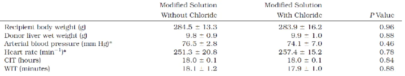
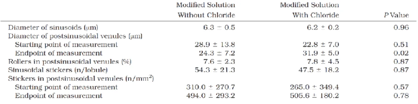
Microcirculation. Assessment of microcirculation and leukocyte-endothelial interaction was performed in an additional series with an intermediate CIT (18 h) and an intermediate WIT (18.0 ± 1.1 min; Table 3). In this series systemic hemodynamic parameters, i.e. arterial blood pressure and heart rate were also measured since these systemic parameters have a relevant impact on hepatic microcirculation. There were no significant differences between the rats of both test groups in regard to systemic hemodynamics, body weight, and liver wet weight (Table 3).
Table 3. General, physiological, and LTx data for the test groups participating in the in vivo microscopy experiments. The experiments were performed with 7 or 8 Lewis rats in each group. *At the beginning of the in vivo microscopy analysis.
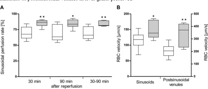
In vivo microscopy starting 30 min after reperfusion exhibited obvious pathological alterations such as intraparenchymal hemorrhages, areas with reduced RBC velocities, and stasis of blood flow in both test groups. These disturbances of microcirculation were observed in both sinusoids and postsinusoidal venules. However, number and size of malperfused areas were clearly decreased in rats that underwent LTx with grafts preserved with the chloride-containing preservation solution. This impression was confirmed by quantitative analysis (carried out in a blinded fashion) exhibiting clearly higher sinusoidal perfusion rates in liver grafts preserved with the chloride-containing new solution at start point, end point, and during 60 min (mean) of in vivo microscopy (Figure 2A). In addition, mean RBC velocities in sinusoids and postsinusoidal venules were clearly higher with the chloride-containing new solution (Figure 2B). The most significant RBC velocity difference was observed at the end point of in vivo microscopy (90 min after reperfusion) in postsinusoidal venules (339.0 ± 95.9 µm/s vs.
182.1 ± 41.1 µm/s; p = 0.001). While sinusoidal perfusion rates and RBC velocities remained