SEMMELWEIS EGYETEM
DOKTORI ISKOLA
Ph.D. értekezések 2162.
BIRTALAN EDE
Fül-Orr-Gégészet, Fej-Nyaksebészet című program
Programvezető: Dr. Tamás László, egyetemi tanár Témavezető: Dr. Tamás László, egyetemi tanár
PROGNOSTIC BIOMARKERS OF HEAD AND NECK CANCER
PhD thesis
Ede Birtalan, MD
Semmelweis University
Doctoral School of Clinical Medicine
Supervisor: László Tamás, MD, Ph.D Official Reviewers: Péter Móricz, MD, Ph.D
Orsolya Dohán, MD, Ph.D Head of the Final Examination Committee:
Henriette Farkas, MD, D.Sc Members of the Final Examination Committee:
Tibor Glasz MD, Ph.D
Zsuzsanna Orosz, MD, Ph.D
Budapest
2018
TABLEOFCONTENTS
1 List of abbreviations ...5
2 Introduction ...7
2.1 Epidemiology of head and neck cancer ...8
2.2 Pathogenesis of head and neck squamous cell carcinoma (HNSCC) ...10
2.2.1 Etiology and risk factors ...10
2.2.2 Premalignant lesions ...12
2.2.3 The main molecular and genetic alterations in HNSCC ...13
2.3 Clinical presentation of HNSCC ...15
2.3.1 Recent changes in tumor, lymph node, metastasis (TNM) classification ...15
2.3.2 Presentation of HNSCC ...18
2.4 Management of head and neck cancer ...20
2.4.1 Surgery of head and neck cancer ...21
2.4.2 Radiation therapy ...22
2.4.3 Chemoradiotherapy and chemotherapy ...25
2.4.4 Biological therapies in HNSCC (other than immunotherapy) ...28
2.4.4.1 GFR-RAS-RAF-MEK-ERK inhibitors ...28
2.4.4.2 PI3K-AKT-mTOR inhibitors ...29
2.4.4.3 Angiogenesis inhibitors ...30
2.4.5 Immunotherapy ...30
2.4.5.1 Checkpoint inhibitors ...31
2.4.5.2 Other immunotherapeutic approaches ...33
3 Objectives ...34
4 Methods ...35
4.1 Patients ...35
4.2 Study design ...37
4.3 Tissue microarray construction ...38
4.4 Immunohistochemistry and evaluation of slides ...38
4.4.1 P16INK4 staining ...38
4.4.2 PD-L1 staining ...40
4.4.3 PD-L2 staining ...40
4.4.4 PD-1 staining ...41
4.4.5 CTLA-4 staining ...41
4.4.6 CD-8 staining ...41
4.5 High-risk HPV DNA real-time polymerase chain reaction ...41
4.6 Tumor infiltrating lymphocyte ratio ...42
4.7 Statistical analysis ...42
5 Results ...43
5.1 P16INK4-expression and high risk HPV DNA status ...43
5.2 P16INK4/HPV DNA status and response to induction chemotherapy ...43
5.3 The impact of TIL rate ...44
5.4 CD-8 expression ...45
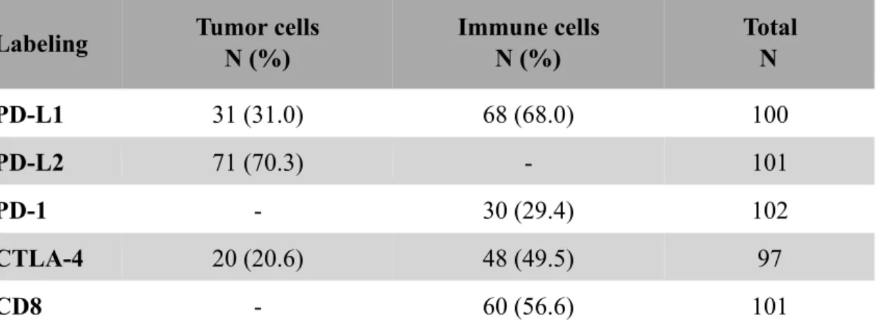
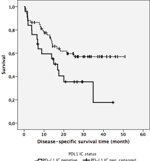
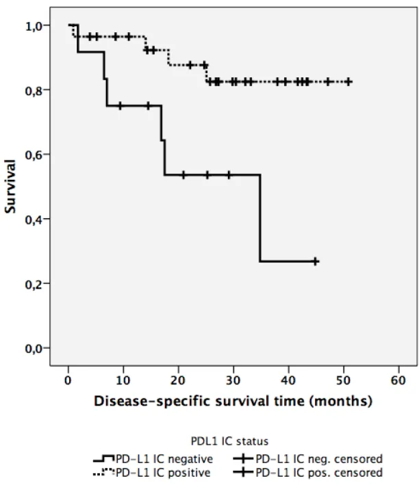
5.5 PD-L1 expression on immune cells (PD-L1IC) ...45
5.6 PD-L1 expression on tumor cells (PD-L1TC) was not associated with disease-specific survival. ...50
5.7 PD-1 expression ...50
5.8 PD-L2 expression ...50
5.9 CTLA-4 expression on immune cells (CTLA-4IC) and tumor cells (CTLA-4TC) ...50
5.10 Anatomical localization and survival ...51
6 Discussion ...52
7 Conclusions ...58
8 Summary ...69
9 Összefoglalás ...60
10 Bibliograpy ...61
11 Bibliography of the candidate’s publications ...78
12 Acknowledgements ...80
13 Supplementary tables ...83
1 LISTOFABBREVIATIONS
AJCC American Joint Committee on Cancer CD cluster of differentiation
CDKN2A cyclin-dependent kinase inhibitor 2A CRT chemoradiotherapy
CTLA-4 cytotoxic T-lymhocyte-associated protein 4 CUL3 cullin-3 gene
DC dendritic cell
DDX3X DEAD-box helicase 3, X-linked DNA deoxyribonucleic acid
DOI depth of invasion
DSS disease-specific survival
EGFR epidermal growth factor receptor ENE (tumor) extranodal extension
EORTC European Organization for Research and Treatment of Cancer ERK extracellular signal–regulated kinase
EU European Union
FFPE formalin-fixed, paraffin-embedded FGFR2/3 fibroblast growth factor receptor 2/3 5-FU 5-fluorouracil
HNSCC head and neck squamous cell carcinoma HPV human papillomavirus
HR hazard ratio
IHC immunohistochemistry
MLL2 histone-lysine N-methyltransferase 2B MTX methotrexate
NMSC nonmelanoma skin cancer NPC nasopharyngeal cancer
NSD1 nuclear receptor binding SET domain protein 1
OCC oral cavity cancer OPC oropharyngeal cancer
PD-1 programmed cell death protein 1 PD-L1 programmed death-ligand 1 PD-L2 programmed death-ligand 2
PIK3CA phosphatidylinositol-4,5-bisphosphate 3-kinase catalytic subunit alpha PNI perineural invasion
RECIST Response Evaluation Criteria in Solid Tumors RTOG Radiation Therapy Oncology Group
R/M recurrent or metastatic
SHP-2 Src homology 2 domain-containing tyrosine phosphatase 2 TIL tumor infiltrating lymphocyte
TIMC tumor-infiltrating mononuclear cell
TNM tumor, lymph node, metastasis (classification) UICC Union for International Cancer Control VEGF vascular endothelial growth factor
2 INTRODUCTION
There are multiple definitions to grasp what head and neck cancer means both on-line and in written literature. A rather general interpretation from the Mayo Clinic reads as follows: Head and neck cancers are a broad category of cancers that occur in the head and neck region. (http://www.mayoclinic.org/diseases-conditions/head-and-neck- cancers/home/ovc-20246134) Another slightly more detailed definition originates from the National Cancer Institute of the United States that reads: Head and neck cancers are cancers that start in the tissues and organs of the head and neck, including cancers of the larynx, throat, lips, mouth, nose, and salivary glands. (https://www.cancer.gov/
types/head-and-neck). However, my favorite definition can be found on Medline (National Library of Medicine, National Cancer Institute) which declares: Head and neck cancer includes cancers of the mouth, nose, sinuses, salivary glands, throat, and lymph nodes in the neck. Most begin in the moist tissues that line the mouth, nose, and throat. (https://medlineplus.gov/headandneckcancer.html)
By its nature, head and neck cancer often occurs at unveiled parts of the human body. It is generally associated with a certain degree of functional impairment causing significant mental and physical distress to its victims and a substantial economic burden to the whole society.
2.1 Epidemiology of head and neck cancer
Head and neck cancer represents a major global health issue ranking as the 6th most common cause of cancer related deaths worldwide (Ferlay et al. 2010, Torre et al.
2015). There has been several estimations of head and neck cancer incidence. The GLOBOCAN Estimates of the worldwide incidence and mortality from several cancers including cancers of the head and neck region is widely considered a reliable calculation published by the International Agency for Research on Cancer (Ferlay et al. 2010).
These statistics do not contain head and neck cancer as an independent category.
Instead, data on cancers of anatomical subsets (e.g. larynx, nasopharynx, etc.) are presented separately. Based on figures from the GLOBOCAN 2012, cancers of the lip, oral cavity, nasopharynx, other pharynx and larynx taken together and regarded as head and neck cancer account for a substantial number of cancer incidence and mortality worldwide. Calculations were made in more developed and in less developed areas separately. The following numbers are age-standardized rates per 100,000 and are standardized to the world standard population (Torre et al. 2015).
Concerning incidence in male, head and neck cancer reached the figures of 17.4 and 13.3 per 100.000 in more developed and less developed areas, respectively. Mortality rates in male were 6.9 and 8.3 per 100.000 in more developed and less developed areas, respectively (Torre et al. 2015).
Looking at the data on females, a remarkably lower incidence and death rate could be seen. The incidence of head and neck cancer was 4.2 vs. 4.4 whereas mortality rate was found to be 1.2 vs. 2.7 per 100.000 in more developed and less developed areas, respectively. Compared with the most common causes of cancer-associated deaths in males, head and neck cancer occupies the 7th and 6th position in the ranking in more developed and less developed countries, respectively. Considering females, head and neck cancer-related mortality is the 15th in more developed areas and 9th in less developed ones on a worldwide scale.
International statistics show a very high occurrence rate of head and neck cancer in Hungary compared to other countries. In 2004, when 10 countries joined the European
Union (EU), Hungary led the total cancer-associated mortality statistics with 258.5 cancerous deaths/100.000 men. Concerning mouth or pharynx localization, Hungary topped the list of EU members with 21.9 deaths/100.0000 that was 27.5% higher than the figure of the second country (Slovakia). Similarly, Hungarian men led the larynx- related cancer mortality as well. (Levi et al. 2004). Hungarian women came out as second (133.5/100.000) after Denmark (136.7/100.000) with regard to total cancer related deaths, whereas Hungary proved to lead the mouth or pharynx-associated cancer and the laryngeal cancer mortality among women as well (Levi et al. 2004).
The contrast between HPV-related and non-HPV-related head and neck cancer cannot be disregarded when assessing data on epidemiology of this disease. As it unfolds in a much more detailed way in the following chapters, HPV-associated oropharyngeal cancer represents a distinct biological and clinical entity. Likewise, the occurrence of it has to be discussed separately from tobacco-related head and neck cancer.
During the last years, compelling evidence has accumulated that reflect a drastically increased incidence of oropharyngeal cancers (OPCs), especially in North America and in northern Europe (Gillison, Chaturvedi et al. 2015).
The proportion of HPV-related cancers within all oropharyngeal malignancies varies in different countries and regions. In Sweden it is 90%, in the United States 50% whereas a large, international study concluded that on average 24.3% of OPCs are HPV- associated (Nasman et al. 2016).
Worldwide cancer registry data (Cancer Incidence in Five Continents) can be used to further elucidate global incidence trends by comparing incidence trends from 1983 to 2002 for upper aerodigestive tract malignancies that are etiologically associated with HPV infection (e.g.: OPC) versus those associated with tobacco smoking (e.g.: oral cavity and lung squamous cell carcinomas) (Chaturvedi et al. 2015). This study found that OPC incidence increased especially among young men (< 60 years old) in developed countries, despite concomitant declines in incidence for oral cavity and lung squamous cell carcinomas. These contrasts suggest a role of HPV infection in increasing OPC incidence rates among men. However, among women, incidence rates increased for all three cancers, supporting a dominant effect of smoking. These figures are
consistent with a hypothesis of a greater impact of HPV infection on OPC incidence trends for men over the last several decades, in contrast to the effect of smoking for women (Chaturvedi et al. 2015).
Taken together, it seems that the growing number of HPV-associated OPCs boosts the incidence figures outweighing the beneficial effect of slightly decreased tobacco consumption.
Nevertheless, despite improving diagnostics and intense research the 5-year overall survival of head and neck cancer in general remains relatively poor, around 60%. (Jay et al. 2015).
2.2 Pathogenesis of head and neck squamous cell carcinoma (HNSCC)
2.2.1 Etiology and risk factors
The main risk factors of head and neck cancer are smoking, alcohol consumption, persistent high-risk HPV infection and poor oral hygiene.
Smoking
Smoking is an independent causative factor of head and neck cancer (Maasland et al.
2014). Patients who continue smoking during radiotherapy are thought to have a failure of local control (hazard ratio (HR) 1.5) and poorer survival (HR: 1.7), but recent data suggests that baseline smoking status may play a more important role (Zevallos et al.
2016).Smoking cessation before surgery reduces the risk of complications related to anesthetics and is taught to improve wound healing, especially after reconstructive surgery (Tang et al. 2016). After quitting tobacco usage, it takes 20 years until the risk of developing oral cavity cancer sinks to the level of non smokers (Marron et al. 2010).
Alcohol
Alcohol consumption is an other main risk factor of head and neck cancer.
Simultaneous smoking and abusive drinking has a synergistic effect on deteriorating prognosis (Tan et al. 1997). Those patient who do not quit heavy drinking after
treatment for head and neck cancer have significantly worse survival (Mayne et al.
2009). The positive effect of alcohol cessation on the risk of head and neck cancer appears after 20 years (Marron et al. 2010).
High-risk HPV infection
The causative relation between infection by high-risk HPV subtypes and head and neck cancer was proved on the verge of the millennium (Gillison, Koch et al. 2000). Now, it is widely accepted that HPV infection is a causative agent in case of oropharyngeal malignancies only (Dillon and Harrington 2015, Castellsague et al. 2016).
The initial infection occurs during oro-genital sexual intercourse. However, there has been reports of oro-oral transmission as well (D'Souza et al. 2009). It is presumed that persistent oropharyngeal infection by high-risk HPV subtypes (mostly HPV-16) poses a risk of developing oropharyngeal cancer. Thus, persistent HPV infection and the transmission of it has drawn much attention recently.
Data of recent analysis (National Health and Nutrition Examination Survey, 2009 to 2012, https://www.cdc.gov/nchs/nhanes/index.htm) demonstrated a three-fold greater increase in high-risk oral HPV prevalence per sexual partner for men compared to that for women. That is consistent with reported higher transmission rates for HPV from female to male than vice versa (Gillison, Chaturvedi et al. 2015). The study found a plateau in prevalence among men at approximately 15 oral sexual partners in contrast to approximately five partners among women (Gillison Chaturvedi et al. 2015, Giuliano et al. 2015). Taking into consideration that the viral load on the surface of the infected cervix is higher than of the infected penis, male predominance could be explained by males acquiring a higher number of virus particles (assuming a heterosexual intercourse) (Marur et al. 2010). Ultimately, oropharyngeal cancer predominantly occurs in male.
Poor oral hygiene
In recent decades, many studies have concluded poor oral hygiene to be a significant risk factor for oral and oropharyngeal cancer (Maier et al. 2016). However, bad oral
condition often coexists with positive anamnesis for alcohol consumption and tobacco smoking.
Other risk factors
On one hand, minor risk factors include inherited diseases e.g.: Fanconi anaemia, ataxia telangiectasia, Bloom’s syndrome and Li–Fraumeni syndrome (Shaw and Beasley 2016).
Secondly, acquired immunodeficiency because of poor nutrition, advanced age, immunosuppressive therapy after transplant or acquired immunodeficiency syndrome can increase the risk of developing head and neck cancer (Shaw and Beasley 2016).
2.2.2 Premalignant lesions
Leukoplakia and erythroplakia are well-known premalignant lesions of the upper respiratory and digestive tract. A large meta-analysis by Mehanna et al. assessed the malignant transformation rate of 992 patients with histologically confirmed oral dysplasia. They concluded the mean overall transformation rate to be 12.1% (Mehanna et al. 2009).
Others found that histologically confirmed dysplastic lesions that were not removed displayed a considerably higher transformation rate compared to those that were excised (Ho et al. 2013). An other large meta-analysis on laryngeal dysplastic lesions of 942 patients showed transformation in 14% after a mean interval of 5.8 years, adding that severity of dysplasia correlated with risk of transformation (Weller et al. 2010).
However, in population-based studies of oral leukoplakia without histological inclusion criteria the risks are much lower; 40-50% vanish spontaneously and less than 1%
transform (Lodi et al. 2006, Roosaar et al. 2016).
The transformation potential of oral lichen planus is controversial. On the contrary, proliferative verrucous leukoplakia presenting with exophytic widespread progressive leukoplakia is taught to have a very high (up to 50-80%) transformation rate, thus a poor overall prognosis (Shaw and Beasley 2016).
Surprisingly, no precursor lesion has been detected in connection with HPV-associated oropharyngeal cancer, that would enable screening of patients, despite the assumingely long latency that occurs between viral exposure and manifest tumor formation (Hayes et al. 2015).
2.2.3 The main molecular and genetic alterations in HNSCC
The idea that HPV-associated head and neck cancer is a distinct biological entity is supported by a compelling body of evidence and is widely accepted. The genetic landscape of HPV-negative and HPV-driven HNSCC was assessed by many large-scale studies such as the The Cancer Genome Atlas and Chicago HNC Genomics cohorts.
Data show a surprisingly wide range of genetic alterations that are common in both non- HPV-related and HPV-related head and neck cancers. These common patterns are amplifications (e.g.: 1q, 3q, 5p, 8q) deletions (e.g.: 3p, 5q, 11q) and other similarities including the generally similar mutation rate and the number of copy number changes (Hayes et al. 2015). According to Seiwert et al. the overall mutational burden in HPV- negative and HPV-positive HNSCC was similar with an average of 15.2 versus 14.4 somatic exonic mutations in the targeted 617 cancer associated genes (Seiwert, Zuo et al. 2015).
However, the genetic landscape of HPV-negative and HPV-positive HNSCC differs significantly. The main mutational spectrum of HPV-negative tumors showed concordance with published lung squamous cell carcinoma analyses with enrichment for mutations in p53, cyclin-dependent kinase Inhibitor 2A (CDKN2A), histone-lysine N-methyltransferase 2B (MLL2), cullin-3 (CUL3), nuclear receptor binding SET domain protein 1 (NSD1), phosphatidylinositol-4,5-bisphosphate 3-kinase catalytic subunit alpha (PIK3CA), and NOTCH genes (Seiwert, Zuo et al. 2015). In contrast, HPV-positive tumors showed unique mutations in DEAD-box helicase 3, X-linked (DDX3X), fibroblast growth factor receptor 2/3 (FGFR2/3) and aberrations in PIK3CA, KRAS, MLL2, and enrichment in NOTCH1 (Seiwert, Zuo et al. 2015).
HPV-negative tumors display a clear prominence of amplification of 3q at the locus for oncogene PIC3CA and other transcription factors (Hayes et al. 2015). Although PIK3CA is altered in tumors without regard to viral association, two specific cytosine>thymine mutations in viral-associated tumors occur predominantly in two hotspots within the helical domain. These result in amino acid substitutions that are implicated in PIK3CA kinase and oncogene activation (Hayes et al. 2015).
Surprisingly, although squamous cell carcinoma of any site rarely demonstrates KRAS mutations, they are reported in HPV-positive HNSCC (Seiwert, Zuo et al. 2015).
Although the data are sparse, the interaction between smoking and KRAS mutation suggests a mechanism through which tobacco might augment risk in HPV-positive HNSCC (Hayes et al. 2015).
Concerning epidermal growth factor receptor (EGFR) protein, HPV-positive tumors have generally shown low or absent levels of protein expression or EGFR gene amplification (Keck et al. 2015).
An other substantial structural difference is the event of viral DNA integration in HPV- positive tumors. Although the predicted impact of integration is generally to silence the gene, the nature of HPV DNA integration remains controversial and is the subject of ongoing investigation (Lawrence et al. 2015). There are data that suggest the most tumors have a single primary integration site, although the integration event itself may be complex at sites of gene amplification (Akagi et al. 2014). Nevertheless, an alternative possibility is that the gene disruption may be a passenger event only and the targeting of a gene may be nonspecific and may result from a stochastic event. The lack of recurrent events gives suggests a lower relevance of the integration site to tumor initiation and progression (Hayes et al. 2015).
2.3 Clinical presentation of HNSCC
2.3.1 Recent changes in tumor, lymph node, metastasis (TNM) classification
Newly available data and the ever increasing number of HPV-positive oropharyngeal cancer cases worldwide produced an urging need for revision and reassessment of TNM classification system in head and neck oncology. This common interest resulted in the publication of the eighth edition of the American Joint Committee on Cancer (AJCC) Cancer Staging Manual in the fall of 2016 (Lydiatt et al. 2017). The 8th manual incorporates significant changes compared to the previous one including a separate staging algorithm for HPV-associated cancer of the oropharynx; changes to the tumor T categories in the nasopharynx, oral cavity, and skin; and the addition of tumor extranodal extension (ENE) to the lymph node category for most anatomical sites (Lydiatt et al. 2017). Authors of both the AJCC and Union for International Cancer Control (UICC) guidelines strove to achieve global applicability and harmony between the two manuals. However, the manuals slightly differ from each other. In the AJCC version the non-HPV-associated pathologic category N criteria contains a group within N2, that is ENE-positive and less than 3 cm in greatest dimension that is considered N2a. On the other hand, the UICC manual does not contain this category but classifies all ENE-positive lymph nodes as N3b. Here, changes according to the AJCC guideline are to be briefly summarized. There are two major structural changes. First, HPV- associated OPC is addressed in a separate chapter based on p16 immunohistochemistry (IHC). On top of that, separate chapters for non-HPV-associated, p16-negative OPC/
hypopharyngeal cancer and for nasopharyngeal cancer are included (Lydiatt et al. 2017).
Secondly, the head and neck section addresses nonmelanoma skin cancer (NMSCs) of the head and neck.
Changes related to HPV-associated oropharyngeal cancers
As mentioned above, distinct biological nature and recent clinical findings urged a change in OPC staging based on HPV status. The biomarker used by the manual to
determine HPV status is the p16 protein expression by IHC, that is surrogate marker of high-risk HPV-induced carcinogenesis with excellent prognostic capacity and nearly 100% sensitivity and 60-80% specificity (Kreimer et al. 2010). Furthermore, it is relatively cheap, widely accessible and is strait forward to interpret (Lydiatt et al. 2017).
P16 positivity requires a diffuse, >75% cancer cell positivity with at least moderate intensity and positive nuclear staining (Lewis and Chernock 2014).
T categories remained merely the same except for 2 differences. Firstly, Tis (in situ carcinoma) does not exist in the p16-positive classification. Secondly, T4a and T4b melted into one category. N staging underwent significant modification and simplification. N1 encompasses any single or multiple ipsilateral lymph node metastases each smaller than 6 cm in greatest diameter. Contralateral or bilateral but smaller than 6 cm metastases on the neck were categorized as N2 without any further specification. N3 was kept for metastases greater than 6 cm.
In turn, N categories of non-HPV-associated OPC became somewhat more complex.
One reason for that was the prognostic role of extranodal extension (ENE). The clinical N category for these cancers classifies all ENE-positive metastases as N3b, thus dissolving the previously homogenous N3 group. It is important to mention that ENE by imaging only is insufficient for ENE stratification. The unambiguous radiological finding has to be supported by physical examination signs as well, such as invasion of skin, infiltration of musculature, tethering to adjacent structures, or dysfunction of a nerve (Lydiatt et al. 2017).
Cancer of unknown primary tumor
Another change from prior editions of the TNM system is the elimination of the T0 category in sites other than the nasopharynx, HPV-associated OPC and salivary gland cancers. If no primary lesion can be identified, then the lymph node metastasis may have emerged from any mucosal site, meaning there is no point in retaining the T0 group except for the virally associated cancers of the oropharynx and nasopharynx (Lydiatt et al. 2017).
Changes in the T category of oral cavity cancers (OCCs)
The new T division of OCC introduced the term ‘depth of invasion’ (DOI) as a new prognostic factor. Recent data suggested that DOI is a better predictive parameter than tumor thickness (Shim et al. 2010). Although DOI was available for analysis in the sixth edition of TNM, the eight edition provided a precise definition. Clinically, DOI was divided into three categories: less invasive lesions (<5 mm), moderate depth lesions (from >5 to <10 mm) and deeply invasive cancers (>10 mm). Pathologically, DOI is measured from the level of the basement membrane of the closest adjacent normal mucosa. A “plumb line” is dropped from this plane to the deepest point of tumor invasion. The pathologic T category increases with every 5 mm (Lydiatt et al. 2017).
Staging of nonmelanoma skin cancer (NMSC)
As stated above, NMSC became part of head and neck chapter in the eight version of the manual. Most of the staging criteria remained the same except for the addition of DOI beyond 6 mm and perineural invasion (PNI) as parameters of the T category, both of which distinguish a lesion as T3, even if the tumor is of limited diameter (Lydiatt et al. 2017). DOI > 6 mm and PNI was associated with increased risk of recurrence and metastasis (Breuninger et al. 2013). Further on, a size criterion of 4 cm (instead of 5 cm as in the sixth edition) was reintroduced to distinguish between T2 and T3 similarly to other head and neck cancers (Lydiatt et al. 2017).
Changes in staging of nasopharyngeal cancers (NPCs)
There are 2 changes made in connection with NPCs. First, it gives a precise definition of the earlier somewhat ambiguous terms “masticator space” and “infratemporal fossa”.
Secondly, involvement of medial pterygoid, lateral pterygoid, and prevertebral muscles have been “down-staged” to T2 (Lydiatt et al. 2017). This was based on a recent analysis showing them to have a more favorable outcome using current treatment (Pan et al. 2016).
Concerning lymph node staging, modifications are to find as well. The unique term used for NPCs N category “supraclavicular fossa” was replaced by contemporary definitions
more suitable to axial cross-sectional imaging. In addition, the previously used low neck involvement (former N3a) and >6 cm size (former N3b) were unified into a single N3 group. Finally, both T4 and N3 would belong to stage IVA (formerly IVA and IVB) in stage categories (Pan et al. 2016, Lydiatt et al. 2017).
2.3.2 Presentation of HNSCC
The most common leading symptoms of head and neck cancers vary according to the anatomical site affected by the disease.
Cancer of the nasal cavity and paranasal sinuses
Any part of the nasal cavity and paranasal sinuses can be affected, but the lateral nasal wall, ethmoids and maxillary sinuses are the most common primary tumor sites. For unknown reasons, the frontal and sphenoid sinuses are rare primary locations (Lund et al. 2016).
The most common initial symptoms such as nasal blockage, blood-stained discharge and loss of smell are often overlooked though their often unilateral nature should raise suspicion. Delayed presentation is common. Subsequent extension to the surrounding structures can produce symptoms such as proptosis, diplopia and epiphora, trismus, facial pain, oro-antral fistula, paraesthesia or other neurological deficits and facial swelling or mass (Lund et al. 2016).
Nasopharyngeal carcinoma
It is more common in men than in women (gender ratio for men:women is 3:1), with a median age of 50 years at the time of presentation. The most common symptoms of nasopharyngeal carcinoma are nasal obstruction, epistaxis, conductive hearing loss due to otitis media with effusion, cranial nerve neuropathies caused by skull base invasion (commonly involved cranial nerves are III, IV, V and VI) (Simo et al. 2016).
Lip and oral cavity cancer
About 90 percent of lip cancers arise in the lower lip with 7 per cent occurring in the upper lip and 3 percent at the oral commissure (Kerawala et al. 2016). The clinical presentation of cancer of the lip is usually an exophytic, crusted lesion with or without invasion into underlying muscle. The adjacent lip often shows features of actinic sun damage such as color change, mucosal thinning and various associated areas of leukoplakia (Wolff et al. 2012).
In the oral cavity, the majority of squamous cell carcinomas are presented as ulcers or masses (Kerawala et al. 2016). Early lesions can appear as flat, discolored areas known as leukoplakia or erythroplakia (Rethman et al. 2010). Advanced tumors can present with additional symptoms because of invasion of neighboring structures causing tooth mobility, trismus, sensory changes and referred otalgia (Kerawala et al. 2016).
Oropharyngeal cancer
Although HNSCC of each anatomical region may firstly present with neck lumps, it is probably the most often seen in oropharyngeal cancer cases. These patients often present with painless, relatively large neck lumps. Other complains such as sore throat or tongue pain, referred ear pain, painful and/or difficult swallowing or a change in voice quality (often mentioned as hot potato voice) are the most common symptoms at presentation (Mehanna, Evans et al. 2016).
Laryngeal cancer
Presentation of laryngeal cancer is highly variable and depends on the site and size of the primary tumor. Tumors of the glottis typically present at an early stage as they cause hoarseness. In comparison, tumors of the supraglottis are likely to present later with symptoms of pain, hoarseness and swallowing difficulty. However, it is not uncommon for patients with laryngeal cancer to delay seeking medical advice and therefore presenting at a much later stage with symptoms of pain, swallowing difficulty, a palpable neck mass or even with airway obstruction and dyspnea (Jones et al. 2016).
Hypopharyngeal cancer
Late presentation is common. Commonly seen symptoms include sore throat, referred ear pain on swallowing and dysphagia that is often progressive, resulting in significant weight loss and malnutrition. Neck mass, hoarseness, voice change and/or upper airway obstruction are late symptoms indicating an advanced disease (Pracy et al. 2016).
Metastatic lymph node of the neck
As mentioned, metastatic lymph node in form of a neck lump may occur as a presenting symptom in HNSCCs independent of the site of primary tumor. For assessment and documentation purposes, the neck is divided into six anatomical regions. Level VII (superior mediastinum) is relevant for some head and neck cancers (Paleri et al. 2016).
Clinical palpation alone is regarded as inaccurate (sensitivity and specificity 70–80 per cent) due to factors e.g. inter-observer variability, shape of neck, absence or presence of significant subcutaneous fat and varying size of involved cervical nodes (Paleri et al.
2016).
2.4 Management of head and neck cancer
The management of head and neck cancer inevitably involves professionals from various fields of medicine and thus it requires a close teamwork to provide the best possible care. The treatment of HNSCC patients is a rapidly evolving and changing area of oncology. A short summary of therapeutic modalities will be described on the following pages. Generally, early stage disease (stage I and II) can be treated by either surgery or radiation whereas patients with locally advanced disease (stage III, IVA and IVB) are candidates of multimodal treatment regimens. Those harboring a distant metastasis (M1 or stage IVC) need to be treated with a systemic approach such as chemotherapy and/or biological therapy.
2.4.1 Surgery of head and neck cancer
A well-known Hungarian head and neck surgeon allegedly said once: “The last real chance of a head and neck cancer patient for cure is the first surgery”. Clearly, this work is not entitled to discuss head and neck surgery in detail. Nevertheless, a few key points have to be mentioned.
The main goal of surgery in head and neck oncology is to provide a complete and microscopic removal of tumorous tissue. Debulking surgery has little to no role in head and neck cancer except for airway preservation and symptom palliation. The quality of resection margin is a critical question and remains a profound prognostic factor (Hinni et al. 2013) and also influences the postoperative management.
The development of transoral, minimally invasive surgical approaches such as transoral robotic surgery (TORS) and transoral laser microsurgery (TLM) are one of the most prominent surgical advances of recent times (Homer and Fardy 2016). However, in case of transoral techniques, comparison is to be made to primary radiotherapy (RT) or chemoradiotherapy (Homer and Fardy 2016). In glottic cancer, it has only been shown that there is equal outcome using TLM or RT for T1a tumors in terms of local control (O'Hara et al. 2013). Evidence for T1b glottic cancers is less convincing (O'Hara et al.
2016) and there is clearly insufficient data for T2 glottic cancers and for supraglottic cancers (Homer and Fardy 2016).
TORS provides improved access to the upper aerodigestive tract such as the supraglottic larynx and the hypopharynx, with superior visibility and maneuverability to that of TLM and allows a multi-planar en bloc resection in the hypopharynx (Lorincz et al.
2015). Using TORS, adjuvant chemotherapy could be spared and adjuvant radiotherapy could be reduced in selected HNSCC patients without jeopardizing oncological outcome (Lorincz et al. 2015, Dabas et al. 2017). Given that TORS is a novel technique, larger studies and longer survival data are needed to establish its safety and role in the armamentarium of head and neck surgery.
For patients with T1/2 tumors (stage I and II), surgery and radiotherapy (RT) is most often efficient and applicable as a single modality. A single exception is the
nasopharyngeal carcinoma (NPC), where the role of surgery is mainly limited to diagnostic acquisition of tissue sample, and RT is the mainstay treatment of early stage disease.
For patients with advanced stage diseases (stage III or higher) multimodal approaches are implemented. Homer and Fardy summarize issues to consider when performing a radical resection for advanced disease as follows: “i.) Can a complete resection be achieved? If this is not realistic, then the morbidity of such surgery can rarely be justified. ii.) Even if complete resection can be achieved, is the mortality risk and morbidity justified by the chances of overall survival? iii.) If radical surgery is to be done, it should be done comprehensively. There should be no compromise in the extent of the resection, when the attendant morbidity is not materially affected by a more radical approach with appropriate reconstruction in expert hands. This may mean pharyngolaryngectomy instead of laryngectomy, mandibulectomy instead of soft tissue resection only in the oral cavity or extending a maxillectomy posteriorly or superiorly.” (Homer and Fardy 2016)
2.4.2 Radiation therapy
Radiotherapy is a key modality in head and neck ongology. The anti-tumor effect of it is reached by multiple mechanisms such as double fracture of DNA chains, production of free radicals, hypoxia, immunization, etc. However, radiation by its nature cannot differentiate between healthy and tumorous tissue hence it is associated with serious short term (e.g. mucositis, skin burnt, soft tissue damage/loss, etc.) and long term (e.g.
decreased saliva production, lost or decreased smelling/gustation, strictures, swallowing impairment, stiffness of the neck, etc.) side effects. These conditions greatly affect patients’ quality of life. To minimize these side effects new techniques have been developed.
3D conformational radiotherapy (3DCRT)
3D conformational radiotherapy (3DCRT) is a sophisticated procedure that starts with the obtained, personalized CT scans. These images are utilized for treatment planning to deliver highly precise conformed dose distribution to the target region and to spare healthy tissues, thus this technique is used to treat patients with the complex tumor shapes (Hodapp 2012, Salimi et al. 2017).
Intensity modulated radiotherapy (IMRT)
Intensity modulated radiotherapy (IMRT) have been developed recently. IMRT techniques employ variable intensity across multiple radiation beams leading to the construction of highly conformal dose distributions (Teoh et al. 2011). Using IMRT, a better preservation of healthy tissues around the tumor can be achieved resulting in less therapy related acute and late toxicities (Staffurth 2010). Besides its advantages, IMRT has disadvantages as well. The planning and quality assurance processes needed for IMRT are more complex and time-consuming compared to conventional conformal RT (Miles et al. 2005, Teoh et al. 2011). In HNSCCs, randomised evidence showed that IMRT can reduce late toxicity parameters such as xerostomia by increasing sparing of the parotid glands compared to conventional techniques (Nutting, Morden et al. 2011).
Volumetric modulated arc therapy (VMAT)
Volumetric modulated arc therapy (VMAT) is another developing, novel radiation technique. It was introduced in 2007 as a method that allowed the simultaneous variation of three parameters during treatment e.g. gantry rotation speed, treatment aperture shape via movement of multileaf collimator leaves and dose rate (Otto 2008).
One of the advantages is that VMAT has considerably shorter delivery time. Cilla et al.
recently observed the swallowing organ sparing potential of VMAT and concluded that VMAT planning directed to spare swallowing structures is a feasible option, providing a significant reduction in normal tissue complication probability and swallowing dysfunction (Cilla et al. 2016).
Other forms of radiotherapy
Patient with tumors close to radiation sensitive essential structures (e.g. brain, spinal chord) and pediatric HNSCC patients may benefit from particle therapy such as protons and stereotactic radiotherapy (Nutting 2016).
Brachytherapy for radiotherapy-resistant HNSCCs can be a feasible option and enables good local control but keeping in mind that many advanced head and neck cancers develop regional or distant metastases, additional treatment should be considered (Hazkani et al. 2016).
The course of radiotherapy
Radiotherapy is delivered in fractions. With standard fractioning a total dose of 66-72 Gy is given. According to the standard regimen radiation is received on weekdays, 2 Gy per day for 7 weeks. In case of accelerated fractionation the therapy is given on each day including weekend in a non-stop manner. The rationale behind that is the inhibition of clonal selection and repopulation of cancer stem cells (Amdur et al. 1989).
However, a recent study observed accelerated fractionation plus concurrent cisplatin treatment versus standard fractionation concomitant chemoradiotherapy and found neither improved outcome nor increased late toxicity in patients with locally advanced head and neck cancer (Nguyen-Tan, et al. 2014).
Another novel delivery technique is the hyperfractionated delivery. This means the delivery of 1.1-1.2 Gy fractions twice a day. By doing so, the total dose can be boosted up to 74-82 Gy without increasing the risk of toxicities.
A phase III randomised study (RTOG 9003) reported that patients treated with hyperfractionation and accelerated fractionation with concomitant boost had significantly better local-regional control than those treated with standard fractionation.
There was also a trend toward improved disease-free survival, although the difference in overall survival was not significant. The altered fractionation groups had significantly greater acute side effects compared to standard fractionation. However, there was no significant increase of late effects (Fu et al. 2000).
2.4.3 Chemoradiotherapy and chemotherapy
In this chapter I intended to give a short list of chemotherapeutics used in head and neck oncology and than summarize the current state of combination therapies. A mention of palliative treatment regimens will take place as well.
Platinating agents
Cisplatin and carboplatin are alkylating-like drugs that preferentially bind to guanine nucleotids causing DNA crosslinks. This further interferes with mitosis, DNA repair, thus induces apoptosis. The main side effects are nephrotoxicity, ototoxicity and myelotoxicity. Carboplatin cause less harm to the kidney and therefore is a feasible option for patients with impaired kidney function.
Taxanes
Docetaxel and paclitaxel are taxanes that belong to alkaloid drugs. Taxanes target tubular proteins that leads to disruption of mitotic spindle assembly at the M-phase of mitosis. These agents are hydrophobic, thus are prone to unleash allergic reactions.
Therefore a premedication (e.g. steroid iv. and antihistamine agent iv.) is given before administration.
5-fluorouracil (5-FU)
5-FU is an antimetabolite and a pyrimidine analog. When 5-FU is built into the DNA chain during DNA replication at the S-phase of mitosis it blocks the thymidylate synthase enzyme leading to DNA and RNA damage. The active form of the drug is a metabolite called ftorafur. The main side effects are: myelotoxicity, neurotoxicity and mucositis.
Methotrexate (MTX)
Methotrexate is an antifolate antimetabolite that impairs de novo biosynthesis of the nucleoside thymidine as well as purine and pyrimidine base biosynthesis, thus it
interferes with DNA synthesis. The main side effects are hepatotoxicity, myelosuppression and stomatitis. In case of intolerable toxicity leucovorin rescue can be administered within 24-36 hours of starting MTX therapy (Ackland and Schilsky 1987).
The role of chemoradiotherapy (CRT)
In 1987, a landmark phase II trial investigated the concomitant use of radiation and cisplatin in locally advanced, unresectable head and neck cancer (Al-Sarraf et al. 1987).
Complete remission was achieved by 69% of patients and a comparison to radiotherapy alone arm suggested improved survival for those receiving the combined treatment.
Since than, the superiority of concomitant CRT over radiotherapy alone in head and neck cancer found proof in numerous clinical trials (Calais et al. 1999, Jeremic et al.
2000, Adelstein et al. 2003). In 2009, a large meta-analysis of prospective clinical trials concluded that those receiving combined radiation and chemotherapy have better local tumor control and improved overall survival compared to those treated with radiation alone (Pignon et al. 2009). Thus, CRT became the standard of care for locally advanced, non-resectable HNSCC.
Nevertheless, the method of delivering CRT continues to be a matter of debate.
The same question arises in case of an adjuvant setting. Adjuvant chemotherapy is indicated in patients at high risk of recurrence after surgical resection, generally defined as having narrow or involved margins at the primary site, multiple nodal metastases, or extracapsular spread (Bernier et al. 2004, Cooper, Pajak et al. 2004).
Is there a benefit in administering a combined regimen postoperatively? Two phase III randomized trials observed this issue: Radiation Therapy Oncology Group (RTOG) 9501 (Cooper, Pajak et al. 2004) and European Organization for Research and Treatment of Cancer (EORTC) 22931 (Bernier et al. 2004). In RTOG 9501, improved locoregional control was observed compared with radiotherapy alone (hazard ratio (HR) for local or regional recurrence, 0.61; P = .01), but no survival benefit was observed (Cooper, Pajak et al. 2004). In EORTC 22931, the progression-free survival (HR, 0.75;
P = .04) and the overall survival (HR, 0.70; P = .02) rates were better in the combined-
therapy group (Bernier et al. 2004). In both studies severe adverse events were more frequent in the combination arm.
The role of induction chemotherapy
Induction chemotherapy is the chemotherapy given prior to definitive local treatment (radiation, chemoradiation or surgery).
In 2007, two large-scale, international trials (TAX 323 and TAX 324) showed improved overall survival and locoregional control with taxane-platina-fluorouracil (TPF) triple combination compared to previously used platina-fluorouracil (PF) treatment. Both induction regimens were followed by chemoradiotherapy and in both trials the incidence of neutropenia and febrile neutropenia was higher in the TPF arm (Posner et al. 2007, Vermorken et al. 2007).
As mentioned before, superiority of CRT over induction chemotherapy plus radiation in terms of local control and survival was showed in 2009, although it was also observed that induction chemotherapy followed by radiation alone was associated with decreased rate of distant metastasis (Pignon et al. 2009). This lead to further studies investigating the potential benefit of induction chemotherapy plus CRT versus CRT alone. Both PARADIGM (Haddad et al. 2013) and DeCIDE (Cohen et al. 2014) phase III trials failed to prove a significant difference, leaving the question unresolved. Both studies failed to recruit the originally planned number of patients, hence they were underpowered.
Palliative chemotherapy
Selected patients with recurrent or metastatic (R/M) HNSCC may receive surgery in case of resectable disease or radiation therapy when the last radiation occurred more than 3 years ago. However in many cases these options are not feasible and palliative chemotherapy is the best therapeutic choice. Platinum-based chemotherapy consisting of either cisplatin or carboplatin is usually considered the first-line treatment for unresectable R/M HNSCC (Schantz et al. 2001). Cisplatin is often combined with fluorouracil. Platinum based chemotherapy showed improved response rate but did not
improve overall survival when compared with single agent methotrexate therapy (Forastiere et al. 1992). The first regimen that could improve overall survival was the combination of cetuximab, an anti-EGFR antibody with platinum based combination chemotherapy, as Vermorken et al. reported in 2008 (Vermorken, Mesia et al. 2008).
Because of that, the first-line chemotherapy in R/M HNSCC is cisplatin or carboplatin plus 5-fluorouracil with or without cetuximab. The second line therapy is weekly given methotrexate.
For selected R/M HNSCC patients enrollment to clinical trials is another chance for improving survival and thus is highly recommended.
2.4.4 Biological therapies in HNSCC (other than immunotherapy)
2.4.4.1 GFR-RAS-RAF-MEK-ERK inhibitors
The GFR-RAS-RAF-MEK-ERK pathway plays an important role in the tumorigenesis and progression of HNSCC. Therefore various drugs have been developed to interrupt this signaling at different levels of signal transduction.
Cetuximab
Cetuximab is the only approved targeted therapy in head and neck cancer in the European Union. Cetuximab binds to EGFR inhibiting signal transduction from the cell membrane level. Unfortunately, reliable predictive biomarkers of cetuximab therapy are missing and further improvement of patient selection is needed. EGFR protein expression was not found to be a predictive of cetuximab therapy and there are several mechanisms assumed to be responsible for cetuximab resistance in HNSCC patients (Cooper and Cohen 2009).
In locoregionally advanced HNSCC radiotherapy plus concomitant cetuximab may be delivered as first line treatment since it improves locoregional control and reduces mortality without increasing toxic effects when compared to high-dose radiation alone (Bonner et al. 2006). As noted above, the combination of cetuximab with platinum
based combination chemotherapy has proven clinical benefit in the R/M setting (Vermorken, Mesia et al. 2008).
EGRF tyrosine kinase inhibitors (TKIs)
EGFR may be blocked at its tyrosine kinase enzyme activity as well. However, erlotinib (Soulieres, Senzer et al. 2004), gefitinib (Rodriguez et al. 2012) and lapatinib (de Souza et al. 2012) failed to show clinical benefit in head and neck cancer. Clinical trials investigating the use of afatinib in HNSCC are currently running (NCT01427478, NCT02979977, NCT01783587 and NCT01856478).
RAS inhibitors
A phase II study is running to investigate the effect of tipifartinib in HRAS mutant HNSCC (NCT02383927).
RAF inhibitors
Vemurafenib and dabrafenib are B-RAF inhibitors that have already showed clinical significance in late-stage melanoma. Their effectivity is investigated in numerous trials involving thyroid cancer but not HNSCC (e.g. NCT01709292, NCT03176485, etc. and NCT01723202, NCT01947023, respectively).
MEK inhibition
Trials investigating the MEK inhibitor trametinib involve oral cavity and pharyngeal cancer patients besides other primary tumor sites (NCT03065387 and NCT01553851).
2.4.4.2 PI3K-AKT-mTOR inhibitors
PI3K blocking is a promising therapeutic target. Buparlisib is a pan-PI3K inhibitor. In a multicentre, randomised, double-blind, placebo-controlled phase II trial (Beril-1) it has been suggested that buparlisib could be an effective second-line treatment for patients with platinum-pretreated recurrent or metastatic squamous cell carcinoma of the head and neck in combination with paclitaxel (Soulieres, Faivre et al. 2017).
Inhibitors of mTOR are not considered that effective yet. Temsirolimus failed to show clinical benefit (TEMHEAD study) in HNSCC (Grunwald et al. 2015).
Everolimus, an other mTOR inhibitor is still investigated in several clinical trials involving head and neck cancer patients (e.g. NCT00858663, NCT00935961, etc.).
2.4.4.3 Angiogenesis inhibitors
Bevacizumab, a vascular endothelial growth factor (VEGF) inhibitor is currently
approved for the treatment of colorectal, breast, lung, renal, ovarian and cervical cancer in Hungary. Its clinical efficacy in HNSCC is a matter of question (NTC00588770).
Direct blockage of VEGF receptor by vandetanib did not fulfill expectations yet (Papadimitrakopoulou et al. 2016). However there are multiple clinical trials that investigate the feasibility of vandetanib in HNSCC (e.g. NCT00450138) or the preventive potential of it in patients with premalignant lesions of HNSCC (NCT01414426).
2.4.5 Immunotherapy
Immuno-oncology has brought a paradigm shift and an entirely new concept in the treatment of numerous malignancies including HNSCC. The immune system normally recognizes and eliminates cancer cells. However, immune evasion plays a key role in the development and evolution of malignancies including HNSCC (Economopoulou et al. 2016). Immune checkpoints were shown to play an important role in the tumor microenvironment serving as a mechanism of tumor immune evasion (Ramsay 2013).
Immune checkpoint inhibitors, such as anti-PD-1, anti-PD-L1 and anti-CTLA-4 antibodies demonstrated clinical benefit and two PD-1 inhibitors were recently approved by the Food and Drug Administration (FDA) in the United States to treat patients with recurrent/metastatic HNSCC. There are several other methods under clinical or preclinical testing that aim to enhance anti-tumor immunity.
2.4.5.1 Checkpoint inhibitors CTLA-4/B7 checkpoint
Cytotoxic T-lymphocyte-associated protein 4 (CTLA-4) is a member of the B7 receptor family expressed on CD4+ , CD8+ , and regulatory T cells (Strauss et al. 2007).
CTLA-4 has two ligands, B7-1 and B7-2. CTLA-4 competes with CD28 to bind with B7-1 and B7-2, although CTLA-4 has much stronger binding affinity for the two ligands than CD28 (Grosso and Jure-Kunkel 2013). While CD28 is a costimulatory receptor, CTLA-4 signaling inhibits T cell activation via cell-cycle arrest and decreased cytokine production (Yu et al. 2015).
In humans, two isoforms of CTLA-4 is known to date. The full-length isoform contains an extracellular ligand-binding domain and an intracellular signal transducing domain;
whereas the soluble isoform consists of the extracellular domain only (Perez-Garcia et al. 2013). Naive effector T cells and regulatory T cells express CTLA-4 at a low level on their surface, but after stimulation by T-cell receptor they upregulate membrane CTLA-4 and secrete soluble CTLA-4 as negative feedback to maintain immune self- tolerance (Greenwald et al. 2013). Therefore CTLA-4 is an early phase regulator of T- cell activation.
CTLA-4 inhibitor ipilimumab was the first checkpoint inhibitor approved by the FDA for the treatment of metastatic melanoma in 2010 (Hodi et al. 2010). In HNSCC there are currently no approved CTLA-4 inhibitors available. However numerous trials are investigating the feasibility of ipilimumab (e.g. NCT02812524, NCT01860430, NCT03003637, etc.) and tremelimumab, an other CTLA-4 inhibitor (e.g.
NCT03019003, NCT02369874, NCT02319044, etc.) in R/M HNSCC.
PD-1/PD-L1 checkpoint
The PD-1/PD-L1 interaction is probably the best characterized immune checkpoint.
PD-1 is expressed on T cells, dendritic cells (DCs), natural killer cells, macrophages and B cells (Chen 2004). PD-L1 can be expressed on T cells, antigen presenting cells such as B cells and myeloid DCs. At very low levels is expressed by tissue macrophages in the lung, kidney, liver, heart and placenta as well (Keir et al. 2008).
After binding to its ligand, PD-1 can recruit SHP-2 (Src homology 2 domain-containing tyrosine phosphatase 2) to the immunoreceptor tyrosine-based inhibitory motif domain of the intracellular part of PD-1, resulting in inhibition of downstream T cell receptor and CD28 signaling, mainly through PI3K/AKT pathway activation (Yokosuka et al.
2012).
Unlike CTLA-4, PD-1/PD-L1 inhibits the effector stage of T-cell activation (Pardoll 2012). However similarly to CTLA-4, activated T cells increase PD-1 expression on their surface (Pardoll 2012). Effects of PD-1/PD-L1 interaction include inhibition of T cell proliferation, survival and effector functions of T cells, induction of apoptosis of tumor-specific T cell and promotion of differentiation of CD4+ T cells into Treg cells.
Furthermore, excessive induction of PD-1 on T cells can result in an exhausted state of T cells (Pardoll 2012, Santarpia et al. 2015).
Checkpoints are intended to regulate immune activation, thus protect the organism from excessive immune response to pathogens and maintain self-tolerance as well as immune homeostasis. However, this mechanism can be exported and is in fact used by cancer cells to hide from the immune system, a phenomenon that is commonly called immune- evasion. The rational of checkpoint inhibitors is that this hiding technique of tumor cells might be turned off unleashing the break on anti-cancer immune activity that may result in more effective fight against cancer cells.
Nivolumab, an anti-PD-1 monoclonal antibody (mAb) was the first checkpoint inhibitor that received approval in Europe for the treatment of late stage melanoma in 2015. The Checkmate 141 trial platinum refractory R/M HNSCC patient were given either nivolumab or the investigator’s choice of treatment (either cetuximab, methotrexate or docetaxel monotherapy) (Szturz and Vermorken 2017). The study was terminated earlier than planned and the FDA gave breakthrough therapy title for nivolumab in R/M HNSCC. The decision was based on the fact that the 1 year overall survival of patients in the nivolumab arm was 36% compared to 16.6% in the other.
The Keynote-012 investigated the anti-PD-1 pemrolizumab in R/M HNSCC. 174 patients who progressed amid or during platinum based chemotherapy were recruited.
Those receiving pembrolizumab displayed 18.2% overall response rate (partial or
complete remission) and 31.3% had a stable disease for at least 6 months. There were no difference based on HPV status (Seiwert, Gupta et al. 2015). Based on these results, the FDA accelerated the approval process and approved pembrolizumab for the therapy of R/M HNSCC in 2016.
There are over 50 different, mainly munticentre trials testing multiple checkpoint inhibitors in various settings of HNSCC.
2.4.5.2 Other immunotherapeutic approaches
HPV-driven tumors provide excellent target for the immune system by their nature. This is exploited by therapeutic vaccines. Numerous phase i/II clinical trials investigate the potential therapeutic use of anti-HPV DNA, peptide or bacteria vaccines. A phase II study researching E6 and E7 peptide vaccines in HPV associated tumors including oropharyngeal cancers is about to supply results (NCT00019110). Besides vaccines, there are phase I/II trials on the field of adoptive T cell transfer and T cell receptor transfer as well (Economopoulou et al. 2016).
There is hope that immunotherapy brings paradigm shift and revolution in medical oncology. For mankind, this would mean a rise of a new era in the long history of the battle against cancer.
3 OBJECTIVES
At the time I joined our research team, there was no established prognostic or predictive marker for head and neck squamous cell carcinomas. Thus, our attention was focused on researching various biomarkers using retrospective analysis of clinical data and tissue samples provided by our institution. We published our results on the potential prognostic value of connexin 43 expression in HNSCCs in 2015 (Danos et al. 2015).
Another field of interest was the prognostic role of the copy number gain of PIK3CA and MET (Brauswetter, Danos et al. 2016). Meanwhile, we turned our attention towards HPV associated oropharyngeal cancer. The question wether p16INK4 immunohistochemistry is a reliable biomarker alone or HPV PCR detection is needed as well was unresolved as we started our investigation of this issue. In 2016 we published, that p16INK4 status alone was an equally precise indicator of prognosis as p16INK4/HPV DNA PCR double testing (Brauswetter, Birtalan et al. 2017). We confirmed that HPV- associated oropharyngeal cancer patients have significantly better disease-specific survival compared with non-HPV-associated cancers and gave a comprehensive analysis of the rate of HPV-associated oropharyngeal malignancies in Hungary (Brauswetter, Birtalan et al. 2017).
Our first question was weather HPV status is a predictive factor as well. In order to answer this question we compared the response rate of p16INK4/HPV-positive versus p16INK4/HPV-negative oropharyngeal cancer patients that were treated with induction chemotherapy.
Our second objective was to investigate the expression of checkpoint inhibitor proteins in HNSCC. In addition to that, we also wanted to find out weather checkpoint inhibitor protein expression is related to subsets of head and neck cancer, such as anatomical localization or subgroups based on p16INK4/HPV status. Thus, we assessed expression of PD-1, PD-L1, PD-L2 and CTLA-4, just as markers of immune activation: CD8- expression and the rate of tumor infiltrating mononuclear cell infiltration.
4 METHODS
4.1 Patients
We enrolled 124 therapy naive, consecutively diagnosed individuals with squamous cell carcinoma of the head and neck. We excluded tumors of nasopharyngeal or paranasal sinus localization. Out of this, 110 patients had available tumor blocks for immunohistochemical staining. For the research of immune checkpoint inhibitors we excluded oral cavity cancer patients (N=3) to increase homogeneity and one other patient whose archival tumor block was consumed by previous research, thus did not met the inclusion criteria any more. Doing so we left 106 individuals in the analysis.
Each patient underwent treatment between 2012 and 2014 at the Department of Oto- Rhino-Laryngology, Head and Neck Surgery, Semmelweis University (Budapest, Hungary). Main characteristics of our cohort can be seen in Table 1.
All procedures performed were in accordance with the ethical standards of the institutional and/or national research committee and with the 1964 Helsinki declaration and its later amendments or comparable ethical standards. This research was approved by the Regional, Institutional Scientific and Research Ethics Committee of Semmelweis University (SE TUKEB 105/2014).
Table 1. Patient characteristics (N=106)
Gender N (%)
female 16 (15.1)
male 90 (84.9)
Age mean
female 62.8 (50-79)
male 60.2 (41-91)
Localization and HPV status N (%)
larynx 42 (39.6)
supraglottic 9 (8.49)
glottic 27 (25.5)
transglottic 6 (5.66)
oropharynx 41 (38.7)
HPV positive 9 (8.49)
HPV negative 32 (30.2)
hypopharynx 23 (21.7)
TNM stage N (%)
I 15 (14.2)
II 15 (14.2)
III 18 (17)
IV A 41 (38.7)
IV B 10 (9.4)
IV C 7 (6.6)
Primary treatment N (%)
surgery 44 (41.5)
radiotherapy alone 21 (19.8
chemoradiotherapy 20 (18.9)
other 6 (5.6)
best supportive care 15 (14.2)
4.2 Study design
Predictive value of p16INK4 and HPV DNA PCR status
First, p16INK4 immunohistochemical staining was performed on each tumor sample (in details, see below). Those tested positive for p16INK4 underwent subsequent real-time high-risk HPV DNA PCR analysis. P16INK4 and HPV DNA PCR double positive samples were regarded as HPV positive. We selected patients who had an oropharyngeal tumor and received induction chemotherapy. Therapeutic response was assessed based on Response Evaluation Criteria in Solid Tumors (RECIST) 1.1. We sought association between p16INK4/HPV status and therapeutic response (complete remission/partial remission/stable disease or progressive disease).
Expression of immune checkpoint inhibitors in subsets of HNSCC
We retrieved clinical parameters (localization, stage, grade, gender, smoking habits, alcohol consumption, response to induction chemotherapy, response to chemoradio- therapy) from our clinical database and utilized information on p16INK4/HPV status gained from the previous analysis.
The expression of PD-1 on immune cells and the expression of PD-L1 and CTLA-4 on both tumor and immune cells were observed. We evaluated PD-L2 expression on tumor cells only. The rate of CD8+ mononuclear cells and the proportion of tumor infiltrating lymphocytes (TILs) was assessed as well.
We primarily aimed to investigate the prognostic impact of PD-1, PD-L1, PD-L2 and CTLA-4 expression as well as TIL density in HNSCC. In particular we focused on differences between subsets of this disease. Subsets were defined either as subgroups according to anatomical localization or subgroups based on p16INK4/HPV status. Thus, our objective was to correlate these findings with clinicopathogical data as well as to analyze the link between these biomarkers and subsets of HNSCC. Through this we investigated whether HNSCC is an immunologically heterogenous disease or not.
4.3 Tissue microarray construction
Formalin-fixed, paraffin-embedded (FFPE) tissue block were retrieved from archives of the 2nd Department of Pathology, Semmelweis University. Consequently, all blocks were created using uniform methods based on the local institutional protocol. TMA blocks containing 2 mm diameter cores were created using the TMA Master instrument (3DHISTECH Kft, Budapest, Hungary). TMA blocks contained 50 or 70 cores each.
To avoid misrepresentation of samples, 2-3 cores were acquired per tumor. Tissue sections (4 um) were cut on adhesion slides and used for immunohistochemical analysis. Similar sections were cut for DNA extraction and real-time PCR testing.
4.4 Immunohistochemistry and evaluation of slides
Immunohistochemistry was performed at the 2nd Department of Pathology, Semmelweis University. After immunostaining, slides were digitalized using a Pannoramic Scan instrument (3DHISTECH, Hungary). Three independent observers blinded to clinical data performed scoring of immunoreactions employing the Pannoramic Viewer software (3DHISTECH, Hungary). In case of inter-observer differences reevaluation took place by all 3 participants and a consensus was reached.
The type and dilution of antibodies used for immunohistochemical staining can be seen in Table 2.
4.4.1 p16INK4 staining (Brauswetter, Birtalan et al. 2017)
BenchMark XT IHC/ISH (Roche, Germany) semi-automated device was used for p16INK4 staining with the application of XT UltraView DAB v3 kit. The protocol of staining method was carried out as described previously (Vankos et al. 2015). Briefly, sections were incubated at 72 °C for 4 min. We used EZ Prep Solution (Ventana Medical Systems, Tucson, AZ, USA) three times to remove paraffin. Cell conditioning solution pH 8 (Ventana Medical Systems) was used for heat induced epitope retrieval at