Electrophysiological correlates of fearful face recognition in schizophrenia
PhD Thesis
Sarolta Komlósi, M.A.
Semmelweis University
Doctoral School of Mental Health Sciences
Advisor: Dr. Pál Czobor, Ph.D., Associate
Professor
Official reviewers: Dr. Ede Frecska, Ph.D. Associate Professor
Dr. Róbert Bódizs, Ph.D. Associate Professor
Head of the final examination board: Dr. Ferenc Túry, Ph.D. Professor Members of the final examination board: Dr. György Purebl, Ph.D. Associate
Professor
Dr. Zsanett Tárnok, Ph.D.
Budapest
2013
2 TABLE OF CONTENTS
LIST OF ABBREVIATIONS ... 4
LIST OF TABLES AND FIGURES ... 5
1. I
NTRODUCTION... 7
1.1. Social cognition in schizophrenia ... 7
1.1.2. Neurocognition, social cognition and the MATRICS initiative ... 9
1.1.3. Neurocognition, social cognition, clinical symptoms and functional outcome ... 10
1.1.4. Impaired emotion perception in schizophrenia ... 12
1.1.5. Facial emotion recognition in schizophrenia ... 13
1.2. The social-emotional processing stream (Ochsner, 2008) ... 14
1.3. Structural and functional impairments related to facial emotion processing in schizophrenia ... 17
1.4. Impaired visual integration of faces in schizophrenia ... 20
1.5. Emotion and cognition ... 22
1.5.1. Emotional attention ... 22
1.5.2. Emotional “automaticity” ... 23
1.6. Electrophysiological correlates of facial emotion recognition ... 26
1.6.1. ERP correlates of facial emotion recognition ... 26
1.6.1.2. ERP deviations of facial emotion recognition in schizophrenia ... 28
1.6.1.3. Topographical distribution: Hypofrontality in schizophrenia ... 29
1.6.1.4. Emotion-related visual mismatch responses in schizophrenia ... 30
1.6.2. Time-frequency domain correlates of facial emotion recognition ... 32
1.6.2.1. Theta-band oscillatory activity involved in emotion recognition ... 33
2. O
BJECTIVES AND COLLABORATIONS... 34
2.1. General objectives and collaborations ... 34
2.2 Experimental paradigms ... 34
2.3. Specific objectives ... 35
3.
M
ETHODS... 37
3.1. Methods Study 1 and Study 2 ... 37
3.1.1. Subjects ... 37
3.1.2. Stimuli ... 39
3.1.3. Procedures ... 39
3.1.4. Instruments and measures ... 41
3.1.5. Recordings ... 42
3.1.6. Data Analysis Study 1 ... 42
3.1.7. Data analysis Study 2 ... 43
3.2. Methods Study 3 ... 48
3.2.1. Stimuli and procedure ... 48
3.2.2. Data Analysis ... 51
3.2.2.1. Generation of difference waveforms ... 51
3.2.2.2. Study Group Comparison ... 54
4. R
ESULTS... 55
4.1. Results Study 1 ... 55
4.1.1. Behavioral results of the „online‟ emotion recognition task ... 55
4.1.1.1. Hit rates in the two study groups ... 55
4.1.1.2. Reaction times in the two study groups ... 55
4.1.2. Electrophysiological results ... 55
4.1.2.1. Preliminary analysis of the N170 for face vs. non-face stimuli ... 55
3
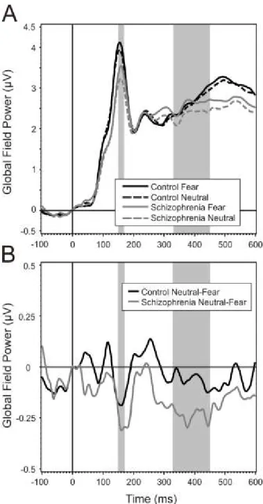
4.1.2.2. Analysis of the difference GFP waveforms ... 56
4.1.2.3. Comparison of the GFP difference waveforms in the two study groups in the mid-latency range ... 58
4.1.2.6. Correlation between psychopathological, behavioral, and electrophysiological results in the schizophrenia group ... 61
4.2. Results Study 2 ... 63
4.2.1. Preliminary analysis: Scalp topography of ERSP ... 63
4.2.2. Behavioral results ... 65
4.2.3. Electrophysiological results: Stimulus-related changes in theta response ... 67
4.2.3.1. Between-group differences in ERSP ... 67
4.2.3.2. Association between theta ERSP and behavioral results ... 69
4.2.4. Electrophysiological results: Phase-locking in the theta band ... 69
4.2.4.1. Between-group differences in theta phase-locking (ITC) ... 69
4.2.4.2. Association between ITC and behavioral results ... 71
4.3. Results Study 3 ... 71
4.3.1. Behavioral Results ... 71
4.3.2. Electrophysiological results ... 71
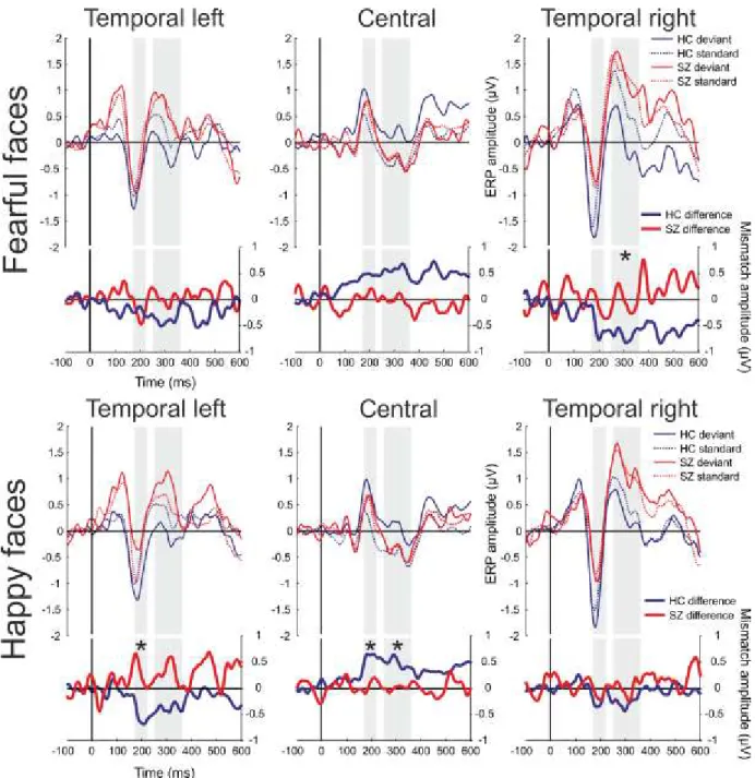
4.3.2.1. Mismatch responses for fearful faces ... 71
4.3.2.2. Mismatch responses for happy faces ... 72
5. D
ISCUSSION... 73
5.1. Discussion of Study 1 ... 73
5.2. Discussion of Study 2 ... 76
5.3. Discussion of Study 3 ... 78
6. G
ENERAL DISCUSSION... 80
6.1. Summary of main findings ... 80
6.1.1. Face-specific processing ... 81
6.1.2. Indices of impaired processing of fearful faces in schizophrenia ... 82
6.1.3. Hyperreactivity to fearful faces in schizophrenia ... 83
6.1.4. Decrease in theta oscillatory activity in fearful face recognition in schizophrenia 84 6.1.5. Combining time-domain and time-frequency analyses ... 85
6.1.6. Processing of unattended facial emotions in schizophrenia ... 86
6.2. Fearful face processing deficit in the context of cognitive and emotional interference 87 6.3. Limitations ... 88
6.4. Future directions ... 90
7. S
UMMARY... 92
8. Ö
SSZEFOGLALÁS... 94
9. R
EFERENCEL
IST... 96
10. L
IST OF PUBLICATIONS... 114
10.1. Publications related to the dissertation ... 114
10.2. Publications unrelated to the dissertation ... 114
11.
A
CKNOWLEDGEMENTS... 116
4 LIST OF ABBREVIATIONS
ACC Anterior cingulate cortex
APA American Psychiatric Association
CNTRICS Cognitive Neuroscience for Treatment Research to
Improve Cognition in Schizophrenia
CPZ Chlorpromazine
DSM-IV Diagnostic and Statistical Manual of Mental Disorders, Fourth Edition
DLPFC Dorsolateral prefrontal cortex
EEG Electroencephalogram
EOG Electrooculogram
ERD Event-related desynchronization
ERS Event-related synchronization
ERO Event-related oscillation
ERSP Event-related spectral perturbation
ERP Event-related potential
FEEST Facial Expressions of Emotion Stimuli and Tests
GFP Global field power
GLM General linear model
HLM Hierarchical linear model
ISI Inter-stimulus interval
ITC Inter-trial coherence
MATRICS NIMH-Measurement and Treatment Research to Improve Cognition in Schizophrenia
PANSS Positive and Negative Symptom Scale
ROI Region of interest
SD Standard deviation
SCL-90 Symptom Checklist – 90
ToM Theory of mind
VPP Vertex positive potential
vMMN Visual mismatch negativity
5 LIST OF TABLES AND FIGURES
Tables
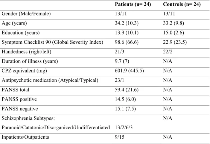
Table 1. Basic demographic and descriptive characteristics of the two study groups ... 38 Table 2. Estimated mean differences in ERP amplitudes between emotions (neutral minus fear) in the 330-450ms time interval by brain regions in the control and schizophrenia group.60 Table 3. Relationship between GFP difference and psychopathological and behavioral indices and medication in the schizophrenia group ... 63 Table 4. Demographic data, clinical characteristics, and behavioral results ... 66
Figures
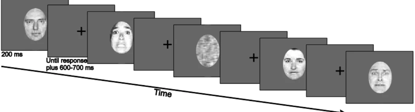
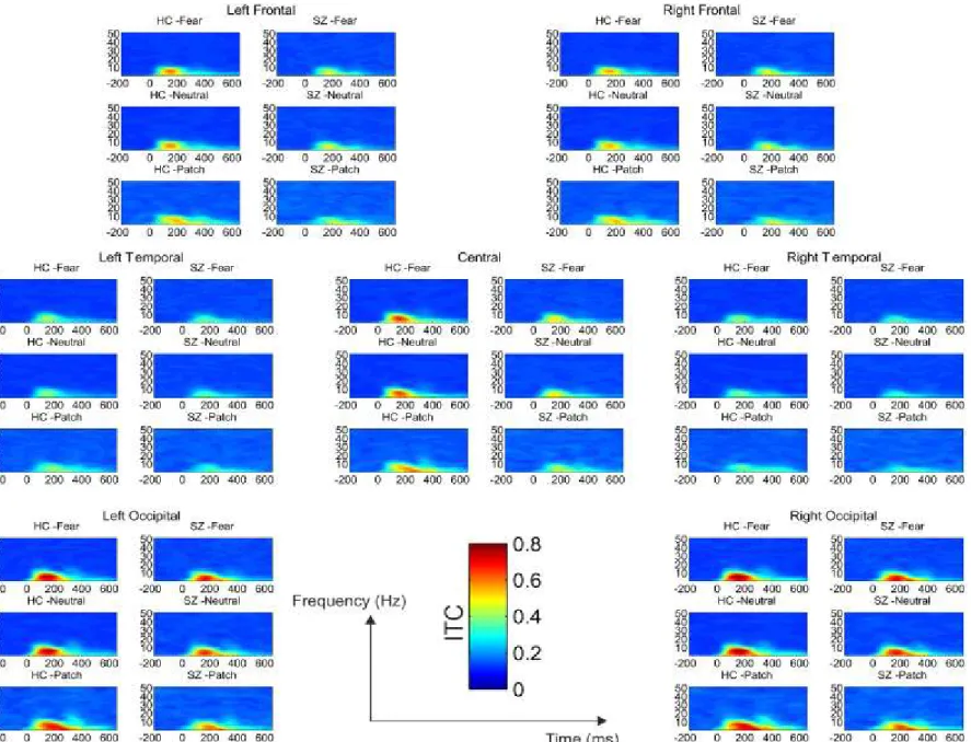
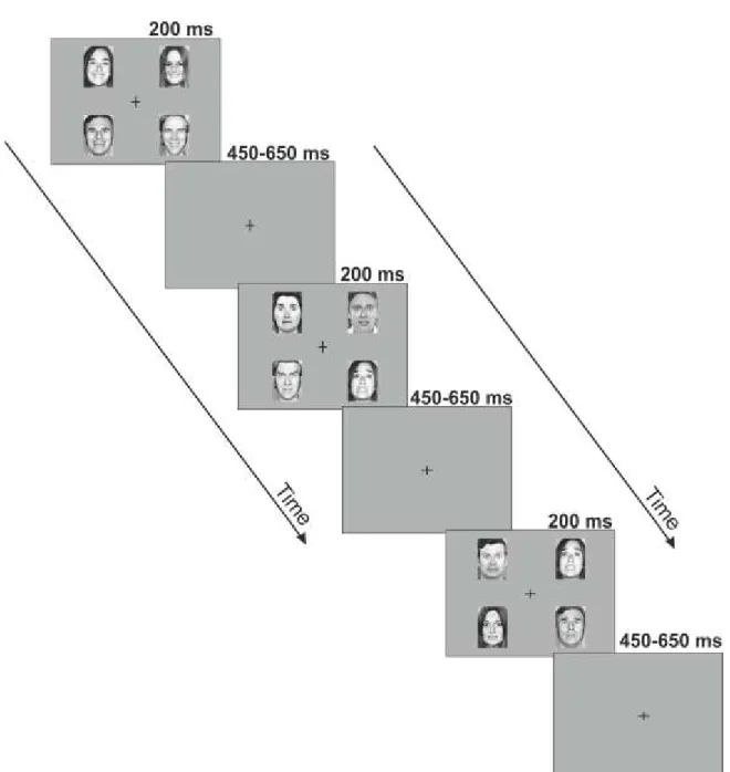
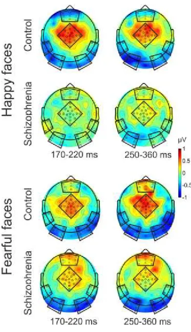
Figure 1. Diagrammatic illustration of the relationship between five proposed core constructs for social and emotional behavior ... 15 Figure 2. Overview of representative experimental trials ... 40 Figure 3. Event-Related Spectral Perturbation (ERSP) by regions of interest (ROI), study group, and stimulus condition ... 46 Figure 4. Inter-Trial Coherence (ITC) by regions of interest (ROI), study group, and stimulus condition ... 47 Figure 5. Schematic illustration of the pattern of emotional stimuli used in the experiment . 50 Figure 6. Event-related potentials and mismatch waveforms by region. ... 52 Figure 7. Scalp topography of the mismatch responses ... 53 Figure 8. Grand Average GFP (Global Field Power) and GFP difference waves in the two groups, Part A and B ... 57 Figure 9. Topographical maps of ERP amplitudes for Neutral and Fearful faces, and Neutral minus Fearful difference waves ... 58 Figure 10. Estimated mean differences in ERP amplitudes between emotions in the 330- 450ms time interval by brain regions in the control and schizophrenia group ... 61 Figure 11. Scalp topography of the Event Related Spectral Perturbation (ERSP) in the two study groups in the three experimental conditions in the 140-200ms time window………….64 Figure 12. Between-group differences in Event-Related Spectral Perturbation (ERSP) in the theta band in the three conditions, and correlations between ERSP values at frontal regions and emotion recognition as indexed by the Facial Expressions of Emotion Stimuli and
Tests (FEEST) scores………68
6
Figure 13. Between-group differences in Inter-Trial Theta Coherence (ITC) in the three conditions ……….70
7
1. I
NTRODUCTIONThe focus of this dissertation, the investigation of the electrophysiological correlates of fearful face recognition impairments in schizophrenia can be best interpreted in the context of social cognition in schizophrenia, an area of research that has come to the attention of cognitive neuroscience research in the past decades. With the shift from positive symptoms as treatment targets to the more devastating negative and cognitive symptoms of the disorder, neuroscience research has adopted the framework of investigating these variables in their association to each other and to functional outcome measures, delineating potential pathways of impairments leading from basic neurocognition, to social cognition and clinical symptoms, and to functional outcome. Facial emotion recognition, as a key component of social cognition, can be placed within this framework. Our aim was to investigate the nature of facial emotion recognition impairment in schizophrenia at a neurophysiological level that would possibly render implications for broader levels of information-processing impairments in schizophrenia.
1.1. Social cognition in schizophrenia
Humans are a highly social species, adapted and attuned to a complex social environment.
From an evolutionary perspective, our survival has depended on the refined social skills we have acquired to navigate through an intricate social world. Individuals with schizophrenia find themselves seriously disadvantaged in their social environment, unable to correctly read and respond to social signals, becoming vulnerable to social stress derived from their complex, social environments (Brune, 2001).
Social cognition refers to the mental operations underlying social interactions, which include processes involved in perceiving, interpreting, and generating responses to the intentions, dispositions, and behaviors of others (Brothers, 1996; Penn et al., 2008). It further involves
„the ability to construct representations of the relation between oneself and others and to use those representations flexibly to guide social behavior” (Adolphs, 2002). Four primary domains of social cogniton have emerged in schizophrenia research: (1) emotion processing, (2) social perception, (3) theory of mind (ToM), and (4) attributional style (Green and Leitman, 2008; Penn et al., 2008). Emotion processing refers to the ability of identifying or discriminating emotional expressions by perceiving or scanning social details and scenes.
8
Social perception refers to the ability of perceiving or scanning social cues and using the context for forming a behavioral response. Theory of mind refers to the ability to represent the mental states of others or to make inferences about others‟ intentions; this includes understanding hints, false beliefs, irony, and metaphor. Attributional style refers to assigning causality (internal, personal, or situational) to events.
There is now evidence that patients with schizophrenia are impaired in each of these social cognitive domains and that these impairments are a hallmark feature of the illness (Green et al., 2012). However, as social cognition is a broad, multifaceted construct, measures of social cognition are varying, lacking a consensus in their methodology. One main question concerns the underlying structure of social cognition in schizophrenia. It has been a question of debate to what extent social cognition is empirically and neurobiologically separable from, but related to non-social neurocognition (Fett et al., 2011; Green and Leitman, 2008). It is not known whether the social cognitive assessments used in schizophrenia reflect a single factor or a cluster of separable factors. A recent study aimed to reveal the underlying factor structure of social cognition in patients with schizophrenia (Mancuso et al., 2011) through an exploratory factor analysis used variabled from a wide range of social cognition tasks. It revealed three dimensions of social cognition to be characteristic of people with schizophrenia, which were the following: (1) hostile attributional style, (2) lower-level social cue detection, and (3) higher-level inferential and regulatory processes. These factors exhibited distinct patterns of correlation with clinical features and functional outcome.
It is still debated whether the aforementioned impairments should be conceptualized as
„deficits”, in a quantitative manner, or rather as „biases”, in a qualitative manner. The heterogeneity existing across the schizophrenia spectrum suggests that both deficits and biases may exist, and contribute to maladaptive social functioning (Tas et al., 2013).
The research on social cognition in schizophrenia can be regarded as a relatively new field that has come into scientific focus with the rapid advent of neuroscience. The need for the explanation of the complex symptomatology seen in schizophrenia has shifted from a classical clinical framework to an integrated neuroscientific framework based on the investigation of information-processing mechanisms. The aim has become to understand the anatomical and physiological structure and function of neural networks which underlie complex social cognition and behavior within the broader perspective of individual cognitive, emotional, and social development. After returning to Bleuler‟s core concept of schizophrenia
9
(Bleuler, 1911), who believed that schizophrenia „is characterized by a specific kind of alteration of thinking and feeling, and of the relations with the outer world that occur nowhere else” (Burns, 2006), schizophrenia has become the „arch” representative of social brain disorder. According to Bleuler, underneath the often obvious and varied symptoms, such as hallucinations and delusions, there existed a less obvious inner unity, which can be characterized by the four „A”s: ambivalence, autism, disturbance of association and affect.
Modern neuroscience aims to explain these symptoms in a neurobiological framework involving social, cognitive, and affective processes, the impairment of which result in disturbed basic everyday functioning.
In sum, social cognitive research in schizophrenia has had two distinct goals: to understand the nature of specific clinical symptoms (e.g., relations to paranoia or thought control) and to understand social cognition‟s role in functional outcome.
Impairment in everyday functioning is profound in schizophrenia, even after successful treatment of clinical symptoms, especially positive psychotic symptoms. A recent literature review (Harvey and Strassnig, 2012) summarized evidence on the empirical association between schizophrenia and vocational disability and has corroborated the fact that patients with schizophrenia have significant impairment across multiple dimensions of functioning, and will typically remain impaired for the duration of normal working ages. Thus, the focus of attention in schizophrenia research and treatment has turned from positive symptoms towards the perhaps most devastating symptoms of the disorder: negative symptoms, and the loss of cognitive and social cognitive skills which result in the alienation of the individual from the social world.
1.1.2. Neurocognition, social cognition and the MATRICS initiative
As a result of the realization of the importance of neurocognition and social cognition in their close relation to functional outcome, NIMH-Measurement and Treatment Research to Improve Cognition in Schizophrenia (MATRICS) (Green and Nuechterlein, 2004) initiative was launched by leading experts in schizophrenia research from academia, the National Institute of Mental Health and the pharmaceutical industry. The rationale of the committee was to identify target domains of neurocognition that have a translational potential and to establish a standardized test battery of the selected domains for clinical trials. The committee identified seven domains of cognitive impairment as target domains in schizophrenia: (1)
10
reasoning and problem solving; (2) processing speed; (3) attention and vigilance; (4) working memory; (5) verbal learning and memory; (6) visual learning and memory. Social cognition was identified as an additional seventh domain (Green et al., 2005; Nuechterlein et al., 2008).
Although the MATRICS committee regarded social cognition as a high priority area, as can be seen in its proportions to neurocognitive domains, its representation in the consensus test battery remained limited because of its inconsistent terminology and differences in the measures of social cognition, making direct comparisons of findings in the field more difficult. Thus, the MATRICS committee delineated three major areas to promote research in social cognition in schizophrenia (Green and Leitman, 2008): first, the agreement on the definition, measures and factors of social cognition; second, improving the interface between social cognition and cognitive neuroscience to identify neural substrates of social cognition performance in schizophrenia; and third, to develop animal models for social cognition.
The MATRICS initiative called general attention to the importance of neurocognitive and social cognitive deficits in schizophrenia and has led to the development and refinement of numerous neurocognitive test batteries assessing cognitive deficits in schizophrenia.
Consequently, the MATRICS initiative facilitated studies and literature reviews investigating the exact nature of the relationships between neurocognition, social cognition, clinical symptoms, and functional outcome. Perhaps most importantly, it has also boosted research in the field of social cognition to further clarify its nature and make it a more methodologically approachable construct for further translational research.
1.1.3. Neurocognition, social cognition, clinical symptoms and functional outcome
The association between social cognition and general cognition or basic neurocognition in schizophrenia has been a question of debate, especially within the context of their relationship to domains of functional outcome. One key question in schizophrenia is the degree of overlap between the circuits that underlie deficits in basic cognition versus those that underlie deficits in social cognition. The question is also raised whether specific types of social cognitive functions arise through computational processes that are similar to those in basic cognition, even though different brain regions may be involved (Green et al., 2005). As „cognition‟ is contained in the term social cognition, it seems obvious that the processing of socially
11
relevant stimuli relies on basic neurocognitive functions, such as attention, memory, and various other cognitive processes, yet research has shown that social cognition and neurocognition are largely distinct domains (Allen et al., 2007; Pinkham et al., 2003; Sergi et al., 2007; van Hooren et al., 2008).
It has been suggested that social cognition functions as a mediator between neurocognition and functional outcome (Addington et al., 2006; Brekke et al., 2005; Meyer and Kurtz, 2009;
Sergi et al., 2007). Still, as social cognition explains additional variance in outcome that cannot be accounted for by neurocognition, it seems to be an independent predictor of functional outcome in itself (Brekke et al., 2005; Pinkham and Penn, 2006). Furthermore, some studies showed that social cognition may even exceed the predictive power of neurocognition and other symptoms of schizophrenia in explaining variance in functional outcome (Fett et al., 2011; Pijnenborg et al., 2009).
With regard to the role of clinical symptoms in their association to social cognition and functional outcomes, the literature provides a generally mixed picture on this relationship.
The relationship of social cognition with positive symptoms (e.g.delusions, thought disorder, hallucinations) and negative symptoms (e.g. avolition, anhedonia, alogia, emotional withdrawal) has been inconsistent across studies. Negative symptoms were found to have the strongest relationship with neuro- and social-cognition (Milev et al., 2005; Ventura et al., 2011), however, recent modeling studies suggest that social cognition is separable from negative symptoms (Rassovsky et al., 2011; Sergi et al., 2007). Though there has been somewhat greater consistency in the associations between attributional style and paranoid delusions or beliefs (Bentall et al., 2001; Combs et al., 2009; Fornells-Ambrojo and Garety, 2009), relations to positive symptoms are similarly inconsistent (Shamay-Tsoory et al., 2007;
Woodward et al., 2009). In a recent structural equation modeling study using a mediation model, Lin and colleagues (Lin et al., 2013) found that mainly negative symptoms mediated the influence of neurocognition and social cognition on functional outcome in schizophrenia.
The authors posited that negative symptoms impair neuro- and social-cognitions possibly through lowered motivation to attend the tasks and in turn make an impact on functioning, or also that negative symptoms decrease the motivation to participate in social activities, which directly influences functional outcome.
12
1.1.4. Impaired emotion perception in schizophrenia
Of the social cognition domains, emotion perception has been identified and studied the most frequently in schizophrenia. Even though on a subjective level patients with schizophrenia report experiencing as much positive and negative emotions as healthy controls and do not seem to have diminished hedonic capacity when providing “online” (e.g. in the moment) self- report in response to stimuli (Kring and Neale, 1996), they do report experiencing greater negative affect than controls when exposed to unpleasant, neutral, and pleasant stimuli (Cohen and Minor, 2010). This chronic elavation in negative mood, anhedonia, is commonly included in the negative symtoms of schizophrenia (Earnst and Kring, 1999), and since Kraepelin (Kraepelin, 1917) and Bleuler (Bleuler, 1911), anhedonia has been regarded as one of the core deficits in schizophrenia.
Recent research has suggested that the reduced capacity to experience positive emotions in schizophrenia might be due to emotion dysregulation mechanisms (Cohen and Minor, 2010;
Horan et al., 2006; Strauss and Herbener, 2011). In experimental settings schizophrenia patients showed difficulty disengaging attention once it had been engaged by a salient unpleasant stimulus in an emotional stroop task (Strauss et al., 2008), indicating a difficulty for them in attenuating negative emotional states, resulting in chronic depression in mood and anhedonia. This might be related to the well-known „negativity bias”, whereby patients show a strong inclination to misidentify neutral stimuli as negatively valenced (Kohler et al., 2003;
Premkumar et al., 2008). Such inabilities to accurately identify emotional valence might be the basis for a bias to interpret situations in a negative light, and thus result in schizophrenia patients experiencing relatively more negative emotions than healthy controls. In sum, research suggests that emotional abnormalities seen in schizophrenia may primarily relate to dysfunctions in negative emotion processing.
A number of studies have investigated the emotion perception deficit in schizophrenia using diverse methodological, clinical, and demographic variables. A meta-analysis (Kohler et al., 2010) of behavioral indices of emotion perception in schizophrenia has shown a large effect size (d=0.91) for emotion perception impairment in patients with schizophrenia as compared to healthy controls, despite heterogeneous moderating effects of illness-related and
13
demographic factors. They concluded that deficits in emotion perception in schizophrenia are an intrinsic and stable aspect of the illness.
A more recent meta-analysis (Irani et al., 2012) aimed to clarify the strength of the effect between emotion perception and functional outcomes. Their results corroborated a significant relationship between emotion perception and functional outcome in individuals with schizophrenia with effect sizes in the medium range. These results also support the notion that studying social cognitive processes such as emotion processing in schizophrenia is promising to provide: (1) a greater understanding of the key mechanisms that might influence the clinical presentation and functional outcome, (2) the identification of endophenotypic markers for genetic research in the vulnerability to schizophrenia, and (3) the delivery of remediating therapies.
1.1.5. Facial emotion recognition in schizophrenia
Within emotion perception, the domain of facial emotion recognition has gained considerable interest, although emotion recognition impairments in the auditory domain have also been extensively studied (Leitman et al., 2010).
Facial emotion recognition can be considered as a main building block of social cognitive abilities. Understanding and expressing facial emotions in nonverbal communication is a key component of interpersonal adaptation. According to Darwin: “The interpretation and expression of affect is fundamental to human experience” (Darwin, 1872). Faces constitute social signals that are granted attentional priority, and this preference is expressed early in life, with infants preferentially attending to face-like stimuli rather than to scrambled or inverted faces (Gliga and Csibra, 2009; Rosa et al., 2011). Through the scanning of other people‟s faces, the social world summons our attention like no other domain: social signals are prioritized by attention, social interactions are intrinsically rewarding, and the maintaining of social relations permeates interpersonal behaviors, fosters collaboration. People are typically the most goal-relevant stimuli in our lives.
The robustness of the impairment of facial affect recognition in schizophrenia has been confirmed in studies which showed that the impairment is stable across the time course of the illness, independent of the clinically effective treatment (Addington and Addington, 1998;
Wolwer et al., 1996a), and is observable in individuals at-risk for schizophrenia (Wolwer et al., 2011) as well as in unaffected biological relatives of patients (Bediou et al., 2007).
14
In order to interpret the neural underpinnings of facial emotion recognition deficit in schizophrenia, I will present a model by Ochsner (Ochsner, 2008) integrating social, cognitive and affective processes in which the ability of facial emotion recognition can be embedded.
The model is regarded only as a broader theoretical framework for the clearer positioning of facial emotion recognition within the social-affective information processing stream, rather than as a specific model from which our electrophysiological hypotheses were derived from.
1.2. The social-emotional processing stream (Ochsner, 2008)
The neural circuitry underlying social cognition is a complex, interrelated neural network, including both cognitive and emotional processing systems. The diverse methods and approaches used in this new field make it difficult to put the disparate pieces of data together to fit into core neurofunctional constructs.
The theoretical framework by Ochsner identifies a set of psychological and neural processes that encode socially and emotionally relevant inputs and guide socially adequate responses.
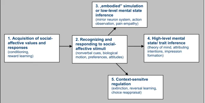
The model enumerates social cognitive and affective abilities grouped into five hierarchically distinct lower-level and higher-level core constructs that are valid and reliable entities in so far as they are suggested to have distinct but related neural correlates. As depicted in Figure 1, these lie along a hierarchy of information processing stages engaged when we initially learn the value of a stimulus (Construct 1), subsequently re-encounter it and recognize its value (Construct 2), understand the feelings and beliefs of a person by relating to his/her mental states in an experiential, bottom-up way (Construct 3), and also in a top-down manner attributing intentions and making inferences (Construct 4), and finally trying to regulate responses to a given stimulus appropriate within the context (Construct 5).
15
Figure 1: Diagrammatic illustration of the relationship between five proposed core constructs for social and emotional behavior. (From: Ochsner: The social-emotional processing stream: Five core constructs and their translational potential for schizophrenia and beyond. Biological Psychiatry, 2008;
64: 48-61.)
Regarding Construct 1, the basic need to learn which stimuli and actions lead to aversive as opposed to appetitive outcomes is mainly mediated through the amygdala. The neural constructs most strongly implicated in affective learning and conditioning are the amygdalae and the striatum, evolutionary old subcortical constructs that receive multi-modal perceptual inputs and also are highly connected to higher-level cortical regions (Holland and Gallagher, 1999). It has been well established that the amygdala is critical for acquiring conditioned fear responses to social stimuli, such as faces or facial expressions of fear and anger (Morris et al., 1998; Morris and Dolan, 2004). The ventral striatum has also been shown to play a crucial role for learning which stimuli or behavioral responses predict rewarding or reinforcing outcomes (Schultz, 2004). Representation of the affective valence of a stimulus is possible via projections from the amygdala and the striatum to the orbitofrontal and ventral medial prefrontal cortex (Rolls, 2000).
In Construct 2, the amygdala not only plays a crucial role in learning the affective valence of stimuli, but also in the recognition of stimuli that directly or indirectly signal the presence of a potential threat, such as fearful facial expressions and the widened eyes that uniquely
2. Recognizing and responding to social- affective stimuli (nonverbal cues, biological motion, preferences, attitudes) 1. Acquisition of social-
affective values and responses
(conditioning, reward learning)
5. Context-sensitve regulation
(extinction, reversal learning, choice reappraisal)
4. High-level mental state/ trait inference (theory of mind, attributing intentions, impression formation)
3. „embodied” simulation or low-level mental state inference
(mirror neuron system, action observation, pain empathy)
16
characterize them (Adolphs et al., 2005). In a broader conceptualization the amygdala is regarded as a “surveillance” system that continuously monitors the environment for affectively salient stimuli and modulates activity in perceptual and memory systems to detect and encode them (Phelps, 2006; Whalen et al., 1998). Thus, facial emotion recognition would be part of Construct 2, faces being the most relevant social-affective stimuli. Another region shown to be important in negative affective experience is the insula, a cortical region connecting the temporal and frontal lobes (Craig, 2004; Critchley et al., 2005). The superior temporal sulcus has also been shown to play an important role in the recognition of social/affective values, especially in recognizing nonverbal social cues, such as faces and biological motion (Allison et al., 2000).
Within this socio-emotional processing stream framework, one step higher in the processing hierarchy, in Construct 3, are the lower-level mentalization processes. Based on these, Construct 4 entails higher-level mentalization processes. Beyond simple recognition judgments of affective values of stimuli, the meanings of the value of the stimuli are embodied in our experience of it. In fact, all of our experiences are embodied in our physical reality, and not just our own first-person experiences, but these are used to help us vicariously understand the experience of others as well as abstract concepts (Barsalou et al., 2003;
Gallese and Lakoff, 2005).
The stimulus-driven activation of shared representations establishes the direct experiential properties of a stimulus (such as perceptual, motor, visceral, or affective), but the activation of higher-level symbolic, abstract, semantic, or categorical representations are needed to be able to interpret the given experience in a given context. Social stimuli are often ambiguous and can be interpreted only if higher-level representations of mental states are activated that take into account the situational /contextual information of the stimulus. Perhaps the most studied faculty of higher-order representations has been the theory of mind. False belief tasks assess the human capacity to understand and explain other peoples‟ behaviour in terms of internal mental states, such as their beliefs, desires, feelings, and goals. Studies using this task have shown active regions in the brain such as dorsal and rostral medial prefrontal cortex, the posterior cingulate precuneus, temporal parietal junction, superior temporal sulcus, and the temporal pole, sometimes referred to as the “mentalizing network” (Gallagher and Frith, 2003). Furthermore, some of the regions of the medial prefrontal cortex that are implicated in
17
mental state inference of others have also been implicated in accessing and making judgments about one‟s own mental states (Ochsner et al., 2005).
The final stage in the social-emotional stream, as described by Construct 5, concerns the ability to regulate one‟s judgments and behavior toward others in a context-appropriate manner. These higher-level processes entail cognitive control: the use of language, memory, and selective attention, to reinterpret or reappraise the meaning of social-emotional stimuli (Ochsner and Gross, 2005). Brain areas involved in cognitive control are the dorsal and lateral prefrontal regions, as well as medial prefrontal regions. Activity in these control systems modulates activity in regions involved in emotion responding, such as the amygdalae or insula.
This above depicted model of the socio-emotional processing stream was proposed to serve as a simplified framework for organizing research on the neural bases of human social cognitive and emotional behaviour and to clarify constructs potentially underlying social cognition and emotion. It also provides a basic context for translational research, as it enables its application to clinical populations to help elucidate potential mechanisms of dysfunction. In the case of schizophrenia, substantial research aims to pin down the neural correlates of the various forms of emotional dysfunction, such as emotion recognition. In the following, a short overview on the impairments of the neural bases of emotion processing in schizophrenia will be given.
1.3. Structural and functional impairments related to facial emotion processing in schizophrenia
Literature on neurobiological impairments in emotion processing in schizophrenia has become abundant, but often inconsistent. Interpretation has been hampered by a range of dissimilar experimental designs and clinical patient samples recruited in the studies. In the following I will present some of the main findings in schizophrenia on the most important neural structures involved in emotion processing, with a main focus on facial emotion processing.
The amygdala, as mentioned earlier, has been shown to play a prominent role in emotion perception. According to a long-held view the vital role of the amygdala has been shown in
18
fear conditioning in rodents, primates and humans (Salzman and Fusi, 2010). However, a recent meta-analysis (Ball et al., 2009) and contemporary reviews hold that amygdala activation is related to both arousal and intensity, regardless of positive or negative emotional valence (Adolphs, 2010; Pessoa and Adolphs, 2010). Thus, a shift can be seen in conceptualizing the amygdala as simply a fear or threat 'processing' module toward the position that amygdala is also tracking stimulus intensity and deals with motivational salience (Adolphs, 2010). Because of its anatomical position and its strong interconnection with other crucial brain areas involved in emotion processing, the amygdala serves as an affective hub of information (Mende-Siedlecki et al., 2013).
Structural imaging suggests that patients with schizophrenia have smaller amygdala volumes than healthy controls (Namiki et al., 2007) and functional imaging data record abnormalities of amygdala activation during emotion processing in schizophrenia. Studies indicate that, compared with healthy controls, patients with schizophrenia and their nonpsychotic siblings fail to activate bilateral amygdala regions during induction of sad mood (Habel et al., 2004).
Lower bilateral amygdala activation in patients relative to healthy controls has been reported during a facial identification task (Hempel et al., 2003) and in an emotional valence and facial discrimination task (Gur et al., 2002; Johnston et al., 2005). However, findings relating to amygdala activation in schizophrenia have not always been consistent. There seem to be conflicting results regarding reduced left (Gur et al., 2002) or right (Johnston et al., 2005) amygdala activation in emotion perception tasks. Moreover, several studies have reported enhanced activity in the amygdala in schizophrenia during the presentation of facial emotional expressions (Holt et al., 2006; Kosaka et al., 2002).
Amygdala dysfunction has been investigated as part of a distributed interactive system of the whole brain. Sepcifically, some studies investigated amygdala signals in their functional connectivity to other brain regions, such as the fronto-parietal network. Results with schizophrenia patients showed that patients display significantly weaker amygdala-prefrontal cortical coupling, specifically during negative emotional distraction (Anticevic et al., 2012).
Furthermore, a functional connectivity study with healthy subjects has shown a decreased connectivity between the amygdala and the facial areas in those participants who were more vigilant for threatening facial features, interpreting weaker connectivity of the amygdala with facial areas as representing social withdrawal (Miyahara et al., 2013). Some authors interpret these findings as evidence for impaired emotional regulatory processes in schizophrenia,
19
which may reflect an impaired capacity to downregulate amygdala responses (Modinos et al., 2010).
In a recent meta-analysis on functional neuroimaging data Li and colleagues (Li et al., 2012) concluded that “a marked underrecruitment of the amygdala, accompanied by a substantial reduction in activation throughout a ventral temporal-basal ganglia-prefrontal cortex “social brain” system may be central to the difficulties patients experience when processing facial emotion”.
The hippocampus, as a "memory-storage" for the brain, could be involved in emotion processing in that it could provide a resource for referencing and placing emotions in the context of previous experiences. Most of the studies reported an underactivation of the hippocampus in schizophrenia patients in facial emotion perception tasks (Takahashi et al., 2004; Williams et al., 2004). However, some studies found increased hippocampus activation in non-paranoid patients (Russell et al., 2007).
The fusiform gyrus is considered to be one of the most specific areas of face recognition in the brain (McCarthy et al., 1997). However, findings show inconsistencies in the involvement of this area in face recognition in schizophrenia. When patients performed a facial emotion discrimination and identification task, their bilateral fusiform gyri were not activated, while the controls showed the expected activation in response to faces in right lateral fusiform gyrus (Quintana et al., 2003). Moreover, underactivation in the right fusiform region was found in patients during remission (Johnston et al., 2005). Greater activation in schizophrenia has been observed in bilateral fusiform gyri during the presentation of neutral faces (Surguladze et al., 2006).
Findings seem to be more consistent in the decreased involvement of the cortical areas in facial emotion perception in schizophrenia. The orbitofrontal cortex forms an interface between emotion and cognition (Rolls, 2000), and, together with the middle temporal lobe, precuneus, and posterior cingulate, is implicated in empathy and in making social judgments (Farrow et al., 2001). In addition, the medial prefrontal cortex facilitates the understanding of the mental state of others, or, as has been referred to earlier, serves as the center for theory of mind. Some researchers have reported hypoactivations in schizophrenia patients in the orbitofrontal cortex during facial emotion perception (Reske et al., 2007), while others
20
observed reduced activation in the medial prefrontal cortex when viewing negative emotional stimuli (Takahashi et al., 2004; Williams et al., 2007). These results are consitent with the general notion of a frontal hypoactivation in schizophrenia (Johnston et al., 2005).
Regarding structural and functional abnormalities in schizophrenia it is important to take into account the fact that schizophrenia is a neurodevelopmental disorder, and several impairments of the nervous system shown to be associated with emotion processing in schizophrenia are related to the maturation of the brain in late adolescence. The connections between the various subcortical regions and the prefrontal cortex and molecular changes within the prefrontal cortex itself also appear to undergo substantial change during adolescence (Weickert et al., 2007). It has been hypothesized that testosterone in adolescence may affect the ability to respond appropriately to emotional faces (Richards et al., unpublished data). If this is correct, then the deficit in emotional face recognition among patients with schizophrenia may emerge shortly after puberty, as the prefrontal cortex and its connections with the amygdala may fail to mature normally (Morris et al., 2009).
1.4. Impaired visual integration of faces in schizophrenia
After having reviewed some of the main impairments in brain structures and their connectivities in schizophrenia related to facial emotion processing, some behavioral evidence will be reviewed on the level of impairment of basic visual perception potentially leading to impaired facial emotion recognition in schizophrenia.
Several studies have explored facial scanning, scanpath patterns and gaze impairment in schizophrenia (Itier and Batty, 2009; Morris et al., 2009). Impairment in eye movements while viewing facial images has been long reported in schizophrenia (Manor et al., 1999).
Examination of patterns of eye fixation to high resolution pictures of human faces versus nonbiological stimuli showed that subjects with schizophrenia exhibit reduced scanpath lengths, shorter saccades, more time spent „gazing‟, and a tendency toward fewer fixations for the face stimulus (Kosmidis et al., 2007). Thus, disturbed spatial and temporal patterns of eye movement in some people with schizophrenia may reflect sub-optimal processing of face stimuli.
21
Other visual scanpath studies of face processing in schizophrenia showed that schizophrenic patients did not concentrate their fixations on salient features of faces, such as eyes (Williams et al., 1999). Evidence indicates that the eye region of faces is used to discriminate fear from other expressions (Adolphs, 2008). It has been shown that the eye region of fearful faces is processed faster and earlier in healthy adults than other regions, and is sufficient to explain the effect of fearful faces in a preconscious processing task (Yang et al., 2007). As patients with schizophrenia have a particular deficit in attending to the eyes, it would be expected that patients have a greater deficit in recognizing fear. Kohler and colleagues (Kohler et al., 2003) using low-and high-intensity images found that patients were more impaired than healthy controls in identifying easier high-intensity expressions, and this difference was most pronounced when recognizing fear. Thus, although patients with schizophrenia are impaired in overall emotion recognition, they appear particularly impaired in recognizing fear, regardless of task difficulty.
Studies investigating the direction of gaze as a referential point in schizophrenia (Hooker and Park, 2005) have shown that schizophrenia patients are as accurate as healthy control subjects at identifying direct gaze when it occurs, but they are more likely to misinterpret averted gaze as directed at them. Results suggested that this tendency to endorse direct gaze was not a consequence of a perceptual deficit in judging angular displacement. Schizophrenia patients seem to have a self-referential bias in judging the direction of gaze that could lead to the misinterpretation of another person's intentions during the course of social interactions.
Altogether, findings suggest that emotion perception deficits in schizophrenia patients may reflect a failure to integrate salient features of a face due to dysfunctions in local processing of detailed, relevant information, and in the networks that synchronize local and global processing of biologically relevant face stimuli. Furthermore, some researchers have suggested that abnormalities in visual scanpaths could be emerging as a novel candidate for a schizophrenia biomarker, as evidence has shown that viewing behaviors in schizophrenia remain atypical regardless of symptom remission and may be present in unaffected relatives of individuals with schizophrenia (Beedie et al., 2011).
22
1.5. Emotion and cognition
The investigation of the neural underpinnings of facial emotion processing inevitably raises the classical neuropsychological question of the interaction between affective and cognitive processes, as this area of research lies at the interface of the two. Faces are not only a subcategory of objects, as they not only provide distinctive information about a person‟s identity, gender, or age, but also convey more subtle signals related to emotion, trustworthiness, attractiveness, or intention of other people. However, little is known about how these various dimensions are coded, through what processes, and how they are integrated into a single face percept reflecting an emotional state.
Traditionally, neuroscientists have described cognition and emotion as separable processes implemented by different regions of the brain, such as the amygdala for emotion and the prefrontal cortex for cognition. On the one hand, emotional processes can influence cognitive processes (in a bottom-up manner); on the other hand, cognitive processes can regulate or modify our emotions (in a top-down manner). However, neurons in these structures often have overalapping representations, whereby single neurons encode multiple cognitive and emotional variables. Current research suggests that these neural stuructures are inextricably linked and represented in dynamic neural networks composed of interconnected prefrontal and limbic brain structures. Recent studies suggest that both the functional and the electrophysiological characteristics of the amygdala and the prefrontal cortex overlap. They intimately depend on each other, mediating cognitive, emotional, physiological, and behavioral responses that are closely linked (Salzman and Fusi, 2010). However, the question remains what the role of these interacting pathways might be in the case of emotionally laden stimulus processing.
1.5.1. Emotional attention
Enhanced sensory processing of emotional stimuli, also termed “emotional attention”
(Pourtois et al., 2005), has been consistently demonstrated in at least two functional properties of brain activity: (1) the amplitude of neural responses to emotional relative to neutral stimuli is consistently enhanced in several areas along sensory pathways, including both specific (e.g.
category-specific) and non-specific (e.g. early sensory cortex) regions (Lindquist et al., 2012), and (2) the time-course of emotion effects suggests a distinctive spatio-temporal dynamic as
23
compared with other attentional modulations (e.g. in fronto-parietal areas), with relatively early responses observed in some limbic regions, such as the amygdala (Luo et al., 2010;
Pourtois et al., 2010) or orbitofrontal cortex (Pourtois et al., 2013). These findings suggest that this boosting of sensory processing by emotion reflects increased processing efficiency for emotionally salient events (Vuilleumier et al., 2002; Vuilleumier, 2005), which may beneficially enhance attention towards them, and may also play a role in the more efficient encoding and subsequent consolidation in memory of emotional events (LaBar and Cabeza, 2006).
A central question about these bottom-up or top-down effects concerns which brain mechanisms are responsible for the preferential selection of emotionally salient stimuli in the environment. There is abundant evidence that visual inputs propagate rapidly throughout the brain and reach high-level cortical regions in the range of 120 ms post-stimulus onset (Bar et al., 2006; Kawasaki et al., 2001) allowing quick perceptual categorization and motor decision processes to be performed in less than 150 ms (Thorpe et al., 1996), that is, before the typical latency associated with voluntary attention control (Hillyard and Anllo-Vento, 1998). Thus, the question arises: Is the perception of emotion-laden stimuli “automatic”? This question has received considerable interest because specific answers (“yes” or “no”) suggest potentially different relationships between emotion and cognition (less or more independence between the two, respectively). Evidence both for and against automaticity have been presented (Pessoa and Adolphs, 2010).
1.5.2. Emotional “automaticity”
One of the most influential theories of rapid emotion effects was based on animal work (LeDoux, 1996; Shi and Davis, 2001) and observations in human patients with blindsight after damage of the primary visual sensory area (Anders et al., 2004; Pegna et al., 2005) which showed that some emotional responses in amygdala and conditioning may still arise for visual stimuli despite damage to cortical relays. Thus, it has been hypothesized that sensory inputs might reach the amygdala through subcortical pathways that bypass cortical processing, through a subcortical “quick and dirty” route via the superior colliculus and pulvinar (Tamietto and de Gelder, 2010). Neuroimaging results also showed activation in these two regions during unconscious processing of emotional stimuli (Liddell et al., 2005;
24
Morris et al., 1998). Also, emotional faces evoke responses in the amygdala when attention is diverted to other stimuli (Anderson et al., 2003; Vuilleumier and Schwartz, 2001). These and many related findings suggest that at least some types of emotional perception occur outside of top-down directed attention.
Other findings have suggested, however, that the perception of emotion-laden items requires attention, as revealed by attentional manipulations that were designed to more strongly consume processing resources, leaving relatively few for the processing of unattended emotional items (Lim et al., 2008; Pessoa et al., 2002).
As an alternative explanation to the subcortical route, most recent intracranial and MEG results converge to suggest a two-stage model of emotional attention and interaction with task relevance (Pourtois et al., 2013). First, the amygdala appears to guide an early (120–140 ms) discrimination between emotional (threat-related) and neutral stimuli even when visual stimuli are weak because they are task-irrelevant (Luo et al., 2010) or outside the current focus of attention (Pourtois et al., 2010). This early effect may take place in parallel to simultenaously with stimulus categorization in the visual cortex, and rely on an initial feedforward sweep of inputs throughout the visual pathways. Second, this early emotion response in the amygdala can trigger an increase of the neural response in the visual cortex (Pourtois et al., 2010; Vuilleumier and Schwartz, 2001), via both direct and indirect pathways projecting back to the cortex (Amaral et al., 2003; Vuilleumier, 2005). This boosting may thus increase processing efficiency and competitive biases for emotional relative to neutral stimuli, resembling an attention gain control effect that may add or combine with other modulatory influences.
Referring to the „automaticity” of the processing of emotional stimuli, Pourtois and colleagues (Pourtois et al., 2013) have proposed that emotional stimuli are “special” only to the extent that they have the propensity to engage dedicated neuronal systems relative to neutral stimuli, which are in turn capable of rapidly influencing perceptual or attentional systems, or both, such that these stimuli may gain additional “weight” in the competition for awareness (Armony and Dolan, 2002; Pourtois et al., 2005). The authors argue that emotional stimuli do not necessarily undergo a privileged route that neutral stimuli would not recruit.
Whereas some neural responses and their subsequent impact on sensory processing might be unique to emotionally significant stimuli, their perceptual analysis is likely to be similar to
25
emotionally neutral stimuli. In sum, a number of results from neuropsychological studies in brain-damaged patients (see Tamietto and de Gelder, 2010; Vuilleumier and Schwartz, 2001;
Williams and Mattingley, 2004), as well as fMRI (Morris et al., 1998; Whalen et al., 1998), ERP (Liddell et al., 2005; Williams et al., 2004) and MEG studies (Bayle et al., 2009) in healthy participants indicate that emotional information is processed (at least to some extent and under certain circumstances) regardless of voluntary top–down attention and even without conscious awareness. Unconscious processing by itself is not necessarily specific to emotional stimuli, since substantial processing of complex, non-emotional information can also take place without conscious awareness and still recruit the corresponding specialized processing pathways (Kouider and Dehaene, 2007; Righart et al., 2011). What is particular to the case of emotion processing is that neural substrates engaged without awareness or attention may include additional structures, relative to neutral stimuli, including the amygdala, which have direct ouputs to influence sensory processing as well as many other brain systems controlling perception and behavior.
The question of emotional attention can be related to psychopathologies seen in mental illnesses through the effects of the disruption of the emotional attention system. This system might be either amplified or attenuated by top–down modulations from higher-order frontal regions involved in emotion regulation processes, such as functional alterations within prefrontal-amygdalar regulatory circuits. In schizophrenia, for example, impairment in these regulatory processes might be associated with different psychopathological conditions which might increase vulnerability to and maintenance of anxiety and negative affect (Bishop et al., 2004; Bishop et al., 2007; Bishop, 2007). Such disruptions might also manifest through changes in the “firing threshold” settings of the amygdala circuitry itself. This could account for attention selection biases towards negative stimuli typically observed in some psychopathological conditions, such as general anxiety or depression (Bar-Haim et al., 2007;
Bishop et al., 2007; De Raedt and Koster, 2010). Alternatively, emotional attention could be exacerbated due to purely intrinsic changes in amygdala and hyper-reactivity of the sensory feedback loops (Pourtois et al., 2013).
26
1.6. Electrophysiological correlates of facial emotion recognition
Electrophysiological measures allow us to gain insight into the neuronal-scale brain avtivity underpinning complex processes such as facial emotion recognition. Scalp EEG signals are produced by partial synchronization of neuronal-scale field potentials across areas of cortex of centimetre-squared scale. Although once viewed as a form of brain „noise‟, it appears increasingly probable that this synchronization optimizes relations between spike-mediated
„top-down‟ and „bottom-up‟ communication, both within and between brain areas. This optimization might have particular importance during motivated anticipation of, and attention to, meaningful events and associations and in the response to their anticipated consequences (Fries et al., 2001; von Stein and Sarnthein, 2000). Analysis of event-related EEG data has traditionally been proceeded in one of two directions. In the time-domain approach, a set of single trials or epochs time-locked to a certain class of events/stimuli are averaged, yielding an event-related potential (ERP) waveform at each recording site. The frequency-domain approach averages changes in the frequency power spectrum of the EEG epoch that are time- locked to (i.e., following) the same events, producing a two-dimensional representation (i.e., time and frequency) that is called the event-related spectral perturbation (ERSP). Both analysis approaches of electrophysiological data can be used to disentangle neuronal processes during facial emotion recognition tasks. In the following we present a theoretical background of both approaches, as both were used in our two studies.
1.6.1. ERP correlates of facial emotion recognition
Use of ERP paradigms to investigate neural activity during emotion processing has become a major approach in cognitive-affective neuroscience, since this method captures the exact time course of the emotional information-processing cascade from early to later processing stages with a millisecond-resolution (Luck et al., 2011). In their seminal work on ERP correlates of emotional face processing Eimer and Holmes (Eimer and Holmes, 2002) reported an earlier component about 120ms post stimulus that can distinguish fearful from neutral faces suggesting that processing of facial emotions begins before face identification. Thus, they
27
identified two distinct stages of facial emotion processing. These include an early initial stage represented by a frontocentrally distributed positivity indexing an initial rapid detection of emotionally significant stimuli; and a later, sustained, and more broadly distributed positivity for fearful faces beyond 250ms post-stimulus reflecting subsequent higher-level, attentional processes, such as the conscious evaluation of emotional content. The authors claim that although selective brain responses to emotional faces are triggered at very short latencies, they are strongly dependent on attention.
The majority of ERP studies on facial affect recognition in schizophrenia have been based on investigating the temporal cascade of information processing, pinpointing salient components of ERPs thought to be related to distinct stages of facial emotion recognition and comparing their amplitude to those produced by healthy controls. These stages of facial emotion recognition are indexed by the following ERP components:
A positive potential about 100 ms post-stimulus, the P100, is recorded at occipital electrodes and is believed to reflect early sensory processing of visual stimuli. As this is considered the earliest component in the information processing stream of faces, it is viewed as a mere sensory component reflecting early visual object recognition that is non-face-specific (Yeap et al., 2006).
The simple perception of a face elicits a negative waveform that peaks approximately around 150-180ms post-stimulus, the N170, and is observed at occipitotemporal sites, reflecting early perceptual processes of structural encoding of the face (Eimer, 2000a; Eimer, 2000b). This is thought to be the first face-secific component and is thought to arise primarily from the fusiform gyrus and can be readily distinguished from the perceptual ERP response to other classes of stimuli (Herrmann et al., 2004b). Thus, it is regarded as the ERP component of
“faceness”, independent of emotion or identity. In addition, the N170 component has been reported to be lateralized, suggesting a right-lateralized topography for face stimuli (Herrmann et al., 2004b).
Emotion effects at early phases of emotion processing, especially those of negative stimuli, have been reported at around 120ms post-stimulus, possibly reflecting rapid emotion processing based on crude visual cues in faces (Horan et al., 2010a; Luo et al., 2010; Stefanics et al., 2012; Vuilleumier and Pourtois, 2007).
28
The role of emotional valence in later, "middle-range" components such as the face-specific N170 or the vertex positive potential (VPP) component – occurring within a time range between 150-200ms post-stimulus - is not sufficiently clear. While some studies support the notion that the N170 is not modulated by emotional content (Eimer et al., 2003), or that it is sensitive only to fearful expressions (Blau et al., 2007; Pourtois et al., 2005), other studies found that the N170 and VPP are affected by both pleasant and unpleasant emotional expressions, as compared to neutral ones (Luo et al., 2010; Stefanics et al., 2012; Williams et al., 2006).
Later components -including the N300, P300, and a sustained P300-like component, the late positive potential (LPP) - have been regarded as classical indices of attention-dependent processing, reflecting a later evaluation of information (Polich, 2007). P300 potentials are conceptualized to represent a neurophysiological correlate of voluntary resource allocation during updating of working memory in an environmental context (e.g. (Donchin and Coles, 1988). They have also been shown to be related to the affective valence of stimuli, and as such can be regarded as indices of emotional attention. Studies have shown that emotional stimuli elicit larger P300 amplitudes compared to neutral ones (Campanella et al., 2002;
Miltner et al., 2005; Schutter et al., 2004) with some studies specifying this effect only to fearful stimuli (Williams et al., 2006), suggesting that signals of danger enhance ongoing stimulus evaluation. As apparent from Hajcak and Olvet's study (Hajcak and Olvet, 2008), even later processing stages are significantly affected by emotional valence: neural activity indexing increased attention, such as the LPP, might persist 800-1000ms after stimulus presentation.
1.6.1.2. ERP deviations of facial emotion recognition in schizophrenia
ERP studies of emotion recognition paradigms with schizophrenia patients yielded controversial results as to where and when abnormal activation patterns occur in the course of emotion processing as compared to healthy controls. Deficits in both early and late ERP components of facial emotion processing have been found.
Deficits in the earliest components, such as the P100 (Wolwer et al., 2012) suggest a deficit in early sensory and perceptual processing.
29
The face-specific N170 has been one of the most researched ERPs in face processing paradigms in schizophrenia. N170 deficits have been found more pronounced over the right scalp (Herrmann et al., 2004b). Deficit in the N170 (Turetsky et al., 2007; Wynn et al., 2008) in face vs. non-face discrimination tasks suggests a dysfunction in face-selective visual processing capacities. Johnston and colleagues (Johnston et al., 2005) reported that schizophrenia patients manifested lower VPP, that represents an anterior counterpart of the N170 early encoding stage of facial processing, and that it was correlated to subsequent P3 amplitude reduction.
Regarding later components, deficits in the N250 (Wynn et al., 2008) suggest a disturbance in later evaluative affect-recognition processes; and in the P300 (Turetsky et al., 2007), they indicate disturbed higher-order cognitive processes associating the structural representation of a face with its affective and contextual information.
Alterations in activation patterns at different processing stages have led to the question where in the time course of emotional information processing the effect of emotions enters and modifies the information processing cascade. The variability of findings has given room for interpreting results as supporting both a bottom-up, initial sensory-encoding-deficit-view (Turetsky et al., 2007), and also a later, top-down contextual-attention deficit view (Horan et al., 2010a). Accordingly, these diverse results among patients with schizophrenia and their interpretations necessitate further research into the neurobiological basis of emotion processing.
1.6.1.3. Topographical distribution: Hypofrontality in schizophrenia
The notion of hypofrontality in schizophrenia has been supported by numerous reports of prefrontal dysfunction in schizophrenia, beginning with the pioneering regional cerebral blood flow study of Ingvar and Franzén (Ingvar and Franzen, 1974) in chronic schizophrenic patients and confirmed with PET by Buchsbaum and colleagues (Buchsbaum et al., 1984).
Based on PET and fMRI studies with sensory and cognitive processing paradigms, a pattern of hypofrontality in schizophrenia has consistently been shown (Buchsbaum, 1995;
Buchsbaum and Hazlett, 1998), according to which patients with schizophrenia show a lower