STRUCTURAL AND MOLECULAR CHANGES OF THE ENTERIC NERVOUS SYSTEM IN RATS WITH CROHN’S DISEASE
Petra Talapka
University of Szeged, Department of Physiology, Anatomy and Neuroscience, Szeged, Hungary
Lajos Nagy
Avidin Ltd. Szeged, Hungary Alexandra Pál
University of Szeged, Department of Physiology, Anatomy and Neuroscience, Szeged, Hungary
László G. Puskás Avidin Ltd., Szeged, Hungary
Marietta Poles
University of Szeged, Department of Physiology, Anatomy and Neuroscience, Szeged, Hungary
Mária Bagyánszki
University of Szeged, Department of Physiology, Anatomy and Neuroscience, Szeged, Hungary
Éva Fekete
University of Szeged, Department of Physiology, Anatomy and Neuroscience, Szeged, Hungary
Nikolett Bódi
University of Szeged, Department of Physiology, Anatomy and Neuroscience, Szeged, Hungary
Abstract
Crohn’s disease (CD) is a chronic relapsing inflammatory bowel disease (IBD).
Development of obstructive strictures associated with CD causes major complica- tions in patients. Our aim was to establish a rat model suitable to investigate the chronic recurring periods of recrudescence and remission in CD. Our findings in- dicate that aligned alteration of enteric neurons, smooth muscle cells (SMCs) and different inflammatory markers have a critical role in formation of intestinal stric- tures. This study demonstrates for the first time that experimentally provoked repeti- tive relapsing inflammations (RRI) develop preconditioning effect by speeding up mucosal healing and restoring impaired morphology of myenteric neurons.
Keywords: Crohn’s disease, Experimental rat model, Inflammatory markers, Intes- tinal strictures, Myenteric neurons
Introduction
IBD is a group of inflammatory conditions of the colon and small intestine.
The major types of inflammatory bowel disease are CD and ulcerative colitis. CD is a multifactorial, relapsing disorder with chronic inflammation involving all layers of the gut wall. Intestinal strictures are a characteristic complication of CD leading to partial or total intestinal obstruction with potentially life-threatening consequenc- es.
The formation of intestinal strictures has been attributed to the excess deposi- tion of extracellular matrix (ECM) components, the expansion of all intestinal tissue layers with disorganisation of intestinal smooth muscles (Graham et al., 1987)1.
Evidence suggests that both the structure and function of the enteric nervous system altered substantially in intestinal inflammation (Lourenssen et al., 2005)2. Marked abnormalities in intestinal motility in patients with CD suggesting that im- pairment in the enteric nervous system (ENS) underline some of the functional ab- normalities observed in patients with IBD. In an animal model of colitis 40% loss of intrinsic neurons has been described early in the time course following induction of colitis (Sanovic et al., 1999)6. Moreover, a complete loss of enteric neurons was observed in the strictures (Marlow and Blennerhassett, 2006)3, suggesting that stric- ture formation is related to the local damage of enteric neurons.
Inflammatory bowel disease is accompanied by altered signalization process- es. Heme oxigenase-1 (HO-1) is inducible by a variety of oxidative stress and is known to play an important role in the protection of tissues from oxidative damage.
Matrix metalloproteinases (MMPs) are involved in the remodelling of ECM under physiological and pathological conditions. Transforming growth factor beta (TGF- beta) is involved in fibrosis, which is the first sign of stricturing in CD. McKaig et al. (2002)4 have shown that the gut mucosa of CD patients expressed significantly higher levels of bioactive TGF-beta 2, however the expression of TGF-beta 3 was significantly lower compared to the controls.
Although there have been numerous studies of the ENS in inflammation, the structural and molecular changes to the ENS under the RRI have not been studied yet. Therefore, our aim was to develop a rat model suitable to investigate the chron- ic recurring periods of recrudescence and remission in CD. Because no effective therapies are available to prevent stricturing, we wanted to gain a better understand- ing of its pathogenesis by investigating the involvement of the enteric neurons, the intestinal smooth muscle cells (SMCs) and different inflammatory markers in the formation of intestinal strictures.
TNBS-treated groups. Control animals received an enema of 0.25 ml of 0.9% sa- line.
Tissue samples were taken from the inflamed segment (IS), and also proximal (PS) and distal (DS) to the inflamed segment in different time points (2, 4, 6, 8, 30, 60, 90, 120 days) after TNBS treatments. Whole-mount preparations were pro- cessed for light microscopic immunohistochemistry using HuC/HuD as a pan- neuronal marker. The Plexus Pattern Analysis software (Román et al., 2004)5 was applied to count the nuclei, and Image J 1.41o (National Institutes of Health, Be- thesda, Maryland, US) were used to measure the area of myenteric neurons. Stric- tured regions of the gut from rats with chronic colitis were selected for transmission electron microscopy. The thickness of lamina muscularis mucosae (LMM) and ex- ternal muscle layers (EML), as well as the expansion of ECM and the width of the tight junctions (TJ) were determined by AnalySIS 3.2 (Soft Imaging Software GmbH, Münster, Germany) and Image J.
Homogenates of intestinal samples were used to study the expression of HO-1, TGF-beta 2, TGF-beta 3 and MMP-9 by real-time polymerase chain reaction (PCR).
Data were expressed as means ± SEM, the statistical analysis was performed by using one-way ANOVA and Newman-Keuls test. The level of significance was a probability P<0.05.
Results
Macroscopic observations after TNBS treatments
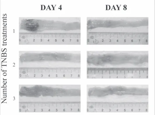
The intestinal inflammation was most pronounced two days after the TNBS administrations, while after 8 days the mucosa seemingly recovered. Therefore, this time point was considered the endpoint of the acute phase of inflammation. Never- theless, after the second and even more after the third TNBS treatment the measure of ulcerousness decreased markedly and the length of the acute phase of inflamma- tion was also reduced. Complete mucosal recovery was noticed already 6 and 4 days after the second and third treatment, respectively (Figure 1). Strictures first appeared 60 days after the TNBS treatment and the frequency of strictures increased until day 120th.
Figure 1 Representative micrographs of the inflamed segment of the colon from TNBS-treated rats after 4 and 8 day induction of colitis. Decrease in the measure of
ulcerousness indicates that mucosal healing speeded up after repeated TNBS treat- ments.
Quantitative and morphometric changes of the myenteric neurons
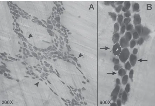
Whole-mounts of colonic segments after HuC/HuD immunohistochemistry were used to evaluate the density (Figure 2. A) and area (Figure 2. B) of myenteric neurons. After the first TNBS treatment the total neuronal number decreased signif- icantly in all three colonic segments only in the acute phase of the inflammation as compared with the control groups. The decreasing was most pronounced in the IS.
Nevertheless, 8 days after the first TNBS treatment no further changes in the neu- ronal number was detected until the end of the investigation (Figure 3. A). Shrink- age of neuronal cell bodies was demonstrated in the acute phase of inflammation induced by the first TNBS treatment. In the chronic phase the restoration of im- paired morphology of myenteric neurons was observed, which was speeded up after repeated treatments. 120 days after the third treatment the soma sizes of myenteric
Figure 2 Representative lightmicrographs of whole-mount preparations of rat colon after HuC/HuD immunohistochemistry. Arrows and arrowheads point stained myenteric neu-
rons. Encircled neurons (asterisk) were used to measure the area of myenteric neurons.
Figure 3 Density (A) and area (B) of HuC/D- immunoreactive myenteric
neurons in the colon of control and TNBS-treated
rats at different time points (4, 8, 90 and 120 days) after single and re- peated treatments. The number of myenteric neu-
rons in the inflamed seg- ment of the colon de- creased significantly al-
ways only in the acute phase of the inflammation independently on the num-
ber of treatments. Shrink- age of neuronal cell bod- ies was demonstrated in the acute phase of inflam- mation induced by the first TNBS treatment. Neverthe-
less, after the second and even more after the third TNBS treatment the soma sizes of myenteric neurons were nearly the same as in
Investigation of the ultrastructure of strictures by transmission electron mi- croscopy
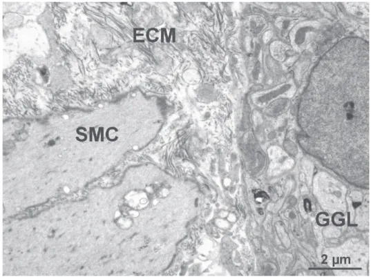
Transmission electron microscopy showed complete reepithelialization in the strictured region. The width of TJs, between adjacent epithelial cells did not change compared to controls. Morphometrical analyzes revealed significant thickening of the LMM and EML. The intercellular spaces with matrix deposition expanded, therefore distance between smooth muscle layers and myenteric ganglia enlarged (Figure 4). Loosed SMCs with morphological sign of apoptosis was frequently seen, while enteric ganglia were seemingly intact.
Figure 4 Representative electron micrograph of the rat colon in a strictured area after 120 days of TNBS treatment. Excess deposition of extracellular matrix (ECM) characterizes the area between smooth muscle layers and myenteric ganglia (GGL).
GGL and smooth muscle cells (SMC) shoved off from each other.
Quantitative changes in the mRNA expression of inflammatory markers
Figure 5 Median values of mRNA expression of HO-1, TGF-beta 2, TGF-beta 3 and MMP9 in the inflamed segment (IS), proximal (PS) and distal (DS) to the in-
flamed segment of the colon in rats with chronic colitis. Hypoxanthine- guaninephosphoribosyltransferase (HPRT) mRNA level was used as an internal
control. Data were expressed as differences from HPRT mRNA level. Data are means ± SEM.
Conclusions
The main focus of the present work was to develop a rat model of chronic in- testinal inflammation which reflects chronic conditions in a patient who is suffering from CD. Here we provide the first experimental data indicating that aligned in- flammatory changes of enteric neurons, SMCs and inflammatory markers deter- mined in the acute phase of the inflammation persist after the resolution of the initi- ating inflammatory events and lasts from day 8th to day 120th. The data presented here suggest that these lasting changes are responsible for stricturing which is the most frequent life-threatening complication in CD patients (Marlow and Blenner- hassett, 2006)3. The repeated administration of TNBS which mimics the remission of inflammation also characteristic in CD patients enhances the frequency of stric- tures indicating that the number of inflammation insults is critical in the pathogene- sis of stricturing. This study demonstrates for the first time that experimentally pro- voked RRI in a rat model of CD develops preconditioning effect by speeding up mucosal healing and restoring impaired morphology of myenteric neurons. De- creased severity of TNBS induced inflammation in RRI might be associated with the persistent upregulation of HO-1 expression throughout the 120 days experi- mental period. Therefore, targeted induction of HO-1 might be of therapeutic inter- est.
References
1Graham MF, Drucker DE, Diegelmann RF, Elson CO. 1987. Collagen synthesis by human intestinal smooth muscle cells in culture. Gastroenterology. 2; 400-405.
2Lourenssen S, Wells RW, Blennerhassett MG. 2005. Differential responses of in- trinsic and extrinsic innervation of smooth muscle cells in rat colitis. Exp Neurol. 2;
497-507.
3Marlow SL, Blennerhassett MG. 2006. Deficient innervation characterizes intesti- nal strictures in a rat model of colitis. Exp Mol Pathol. 80 (1); 54-66.
4McKaig BC, Hughes K, Tighe PJ, Mahida YR. 2002. Differential expression of TGF-beta isoforms by normal and inflammatory bowel disease intestinal myofibro- blasts. Am J Physiol Cell Physiol. 282 (1); 172-182.
5Román V, Bagyánszki M, Krecsmarik M, Horváth A, Resch BÁ, Fekete É. 2004.
Spatial pattern analysis of nitrergic neurons in the developing myenteric plexus of the human fetal intestine. Cytom Part A 57A:108-112.
6Sanovic S, Lamb DP, Blennerhassett MG. 1999. Damage to the enteric nervous system in experimental colitis. Am J Pathol.155 (4); 1051–1057.
„This research was realized in the frames of TÁMOP 4.2.4. A/2-11-1-2012-0001
„National Excellence Program – Elaborating and operating an inland student and researcher personal support system”. The project was subsidized by the European Union and co-financed by the European Social Fund.”