THESES OF DOCTORAL (PhD) DISSERTATION
DR. ROLAND PÓSA
KAPOSVÁR UNIVERSITY FACULTY OF AGRICULTURAL AND
ENVIRONMENTAL SCIENCES
2014
KAPOSVÁR UNIVERSITY
FACULTY OF AGRICULTURAL AND ENVIRONMENTAL SCIENCES
DEPARTMENT OF ANIMAL PHYSIOLOGY AND HYGIENE
The Head of Doctoral (Ph.D.) School:
DR. MELINDA KOVÁCS correspondent member of the HAS
Supervisors:
DR. MELINDA KOVÁCS correspondent member of the HAS
DR. TIBOR MAGYAR DSc, honorary professor
Institute for Veterinary Medical Research Centre for Agricultural Research Hungarian Academy of Sciences
Budapest
COMPUTED TOMOGRAPHY BASED EXAMINATION OF THE COMPLEX RESPIRATORY DISEASES OF
SWINE
Written by:
DR. ROLAND PÓSA
Kaposvár
2014
3
ANTECEDENTS OF THE DISSERTATION, OBJECTIVES
The past few decades have seen a rapid increase of productivity also in the pig production sector, accompanied by the size increase and concentration of pig herds and the spread of intensive management technologies. These changes, however, have brought along a drastic increase in the prevalence of respiratory diseases. The overwhelming majority of porcine respiratory diseases are disease entities developing in the simultaneous presence of multiple pathogens. In the case of these syndromes, the appearance of clinical signs and the magnitude of economic losses are decisively influenced by the predisposing factors (primarily the management, care and feeding conditions).
Today, the multifactorial diseases belonging to this category are referred to in the special literature as porcine respiratory disease complex (PRDC).
As one of the most prevalent respiratory pathogens, M. hyopneumoniae has played a decisive role in the aetiology of respiratory diseases for several decades, usually in association with other pathogenic bacteria (A.
pleuropneuminae, B. bronchiseptica and P. multocida), resulting in a complex respiratory syndrome. The prevalence of other respiratory pathogens varies more widely; however, over the past 20 years PRRS virus, porcine circovirus type 2 and the newly emerging swine influenza virus strains have caused the biggest problems to swine health professionals in the leading pig-producing countries of the world.
Atrophic rhinitis is also a long-known and widely distributed pig disease, in the aetiology of which the interaction between toxigenic strains of two pathogenic bacteria, Bordetella bronchiseptica and Pasteurella multocida, is thought to play the primary role. These two pathogens are frequently isolated from the respiratory tract of pigs and they play a major role in the development of PRDC. Based on the data of the literature, the
4
toxigenic strains of P. multocida are more often found in lesions restricted to the upper airways, whereas in the pathological processes taking place in the lungs mostly P. multocida strains not capable of toxin production are involved.
Microscopic filamentous fungi contaminating feed and food raw materials produce numerous toxins (mycotoxins) which, when entering the soil-plant-animal-human food chain, are a source of major public health risks. In addition, they cause substantial economic losses in the animal production sector. Fumonisins (FB1, FB2, FB3, FB4) constitute a relatively recently identified group of mycotoxins, which was discovered in 1988.
After American authors had first described the syndrome termed porcine pulmonary oedema (PPE), confirmed to be attributable to fumonisin B1 (FB1) toxin, together with the main pathological lesions associated with it (pulmonary oedema, liver and renal degeneration), research studies aimed at getting a closer insight into the pathological processes were started, but to date, few studies have been done to determine the potential predisposing role of FB1 toxin in syndromes induced by the most important porcine respiratory pathogens. The interaction between this mycotoxin and the above-mentioned pathogens was studied in pigs experimentally infected with serotype A of P.
multocida and treated with FB1 toxin (Halloy et al., 2005), as well as in pigs experimentally infected with PRRS virus and treated with FB1 toxin (Ramos et al., 2010).
At the Department of Physiology and Animal Hygiene of Kaposvár University, in co-operation with the Institute of Diagnostic Imaging and Radiation Oncology of the same University, animal experiments aimed at monitoring the pathological processes that take place in the lungs of pigs have been conducted since 1997. During these experiments, the basic
5
techniques of using modern diagnostic imaging procedures (CT, MRI) for the above purpose were developed.
At Kaposvár University, CT studies to monitor the development of pulmonary oedema and pulmonary fibrosis caused by fumonisin B1 were started at the end of the 1990s. The Animal Breeding and Animal Hygiene Research Group of the Hungarian Academy of Sciences and Kaposvár University conducted several experiments to study the changes caused by FB1 toxin in pigs depending on the dose and the exposure time, to determine the still tolerable FB1 limits and the ‘no observed effect level’ (NOAEL) (Zomborszky-Kovács et al., 2000, 2002). Based on these results, the maximum allowable limit of FB1 was determined and declared as 5 mg/kg first in the Hungarian Feed Code (Codex Pabularis Hungaricus) and subsequently in the recommendation of the European Union (Commission Recommendation, 2006).
Co-operation with the Institute for Veterinary Medical Research, Centre for Agricultural Research, Hungarian Academy of Sciences dates back to 2000. In its framework the impact of porcine atrophic rhinitis on production was studied and the pathogenesis of the disease was monitored by CT. A specific pathogen free (SPF) piglet-rearing method has been developed, by which freedom from B. bronchiseptica and P. multocida can be achieved. The method is suitable for performing infection experiments that require animals not infected by the given pathogens (Magyar et al., 2003).
Also to date, few studies have been done to determine the potential predisposing role of FB1 toxin in syndromes induced by the most important porcine respiratory pathogens and there is only scant literature on lung examinations using radiography and modern imaging modalities with the objective to monitor swine respiratory diseases. The scientific work forming
6
the basis of the present doctoral thesis was planned as a continuation of the earlier successful co-operation mentioned above.
Our principal objective was to obtain, by the use of CT as a modern diagnostic imaging modality, new data on the pathogenesis, pathological features, gross pathological lesions and histopathological changes of some common swine respiratory diseases of infectious origin, as well as on the predisposing and presumably pathogenesis-modifying role played by the FB1
mycotoxin in these processes. We selected three respiratory pathogens for the model experiments: B. bronchiseptica, a toxigenic strain of P. multocida and M. hyopneumoniae.
An additional objective was to develop further the use of the diagnostic imaging modality in the study of swine respiratory diseases, including the elaboration of a numerical model that could facilitate the accurate evaluation of pathological processes taking place in the lungs of pigs.
METHODOLOGICAL SUMMARY
The experimental series consisted of three separate experiments.
In Experiment 1 a single-pathogen infection based on B.
bronchiseptica was used, without FB1 toxin treatment, in order to demonstrate and monitor the development of pneumonia induced by the pathogen in young piglets. The methods of CT studies were also elaborated during this experiment.
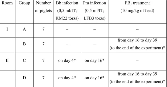
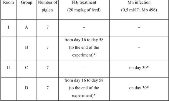
In Experiment 2 we demonstrated and studied the lung lesions induced in young pigs by the interaction of two pathogens (B. bronchiseptica and P. multocida) and the mycotoxin FB1, while in Experiment 3 the lesions due to the interaction of M. hyopneumoniae and the FB1 mycotoxin were studied.
7
The experimental design used in the three experiments is illustrated in Tables 1–3.
Table 1: Arrangement of treatment groups in Experiment 1 Room Group Number of
piglets
Bb infection (0.5 ml/IT; KM22)
I A 10 –
II B 20 on day 4*
Table 2: Arrangement of treatment groups in Experiments 2
Room Group Number of piglets
Bb infection (0,5 ml/IT;
KM22 törzs)
Pm infection (0,5 ml/IT;
LFB3 törzs)
FB1 treatment (10 mg/kg of feed)
I A 7 – – –
B 7 – – from day 16 to day 39
(to the end of the experiment)*
II C 7 on day 4* on day 16* –
D 7 on day 4* on day 16* from day 16 to day 39 (to the end of the experiment)*
8
Table 3: Arrangement of treatment groups in Experiment 3
Room Group Number of piglets
FB1 treatment (20 mg/kg of feed)
Mh infection (0,5 ml/IT; Mp 496)
I A 7 – –
B 7
from day 16 to day 58 (to the end of the
experiment)*
–
II C 7 – on day 30*
D 7
from day 16 to day 58 (to the end of the
experiment)*
on day 30*
Bb = Bordetella bronchiseptica Pm = Pasteurella multocida serotype D Mh = Mycoplasma hyopneumoniae
IT = intratracheal infection through an endotracheal tube
* the piglets arrived on day 0, the indicated numbers represent the days of experiment
Experiments 1 and 2 lasted 39 days whereas Experiment 3 lasted 58 days, which duration was chosen on the basis of data available in the literature on the selected pathogens.
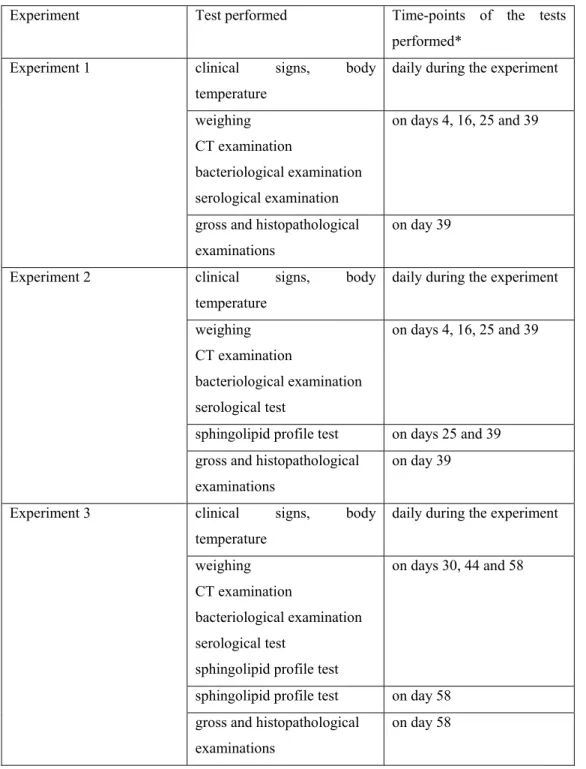
Of the modern imaging modalities, we used CT for monitoring the changes induced in the lungs. This was complemented by bacteriological and serological tests as well as sphingolipid profile tests of the blood during the experiment and by gross and histopathological examinations carried out at the end of the experiment (Table 4).
9
Table 4: Examinations/tests performed and the time of their performance in the three experiments
Experiment Test performed Time-points of the tests performed*
Experiment 1 clinical signs, body temperature
daily during the experiment
weighing CT examination
bacteriological examination serological examination
on days 4, 16, 25 and 39
gross and histopathological examinations
on day 39
Experiment 2 clinical signs, body temperature
daily during the experiment
weighing CT examination
bacteriological examination serological test
on days 4, 16, 25 and 39
sphingolipid profile test on days 25 and 39 gross and histopathological
examinations
on day 39
Experiment 3 clinical signs, body temperature
daily during the experiment
weighing CT examination
bacteriological examination serological test
sphingolipid profile test
on days 30, 44 and 58
sphingolipid profile test on day 58 gross and histopathological
examinations
on day 58
*the piglets arrived on day 0, the indicated numbers represent the days of experiment
10 RESULTS
During the experiment, we did not find significant differences among the treatment groups in body weight gain.
Experiment 1
During the experiment, there were no significant differences between the treatment groups in terms of body weight gain. On day 4 after infection, sneezing, stertorous breathing and mild coughing accompanied by mild serous nasal discharge could be observed in Group B. CT scans taken on day 16 after infection confirmed colonisation of the lungs of young piglets by B.
bronchiseptica, as pathological lesions were found in 95% of the infected animals. The gross pathological findings were consistent with the changes seen on the CT scans and confirmed their localisation. Lesions were primarily seen in the cranial and middle lobes as well as in the cranial third of the caudal lobe. The affected lungs exhibited acute catarrhal inflammation with areas showing signs of chronicity and mottled appearance due to haemorrhages, with pleuritis occurring as a secondary complication in some animals.
Experiment 2
During the experiment, a total of three piglets died, none of them due to causes associated with their rearing. One piglet of Group C died one day after infection with P. multocida. Two piglets of Group D died 8 and 18 days, respectively, after P. multocida infection. During postmortem examination, bronchopneumonia of varying severity, acute catarrhal
11
inflammation with areas showing signs of chronicity and mottled appearance due to haemorrhages, with pleuritis as a secondary complication, were found in the 3 pigs that died and in 5 other animals (2 pigs of Group C and 3 pigs of Group D).
Analysis of the CT scans demonstrated lung lesions in 3 pigs of Group C and 5 pigs of Group D on day 16 of the experiment (at the second CT examination). Lesions indicative of pneumonia were much more marked on day 25 of the experiment (at the time of the third CT examination), 9 days after P. multocida infection and the start of FB1 toxin feeding. After P.
multocida infection, lesions were found only in one piglet each of Groups C and D. Two piglets (one piglet each of Groups C and D) showed mild changes after B. bronchiseptica infection; however, these changes were no longer demonstrable at the end of the experiment either on the CT scans or during necropsy. The lesions developing after B. bronchiseptica infection were of diffuse character, extending to entire lobes or lobules, whereas those induced by P. multocida infection were smaller, well demarcated and of focal nature.
Experiment 3
During the experiment, we did not find significant differences among the treatment groups in body weight gain. After infection (from postinfection day 31) elevated body temperature (39.5–40.8 °C) occurred in Groups C and D, and from day 37 clinical signs (coughing, sneezing, hoarse voice, dyspnoea) could be observed. During the experiment, a single death occurred in the infected and toxin-treated group. On the CT scans taken 14 days after infection, well-visible lung changes indicative of inflammation were observed in all piglets of the two infected groups. These lesions initially
12
occurred around the smaller airways, but subsequently they increased in size and at the end of the experiment (on day 58) they were found to be extensive in several animals. The most severely affected parts of the lungs were the cranial parts of the lung lobes. At necropsy, lung areas showing acute and subacute catarrhal inflammation were found in the affected animals with an extension identical with that seen on the CT scans.
Mycoplasma hyopneumoniae infection was able to induce lung changes in the growing piglets even in itself (as monoinfection). However, the ingestion of FB1 toxin aggravated these lesions. Both the mortality rate and the severity and extent of the lung lesions observed were the highest in Group D. From the results, it can be concluded that M. hyopneumoniae infection combined with the consumption of FB1 toxin raises the probability of pneumonia developing in the pigs and increases the severity and extent of lung lesions.
The CT scans taken of lungs kept at a specific pressure during the time of CT examination proved to be suitable for detecting the lung lesions. Using the measurement method elaborated by us, we could demonstrate a significant difference between the infected and the non-infected pigs in the mean density values of the lungs.
The feeding of a diet containing 20 ppm FB1 toxin induced only histopathological changes and mild macroscopic lesions such as oedema in the lungs of pigs in the treated groups. By analysing the CT scans, oedema of such minor extent could not be expressly detected.
13 CONCLUSIONS
By the piglet-rearing method applied in the study we could prevent the infection of experimental animals by respiratory pathogens, and thus we could study the pathogenesis of lung lesions produced by respiratory pathogens introduced into the respiratory tract by experimental infection.
We successfully demonstrated that both B. bronchiseptica and M.
hyopneumoniae could cause pneumonia in young and growing piglets also when used alone (in the form of monoinfection).
In our experiments, we did not find significant differences (p<0.05) among the groups in average body weight gain (Experiments 1–3), although in the groups subjected to experimental infection combined with FB1 toxin treatment the growth of pigs was inferior to that seen in the other three groups (Experiments 2–3).
In addition, it can be stated that in pigs subjected to dual infection by B. bronchiseptica plus P. multocida or to infection by M. hyopneumoniae, the consumption of FB1 toxin in a concentration of 10 mg/kg or 20 mg/kg of feed aggravated the course of bacterial diseases, as the clinical signs were the most pronounced in the experimentally infected groups fed FB1 toxin, in these groups mortality also occurred, and in these groups increased the probability of development of pneumonia, as the number of pigs with lung lesions was higher.
We have elaborated a possible application of computed tomography for studying the time-course of development and the pathogenesis of pneumonia. This method may provide useful information for the study of other respiratory diseases as well. A great advantage of computed tomography is that it enables us to monitor the localisation, extent and character of the developing pathological lung lesions in live animals,
14
simultaneously with the appearance of the disease. By the evaluation of CT scans taken at different time-points we can obtain new scientific insights into the pathogenesis of different respiratory diseases or, in the framework of applied experiments, we can compare the efficacy of therapeutic interventions on the disease entities concerned.
By applying a new model for the evaluation of lesions developing in the lungs, in Experiment 3 we expressed the mean density values in numerical terms and demonstrated a significant difference between the infected and the non-infected animals.
15 NEW SCIENTIFIC RESULTS
1. CT could be applied and a useful tool for studying the pathological conditions in the lower respiratory tract of swine.
2. Elaboration of a new method for the examination of the lung of swine with CT as well as for the evaluation of the images that made the quantitative analysis of the pathological processes possible.
3. B. bronchiseptica mono-infection was able to produce lung lesions in young pigs.
4. In case of dual infection by B. bronchiseptica and P. multocida, dietary exposure to the mycotoxin FB1 (10 mg/kg of feed) above the advised level of 5 mg/kg of feed raised the risk of pneumonia and increased the extent and severity of the pathological changes produced.
5. In case of infection by M. hyopneumoniae, dietary exposure to the mycotoxin FB1 (20 mg/kg of feed) above the advised level of 5 mg/kg of feed raised the risk of pneumonia and increased the extent and severity of the pathological changes produced.
16
SCIENTIFIC PAPERS AND LECTURES ON THE SUBJECT OF THE DISSERTATION
Peer-reviewed papers published in foreign scientific journals
Pósa R., Kovács M., Szabó-Fodor J., Mondok J., Bogner P., Repa I., Magyar T. Effect of Mycoplasma hyopneumoniae and fumonisin B1 toxin on the lung in pigs. Italian Journal of Animal Science. 2009. 8(3):172-174.
Pósa R., Kovács M., Donkó T., Repa I., Magyar T. Non invasive (CT) investigation of the lung in Bordetella bronchiseptica infected pigs.
Agriculturae Conspectus Scientificus. 2011. 76(4): 357-359.
Pósa R., Donkó T., Bogner P., Kovács M., Repa I., Magyar T. Interaction of Bordetella bronchiseptica, Pasteurella multocida and fumonisin B1 in the porcine respiratory tract followed up by computed tomography. Canadian Journal of Veterinary Research. 2011. 75(3):176–182.
Pósa R., Magyar T., Stoev S.D., Glávits R., Donkó T., Repa I., Kovács M.
Use of computed tomography and histopathologic review for lung lesions produced by the interaction between Mycoplasma hyopneumoniae and fumonisin mycotoxins in pigs. Veterinary Pathology. 2013. 50:(6):971-979.
17 Abstracts
Pósa R., Donkó T., Bogner P., Kovács M., Repa I., Magyar T. Synergy between Bordetella bronchiseptica, Pasteurella multocida and fumonisin B1
toxin in the porcine respiratory tract. Proceedings of IPVS Congress, Durban, 2008. pp.:195.
Pósa R., Kovács M., Szabó-Fodor J., Mondok J., Bogner P., Repa I., Magyar T. Interaction between Mycoplasma hyopneumoniae and fumonisin B1 toxin in the porcine respiratory tract. In: D’Allaire S, Friendship R (ed) Proceedings 21th International Pig Veterinary Society Congress (IPVSC), Vancouver, Canada. 2010. pp.:654:348.
Oral presentations
Pósa R., Donkó T., Bogner P., Kovács M., Repa I., Magyar T. A fumonizin B1 hatásának vizsgálata Bordetella bronchiseptica és Pasteurella multocida kórokozókkal fertőzött sertésekben. MTA Állatorvostudományok Bizottsága, Akadémiai beszámoló. Budapest, Hungary, 28 January 2009.
Pósa R., Kovács M., Donkó T., Szabó-Fodor J., Mondok J., Bogner P., Repa I., Magyar T. A Mycoplasma hyopneumoniae és a fumonizin B1 mikotoxin kölcsönhatása sertések tüdejében. MTA Állatorvos-tudományi Bizottsága, Akadémiai beszámoló. Budapest, Hungary, 27 January 2010.