International Journal of
Molecular Sciences
Article
Novel High Affinity Sigma-1 Receptor Ligands from Minimal Ensemble Docking-Based Virtual Screening
Szabolcs Dvorácskó1, LászlóLázár2 , Ferenc Fülöp2,† , Márta Palkó2 , Zita Zalán2, Botond Penke3, Lívia Fülöp3,* , Csaba Tömböly1 and Ferenc Bogár3,4,*
Citation: Dvorácskó, S.; Lázár, L.;
Fülöp, F.; Palkó, M.; Zalán, Z.; Penke, B.; Fülöp, L.; Tömböly, C.; Bogár, F.
Novel High Affinity Sigma-1 Receptor Ligands from Minimal Ensemble Docking-Based Virtual Screening.Int. J. Mol. Sci.2021,22, 8112. https://doi.org/10.3390/
ijms22158112
Academic Editors: Carmen Abate and Tangui Maurice
Received: 4 July 2021 Accepted: 26 July 2021 Published: 29 July 2021
Publisher’s Note:MDPI stays neutral with regard to jurisdictional claims in published maps and institutional affil- iations.
Copyright: © 2021 by the authors.
Licensee MDPI, Basel, Switzerland.
This article is an open access article distributed under the terms and conditions of the Creative Commons Attribution (CC BY) license (https://
creativecommons.org/licenses/by/
4.0/).
1 Biological Research Centre, Institute of Biochemistry, Eötvös Loránd Research Network (ELKH), H-6726 Szeged, Hungary; dvoracsko.szabolcs@brc.hu (S.D.); tomboly.csaba@brc.hu (C.T.)
2 Institute of Pharmaceutical Chemistry, University of Szeged, H-6720 Szeged, Hungary;
lazar.laszlo@szte.hu (L.L.); fulop@pharm.u-szeged.hu (F.F.); palko.marta@szte.hu (M.P.);
lazarne.zalan.zita@szte.hu (Z.Z.)
3 Department of Medical Chemistry, University of Szeged, H-6720 Szeged, Hungary;
penke.botond@med.u-szeged.hu
4 MTA-SZTE Biomimetic Systems Research Group, Eötvös Loránd Research Network (ELKH), H-6720 Szeged, Hungary
* Correspondence: fulop.livia@med.u-szeged.hu (L.F.); bogar@sol.cc.u-szeged.hu (F.B.)
† Prof. Ferenc Fülöp passed away in July 2021.
Abstract:Sigma-1 receptor (S1R) is an intracellular, multi-functional, ligand operated protein that also acts as a chaperone. It is considered as a pluripotent drug target in several pathologies. The publication of agonist and antagonist bound receptor structures has paved the way for receptor-based in silico drug design. However, recent studies on this subject payed no attention to the structural differences of agonist and antagonist binding. In this work, we have developed a new ensemble docking-based virtual screening protocol utilizing both agonist and antagonist bound S1R structures.
This protocol was used to screen our in-house compound library. The S1R binding affinities of the 40 highest ranked compounds were measured in competitive radioligand binding assays and the sigma-2 receptor (S2R) affinities of the best S1R binders were also determined. This way three novel high affinity S1R ligands were identified and one of them exhibited a notable S1R/S2R selectivity.
Keywords:novel sigma-1 receptor ligands; virtual screening protocol; ensemble docking; radioligand binding assay
1. Introduction
Sigma receptor was first identified in 1976 by Martin et al. as an opioid receptor subtype [1]. However it turned out in the early eighties that the pharmacological character of sigma receptor diverges from the other opioid receptors [2,3]. This and the subsequent scientific efforts finally led to the identification of the sigma non-opioid intracellular receptor family [4] with its two members, sigma-1 (S1R) and sigma-2 (S2R) receptors [5].
S1Rs are broadly spread in the whole organism (central nervous system (CNS), heart, liver, kidney, lung, muscles [6]). It localizes mainly in the mitochondria-associated endoplas- mic reticulum (ER) membrane (MAM). The receptor can also be dynamically translocated inside the cells. It is an intracellular, multi-functional, ligand operated protein that also acts as a chaperone [6].
S1R has a unique amino acid sequence with no mammalian homologues, the canonical isoform contains 223 amino acid residues [7]. Its ligand bound form showed a homotrimeric structure with only one transmembrane helix for each monomeric subunit [8].
The other member of sigma receptor family, S2R is primarily implicated in cancer. It is used as a biomarker for proliferation and its agonists are potent anticancer agents [9].
Sequence of S2R has only been identified recently [10], but its structure is not known yet. In spite of their sequential and structural dissimilarities, S1 and S2 receptors often
Int. J. Mol. Sci.2021,22, 8112. https://doi.org/10.3390/ijms22158112 https://www.mdpi.com/journal/ijms
Int. J. Mol. Sci.2021,22, 8112 2 of 17
have common ligands with similar receptor affinities. This can be demonstrated using the S2RSLDB (Sigma 2 Receptor Selective Ligand Database [11]). More than half (392) of the 651 S2R selective ligands in the database have a binding affinity that is only less than 10 times larger to S2R than to S1R. Therefore, finding highly selective ligands of either sigma receptor has remained a challenging task [12,13].
Although S1R might participate in a variety of specific physiological functions, its genuine role has been yet poorly understood. Interestingly, the S1R knock-out mice are viable and fertile and do not show any apparent phenotype changes compared to wild-type mice [14]. The exact physiological role of S1R is difficult to determine as many S1R ligands also bind to other receptor targets (e.g., kappa-receptor, NMDA receptor). S1R interacts with a large range of client proteins [15,16], e.g., with several voltage- and ligand-gated ion channels, modulating their activity. The main functions of S1R are probably modulation of Ca2+release, modulation of cardiac myocyte contractility and inhibition of voltage gated K+channels [17]. The receptor apparently co-localizes with the IP3 receptors on the ER membrane and is responsible for the calcium exchange between ER and mitochondria [18].
S1R was found together with voltage gated K+channels in the membranes and thus it was considered to be an auxiliary subunit of the protein complex [19]. S1R antagonists show GTP-sensitive high affinity binding indicating an existing link between S1R and G-proteins [20]. S1 receptors are considered to be linked to a wide variety of signal transduction pathways. S1R may be involved in the prevention of ER-stress [21]. S1R possesses profound effect on neuronal excitatibility and neurotransmission [22].
The molecular functions of S1R and the role in cellular stress signaling have been recently reviewed in details [16,23]. The multiplicity of intracellular partners and signaliza- tion pathways may explain the multiplicity of actions of S1R in different types of cells as well as the involvement of the receptor in a series of physiopathological processes [21]. S1R has been considered as a ‘pluripotent modulator of the living system’ [24], an intracellular signal transduction amplifier [15].
The biochemical pharmacology and pharmacological profile have been reviewed recently in detail [16,23,25,26]. Although S1Rs are spread both in the CNS and the periphery, CNS is the primary site of S1R activity and effects [6]. S1Rs are expressed abundantly in neurons, oligodendrocytes, and ependymocytes. S1R accumulates to nuclear inclusions in neurons in neurodegenerative diseases [27]. S1R is the most important factor for fine- tuning of the cellular calcium homeostasis and thus it is the most promising therapeutic target [21]. Experimental results demonstrated that neuronal S1Rs show protective roles in neurodegeneration and are involved in neurorestauration [28–31]. S1R activity is necessary for physiological brain plasticity [32]. S1R ligands (e.g., dimethyl tryptamine, DMT) can induce psychotomimetic effects and antidepressant activity. Drug screening experiments demonstrated that S1R may be a target for the therapy of VWM leukodystrophy [33].
S1R activation can protect neurons against ER-stress mediated apoptosis with cerebral ischemia/reperfusion injury [34]. Upregulation of S1Rs ameliorates cell death signaling and execute protective function in ER-stress [35]. As neuroinflammation is a key abnormality in the progression of Alzheimer’s disease (AD), the anti-inflammatory activity of S1R might control the disease-related inflammatory process. Activation of S1R promotes the repair of microglia and restores their physiological functions [36,37].
Cardiac S1Rs regulate response to ER stress and modulate Ca signaling in cardiomy- ocytes and thus are potential novel targets for special treatment of cardiovascular dis- eases [38].
S1R ligands are historically classified as agonists, antagonists, and allosteric modula- tors, although this classification differs from the canonical pharmacological nomenclature.
The receptor has a unique property: it does not possess identified intrinsic activity, thus the concept of S1R ‘agonism’ and ‘antagonism’ has no consensual definition [39]. Therefore, the pharmacological character of S1R ligands is determined not only by their inherent properties, but also by the choice and setup of the in vitro or in vivo experiment applied.
According to the conventionally accepted classification, ligands inducing characteristic
Int. J. Mol. Sci.2021,22, 8112 3 of 17
pharmacological responses typical of known agonists or S1R overexpression are character- ized as agonists. The identification of the characteristic responses goes back to SKF-10047, the first identified S1R ligand of this kind [40]. S1R antagonists are ligands without inherent neurophysiological effects, but inhibit the effects induced by agonists, or ligands causing similar consequences that appear in S1R knockout animals. Agreeing with Oyer et al. [39], instead of agonist/antagonist classification the term positive and negative ‘modulator’
might be more accurate for defining substances with affinity to S1R.
S1R ligands regulate the self-association of the receptor molecules: agonists preferring the lower level of association; antagonists shift the equilibrium towards larger aggre- gates [16,26]. Agonists act generally neuroprotective, pro-survival and anti-apoptotic via different signaling pathways [15].
The amino acid sequence of S1R was identified in 1996 [7]. It showed no significant ho- mology with any known mammalian proteins. Therefore, besides the traditional methods, dominantly the ligand based computational design tools could be used for the identification of novel S1R ligands. Design of small molecular S1R ligands was performed first by using the Glennon-model for the receptor binding site and pharmacophore identification [41,42].
Several other pharmacophore models were built later that are reviewed in [43].
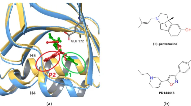
The first X-ray structures of S1R (Protein Data Bank (PDB) [44] IDs: 5HK1 (Figure1a), 5HK2) were published by Schmidt el al. in 2016 [4]. The experiments uncovered a trimeric structure of the ligand-bound receptor, each monomeric unit being anchored with a single helix in the membrane. The ligands in these complexes were PD144418 (Figure1b), a high-affinity selective S1R antagonist and 4IBP with a not well-defined pharmacological character. The binding pocket of the receptor is situated in a beta barrel-like unit which shows high structural similarity to oligomeric enzymes from the cupin family [4].
The general scheme of ligand binding in accordance with the Glennon-model involves a positively charged nitrogen, which forms an electrostatic interaction with residue E172.
This charged site sits between two dominantly hydrophobic regions: a longer one that occupies the region of theβ-barrel that is proximal to the membrane and a shorter one that occupies space near the other end of theβ-barrel.
Int. J. Mol. Sci. 2021, 22, x FOR PEER REVIEW 3 of 18
properties, but also by the choice and setup of the in vitro or in vivo experiment applied.
According to the conventionally accepted classification, ligands inducing characteristic pharmacological responses typical of known agonists or S1R overexpression are charac- terized as agonists. The identification of the characteristic responses goes back to SKF- 10047, the first identified S1R ligand of this kind [40]. S1R antagonists are ligands without inherent neurophysiological effects, but inhibit the effects induced by agonists, or ligands causing similar consequences that appear in S1R knockout animals. Agreeing with Oyer et al. [39], instead of agonist/antagonist classification the term positive and negative ‘mod- ulator’ might be more accurate for defining substances with affinity to S1R.
S1R ligands regulate the self-association of the receptor molecules: agonists prefer- ring the lower level of association; antagonists shift the equilibrium towards larger aggre- gates [16,26]. Agonists act generally neuroprotective, pro-survival and anti-apoptotic via different signaling pathways [15].
The amino acid sequence of S1R was identified in 1996 [7]. It showed no significant homology with any known mammalian proteins. Therefore, besides the traditional meth- ods, dominantly the ligand based computational design tools could be used for the iden- tification of novel S1R ligands. Design of small molecular S1R ligands was performed first by using the Glennon-model for the receptor binding site and pharmacophore identifica- tion [41,42]. Several other pharmacophore models were built later that are reviewed in [43].
The first X-ray structures of S1R (Protein Data Bank (PDB) [44] IDs: 5HK1 (Figure 1a), 5HK2) were published by Schmidt el al. in 2016 [4]. The experiments uncovered a trimeric structure of the ligand-bound receptor, each monomeric unit being anchored with a single helix in the membrane. The ligands in these complexes were PD144418 (Figure 1b), a high- affinity selective S1R antagonist and 4IBP with a not well-defined pharmacological char- acter. The binding pocket of the receptor is situated in a beta barrel-like unit which shows high structural similarity to oligomeric enzymes from the cupin family [4].
The general scheme of ligand binding in accordance with the Glennon-model in- volves a positively charged nitrogen, which forms an electrostatic interaction with residue E172. This charged site sits between two dominantly hydrophobic regions: a longer one
(a) (b)
Figure 1. Ligand binding of S1R: (a) Superposition of agonist- ((+)-pentazocine, red) and antagonist-(PD144418, green) bound S1R monomers (PDB IDs: 6DK1 chain C (blue) and 5HK1 chain A (gold)). Backbone atoms of residues 170–176 and 197–217 were used if fitting. PD144418 and (+)-pentazocine occupy two different subpockets, P1 and P2, between the C- terminal helixes H4 and H5. (b) Chemical structure of ligands (+)-pentazocine and PD144418.
Figure 1.Ligand binding of S1R: (a) Superposition of agonist- ((+)-pentazocine, red) and antagonist-(PD144418, green) bound S1R monomers (PDB IDs: 6DK1 chain C (blue) and 5HK1 chain A (gold)). Backbone atoms of residues 170–176 and 197–217 were used if fitting. PD144418 and (+)-pentazocine occupy two different subpockets, P1 and P2, between the C-terminal helixes H4 and H5. (b) Chemical structure of ligands (+)-pentazocine and PD144418.
Int. J. Mol. Sci.2021,22, 8112 4 of 17
In 2018, Schmidt et al. published the X-ray structures of an agonist ((+)-pentazocine, PDB: 6DK1, see Figure1) and two antagonists (haloperidol and NE-100, PDB: 6DK0 and 6DJZ, respectively) bound to S1R [45]. Interestingly, the trimeric structure was preserved even in the case of agonist binding. These structures showed high similarity to the previous ones but also revealed slight differences between agonist and antagonist binding. The longer hydrophobic regions of the antagonists (PD144418, haloperidol, and NE-100) and agonist ((+)-pentazocine) occupy two different subpockets, P1 and P2, between the C- terminal helixes H4 and H5 siting on the ER membrane surface (see Figure1a). The different binding poses also induce structural differences primarily in these helices. Unfortunately, only a single agonist bound X-ray structure ((+)-pentazocine) is available to date. Schmidt et al. [45] presumed that structurally similar agonists, such as (+)-SKF-10,047 adopt a similar binding pose to (+)-pentazocine. In addition, based on their docking studies they suggested that the agonist PRE-084 also adopt a similar pose occupying subpocket P2.
However, there is no experimental evidence that connects generally the occupation either of the subpockets and the pharmacological character of the ligand. The binding mode is not known for such structurally divergent S1R agonists like fluvoxamine, cutamesine, or donepezil. The existence of ligands occupying both P1 and P2 subpockets also cannot be excluded.
These structural data made possible the rational design of novel S1R ligands. Pasqual et al. developed a new pharmacophore model based on the PDB structure 5HK1 [22].
Recently, a new docking-based protocol was proposed to predict the affinity of small compounds against S1R [46], which used the 5HK2 structure. Greenfield et al. published an 5HK1-based virtual screening (VS) protocol for the development of novel positive modulators of S1R with neuroprotective effects [47]. These studies were based on X-ray structures with ligands of antagonist-like binding and therefore the ligands that resulted from their VS probably also prefer the same binding mode.
Our aim was to develop a new VS protocol without this bias; using both agonist- (PDB:
6DK1) and antagonist-bound (PDB: 5HK1) X-ray structures in an ensemble docking-based VS protocol. Ensemble docking procedure was introduced by Carlson et al. [48] in 1999 and intended to include the dynamic fluctuations of a protein in computer-aided drug design (for a recent review, see [49]). In this method structures usually collected from molecular dynamics simulations form an ‘ensemble’ of receptor conformations that is used in ligand docking and can improve the performance of virtual screening (see e.g., [50]).
Here we use a different method, which selects an optimized minimal ensemble (with only two receptor structures), one from the agonist and another from the antagonist bound X-ray structures. This approach is closely related to the “slow heuristic” knowledge- based ensemble optimization procedure proposed by Swift et al. [51]. For the validation of this protocol, we used high-affinity ligands collected from the BindingDB [52] and a corresponding decoy set generated from the DUD-E [53] database. We have screened our in-house compound library of ~4000 substances with this new method. The S1R binding affinity of the top-ranked ~1% of the racemic compounds was measured with competitive radioligand binding assay. Finally, the binding affinity and the S1R/S2R selectivity of the enantiomers of the best-ranked racemic compounds were also quantified. We have identified three high-affinity compounds that are putative neuroprotective drug candidates for treating AD and other neurodegenerative diseases.
2. Results and Discussion
2.1. Receptor Model Selection and Validation
From the available ligand-bound S1R structures, we selected two, 5HK1 and 6DK1 [8,45].
In the former complex, the antagonist PD144418 and in the latter the agonist (+)-pentazocine are bound to S1R. The selection of 5HK1 is based on two recent virtual screening studies mentioned earlier [46,47] where this structure performed satisfactorily. The selection of 6DK1 has no alternatives, as this is the only structure where the hydrophobic pocket, P2 is occupied. Both structures are trimeric, therefore we evaluated the performance of each
Int. J. Mol. Sci.2021,22, 8112 5 of 17
monomers applying the Glide XP docking protocol of the Schrödinger program suite [54].
For this purpose, we used an active set of twenty diverse compounds selected from 190 S1R ligands with subnanomolar affinity collected from the BINDINGDB database [52]. The corresponding decoy set of 1000 singly charged compounds were generated at the on-line surface of the DUD-E database [53].
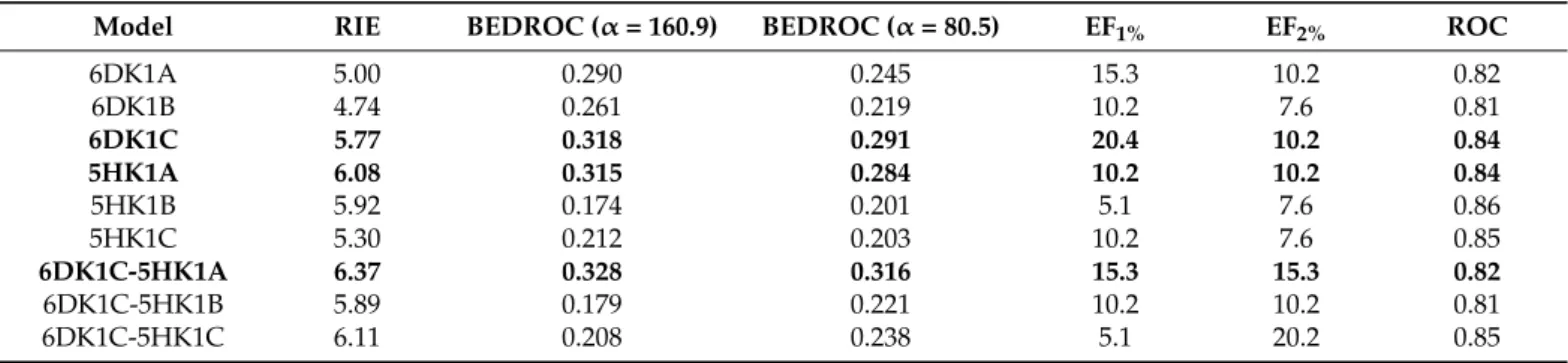
Unified active and decoy sets were docked to each of the six S1R monomers, and the enrichment of the actives were characterized with three measures suitable for the evaluation of the early enrichment (RIE, BEDROC, enrichment factor (EF) in the top 1% and 2%). The chain A of the 5HK1 structure (5HK1A) outperforms the other two chains in the trimer with a RIE value of 6.08 and a BEDROC(α= 160.9) of 0.315 (see Table1). In the case of 6DK1, the best values belong to chain C (6DK1C) being 5.77 and 0.318, respectively. The enrichment factor calculated for the top 1% (EF1%) shows the same tendency for 6DK1C but cannot distinguish the best performer for the three chains of 5HK1A. Following the slow heuristic method of Swift et al. [51] we selected the chain 6DK1C as the first member of the ensemble and calculated the measures for the three pairs formed with the chains of the 5HK1A structure.
Table 1.Calculated measures of the VS efficiency using a protocol based on single chains from structures 5HK1 or 6DK1 and ensemble docking to the chain pairs 6DK1C-5HK1A, 6DK1C-5HK1B, and 6DK1C-5HK1C.
Model RIE BEDROC (α= 160.9) BEDROC (α= 80.5) EF1% EF2% ROC
6DK1A 5.00 0.290 0.245 15.3 10.2 0.82
6DK1B 4.74 0.261 0.219 10.2 7.6 0.81
6DK1C 5.77 0.318 0.291 20.4 10.2 0.84
5HK1A 6.08 0.315 0.284 10.2 10.2 0.84
5HK1B 5.92 0.174 0.201 5.1 7.6 0.86
5HK1C 5.30 0.212 0.203 10.2 7.6 0.85
6DK1C-5HK1A 6.37 0.328 0.316 15.3 15.3 0.82
6DK1C-5HK1B 5.89 0.179 0.221 10.2 10.2 0.81
6DK1C-5HK1C 6.11 0.208 0.238 5.1 20.2 0.85
The pair 6DK1C-5HK1A showed the best early enrichment (BEDROC(α= 160.9)
= 0.328) although its EF1%is somewhat worse than that obtained for the 6DK1C alone (see Table1). These structures were added to our minimal ensemble in order to simultaneously increase the early enrichment of our VS and extend the chemical space of the compounds resulted from it. The ensemble ranking provided better early enhancement measures, but the global performance of ensemble docking became slightly weaker than the contributing models alone, as it is shown by the ROC curves in Figure2a and ROC values in Table1.
The calculated measures could be successfully used to select the best receptor com- binations but they provide no information on the target preference of the compounds of the test set. To demonstrate the importance of involving both receptor models instead of either of them, we identified the target for each of the best poses obtained from ensemble docking. For 9 of the 20 active and 594 of 1000 decoy compounds were hosted by the 6DK1C and the remaining ones by the 5HK1A S1R structure. These numbers indicate a well-balanced sharing between the two target-structures involved in our minimal ensemble docking setup. In addition, two of the active (A9 and A14) and more than 100 of the decoy compounds from those having their highest ranked pose in 6DK1C entered the hydropho- bic pocket, P2. However, none of the actives and only around 30 of the decoys occupied partially the pocket P2 from the compounds preferring 5HK1. This shows that agonist-like binding poses can be accommodated almost equally in both receptor models; however, poses similar to that of (+)-pentazocine appear more frequently in the case of 6DK1C.
Int. J. Mol. Sci.2021,22, 8112 6 of 17
Int. J. Mol. Sci. 2021, 22, x FOR PEER REVIEW 6 of 18
decoy compounds from those having their highest ranked pose in 6DK1C entered the hy- drophobic pocket, P2. However, none of the actives and only around 30 of the decoys occupied partially the pocket P2 from the compounds preferring 5HK1. This shows that agonist-like binding poses can be accommodated almost equally in both receptor models;
however, poses similar to that of (+)-pentazocine appear more frequently in the case of 6DK1C.
(a) (b)
Figure 2. (a) Global performance of ensemble docking characterized by the ROC-curve for 5HK1A (black), 6DK1C (red) and ensemble (blue) models compared to random selection (green). (b) Comparison of top ranked poses of (+)-pentazocine and PD144418 of ensemble docking (green) to their X-ray structure (gray).
After validating our minimal ensemble screening protocol, we also measured the ac- curacy with which it reproduces the X-ray pose of the original ligands. Both (+)-pentazo- cine as well as PD144418 were docked with our method. The top ranked poses of both ligands were hosted by their corresponding X-ray protein structure. The RMSD values of the heavy atoms of these ligands in their X-ray and docked poses (Figure 2b) are 0.151 Å for PD144418 and 0.448 Å for (+)-pentazocine.
2.2. Setup and Validation of In Vitro Competitive Binding Assays
To measure the binding affinities of selected ligands to S1R and S2R we performed competition binding experiments in guinea pig and rat liver membrane homogenates.
Guinea pig liver tissue preparation abundantly contains sigma-1 receptor as compared to other tissues; therefore, it is an appropriate model to investigate the sigma-1 receptor binding properties of synthetic compounds [55–60]. The expression level of sigma-2 re- ceptors is higher in rat liver than in guinea pig liver or rat brain [61], therefore, rat liver membrane homogenate was established as the tissue model in sigma-2 receptor binding assays [5,60–63].
The selective S1R agonist [3H]-(+)-pentazocine possessed a saturable, high affinity binding to a single class of sites with an equilibrium dissociation rate constant (Kd) of 1.8 nM and the maximal density of binding sites (Bmax) of 1072 fmol/mg protein in guinea pig liver membrane preparations at 37 °C. We also determined the Kd of the non-selective sigma receptor ligand, [3H]-DTG in rat liver membranes in the presence of (+)-pentazocine (100 nM) to mask S1R sites. The Kd value was found to be 47 nM (see Figure S1 in Supple- mentary Information).
Figure 2.(a) Global performance of ensemble docking characterized by the ROC-curve for 5HK1A (black), 6DK1C (red) and ensemble (blue) models compared to random selection (green). (b) Comparison of top ranked poses of (+)-pentazocine and PD144418 of ensemble docking (green) to their X-ray structure (gray).
After validating our minimal ensemble screening protocol, we also measured the accu- racy with which it reproduces the X-ray pose of the original ligands. Both (+)-pentazocine as well as PD144418 were docked with our method. The top ranked poses of both ligands were hosted by their corresponding X-ray protein structure. The RMSD values of the heavy atoms of these ligands in their X-ray and docked poses (Figure2b) are 0.151 Å for PD144418 and 0.448 Å for (+)-pentazocine.
2.2. Setup and Validation of In Vitro Competitive Binding Assays
To measure the binding affinities of selected ligands to S1R and S2R we performed competition binding experiments in guinea pig and rat liver membrane homogenates.
Guinea pig liver tissue preparation abundantly contains sigma-1 receptor as compared to other tissues; therefore, it is an appropriate model to investigate the sigma-1 receptor binding properties of synthetic compounds [55–60]. The expression level of sigma-2 receptors is higher in rat liver than in guinea pig liver or rat brain [61], therefore, rat liver membrane homogenate was established as the tissue model in sigma-2 receptor binding assays [5,60–63].
The selective S1R agonist [3H]-(+)-pentazocine possessed a saturable, high affinity binding to a single class of sites with an equilibrium dissociation rate constant (Kd) of 1.8 nM and the maximal density of binding sites (Bmax) of 1072 fmol/mg protein in guinea pig liver membrane preparations at 37◦C. We also determined the Kdof the non-selective sigma receptor ligand, [3H]-DTG in rat liver membranes in the presence of (+)-pentazocine (100 nM) to mask S1R sites. The Kd value was found to be 47 nM (see Figure S1 in Supplementary Information).
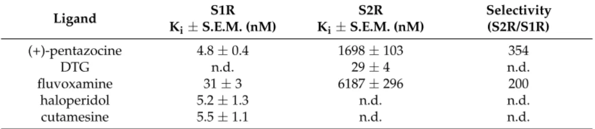
The binding affinities of the compounds for S1R and S2R were determined using in vitro competitive binding assays. The assay conditions were validated with the fol- lowing S1R and S2R ligands: (+)-pentazocine, fluvoxamine, haloperidol, cutamesine, and DTG. The displacement curves and the measured binding parameters are presented in Figure3and Table2, respectively. Competition binding assays in guinea pig liver mem- brane homogenate against the S1R specific radioligand [3H](+)-pentazocine revealed that all four compounds exhibited nanomolar S1R affinities and induced a similar maximal displacement (100%). The order of potencies of the prototypic sigma ligands were as (+)-pentazocine > haloperidol > cutamesine > fluvoxamine that is consistent with previous S1R pharmacology findings [40,64–67]. In homologous displacement experiments for the S2R, the non-selective S1R and S2R ligand, DTG exhibited a Kivalue of 29 nM in rat liver
Int. J. Mol. Sci.2021,22, 8112 7 of 17
membranes. In order to mask the S1R binding sites of the rat liver membrane preparation 100 nM (+)-pentazocine was applied [5,60].
Int. J. Mol. Sci. 2021, 22, x FOR PEER REVIEW 7 of 18
The binding affinities of the compounds for S1R and S2R were determined using in vitro competitive binding assays. The assay conditions were validated with the following S1R and S2R ligands: (+)-pentazocine, fluvoxamine, haloperidol, cutamesine, and DTG.
The displacement curves and the measured binding parameters are presented in Figure 3 and Table 2, respectively. Competition binding assays in guinea pig liver membrane ho- mogenate against the S1R specific radioligand [3H](+)-pentazocine revealed that all four compounds exhibited nanomolar S1R affinities and induced a similar maximal displace- ment (100%). The order of potencies of the prototypic sigma ligands were as (+)-pentazo- cine > haloperidol > cutamesine > fluvoxamine that is consistent with previous S1R phar- macology findings [40,64–67]. In homologous displacement experiments for the S2R, the non-selective S1R and S2R ligand, DTG exhibited a Ki value of 29 nM in rat liver mem- branes. In order to mask the S1R binding sites of the rat liver membrane preparation 100 nM (+)-pentazocine was applied [5,60].
(a) (b)
Figure 3. Displacement curves for reference compounds against (a) [3H]-(+)-pentazocine binding to S1R in guinea pig liver and (b) [3H]-DTG binding to S2R sites in rat liver membranes. Data are mean percentage of specific binding ± SEM from minimum three independent experiments, each performed in duplicate.
The S1R ligands, (+)-pentazocine and fluvoxamine were able to compete with [3H]DTG with apparently high inhibitory constants in the micromolar range; however, they only partially (c.a. 70–80%) displaced [3H]DTG. Furthermore, in our experimental model, (+)-pentazocine and fluvoxamine displayed high overall selectivities as S1R lig- ands, which is in a good agreement with the results of Narita et al. [65] and Lever et al.
[66].
Table 2. Sigma-1 and sigma-2 receptor binding affinity (Ki) and S2R/S1R selectivity for known sigma receptor ligands
Ligand S1R
Ki ± S.E.M. (nM)
S2R Ki ± S.E.M. (nM)
Selectivity (S2R/S1R)
(+)-pentazocine 4.8 ± 0,4 1698 ± 103 354
DTG n.d. 29 ± 4 n.d.
fluvoxamine 31 ± 3 6187 ± 296 200
haloperidol 5.2 ± 1,3 n.d. n.d.
cutamesine 5.5 ± 1.1 n.d. n.d.
n.d.: not determined.
2.3. Screening of Our In-House Library
According to our results the minimal ensemble docking procedure described above provided high early enrichment in our test set. This validates our method for virtual screening of large compound libraries, as well. Our in-house compound library contains
~4000 dominantly drug-like molecules. The compound library contains mainly various Figure 3.Displacement curves for reference compounds against (a) [3H]-(+)-pentazocine binding to S1R in guinea pig liver and (b) [3H]-DTG binding to S2R sites in rat liver membranes. Data are mean percentage of specific binding±SEM from minimum three independent experiments, each performed in duplicate.
Table 2.Sigma-1 and sigma-2 receptor binding affinity (Ki) and S2R/S1R selectivity for known sigma receptor ligands.
Ligand S1R
Ki±S.E.M. (nM)
S2R Ki±S.E.M. (nM)
Selectivity (S2R/S1R)
(+)-pentazocine 4.8±0.4 1698±103 354
DTG n.d. 29±4 n.d.
fluvoxamine 31±3 6187±296 200
haloperidol 5.2±1.3 n.d. n.d.
cutamesine 5.5±1.1 n.d. n.d.
n.d.: not determined.
The S1R ligands, (+)-pentazocine and fluvoxamine were able to compete with [3H]DTG with apparently high inhibitory constants in the micromolar range; however, they only partially (c.a. 70–80%) displaced [3H]DTG. Furthermore, in our experimental model, (+)- pentazocine and fluvoxamine displayed high overall selectivities as S1R ligands, which is in a good agreement with the results of Narita et al. [65] and Lever et al. [66].
2.3. Screening of Our in-House Library
According to our results the minimal ensemble docking procedure described above provided high early enrichment in our test set. This validates our method for virtual screening of large compound libraries, as well. Our in-house compound library contains
~4000 dominantly drug-like molecules. The compound library contains mainly various derivatives of 1,2- and 1,3-bifunctional compounds (derivatives of amino acids, amino alcohols, hydroxy acids, diamines etc. with acyclic, aromatic, alicyclic and heterocyclic scaffolds). Based on the screening, the highest ranked 40 molecules (top 1%) were selected for experimental testing. These molecules had their docking scores between−9.38 kcal/mol and−13.76 kcal/mol.
As a first step of the experimental evaluation, competition binding experiments were carried out by incubating guinea pig liver membranes with 2.4 nM of [3H](+)-pentazocine in the presence of increasing concentrations (between 10−11M and−10−5M) of the selected unlabeled ligands.
Table 3lists twelve compounds that showed activity in this range of concentra- tion. Corresponding displacement curves can be found in the Supplementary Materials (Supplemental Figure S2). The best compounds L1, L2, and L3 have inhibitory constants of 32 nM, 91 nM, and 110 nM, respectively.
Int. J. Mol. Sci.2021,22, 8112 8 of 17
Table 3. Sigma-1 receptor binding parameters for racemic ligands in guinea pig liver membrane homogenates and the corresponding docking scores.
ID Measured Ki(nM)
Docking Score
(kcal/mol) Chemical Name Structure of Best Docked
Stereoisomer
L11 32.0 −11.62
N-Benzyl-6,7-dimethoxy-1,2,3,4-tetrahydro-1- isoquinolineethanamine
dihydrochloride
Int. J. Mol. Sci. 2021, 22, x FOR PEER REVIEW 9 of 18
Table 3. Sigma-1 receptor binding parameters for racemic ligands in guinea pig liver membrane homogenates and the corresponding docking scores
ID Measured Ki (nM)
Docking Score
(kcal/mol) Chemical Name Structure of Best Docked Stereoi-
somer L1 1 32.0 −11.62 N-Benzyl-6,7-dimethoxy-1,2,3,4-tetrahydro-1-iso-
quinolineethanamine dihydrochloride
L2 1 91.0 −12.41 3-Amino-N-(2-fluoro-3-(trifluoromethyl)benzyl]-3- phenylpropanamide hydrochloride
L3 2 110.0 −11.95 1-[(4-Methoxyphenoxy)methyl]-2-(1,2,3,4-tetrahy- droisoquinolin-2-yl)ethanol
L4 1 420.0 −12.47 3-Amino-N-(3-fluoro-5-(trifluoromethyl)benzyl]-3- phenylpropanamide hydrochloride
L5 1 463.0 −10.74
(±)-diendo-3′-amino-1-benzyl-5′,8′-methano- 4′a,5′,8′,8′a-tetrahydrospiro[piperidine-4,2′(1′H)-
quinazolin]-4′(3′H)-one
L6 2 1036.0 −11.12 N-Benzyloxycarbonyl-(9-methyl-2,3,4,9-tetrahy- dro-1H-pyrido[3,4-b]indol-1-yl)methanamine
L7 2 1381.0 −11.56 (1S,3R,4R,6R)-3-(benzylamino)methyl)-7,7-dime- thylbicyclo[4.1.0]heptane-3,4-diol
L8 2 1534.0 −10.68 3-Amino-N-(2-fluoro-3-(trifluoromethyl)benzyl]-2- methylpropanamide trifluoroacetate
L9 1 1716.0 −10.87 N-Benzyloxycarbonyl-(2,3,4,9-tetrahydro-1H-pyr- ido[3,4-b]indol-1-yl)methanamine
L10 1 2154.0 −11.58
(4R*,11bR*)-9,10-Diethoxy-4-[4-(dimethyla- mino)phenyl]-1,3,4,6,7,11b-hexahydro-2H-pyrim-
ido[6,1-a]isoquinoline
L11 1 2381.0 −10.55 1-{[(Benzyloxycarbonyl)amino]methyl}-6,7-di- methoxy-1,2,3,4-tetrahydroisoquinoline
L12 2 3266.0 −9.69
(1R*,9bR*)-1-{[(4-Chlorophenyl)thio]methyl}-7,8- diethoxy-1,4,5,9b-tetrahydro-2H-azeto[2,1a]-iso-
quinoline
1 Preferred target: 6DK1C. 2 Preferred target: 5HK1A.
L21 91.0 −12.41
3-Amino-N-(2-fluoro-3-(trifluoromethyl)benzyl]-3- phenylpropanamide
hydrochloride
Int. J. Mol. Sci. 2021, 22, x FOR PEER REVIEW 9 of 18
Table 3. Sigma-1 receptor binding parameters for racemic ligands in guinea pig liver membrane homogenates and the corresponding docking scores
ID Measured Ki (nM)
Docking Score
(kcal/mol) Chemical Name Structure of Best Docked Stereoi-
somer L1 1 32.0 −11.62 N-Benzyl-6,7-dimethoxy-1,2,3,4-tetrahydro-1-iso-
quinolineethanamine dihydrochloride
L2 1 91.0 −12.41 3-Amino-N-(2-fluoro-3-(trifluoromethyl)benzyl]-3- phenylpropanamide hydrochloride
L3 2 110.0 −11.95 1-[(4-Methoxyphenoxy)methyl]-2-(1,2,3,4-tetrahy- droisoquinolin-2-yl)ethanol
L4 1 420.0 −12.47 3-Amino-N-(3-fluoro-5-(trifluoromethyl)benzyl]-3- phenylpropanamide hydrochloride
L5 1 463.0 −10.74
(±)-diendo-3′-amino-1-benzyl-5′,8′-methano- 4′a,5′,8′,8′a-tetrahydrospiro[piperidine-4,2′(1′H)-
quinazolin]-4′(3′H)-one
L6 2 1036.0 −11.12 N-Benzyloxycarbonyl-(9-methyl-2,3,4,9-tetrahy- dro-1H-pyrido[3,4-b]indol-1-yl)methanamine
L7 2 1381.0 −11.56 (1S,3R,4R,6R)-3-(benzylamino)methyl)-7,7-dime- thylbicyclo[4.1.0]heptane-3,4-diol
L8 2 1534.0 −10.68 3-Amino-N-(2-fluoro-3-(trifluoromethyl)benzyl]-2- methylpropanamide trifluoroacetate
L9 1 1716.0 −10.87 N-Benzyloxycarbonyl-(2,3,4,9-tetrahydro-1H-pyr- ido[3,4-b]indol-1-yl)methanamine
L10 1 2154.0 −11.58
(4R*,11bR*)-9,10-Diethoxy-4-[4-(dimethyla- mino)phenyl]-1,3,4,6,7,11b-hexahydro-2H-pyrim-
ido[6,1-a]isoquinoline
L11 1 2381.0 −10.55 1-{[(Benzyloxycarbonyl)amino]methyl}-6,7-di- methoxy-1,2,3,4-tetrahydroisoquinoline
L12 2 3266.0 −9.69
(1R*,9bR*)-1-{[(4-Chlorophenyl)thio]methyl}-7,8- diethoxy-1,4,5,9b-tetrahydro-2H-azeto[2,1a]-iso-
quinoline
1 Preferred target: 6DK1C. 2 Preferred target: 5HK1A.
L32 110.0 −11.95 1-[(4-Methoxyphenoxy)methyl]-2-(1,2,3,4-tetrahydroisoquinolin-2- yl)ethanol
Int. J. Mol. Sci. 2021, 22, x FOR PEER REVIEW 9 of 18
Table 3. Sigma-1 receptor binding parameters for racemic ligands in guinea pig liver membrane homogenates and the corresponding docking scores
ID Measured Ki (nM)
Docking Score
(kcal/mol) Chemical Name Structure of Best Docked Stereoi-
somer L1 1 32.0 −11.62 N-Benzyl-6,7-dimethoxy-1,2,3,4-tetrahydro-1-iso-
quinolineethanamine dihydrochloride
L2 1 91.0 −12.41 3-Amino-N-(2-fluoro-3-(trifluoromethyl)benzyl]-3- phenylpropanamide hydrochloride
L3 2 110.0 −11.95 1-[(4-Methoxyphenoxy)methyl]-2-(1,2,3,4-tetrahy- droisoquinolin-2-yl)ethanol
L4 1 420.0 −12.47 3-Amino-N-(3-fluoro-5-(trifluoromethyl)benzyl]-3- phenylpropanamide hydrochloride
L5 1 463.0 −10.74
(±)-diendo-3′-amino-1-benzyl-5′,8′-methano- 4′a,5′,8′,8′a-tetrahydrospiro[piperidine-4,2′(1′H)-
quinazolin]-4′(3′H)-one
L6 2 1036.0 −11.12 N-Benzyloxycarbonyl-(9-methyl-2,3,4,9-tetrahy- dro-1H-pyrido[3,4-b]indol-1-yl)methanamine
L7 2 1381.0 −11.56 (1S,3R,4R,6R)-3-(benzylamino)methyl)-7,7-dime- thylbicyclo[4.1.0]heptane-3,4-diol
L8 2 1534.0 −10.68 3-Amino-N-(2-fluoro-3-(trifluoromethyl)benzyl]-2- methylpropanamide trifluoroacetate
L9 1 1716.0 −10.87 N-Benzyloxycarbonyl-(2,3,4,9-tetrahydro-1H-pyr- ido[3,4-b]indol-1-yl)methanamine
L10 1 2154.0 −11.58
(4R*,11bR*)-9,10-Diethoxy-4-[4-(dimethyla- mino)phenyl]-1,3,4,6,7,11b-hexahydro-2H-pyrim-
ido[6,1-a]isoquinoline
L11 1 2381.0 −10.55 1-{[(Benzyloxycarbonyl)amino]methyl}-6,7-di- methoxy-1,2,3,4-tetrahydroisoquinoline
L12 2 3266.0 −9.69
(1R*,9bR*)-1-{[(4-Chlorophenyl)thio]methyl}-7,8- diethoxy-1,4,5,9b-tetrahydro-2H-azeto[2,1a]-iso-
quinoline
1 Preferred target: 6DK1C. 2 Preferred target: 5HK1A.
L41 420.0 −12.47
3-Amino-N-(3-fluoro-5-(trifluoromethyl)benzyl]-3- phenylpropanamide
hydrochloride
Int. J. Mol. Sci. 2021, 22, x FOR PEER REVIEW 9 of 18
Table 3. Sigma-1 receptor binding parameters for racemic ligands in guinea pig liver membrane homogenates and the corresponding docking scores
ID Measured Ki (nM)
Docking Score
(kcal/mol) Chemical Name Structure of Best Docked Stereoi-
somer L1 1 32.0 −11.62 N-Benzyl-6,7-dimethoxy-1,2,3,4-tetrahydro-1-iso-
quinolineethanamine dihydrochloride
L2 1 91.0 −12.41 3-Amino-N-(2-fluoro-3-(trifluoromethyl)benzyl]-3- phenylpropanamide hydrochloride
L3 2 110.0 −11.95 1-[(4-Methoxyphenoxy)methyl]-2-(1,2,3,4-tetrahy- droisoquinolin-2-yl)ethanol
L4 1 420.0 −12.47 3-Amino-N-(3-fluoro-5-(trifluoromethyl)benzyl]-3- phenylpropanamide hydrochloride
L5 1 463.0 −10.74
(±)-diendo-3′-amino-1-benzyl-5′,8′-methano- 4′a,5′,8′,8′a-tetrahydrospiro[piperidine-4,2′(1′H)-
quinazolin]-4′(3′H)-one
L6 2 1036.0 −11.12 N-Benzyloxycarbonyl-(9-methyl-2,3,4,9-tetrahy- dro-1H-pyrido[3,4-b]indol-1-yl)methanamine
L7 2 1381.0 −11.56 (1S,3R,4R,6R)-3-(benzylamino)methyl)-7,7-dime- thylbicyclo[4.1.0]heptane-3,4-diol
L8 2 1534.0 −10.68 3-Amino-N-(2-fluoro-3-(trifluoromethyl)benzyl]-2- methylpropanamide trifluoroacetate
L9 1 1716.0 −10.87 N-Benzyloxycarbonyl-(2,3,4,9-tetrahydro-1H-pyr- ido[3,4-b]indol-1-yl)methanamine
L10 1 2154.0 −11.58
(4R*,11bR*)-9,10-Diethoxy-4-[4-(dimethyla- mino)phenyl]-1,3,4,6,7,11b-hexahydro-2H-pyrim-
ido[6,1-a]isoquinoline
L11 1 2381.0 −10.55 1-{[(Benzyloxycarbonyl)amino]methyl}-6,7-di- methoxy-1,2,3,4-tetrahydroisoquinoline
L12 2 3266.0 −9.69
(1R*,9bR*)-1-{[(4-Chlorophenyl)thio]methyl}-7,8- diethoxy-1,4,5,9b-tetrahydro-2H-azeto[2,1a]-iso-
quinoline
1 Preferred target: 6DK1C. 2 Preferred target: 5HK1A.
L51 463.0 −10.74 (±)-diendo-30-amino-1-benzyl-50,80-methano-40a,50,80,80a- tetrahydrospiro[piperidine-4,20(10H)-quinazolin]-40(30H)-one
Int. J. Mol. Sci. 2021, 22, x FOR PEER REVIEW 9 of 18
Table 3. Sigma-1 receptor binding parameters for racemic ligands in guinea pig liver membrane homogenates and the corresponding docking scores
ID Measured Ki (nM)
Docking Score
(kcal/mol) Chemical Name Structure of Best Docked Stereoi-
somer L1 1 32.0 −11.62 N-Benzyl-6,7-dimethoxy-1,2,3,4-tetrahydro-1-iso-
quinolineethanamine dihydrochloride
L2 1 91.0 −12.41 3-Amino-N-(2-fluoro-3-(trifluoromethyl)benzyl]-3- phenylpropanamide hydrochloride
L3 2 110.0 −11.95 1-[(4-Methoxyphenoxy)methyl]-2-(1,2,3,4-tetrahy- droisoquinolin-2-yl)ethanol
L4 1 420.0 −12.47 3-Amino-N-(3-fluoro-5-(trifluoromethyl)benzyl]-3- phenylpropanamide hydrochloride
L5 1 463.0 −10.74
(±)-diendo-3′-amino-1-benzyl-5′,8′-methano- 4′a,5′,8′,8′a-tetrahydrospiro[piperidine-4,2′(1′H)-
quinazolin]-4′(3′H)-one
L6 2 1036.0 −11.12 N-Benzyloxycarbonyl-(9-methyl-2,3,4,9-tetrahy- dro-1H-pyrido[3,4-b]indol-1-yl)methanamine
L7 2 1381.0 −11.56 (1S,3R,4R,6R)-3-(benzylamino)methyl)-7,7-dime- thylbicyclo[4.1.0]heptane-3,4-diol
L8 2 1534.0 −10.68 3-Amino-N-(2-fluoro-3-(trifluoromethyl)benzyl]-2- methylpropanamide trifluoroacetate
L9 1 1716.0 −10.87 N-Benzyloxycarbonyl-(2,3,4,9-tetrahydro-1H-pyr- ido[3,4-b]indol-1-yl)methanamine
L10 1 2154.0 −11.58
(4R*,11bR*)-9,10-Diethoxy-4-[4-(dimethyla- mino)phenyl]-1,3,4,6,7,11b-hexahydro-2H-pyrim-
ido[6,1-a]isoquinoline
L11 1 2381.0 −10.55 1-{[(Benzyloxycarbonyl)amino]methyl}-6,7-di- methoxy-1,2,3,4-tetrahydroisoquinoline
L12 2 3266.0 −9.69
(1R*,9bR*)-1-{[(4-Chlorophenyl)thio]methyl}-7,8- diethoxy-1,4,5,9b-tetrahydro-2H-azeto[2,1a]-iso-
quinoline
1 Preferred target: 6DK1C. 2 Preferred target: 5HK1A.
L62 1036.0 −11.12 N-Benzyloxycarbonyl-(9-methyl-2,3,4,9-tetrahydro-1H-pyrido[3,4- b]indol-1-yl)methanamine
Int. J. Mol. Sci. 2021, 22, x FOR PEER REVIEW 9 of 18
Table 3. Sigma-1 receptor binding parameters for racemic ligands in guinea pig liver membrane homogenates and the corresponding docking scores
ID Measured Ki (nM)
Docking Score
(kcal/mol) Chemical Name Structure of Best Docked Stereoi-
somer L1 1 32.0 −11.62 N-Benzyl-6,7-dimethoxy-1,2,3,4-tetrahydro-1-iso-
quinolineethanamine dihydrochloride
L2 1 91.0 −12.41 3-Amino-N-(2-fluoro-3-(trifluoromethyl)benzyl]-3- phenylpropanamide hydrochloride
L3 2 110.0 −11.95 1-[(4-Methoxyphenoxy)methyl]-2-(1,2,3,4-tetrahy- droisoquinolin-2-yl)ethanol
L4 1 420.0 −12.47 3-Amino-N-(3-fluoro-5-(trifluoromethyl)benzyl]-3- phenylpropanamide hydrochloride
L5 1 463.0 −10.74
(±)-diendo-3′-amino-1-benzyl-5′,8′-methano- 4′a,5′,8′,8′a-tetrahydrospiro[piperidine-4,2′(1′H)-
quinazolin]-4′(3′H)-one
L6 2 1036.0 −11.12 N-Benzyloxycarbonyl-(9-methyl-2,3,4,9-tetrahy- dro-1H-pyrido[3,4-b]indol-1-yl)methanamine
L7 2 1381.0 −11.56 (1S,3R,4R,6R)-3-(benzylamino)methyl)-7,7-dime- thylbicyclo[4.1.0]heptane-3,4-diol
L8 2 1534.0 −10.68 3-Amino-N-(2-fluoro-3-(trifluoromethyl)benzyl]-2- methylpropanamide trifluoroacetate
L9 1 1716.0 −10.87 N-Benzyloxycarbonyl-(2,3,4,9-tetrahydro-1H-pyr- ido[3,4-b]indol-1-yl)methanamine
L10 1 2154.0 −11.58
(4R*,11bR*)-9,10-Diethoxy-4-[4-(dimethyla- mino)phenyl]-1,3,4,6,7,11b-hexahydro-2H-pyrim-
ido[6,1-a]isoquinoline
L11 1 2381.0 −10.55 1-{[(Benzyloxycarbonyl)amino]methyl}-6,7-di- methoxy-1,2,3,4-tetrahydroisoquinoline
L12 2 3266.0 −9.69
(1R*,9bR*)-1-{[(4-Chlorophenyl)thio]methyl}-7,8- diethoxy-1,4,5,9b-tetrahydro-2H-azeto[2,1a]-iso-
quinoline
1 Preferred target: 6DK1C. 2 Preferred target: 5HK1A.
L72 1381.0 −11.56 (1S,3R,4R,6R)-3-(benzylamino)methyl)-7,7-
dimethylbicyclo[4.1.0]heptane-3,4-diol
Int. J. Mol. Sci. 2021, 22, x FOR PEER REVIEW 9 of 18
Table 3. Sigma-1 receptor binding parameters for racemic ligands in guinea pig liver membrane homogenates and the corresponding docking scores
ID Measured Ki (nM)
Docking Score
(kcal/mol) Chemical Name Structure of Best Docked Stereoi-
somer L1 1 32.0 −11.62 N-Benzyl-6,7-dimethoxy-1,2,3,4-tetrahydro-1-iso-
quinolineethanamine dihydrochloride
L2 1 91.0 −12.41 3-Amino-N-(2-fluoro-3-(trifluoromethyl)benzyl]-3- phenylpropanamide hydrochloride
L3 2 110.0 −11.95 1-[(4-Methoxyphenoxy)methyl]-2-(1,2,3,4-tetrahy- droisoquinolin-2-yl)ethanol
L4 1 420.0 −12.47 3-Amino-N-(3-fluoro-5-(trifluoromethyl)benzyl]-3- phenylpropanamide hydrochloride
L5 1 463.0 −10.74
(±)-diendo-3′-amino-1-benzyl-5′,8′-methano- 4′a,5′,8′,8′a-tetrahydrospiro[piperidine-4,2′(1′H)-
quinazolin]-4′(3′H)-one
L6 2 1036.0 −11.12 N-Benzyloxycarbonyl-(9-methyl-2,3,4,9-tetrahy- dro-1H-pyrido[3,4-b]indol-1-yl)methanamine
L7 2 1381.0 −11.56 (1S,3R,4R,6R)-3-(benzylamino)methyl)-7,7-dime- thylbicyclo[4.1.0]heptane-3,4-diol
L8 2 1534.0 −10.68 3-Amino-N-(2-fluoro-3-(trifluoromethyl)benzyl]-2- methylpropanamide trifluoroacetate
L9 1 1716.0 −10.87 N-Benzyloxycarbonyl-(2,3,4,9-tetrahydro-1H-pyr- ido[3,4-b]indol-1-yl)methanamine
L10 1 2154.0 −11.58
(4R*,11bR*)-9,10-Diethoxy-4-[4-(dimethyla- mino)phenyl]-1,3,4,6,7,11b-hexahydro-2H-pyrim-
ido[6,1-a]isoquinoline
L11 1 2381.0 −10.55 1-{[(Benzyloxycarbonyl)amino]methyl}-6,7-di- methoxy-1,2,3,4-tetrahydroisoquinoline
L12 2 3266.0 −9.69
(1R*,9bR*)-1-{[(4-Chlorophenyl)thio]methyl}-7,8- diethoxy-1,4,5,9b-tetrahydro-2H-azeto[2,1a]-iso-
quinoline
1 Preferred target: 6DK1C. 2 Preferred target: 5HK1A.
L82 1534.0 −10.68
3-Amino-N-(2-fluoro-3-(trifluoromethyl)benzyl]-2- methylpropanamide
trifluoroacetate
Int. J. Mol. Sci. 2021, 22, x FOR PEER REVIEW 9 of 18
Table 3. Sigma-1 receptor binding parameters for racemic ligands in guinea pig liver membrane homogenates and the corresponding docking scores
ID Measured Ki (nM)
Docking Score
(kcal/mol) Chemical Name Structure of Best Docked Stereoi-
somer L1 1 32.0 −11.62 N-Benzyl-6,7-dimethoxy-1,2,3,4-tetrahydro-1-iso-
quinolineethanamine dihydrochloride
L2 1 91.0 −12.41 3-Amino-N-(2-fluoro-3-(trifluoromethyl)benzyl]-3- phenylpropanamide hydrochloride
L3 2 110.0 −11.95 1-[(4-Methoxyphenoxy)methyl]-2-(1,2,3,4-tetrahy- droisoquinolin-2-yl)ethanol
L4 1 420.0 −12.47 3-Amino-N-(3-fluoro-5-(trifluoromethyl)benzyl]-3- phenylpropanamide hydrochloride
L5 1 463.0 −10.74
(±)-diendo-3′-amino-1-benzyl-5′,8′-methano- 4′a,5′,8′,8′a-tetrahydrospiro[piperidine-4,2′(1′H)-
quinazolin]-4′(3′H)-one
L6 2 1036.0 −11.12 N-Benzyloxycarbonyl-(9-methyl-2,3,4,9-tetrahy- dro-1H-pyrido[3,4-b]indol-1-yl)methanamine
L7 2 1381.0 −11.56 (1S,3R,4R,6R)-3-(benzylamino)methyl)-7,7-dime- thylbicyclo[4.1.0]heptane-3,4-diol
L8 2 1534.0 −10.68 3-Amino-N-(2-fluoro-3-(trifluoromethyl)benzyl]-2- methylpropanamide trifluoroacetate
L9 1 1716.0 −10.87 N-Benzyloxycarbonyl-(2,3,4,9-tetrahydro-1H-pyr- ido[3,4-b]indol-1-yl)methanamine
L10 1 2154.0 −11.58
(4R*,11bR*)-9,10-Diethoxy-4-[4-(dimethyla- mino)phenyl]-1,3,4,6,7,11b-hexahydro-2H-pyrim-
ido[6,1-a]isoquinoline
L11 1 2381.0 −10.55 1-{[(Benzyloxycarbonyl)amino]methyl}-6,7-di- methoxy-1,2,3,4-tetrahydroisoquinoline
L12 2 3266.0 −9.69
(1R*,9bR*)-1-{[(4-Chlorophenyl)thio]methyl}-7,8- diethoxy-1,4,5,9b-tetrahydro-2H-azeto[2,1a]-iso-
quinoline
1 Preferred target: 6DK1C. 2 Preferred target: 5HK1A.
L91 1716.0 −10.87 N-Benzyloxycarbonyl-(2,3,4,9-tetrahydro-1H-pyrido[3,4-b]indol-1- yl)methanamine
Int. J. Mol. Sci. 2021, 22, x FOR PEER REVIEW 9 of 18
Table 3. Sigma-1 receptor binding parameters for racemic ligands in guinea pig liver membrane homogenates and the corresponding docking scores
ID Measured Ki (nM)
Docking Score
(kcal/mol) Chemical Name Structure of Best Docked Stereoi-
somer L1 1 32.0 −11.62 N-Benzyl-6,7-dimethoxy-1,2,3,4-tetrahydro-1-iso-
quinolineethanamine dihydrochloride
L2 1 91.0 −12.41 3-Amino-N-(2-fluoro-3-(trifluoromethyl)benzyl]-3- phenylpropanamide hydrochloride
L3 2 110.0 −11.95 1-[(4-Methoxyphenoxy)methyl]-2-(1,2,3,4-tetrahy- droisoquinolin-2-yl)ethanol
L4 1 420.0 −12.47 3-Amino-N-(3-fluoro-5-(trifluoromethyl)benzyl]-3- phenylpropanamide hydrochloride
L5 1 463.0 −10.74
(±)-diendo-3′-amino-1-benzyl-5′,8′-methano- 4′a,5′,8′,8′a-tetrahydrospiro[piperidine-4,2′(1′H)-
quinazolin]-4′(3′H)-one
L6 2 1036.0 −11.12 N-Benzyloxycarbonyl-(9-methyl-2,3,4,9-tetrahy- dro-1H-pyrido[3,4-b]indol-1-yl)methanamine
L7 2 1381.0 −11.56 (1S,3R,4R,6R)-3-(benzylamino)methyl)-7,7-dime- thylbicyclo[4.1.0]heptane-3,4-diol
L8 2 1534.0 −10.68 3-Amino-N-(2-fluoro-3-(trifluoromethyl)benzyl]-2- methylpropanamide trifluoroacetate
L9 1 1716.0 −10.87 N-Benzyloxycarbonyl-(2,3,4,9-tetrahydro-1H-pyr- ido[3,4-b]indol-1-yl)methanamine
L10 1 2154.0 −11.58
(4R*,11bR*)-9,10-Diethoxy-4-[4-(dimethyla- mino)phenyl]-1,3,4,6,7,11b-hexahydro-2H-pyrim-
ido[6,1-a]isoquinoline
L11 1 2381.0 −10.55 1-{[(Benzyloxycarbonyl)amino]methyl}-6,7-di- methoxy-1,2,3,4-tetrahydroisoquinoline
L12 2 3266.0 −9.69
(1R*,9bR*)-1-{[(4-Chlorophenyl)thio]methyl}-7,8- diethoxy-1,4,5,9b-tetrahydro-2H-azeto[2,1a]-iso-
quinoline
1 Preferred target: 6DK1C. 2 Preferred target: 5HK1A.
L101 2154.0 −11.58 (4R*,11bR*)-9,10-Diethoxy-4-[4-(dimethylamino)phenyl]- 1,3,4,6,7,11b-hexahydro-2H-pyrimido[6,1-a]isoquinoline
Int. J. Mol. Sci. 2021, 22, x FOR PEER REVIEW 9 of 18
Table 3. Sigma-1 receptor binding parameters for racemic ligands in guinea pig liver membrane homogenates and the corresponding docking scores
ID Measured Ki (nM)
Docking Score
(kcal/mol) Chemical Name Structure of Best Docked Stereoi-
somer L1 1 32.0 −11.62 N-Benzyl-6,7-dimethoxy-1,2,3,4-tetrahydro-1-iso-
quinolineethanamine dihydrochloride
L2 1 91.0 −12.41 3-Amino-N-(2-fluoro-3-(trifluoromethyl)benzyl]-3- phenylpropanamide hydrochloride
L3 2 110.0 −11.95 1-[(4-Methoxyphenoxy)methyl]-2-(1,2,3,4-tetrahy- droisoquinolin-2-yl)ethanol
L4 1 420.0 −12.47 3-Amino-N-(3-fluoro-5-(trifluoromethyl)benzyl]-3- phenylpropanamide hydrochloride
L5 1 463.0 −10.74
(±)-diendo-3′-amino-1-benzyl-5′,8′-methano- 4′a,5′,8′,8′a-tetrahydrospiro[piperidine-4,2′(1′H)-
quinazolin]-4′(3′H)-one
L6 2 1036.0 −11.12 N-Benzyloxycarbonyl-(9-methyl-2,3,4,9-tetrahy- dro-1H-pyrido[3,4-b]indol-1-yl)methanamine
L7 2 1381.0 −11.56 (1S,3R,4R,6R)-3-(benzylamino)methyl)-7,7-dime- thylbicyclo[4.1.0]heptane-3,4-diol
L8 2 1534.0 −10.68 3-Amino-N-(2-fluoro-3-(trifluoromethyl)benzyl]-2- methylpropanamide trifluoroacetate
L9 1 1716.0 −10.87 N-Benzyloxycarbonyl-(2,3,4,9-tetrahydro-1H-pyr- ido[3,4-b]indol-1-yl)methanamine
L10 1 2154.0 −11.58
(4R*,11bR*)-9,10-Diethoxy-4-[4-(dimethyla- mino)phenyl]-1,3,4,6,7,11b-hexahydro-2H-pyrim-
ido[6,1-a]isoquinoline
L11 1 2381.0 −10.55 1-{[(Benzyloxycarbonyl)amino]methyl}-6,7-di- methoxy-1,2,3,4-tetrahydroisoquinoline
L12 2 3266.0 −9.69
(1R*,9bR*)-1-{[(4-Chlorophenyl)thio]methyl}-7,8- diethoxy-1,4,5,9b-tetrahydro-2H-azeto[2,1a]-iso-
quinoline
1 Preferred target: 6DK1C. 2 Preferred target: 5HK1A.
L111 2381.0 −10.55 1-{[(Benzyloxycarbonyl)amino]methyl}-6,7-dimethoxy-1,2,3,4- tetrahydroisoquinoline
Int. J. Mol. Sci. 2021, 22, x FOR PEER REVIEW 9 of 18
Table 3. Sigma-1 receptor binding parameters for racemic ligands in guinea pig liver membrane homogenates and the corresponding docking scores
ID Measured Ki (nM)
Docking Score
(kcal/mol) Chemical Name Structure of Best Docked Stereoi-
somer L1 1 32.0 −11.62 N-Benzyl-6,7-dimethoxy-1,2,3,4-tetrahydro-1-iso-
quinolineethanamine dihydrochloride
L2 1 91.0 −12.41 3-Amino-N-(2-fluoro-3-(trifluoromethyl)benzyl]-3- phenylpropanamide hydrochloride
L3 2 110.0 −11.95 1-[(4-Methoxyphenoxy)methyl]-2-(1,2,3,4-tetrahy- droisoquinolin-2-yl)ethanol
L4 1 420.0 −12.47 3-Amino-N-(3-fluoro-5-(trifluoromethyl)benzyl]-3- phenylpropanamide hydrochloride
L5 1 463.0 −10.74
(±)-diendo-3′-amino-1-benzyl-5′,8′-methano- 4′a,5′,8′,8′a-tetrahydrospiro[piperidine-4,2′(1′H)-
quinazolin]-4′(3′H)-one
L6 2 1036.0 −11.12 N-Benzyloxycarbonyl-(9-methyl-2,3,4,9-tetrahy- dro-1H-pyrido[3,4-b]indol-1-yl)methanamine
L7 2 1381.0 −11.56 (1S,3R,4R,6R)-3-(benzylamino)methyl)-7,7-dime- thylbicyclo[4.1.0]heptane-3,4-diol
L8 2 1534.0 −10.68 3-Amino-N-(2-fluoro-3-(trifluoromethyl)benzyl]-2- methylpropanamide trifluoroacetate
L9 1 1716.0 −10.87 N-Benzyloxycarbonyl-(2,3,4,9-tetrahydro-1H-pyr- ido[3,4-b]indol-1-yl)methanamine
L10 1 2154.0 −11.58
(4R*,11bR*)-9,10-Diethoxy-4-[4-(dimethyla- mino)phenyl]-1,3,4,6,7,11b-hexahydro-2H-pyrim-
ido[6,1-a]isoquinoline
L11 1 2381.0 −10.55 1-{[(Benzyloxycarbonyl)amino]methyl}-6,7-di- methoxy-1,2,3,4-tetrahydroisoquinoline
L12 2 3266.0 −9.69
(1R*,9bR*)-1-{[(4-Chlorophenyl)thio]methyl}-7,8- diethoxy-1,4,5,9b-tetrahydro-2H-azeto[2,1a]-iso-
quinoline
1 Preferred target: 6DK1C. 2 Preferred target: 5HK1A.
L122 3266.0 −9.69 (1R*,9bR*)-1-{[(4-Chlorophenyl)thio]methyl}-7,8-diethoxy-1,4,5,9b- tetrahydro-2H-azeto[2,1a]-isoquinoline
Int. J. Mol. Sci. 2021, 22, x FOR PEER REVIEW 9 of 18
Table 3. Sigma-1 receptor binding parameters for racemic ligands in guinea pig liver membrane homogenates and the corresponding docking scores
ID Measured Ki (nM)
Docking Score
(kcal/mol) Chemical Name Structure of Best Docked Stereoi-
somer L1 1 32.0 −11.62 N-Benzyl-6,7-dimethoxy-1,2,3,4-tetrahydro-1-iso-
quinolineethanamine dihydrochloride
L2 1 91.0 −12.41 3-Amino-N-(2-fluoro-3-(trifluoromethyl)benzyl]-3- phenylpropanamide hydrochloride
L3 2 110.0 −11.95 1-[(4-Methoxyphenoxy)methyl]-2-(1,2,3,4-tetrahy- droisoquinolin-2-yl)ethanol
L4 1 420.0 −12.47 3-Amino-N-(3-fluoro-5-(trifluoromethyl)benzyl]-3- phenylpropanamide hydrochloride
L5 1 463.0 −10.74
(±)-diendo-3′-amino-1-benzyl-5′,8′-methano- 4′a,5′,8′,8′a-tetrahydrospiro[piperidine-4,2′(1′H)-
quinazolin]-4′(3′H)-one
L6 2 1036.0 −11.12 N-Benzyloxycarbonyl-(9-methyl-2,3,4,9-tetrahy- dro-1H-pyrido[3,4-b]indol-1-yl)methanamine
L7 2 1381.0 −11.56 (1S,3R,4R,6R)-3-(benzylamino)methyl)-7,7-dime- thylbicyclo[4.1.0]heptane-3,4-diol
L8 2 1534.0 −10.68 3-Amino-N-(2-fluoro-3-(trifluoromethyl)benzyl]-2- methylpropanamide trifluoroacetate
L9 1 1716.0 −10.87 N-Benzyloxycarbonyl-(2,3,4,9-tetrahydro-1H-pyr- ido[3,4-b]indol-1-yl)methanamine
L10 1 2154.0 −11.58
(4R*,11bR*)-9,10-Diethoxy-4-[4-(dimethyla- mino)phenyl]-1,3,4,6,7,11b-hexahydro-2H-pyrim-
ido[6,1-a]isoquinoline
L11 1 2381.0 −10.55 1-{[(Benzyloxycarbonyl)amino]methyl}-6,7-di- methoxy-1,2,3,4-tetrahydroisoquinoline
L12 2 3266.0 −9.69
(1R*,9bR*)-1-{[(4-Chlorophenyl)thio]methyl}-7,8- diethoxy-1,4,5,9b-tetrahydro-2H-azeto[2,1a]-iso-
quinoline
1 Preferred target: 6DK1C. 2 Preferred target: 5HK1A.
1Preferred target: 6DK1C.2Preferred target: 5HK1A.
Int. J. Mol. Sci.2021,22, 8112 9 of 17
These ligands have common structural elements: a basic amine site (present in all high affinity S1R ligands), that is necessary for the formation of an electrostatic bond with Glu172 of the receptor. It is typically flanked by two different hydrophobic groups. This common structure matches to the Glennon pharmacophore model (amine site together with a primary and a secondary hydrophobic region which is necessary for the proper fitting into the binding site of S1R [42]). The hydrophobic parts are mostly aromatic and/or rigid heterocyclic sites.
Five of the 40 selected compounds possessed nanomolar activities, the remaining ones bound weaker. We also investigated the preferred receptor models for these compounds (see footnotes in Table3). In case of five molecules, the top ranked pose belongs to 5HK1A and in case of seven ones to 6DK1C. Interestingly, only one of the latter set (L7) entered the P2 pocket preferred by the agonist (+)-pentazocine (see supplemental Figure S3). It has a binding constant in the low micromolar range (1310 nM). The experimental binding constants show moderate correlation with docking scores (see supplemental Figure S4) the corresponding coefficient of determination is R2= 0.41.
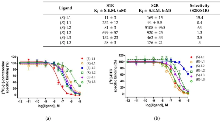
2.4. Binding Affinity of the Enantiomers of the Best Compounds to S1R and S2R
The three highest ranked compounds are chiral molecules, which have two enan- tiomeric forms. The enantiomers were separated and their binding affinities to S1R and S2R were measured separately (Table4and Figure4). The binding constants to S1R were determined similarly to those of the racemic compounds. In the case of S2R, competition binding experiments were carried out by incubating rat liver membranes with 17 nM of [3H]DTG (Kd= 47 nM) in the presence of 100 nM (+)-pentazocine to mask S1R binding sites.
Table 4. Sigma-1 and Sigma-2 receptor binding parameters and S2R/S1R selectivity for a panel of ligands.
Ligand S1R
Ki±S.E.M. (nM)
S2R Ki±S.E.M. (nM)
Selectivity (S2R/S1R)
(S)-L1 11±3 169±15 15.4
(R)-L1 252±12 94±5.5 0.4
(S)-L2 81±3 5108±960 63
(R)-L2 699±57 920±25 1.3
(S)-L3 132±23 463±33 3.5
(R)-L3 58±3 176±21 3
Int. J. Mol. Sci. 2021, 22, x FOR PEER REVIEW 11 of 18
(a) (b)
Figure 4. Displacement curves for corresponding ligands against (a) [3H]-(+)-pentazocine binding to S1R sites in guinea pig liver membranes and (b) [3H]-DTG binding to S2R sites in rat liver brain membranes using (+)-pentazocine (100 nM) to mask S1R sites. Data are mean percentage of specific binding ± SEM from minimum three independent experiments, each performed in duplicate.
2.5. S1R Binding Poses of Enantiomers of the New Compounds
The highest ranked binding poses of the enantiomer pairs of the new compounds L1, L2, and L3 were extracted from Glide XP docking results (Figure 5). Each of them resem- bles the known binding pose of the antagonist. Only (R)-L1 enters, at least partially, the binding pocket P2 occupied by (+)-pentazocine in the original X-ray structure. In the case of L1, these poses belong to the complex formed with the 6DK1A S1R model. (S)-L1 has a better docking score (−11.62 kcal/mol) than (R)-L1 (−10.97 kcal/mol) in accordance with the measured Ki values (Table 4). The most preferred binding mode of (R)/(S)-L2 belongs to chain 5HK1A/6DK1C, respectively, with a docking score of −12.41/−12.28 kcal/mol.
Their order is opposite to that obtained from our measurements. Finally, both enantiomers of L3 compound prefer 5HK1A binding and docking score values of −10.88 kcal/mol and
−11.95 kcal/mol for the (S) and (R) enantiomers properly reflect their experimental binding affinities.
(a) (b) (c)
Figure 5. Comparison of the binding poses of the (S) (gold) and (R) (green) enantiomers of the highest ranked new S1R ligands (a) L1, (b) L2, and (c) L3. The binding pose of (+)-pentazocine (red) is also shown.
Figure 4.Displacement curves for corresponding ligands against (a) [3H]-(+)-pentazocine binding to S1R sites in guinea pig liver membranes and (b) [3H]-DTG binding to S2R sites in rat liver brain membranes using (+)-pentazocine (100 nM) to mask S1R sites. Data are mean percentage of specific binding±SEM from minimum three independent experiments, each performed in duplicate.
(S)-L1 (Ki= 11 nM) showed much higher affinity for S1R than S2R sites and exhibited a similar, 15-fold S1R selectivity, as was previously shown by the fluoroethyl analog of
![Figure 4. Displacement curves for corresponding ligands against (a) [ 3 H]-(+)-pentazocine binding to S1R sites in guinea pig liver membranes and (b) [ 3 H]-DTG binding to S2R sites in rat liver brain membranes using (+)-pentazocine (100 nM) to mask S1R](https://thumb-eu.123doks.com/thumbv2/9dokorg/745053.30959/10.892.85.816.538.786/figure-displacement-corresponding-ligands-pentazocine-membranes-membranes-pentazocine.webp)
