Nonsuppurative (Aseptic) Meningoencephalomyelitis Associated with Neurovirulent Astrovirus Infections in Humans and Animals
Gábor Reuter,a,b Péter Pankovics,a,b Ákos Borosa,b
"Regional Laboratory of Virology, National Reference Laboratory of Gastroenteric Viruses, ÁNTSZ Regional Institute ofState Public HealthService, Pécs, Hungary
bDepartment of Medical Microbiology and Immunology, Medical School, University of Pécs, Pécs, Hungary
SUMMARY Astroviruses are thought to be enteric pathogens. Since 2010, a certain group of astroviruses has increasingly been recognized, using up-to-date random amplification and high-throughput next-generation sequencing (NGS) methods, as potential neurovirulent (Ni) pathogens of severe central nervous system (CNS) infections, causing encephalitis, meningoencephalitis, and meningoencephalomyelitis. To date, neurovirulent astrovirus cases or epidemics have been reported for humans and domesticated mammals, including mink, bovines, ovines, and swine. This comprehensive review summarizes the virology, epidemiology, pathology, diagnosis, therapy, and future perspective related to neurovirulent astroviruses in humans and mammals, based on a total of 30 relevant articles available in PubMed (searched by use of the terms
"astrovirus/encephalitis" and "astrovirus/meningitis" on 2 March 2018). A paradigm shift should be considered based on the increasing knowledge of the causality-effect association between neurotropic as-troviruses and CNS infection, and attention should be drawn to the role of astroviruses in unknown CNS diseases.
KEYWORDS animal, astrovirus, encephalitis, human, meningitis, meningoencephalomyelitis, neurotropic, neurovirulent
INTRODUCTION
Encephalitis is an inflammation of the brain tissue associated with clinical evidence of nervous system dysfunction. Encephalitis and central nervous system (CNS) infections are caused by a wide spectrum of known and unknown infectious agents, including bacteria, viruses, fungi, and parasites, in humans and animals (1, 2). Approximately 25% of the notifiable infectious diseases listed by the World Organization for Animal Health (OIE) are caused by neurotropic microbes (3). The microbial diagnosis of acute encephalitis and CNS diseases is challenging, especially for immunocompromised individuals, because of the atypical clinical signs and the potential presence of novel or uncommon infectious agents (4). Despite the application of various classical and modern diagnostic methods (e.g., culture, immunohistochemical, serologic, or molecular methods), a causative agent is not identified for a substantial proportion of encephalitis cases (5, 6). Failure to identify the etiology of neuroinfections impairs or delays appropriate therapeutics and influences the morbidity and mortality of these diseases (7).
Nonsuppurative (aseptic) encephalitis is an inflammation of the brain observed in postmortem histopathological examinations and is indicative of an infectious viral etiology. Perivascular cuffs with mononuclear cells, gliosis, and neuronal necrosis are common characteristic features of the disease (8). New unbiased molecular technologies, such as random amplification and high-throughput next-generation sequencing (NGS) methods, are valuable tools for differential diagnosis of unexplained diseases. An excellent example is that this technique raised the possibility that astroviruses may be associated with encephalitis in humans (9-11). Growing evidence of neurotropic/ neurovirulent astrovirus (Ni-AstV) strains has arisen for cases of encephalitis, meningitis, and meningoencephalomyelitis in humans and animals, including mink (12), bovines (13), ovines (14), and swine (15). If astrovirus infection reaches the CNS, the virus may infect multiple types of neurons (e.g., Purkinje cells, interneurons, and CA pyramidal neurons) and glial cells (e.g., astrocytes) in different parts of the CNS, including the brainstem, cerebellar/cerebral cortex, hippocampus, and spinal cord. Astrovirus infection may also cause neuronal degeneration, necrosis, neuronophagia, and gliosis (15-18). This means that the term "neuroinvasive astrovirus" may also be appropriate.
These unexpected initial results of astrovirus neuroinfection are starting to change our view about the astrovirus disease spectrum and pathogenesis, although there are several unanswered questions. At the same time, the evidence of astrovirus CNS infections opens an important and interesting new research field in the pathology of astroviruses.
NEUROVIRULENT ASTROVIRUS INFECTIONS IN HUMANS
Astroviruses are nonenveloped, single-stranded, positive-sense RNA viruses that are typically transmitted by the fecal-oral route and may cause gastrointestinal disease (19). One of the two astrovirus genera, Mamastrovirus, comprises viruses infecting mammals, including 4 species with human astroviruses. The first species, Mamastrovirus 1 (MAstV1), corresponds to "classical" (fecal origin) human astrovirus types 1 to 8 (HAstVs 1 to 8). Since 2008, a further three species (MAstV 6,8, and 9) of human astrovirus have been identified. MAstV6 includes human astrovirus clades MLB1 ("MLB" stands for Melbourne, the place of the first detection), MLB2, and MLB3 (20-22). The MAstV8 species includes astrovirus clades VA2 ("VA" stands for Virginia; also known as HMO-A [human-mink-ovine-like astroviruses]) (21) and VA4 (22).
Finally, the MAstV9 species includes astrovirus clades VA1 (or HMO-C) (23) and VA3 (or HMO-B) (19, 21, 24). One further human astrovirus, VA5 (25), is presently unassigned (19). Although these novel astroviruses were initially detected in children with gastroenteritis, no definitive association has yet been established (19, 26, 27).
Both classical and, especially, novel astroviruses have been identified in unexpected CNS infections, indicating that some of these viruses are able to enter the host through the mucosal surfaces and infect extraintestinal cells and organs. In 2010, astrovirus-associated encephalitis was reported for the first time, in a 15-year-old immunocompromised boy with X-linked agammaglobulinemia in the United States (9). The patient had suicidal and homicidal ideation, headache, memory loss, and ataxia. He also had progressive cognitive decline and movement and com- munication problems, became comatose, and died 71 days after admission (9). The possible connection to a mink farm and the phylogenetic relationship of HAstV-PS to mink astroviruses led the authors to assume that the mink was a potential source of exposure (9). However, we cannot exclude the possibility of human-to-human transmission of the infection in this case. To date, a further eight human cases of astrovirus encephalitis have been reported, related to genotype VA1/HMO-C (n = 5) (9, 28-31), the MLB group (MLB1 and MLB2) (32, 33), and the
"classical" HAstV4 species (34), although in the latter case, due to the undetermined complete capsid coding region (ORF2), the exact classification of the virus is not possible. The nine cases of neurotropic human astrovirus infections are summarized in Table 1. Except for an adult immuno-competent woman with MLB2 self-limited CNS infection, all other infections occurred in immunocompromised patients with severe underlying diseases. Six of the infections were fatal (19).
These results led to the conclusion that astroviruses may be a cause of previously unrecognized and severe neuroinfections, at least in immunocompromised humans, with a potential tropism for the CNS as a novel human disease (19, 35).
NEUROVIRULENT ASTROVIRUS INFECTIONS IN ANIMALS
Astrovirus-associated encephalitis is not restricted to humans. Based on pioneer studies, neurovirulent astroviruses have been reported
from four domesticated mammals: mink (Mustela vison)(12), bovines (Bos taurus)(13), ovines (Ovis aries)(14), and swine (Sus scrofa domestica)(15).
Mink
The first report of neurovirulent astrovirus infections ("shaking mink syndrome") from animals was reported retrospectively in 2010 for farmed minks in Denmark by the NGS method (12). Shaking, staggering gait, ataxia, and paraplegia were the characteristic clinical signs (Table 2). The symptoms were reproduced by the inoculation of brain homogenate from mink displaying "shaking mink syndrome" into experimentally infected healthy mink kits (12). A similar neurological disease was observed in farmed minks in Denmark in 2000 and 2002 and in Sweden and Finland in 2001 (12, 16), which suggests the epidemic spread of this disease.
Bovines
Many more data and cases are available about neurotropic astroviruses in bovines. While there are some historical reports since more than 60 years ago about "European sporadic bovine encephalitis/encephalomyelitis" with unknown origin in bovines (3642), the first confirmed cases of bovine neurovirulent astrovirus infections were reported only in 2013, from the United States (13). The neurovirulent astrovirus (bovine astrovirus [BoAstV] strain NeuroS1; accession number KF233944) was identified by NGS as the solely detectable virus in the CNS, and its presence was subsequently confirmed retrospectively for 3 of 32 other bovines that had bovine encephalitides (meningoen-cephalomyelitis and ganglioneuritis) of unknown etiology by reverse transcription-PCR (RT-PCR) and in situ RNA hybridization (ISH) (13). Viral RNA was detected in the spinal cord, brainstem, and cerebellum (13)(Table 2). The initial animal had lateral recumbency with opisthotonus and limb rigidity (13). Independently, shortly thereafter, similar series of cases identified by the NGS method were reported from Switzerland (17). In 2014, 5 (22.7%) of 22 cattle with nonsuppurative encephalitis of unknown etiology were associated with neurotropic astrovirus infections (17).
The presence of astroviral RNA (BoAstV-CH13; accession number KM035759) at the site of the affected neurons was also confirmed by ISH in all 5 cases (17)(Table 2). This astrovirus has been present in the Swiss bovine population since at least 1995, particularly within the age group of
1.5 to 2.5 years (17).
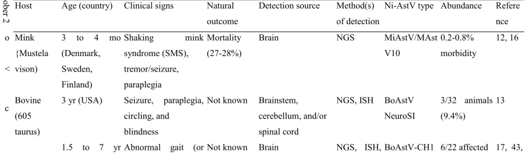
TABLE 2 Neurovirulent astrovirus infections associated with CNS disease in animals, in order of publication year (2004 to 2017)°
ober 2 Host Age (country) Clinical signs Natural
outcome
Detection source Method(s) of detection
Ni-AstV type Abundance Refere nce o Mink
{Mustela
3 to 4 mo (Denmark,
Shaking mink
syndrome (SMS),
Mortality (27-28%)
Brain NGS MiAstV/MAst
V10
0.2-0.8%
morbidity
12, 16
< vison) Sweden, Finland)
tremor/seizure, paraplegia
c Bovine (605
3 yr (USA) Seizure, paraplegia, circling, and
Not known Brainstem,
cerebellum, and/or
NGS, ISH BoAstV NeuroSI
3/32 animals (9.4%)
13
taurus) blindness spinal cord
1.5 to 7 yr Abnormal gait (or Not known Brain NGS, ISH, BoAstV-CH1 6/22 affected 17, 43,
Astrovirus type (method used for pathogen identification), strain name, available
Patient (country) Underlying disease and conditions Clinical diagnosis Symptoms Outcome GenBank accession no. Potential source Reference
15-yr-old boy (USA) X-linked agammaglobulinemia (immunocompromised)
Encephalitis Suicidal and homicidal ideation, headache, memory loss, ataxia, progressive cognitive decline
Fatal (within 71 days) VA1/HMO-C (NGS), HuAstV-PS (Puget Sound) or HuAstV-SG, GQ891990
Exposure to mink? 9
3-mo-old boy (Switzerland) Severe combined immunodeficiency, hematopoietic stem cell transplantation, chemotherapy (immunocompromised)
Meningoencephalitis Multiorgan dysfunction (hepatic and respiratory)
Fatal (within 17 days posttransplantation)
HuAstV4 (NGS), HQ396880-HQ396890 Nosocomial 34
18-mo-old boy (UK) Cartilage hair hypoplasia with hereditary immunodeficiency, hematopoietic stem cell transplantation, chemotherapy (immunocompromised)
Encephalopathy, encephalitis Irritability, dystonia, reduced consciousness Fatal (within 9 mo) VA1/HMO-C (NGS), VA1/HMO-C UK1(a) Londonl, KJ920196 Community acquired? 28
42-yr-old man (UK) Chronic lymphocytic leukemia (CLL); hematopoietic stem cell transplantation, chemotherapy (immunocompromised)
Progressive encephalitis, bilateral hearing loss
Tinnitus, sensorineural deafness, central dyspnea, hypotension, nausea, Irritability, agitation
Fatal (within 7.5 mo) VA1/HMO-C (NGS), VA1/HMO-C-UK1, KM 358468 Community acquired? 29
14-yr-old boy (France) X-linked agammaglobulinemia (immunocompromised)
Progressive encephalitis Progressive cognitive decline, recurring seizures, ataxia, erratic myoclones, dysarthria
Alive at time of publication VA1/HMO-C (NGS), VA1/HMO-C-PA, KM401565 Data not available 30
4-yr-old boy (Japan) Congenital aplastic anemia, hematopoietic stem cell transplantation, chemotherapy
(immunocompromised)
Encephalopathy Cluster of convulsion, fever, diarrhea Recovered MLB1 (NGS), MLB1-NAGANO-1545, LC064152 Unknown 32
21-yr-old women Healthy Acute meningitis Severe headache, fever, neck Recovered MLB2 (NGS), MLB2/human/Geneva/2014, Unknown (contact 33
(Switzerland) stiffness KT224358 with children?)
37-year-old (Switzerland) Acute myeloid leukemia, relapse hematopoietic stem cell transplantation, chemotherapy (immunocompromised)
Meningitis Headache, meningeal
Involvement, vertigo, limb weakness, lightheadedness
Fatal (within 9 mo) MLB2 (RT-PCR) Unknown (contact with children?) 33
8-mo-old girl (UK) Acute myeloid leukemia, hematopoietic stem cell transplantation, chemotherapy (immunocompromised)
Encephalitis Encephalopathy, uncontrolled dystonic movement, poor respiratory effort
Fatal VA1/HMO-C (NGS) Unknown, no history of exposure
to animals
31
TABLE 1 Neurovirulent astrovirus infection cases associated with CNS disease in humans, in order of publication year (2010 to 2017)°
"Abbreviations: VA/HMO, Virginia/human-mink-ovine-like astrovirus; NGS, next-generation sequencing.
(Switzerland) recumbency), tissue/medulla RT-PCR 3 and/or 45
_ behavioral changes,
and
oblongata BoAstV-CH1
5
animals, 0/33
c hyperreactivity or
depression
healthy animals
m 4 yr
(Switzerland)
Unspecified CNS symptoms
Not known Medulla oblongata, cerebellar
NGS, RT-PCR
BoAstV-CH1 5
Single case 43
cortex, midbrain,
cerebral n
>
o
cortex
o o
1.5 to 10 yr Encephalitis Deceased Brain, medulla oblongata
NGS, RT-PCR
BoAstV-CH1 3/BoAstV
2/16 affected 44
O (Switzerland) NeuroSI animals, 0/50
C O
healthy animals 1 to >10 yr Encephalitis,
meningoencephalitis
Not known Brain, brainstem, cerebellum,
ISH, RT-PCR
BoAstV-CH1 3/BoAstV
12/14 affected
18
(Switzerland) cerebrum,
hippocampus
NeuroSI animals
15 mo
(Germany)
Central blindness, circling,
Deceased Brain, spinal cord, spleen,
NGS, RT-PCR, ISH
BoAstV-BH8 9/14
Single case 47
inappétence,
somnolence
liver, pancreas
7 mo to 2 yr (Canada)
Encephalitis, seizures Mortality (100%)
Brain RT-PCR,
ISH
BoAstV-CH1 3/BoAstV
2 single cases; 4/9
48, 49
NeuroSI affected
animals
1 to 11 yr
(Switzerland)
Encephalitis, gait abnormalities,
Deceased Gray matter of the CNS
ISH, IHC BoAstV-CH1 3/BoAstV
33/97 affected
8
behavioral changes (brainstem),
cerebellum,
NeuroSI animals, 0/52
cerebrum,
hippocampus
healthy animals Ovine
[Ovis aries)
4 yr or 10 days Trembling,
whole-body tremor,
Not known Cerebrum,
cerebellum, obex,
NGS, RT-PCR
MAstV-13 Single cases 14
(Scotland) circling, recumbency spinal cord, tonsil
7 yr
(Switzerland)
Clinical signs of nonsuppurative
Deceased Medulla oblongata, RT-PCR, IHC
MAstV13-CH 16
1/48 affected 51
encephalitis cerebellum,
thalamus,
animals
hippocampus,
cortex,
caudate nucleus
Swine [Sus scrofa
1-6 days
(Sweden)
Congenital tremor type All
Not known Cerebrum, brainstem,
RT-PCR PoAstV-2 and 3/3 affected animals,
52
domestica) cerebellum PoAstV-5 3/3 healthy
animals
3-5 wk (Hungarian pigs
Paraplegia, pitching, flaccid
Mortality (100%)
Brainstem,
cerebellum, spinal
RT-PCR, ISH
PoAstV-3 1.5-4%
morbidity
15
with presumed paralysis, loss of consciousness
cord, tonsils, serum, lung,
postweaning
stress)
nasal mucosa
5 wk (pigs and sows,
Hind-limb weakness, Mortality (75-100%)
Cerebrum,
cerebellum, spinal
NGS, RT-PCR
PoAstV-3 3/4 affected sows,
53
USA) quadriplegia,
convulsions
cord 1/2 affected
pigs
"Abbreviations: NGS, next-generation sequencing; ISH, in situ hybridization; IHC, immunohistochemistry; RT-PCR, reverse transcription-PCR.
Two years later, the same research group reported additional astrovirus-positive encephalitis cases from cattle, as determined by NGS and RT-PCR (43). This included a 4-year-old Swiss cow that had hyperreactivity and incoordination and was diagnosed with me-ningoencephalitis.
The neurovirulent astrovirus (BoAstV-CH15; accession number KT956903) was genetically different from cattle BoAstV-CH13 strains within the cluster (VA1/HMO). By retrospective analysis, coinfection with BoAstV-CH13 and BoAstV-CH15 was detected in a case from the original 22 brain specimens from cattle with nonsuppurative encephalitis. Another specific study from Switzerland showed that 2 (12.5%) of 16 brain samples from cattle with unknown encephalitides, collected between 1995 and 2015, were astrovirus positive by NGS (44)(Table 2). The neurovirulent astroviruses from the 2- and 6-year-old diseased animals belonged to the BoAstV-CH13/BoAstV NeuroS1 astroviruses within the VA1/HMO cluster. None of 50 healthy control cattle were astrovirus positive by molecular methods (44). Taken together, the data show that around one quarter of the Swiss bovine population with nonsuppurative encephalitis of unresolved etiology had BoAstV-CH13 infection (45).
Based on these results, formalin-fixed, paraffin-embedded (FFPE) brain tissues from histologically confirmed cases of European sporadic bovine encephalitis/encephalomyelitis between 1958 and 1976 were retested by ISH specific for BoAstV-CH13/BoAstV NeuroS1 in Switzerland (18).
Astrovirus positivity was detected in 12 (86%) of the 14 cases (18). Using a combination of newly developed specific nucleic acid and viral protein detection methods (ISH and immunohistochemistry [IHC]), 33 (34%) of the 97 sporadic bovine encephalitis cases in Switzerland between 1985 and 2015 were BoAstV-CH13/BoAstV NeuroS1 positive, but none of the control group samples were positive (8). The mean age of the affected animals was 3.8 years in this study, and interestingly, all animals but one were female (8). Seasonality from the beginning of winter until the end of spring was observed, with peaks in November-December and March-April (8). Cattle with neurovirulent astrovirus had behavioral changes (e.g., aggression or anxiety), gastro-enteric symptoms (e.g., diarrhea or excessive salivation), and nystagmus (8). The most important conclusion from the Swiss studies is that using systematic investigation and specific methods to focus on the selected cases of bovine encephalitis increased the sensitivity; this underlines the importance and correlation of the causal relationship between neurological disease and astrovirus in the "post-Koch-postulate era" (8, 46).
Neurovirulent astrovirus infections in bovines were reported recently from Germany and Canada as well. In Germany, a 15-month-old cow died of encephalitis within 6 days after the onset of clinical signs (47). A neurovirulent astrovirus (BoAstV-BH89/14; accession number LN879482) was identified by NGS (47)(Table 2). Recent ISH and RT-PCR studies from Canada report that neurovirulent BoAstV-CH13/BoAstV NeuroS1 astroviruses are a common cause of encephalitis of unknown etiology (4 of 9 animals tested) in Canadian feedlot cattle (48, 49).
The most frequently observed clinical signs in Swiss bovines (n = 8) with astrovirus-associated encephalitis were decreased awareness of surroundings (87%), cranial nerve dysfunction (62%), such as dysphagia, decreased lingual and jaw tone, and reduced menace response and palpebral reflex, recumbency (62%), weakness, and tremor (50) (Table 2). Cell counts and protein concentration were increased in 44% and 60%
of cerebrospinal fluid (CSF) samples, respectively. Astrovirus RNA was identified in only 1 of the 4 CSF samples. Other laboratory abnormalities (increased total and CSF protein concentrations, nonsuppurative pleocytosis, and increased creatinine kinase level) were non- specific (50). None of the affected bovines had a known immunocompromised status.
Ovines
A recent study reported cases of encephalitis and ganglionitis associated with astrovirus in domestic sheep from Scotland (14). In that study, a 4-year-old Welsh mountain ewe (from 2013) and a 10-day-old lamb (from 2014) from the same farm had progressive neurological signs (e.g., depressed sensorium, tremor, and unusual behavior) (Table 2). Nonsuppurative encephalomyelitis, particularly involving the cerebellar cortex and spinal cord, and dorsal root ganglionitis were detected by neurohistopathological methods. Nearly identical ovine astrovirus sequences (accession numbers LT706530 and LT706531) within cluster VA1/HMO were detected from different tissue samples—including from the
CNS—although the cases presented 9 months apart (14). In addition, an ovine astrovirus sequence (accession number KY859988) was also reported for a 7-year-old Swiss white alpine ewe with nonsuppurative encephalitis in Switzerland (51)(Table 2).
Swine
Astrovirus as a possible cause of congenital tremor type AII in piglets (with brain and spinal cord demyelination) was first suggested in 2014 in a report from Sweden (52). Two lineages of partially characterized (only 296-bp-long RNA-dependent RNA poly-merase [RdRp] gene) porcine astroviruses (accession numbers KC790414 to KC790418), related to porcine astroviruses 2 and 5, were detected by nested RT-PCR in the cerebrum, brainstem, and cerebellum of affected and also healthy control piglets. The affected animals (n = 3) were 4 and 6 days old, while the control animals (n = 3) were only 1 day old (52). Furthermore, coinfection with porcine circovirus type 2 was found in healthy and diseased piglets (52). These preliminary results did not provide strong evidence for astrovirus as a cause of the disease.
Very recently (between 2011 and 2017), outbreaks of meningoencephalomyelitis associated with neurovirulent porcine astrovirus type 3 (PoAstV-3; accession numbers KY073229 to KY073232), within the VA/HMO clade, were detected among 25- to 35-day-old domestic pigs in large, highly prolific swine farms in Hungary (15)(Table 2). Episodes of CNS disease of unknown etiology have persisted for 2 years at the index farm, and despite extensive decontamination efforts, sporadic cases are still present in 2018 (A. Boros and M. Albert, unpublished results). The disease has affected approximately 30 to 40 weaned pigs monthly (1.5% to 2% of the total), up to around 80 pigs (4%) in the autumn and winter seasons (15). Clinical symptoms typically appeared among weaned pigs 1 week after the weaning procedure (15). In stage 1, posterior leg weakness or paraplegia and pitching were visible. In stage 2, paralysis of both legs and skin pain were present. Later, in stage 3, paresis and serious spastic paralysis of muscles appeared, with loss of consciousness (15). Gastroenteric symptoms were not observed. Finally, in stage 3, the affected pigs were unable to eat or drink, and they died within a week due to exsiccosis (dehydration) (15). The neurovirulent astrovirus was detected in the cerebrum, brainstem, and spinal cord by RT-PCR, real-time PCR, and ISH analyses of samples from the affected group but not the healthy control animals (15).
Similar results were reported from the United States, where Ni-PoAstV-3 (accession number KY940545) was identified by NGS analysis of tissues from the CNS of piglets and sows with encephalomyelitis (53)(Table 2).
NEUROHISTOPATHOLOGY OF NEUROTROPIC ASTROVIRUS INFECTIONS
In general, the neurohistopathologic findings are consistent with nonsuppurative viral encephalitis. The most characteristic signs are severe neuronal degeneration, hypertrophic astrocytes, and infiltration of T lymphocytes and macrophages into the brain. Specifically, perivascular cuffs, composed of mononuclear cells, gliosis, and neuronal necrosis, are a common feature of all neurovirulent astrovirus infections (8,13, 15). The infection is present in different parts of the CNS (e.g., cerebrum, brainstem, and cerebellum), with variable severity, in all investigated host species (8, 13, 15). No inclusion bodies are observed.
Human Cases
The available neurohistopathological data are limited for human cases and are specific only to the brain. The human spinal cord was never investigated by histo-pathological methods in cases of neurovirulent astrovirus infection. Histopathology results are available for five VA1/HMO-C cases and the "classical" HAstV4 cases (9, 28-31). Biopsy of the frontal cortex found microgliosis and diffuse astrogliosis of white and gray matter, parenchymal and perivascular CD3+ T-cell infiltrates, axonal swelling, and neuronal loss in a boy with X-linked agammaglobulinemia (9). In another case, neuronal apoptosis, microglial reaction, and astrocytosis were observed in the brain biopsy specimen, without significant lymphocytic inflammation (28). Frontal lobe biopsy found gliosis, infiltration of CD3+/CD8+ lymphocytes, and astrovirus-infected neurons in the third case (29). In the fourth case, right frontal lobe biopsy found acute pan-encephalitis, microglial nodules, and perivascular infiltrates of CD3+/CD8+ lymphocytes (30). Scattered glial cells, perivascular and meningeal chronic inflammatory cell infiltrates, and large reactive astrocytes were identified in the fifth case caused by a VA1/HMO-C virus (31). In the case of HAstV4 infection, ventricular/leptomeningeal inflammatory infiltrates with macrophages and granulocytes and necrosis were detected in the brainstem, cerebellum, basal ganglia, and hippocampus (34).
Animal Cases
Mink. The lesions were most severe in the cerebellum and brainstem (Table 2) and consisted of neuronal necrosis (including neuronophagia), gliosis, perivascular cuffs with lymphocytes, plasma cells, and macrophages, and segmental loss of Purkinje cells (16).
Bovines. In bovines, encephalomyelitis cases were also characterized by neuronal degeneration and necrosis, gliosis, moderate to marked lymphohistiocytic perivascular cuffs, and meningitis with lymphocytes (13, 54). Nuclei of affected neurons were variably pyknotic, karyorrhectic, or karyomegalic, with absent or dispersed chromatin (13). Such lesions were most severe in the brainstem but were also present in the cerebellar cortex, the cerebrum, and the hippocampus (17, 18)(Table 2). The meningoencephalomyelitis was largely confined to gray matter in the CNS, with the most severe lesions in the cerebellar folia, brainstem, and spinal cord (13). Other parts of the brain were minimally (midbrain, thalamus, and basal nuclei) or not (cerebral cortex and underlying corona radiata) involved in inflammatory cell infiltrates (13).
Differences were also reported in certain studies. In one study, the histopathology revealed trigeminal ganglionitis with massive neuronal
necroses in both the brain and the ganglia (47). In another study, all layers of the hippocampus were seriously affected against the brainstem (18).
In addition, viral RNA was also present in the medulla oblongata, cerebellar cortex, midbrain, and cerebral cortex (43)(Table 2). In general, the cerebellar folia had significant lesions with Purkinje cell necrosis and Bergmann glial and microglial proliferation (13). However, another study did not reveal astrovirus-positive Purkinje cells by ISH (8). Variations of the neuroanatomical distribution of the affected regions among individual animals might be associated with the possibility of nonrestricted neurovirulent astrovirus infection of gray matter of the CNS, with no preferred target region (8). It was also observed that the intensity of the lesions varied between individuals at different stages of the disease (8).
This means that the stage of CNS infection may also have an effect on the results (8). This study also supports the probability of causality, as a close correlation of astrovirus and CNS lesions was found in most of the brain regions in cattle (8).
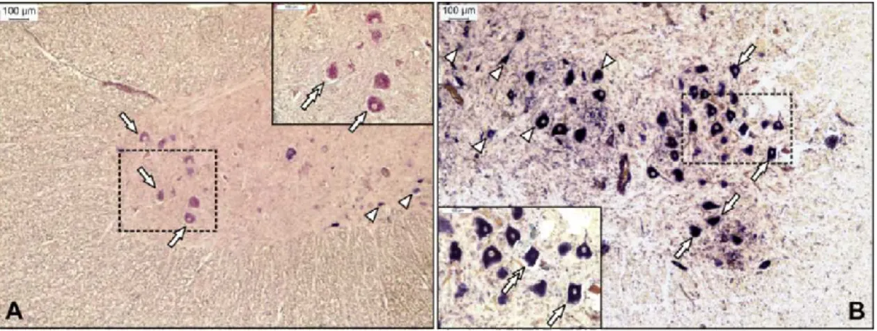
Swine. Histologically, moderate to marked perivascular lymphohistiocytic cell cuffing with marked vasculitis and neuronal degeneration, necrosis, and neurophagia with multifocal microgliosis and satellitosis were found in CNS lesions in pigs (15, 53). Neuronal necrosis was prominent in the gray matter of the cervical spinal cord but was also detected in neurons of the Purkinje layer (cerebellum), the medulla oblongata, cerebellar peduncles, and the midbrain (15). Necrotic neurons were variously swollen and hypereosinophilic or shrunken with tinctorial changes, including faded, ampho-philic, or eosinophilic cytoplasm (15). Nuclei of affected neurons were pyknotic, kary-orrhectic, or losing border definition within the cytoplasm (15, 53). The intensity of the CNS lesions—as in cattle—was variable between individuals at different stages of the disease (Fig. 1).
For type AII congenital tremor, mild to moderate vacuolar changes of the white matter in the cerebrum, brain stem, and especially cerebellum were reported for pigs
(52)(Table 2).
QUANTITATIVE AND MOLECULAR GENETIC ANALYSIS OF NEUROVIRULENT ASTROVIRUSES Quantitative PCR Analysis
Viral loads in human brain biopsy specimens from a boy with X-linked agammaglobulinemia were as high as 1.53 X 10⁷ RNA copies and were higher than those in postmortem samples (brain stem, 1.92 X 10⁴ RNA molecules per reaction; cerebellum, 5.39 X 10² RNA molecules per reaction; and frontal lobe, 1.14 X 10² RNA molecules per reaction). No other postmortem samples were astrovirus positive (9). The brain tissue and the cerebrospinal fluid (CSF) usually contain viral RNA, but significantly lower viral loads were found in stool and serum, the latter of which confirmed viremia (28). The viral loads in brain biopsy specimens can be 103- and 106-fold higher than those in CSF or fecal samples, respectively (28). These and other results (30, 31) highlight that analysis of CSF or stools cannot be sufficient alone and that analysis of profound specimens (antemortem or postmortem brain biopsy specimens) may be required in order to make an appropriate laboratory diagnosis of neurovirulent astrovirus infection.
In ovines, the highest viral loads were detected in regions of the CNS (cerebrum, 2.16 X 10⁷/xl template; and cerebellum, 1.23 X 10⁵/xl template), including the obex (1.34 X 10⁵ to 1.06 X 10⁶/xl template) and spinal cord (1.59 X 10⁵/xl template) (14). Other organs, such as the spleen, ileum, and pooled intestine (3.72 X 10²/xl template), showed remarkably low viral loads in comparison to those in organs of the CNS (14). Furthermore, low to moderate RNA loads were detected in lymphoid tissue of tonsils of sheep (14).
Similar results were reported for domestic pigs. The highest CNS viral loads were detected in the brain stem (1.85 X 10⁶ to 7.43 X 10⁷/xg total RNA) and the cervical, thoracic, and lumbar spinal cord (1.47 X 10⁶ to 3.39 X 10⁷/xg total RNA). However, moderate to high viral RNA loads were also find in the respiratory tract, such as the nasal mucosa (2.33 X 10⁵ to 3.22 X 10⁵/xg total RNA), tonsil (9.08 X 10² to 4.65 X 10⁴/xg total RNA), and lung (1.34 X 10² to 5.01 X 10²/xg total RNA) (15). On the other hand, no or low copy numbers were detected in fecal
FIG 1 Representative sections from the anterior horn of the lumbar spinal cord for Ni-PoAstV-3-infected freshly weaned pigs with encephalomyelitis in stage 1 (posterior weakness) (A) and stage 3 (complete paralysis) (B) of the disease. The Ni-PoAstV-3-positive cells, which are presumed (based on the anatomical positions of the cells) to be motoneurons (arrows) and interneurons (arrowheads), were visualized using a digoxigenin-11-UTP (DIG)-labeled RNA probe and an in situ RNA hybridization technique.
The 189-nt-long DIG-labeled antisense PoAstV-3 RNA probe targets the RdRp region of Ni-PoAstV-3 (accession numbers KY073229 to KY073232). The sections were visualized using an anti-DIG Fab fragment conjugated to alkaline phosphatase (Roche) and NBT/BCIP solution (Roche) supplemented with 1 mM levamisole. The section in panel A was counterstained with FastRed. Note that Ni-PoAStV-3 ISH-positive apoptotic (shrinking) neurons (double arrows in inserts) are observable in all stages of the disease.
samples by nested RT-PCR (15).
Genomic Analysis of Neurovirulent Astroviruses
Due to their relatively recent discovery (in 2010) and the absence of model animals and cell cultures, little is known about the pathogenicity, replication cycle, or genomic features of Ni-AstVs, although the recently described Caco-2 cell-adapted Ni-HAstV strain VA1 may facilitate the cytopathogenic research on Ni-HAstVs (55). The majority of the current knowledge about the genome organization, viral peptide composition, and virion architecture of AstVs originated from experimental analysis of the cultivable turkey (Tu) and "classical" human astroviruses, mainly TuAstV-2, HAstV-1, HAstV-2, and HAstV-8, which have more than 40 years of scientific research background (56, 57).
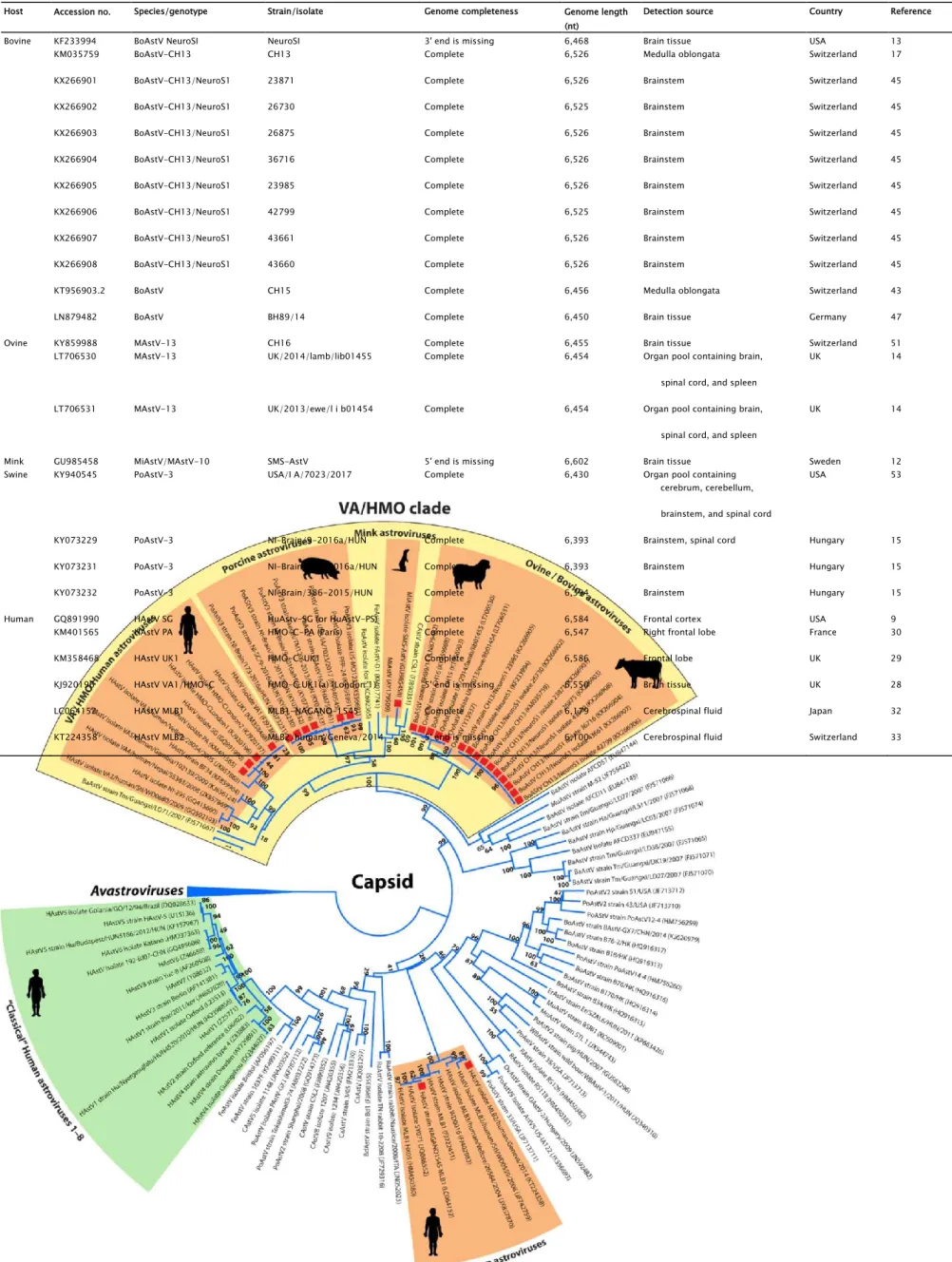
However, in silico genome analyses of Ni-AstVs and genome comparisons with the well-studied "classical" AstVs may reveal conserved sites and possible functional genome regions which may be important for the genome replication and even neurovirulence of Ni-AstVs. In this section, the results of in silico comparative genomic analyses of all Ni-AstV genomes are summarized. There are total of 26 Ni-AstV strains detected in CNS samples with known complete genome sequences or complete coding sequences (CDS) available in the GenBank database (as of 5 March 2018). The sequences originated from human (HAstV; n = 6), mink (MiAstV; n = 1), ovine (OvAstV; n = 3), bovine (BoAstV; n = 12), and porcine (PoAstV; n = 4) hosts located in Europe, the United States, and Japan (Table 3).
Phylogenetically, the majority (24/26 strains) of the Ni-AstVs belong to the VA/HMO phylogenetic clade (MAstV genogroup II), with the exception of Ni-HAstV-MLB strains (Fig. 2). Although other, non-HMO AstVs are also detectable from CNS samples, the dominance of Ni-AstVs in the VA/HMO clade suggests an increased neurotropic potential of this group of astroviruses compared to that of other AstVs. In the VA/HMO clade, the Ni-AstVs of the same host form distinct phylogenetic groups, with the exception of Ni-AstVs of bovine and ovine origins, which are present in a mixed group (Fig. 2) and suggest the capability of cross-species transmission of at least the Ni-BoAstVs (51, 58).
Members of the same phylogenetic group may belong to the same AstV serotype/genotype, although the official classifications of the majority of Ni-AstVs are still absent.
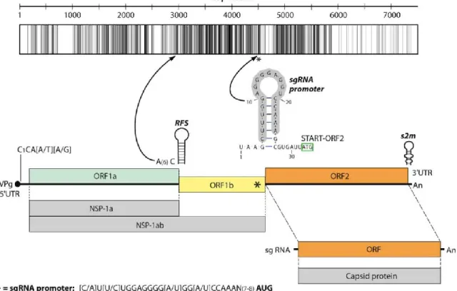
Besides the phylogenetic distance, the genome organizations of all Ni-AstVs are essentially the same and follow the general astrovirus genome architecture, with three open reading frames (ORFs) flanked by 5= and 3= untranslated regions (UTRs) (Fig. 3)(57, 59). The 5=UTRs of AstVs, including the Ni-AstVs, predominantly start with the conserved pentamer CCAAA (60), although there are some Ni-AstVs [such as MiAstV strain SMS-AstV, HAstV strain HMO-C-UK1(a)-London1, and HAstV-MLB2 strain MLB2/human/ Geneva/2014] in which this motif is missing, probably due to the incompletely determined 5= UTR (Fig. 3; Table 3). The full-length 5= UTRs of Ni-AstVs are relatively short, ranging from 14 nucleotides (nt) (HAstV-MLB1 strain NAGANO-1545; accession number LC064152) to 51 nt (BoAstV strain NeuroS1;
accession number KF233994), and may contain a variable number of stem-loops (data not shown) (45).
An identity graph of the nt alignment (Fig. 3) of all (n = 26) Ni-AstVs revealed the presence of conserved regions, including the ribosomal frameshift signal (RFS; adenine hexamer, cysteine, and stem-loop), located in the junction of the ORFs, and the presumed subgenomic RNA (sgRNA) promoter sequence, located in the 3= end of ORF1b (57, 59, 61)(Fig. 3). Due to the presence of RFS, ORF1a and the adjacent ORF1b could be transcribed continuously (ORF1ab), resulting in production of nonstructural polyprotein 1ab (NSP-1ab), while ORF2 and the sgRNA copies could encode the capsid proteins (62). Besides the primary sequence conservation, the promoter sequence of sgRNA could be found in a conserved stem-loop structure (Fig. 3). Most of the conserved nt are located in ORF1b and the 5= end of ORF2 (Fig. 3).
The 3’ UTRs of NI-AstVs are relatively short, ranging from 32 nt (HAstV-MLB2 strain MLB2/human/Geneva/2014; accession number KT224358.1) to 101 nt (HAstV-PA strain Paris; accession number KM401565.1) without the poly(A) tail, and they may contain a variable number of stem-loops (45; data not shown). The highly conserved stem-loop II-like motif (s2m), identified in four different families of positive-sense single-stranded RNA (ssRNA) viruses—Astroviridae, Caliciviridae, Picornaviridae, and Coronaviridae (63, 64)—is predominantly present in the 3= end of the genome for all Ni-AstVs, with the exception of Ni-HAstV-MLB strains NAGANO1545 and MLB2/human/Geneva/2014, similar to other MLB AstVs (22). s2m is highly conserved at both the structural and
FIG 2 Phylogenetic analysis of full-length capsid proteins (ORF2) of representative members of the family Astroviridae. The amino acid sequences were aligned with the MUSCLE algorithm (86), and evolutionary analysis was conducted in MEGA 6 (87), using an ML method based on the LG model with frequencies, a discrete gamma distribution (+G), and evolutionarily invariable sites ( + I). Bootstrap values were determined for 1,000 replicates. The Ni-AstV strains with known full-length capsid proteins are indicated with red squares. The astrovirus clades representing the known Ni-AstVs are highlighted in orange. The Avastroviruses (accession numbers AB033998, FR727149, HQ889774, FR727146, KJ020899, JF414802, Y15936, AF206663, EU143851, FJ434664, and FJ919228) were chosen as a tree outgroup. Abbreviations:
Host Accession no. Species/genotype Strain/isolate Genome completeness Genome length (nt)
Detection source Country Reference
Bovine KF233994 BoAstV NeuroSI NeuroSI 3' end is missing 6,468 Brain tissue USA 13
KM035759 BoAstV-CH13 CH13 Complete 6,526 Medulla oblongata Switzerland 17
KX266901 BoAstV-CH13/NeuroS1 23871 Complete 6,526 Brainstem Switzerland 45
KX266902 BoAstV-CH13/NeuroS1 26730 Complete 6,525 Brainstem Switzerland 45
KX266903 BoAstV-CH13/NeuroS1 26875 Complete 6,526 Brainstem Switzerland 45
KX266904 BoAstV-CH13/NeuroS1 36716 Complete 6,526 Brainstem Switzerland 45
KX266905 BoAstV-CH13/NeuroS1 23985 Complete 6,526 Brainstem Switzerland 45
KX266906 BoAstV-CH13/NeuroS1 42799 Complete 6,525 Brainstem Switzerland 45
KX266907 BoAstV-CH13/NeuroS1 43661 Complete 6,526 Brainstem Switzerland 45
KX266908 BoAstV-CH13/NeuroS1 43660 Complete 6,526 Brainstem Switzerland 45
KT956903.2 BoAstV CH15 Complete 6,456 Medulla oblongata Switzerland 43
LN879482 BoAstV BH89/14 Complete 6,450 Brain tissue Germany 47
Ovine KY859988 MAstV-13 CH16 Complete 6,455 Brain tissue Switzerland 51
LT706530 MAstV-13 UK/2014/lamb/lib01455 Complete 6,454 Organ pool containing brain, UK 14
spinal cord, and spleen
LT706531 MAstV-13 UK/2013/ewe/l i b01454 Complete 6,454 Organ pool containing brain, UK 14
spinal cord, and spleen
Mink GU985458 MiAstV/MAstV-10 SMS-AstV 5' end is missing 6,602 Brain tissue Sweden 12
Swine KY940545 PoAstV-3 USA/I A/7023/2017 Complete 6,430 Organ pool containing USA 53
cerebrum, cerebellum, brainstem, and spinal cord
KY073229 PoAstV-3 NI-Brain/9-2016a/HUN Complete 6,393 Brainstem, spinal cord Hungary 15
KY073231 PoAstV-3 NI-Brain/173-2016a/HUN Complete 6,393 Brainstem Hungary 15
KY073232 PoAstV-3 NI-Brain/386-2015/HUN Complete 6,394 Brainstem Hungary 15
Human GQ891990 HAstV SG HuAstv-SG (or HuAstV-PS) Complete 6,584 Frontal cortex USA 9
KM401565 HAstV PA HMO-C-PA (Paris) Complete 6,547 Right frontal lobe France 30
KM358468 HAstV UK1 HMO-C-UK1 Complete 6,586 Frontal lobe UK 29
KJ920196 HAstV VA1/HMO-C HMO-C UK1(a) (London 1) 5' end is missing 6,550 Brain tissue UK 28
LC064152 HAstV MLB1 MLB1-NAGANO-1545 Complete 6,179 Cerebrospinal fluid Japan 32
KT224358 HAstV MLB2 MLB2/human/Geneva/2014 5' end is missing 6,100 Cerebrospinal fluid Switzerland 33
TABLE 3 Summary of currently known neurovirulent (identified from CNS samples) astroviruses with complete genome or coding sequences obtained from humans and animals
BaAstV, bat astrovirus; BdAstV, bottlenose dolphin astrovirus; BoAstV, bovine astrovirus; CAstV, California sea lion astrovirus; CaAstV, canine astrovirus; ErAstV, European roller astrovirus; FeAstV, feline astrovirus; FoAstV, fox astrovirus; HAstV, human astrovirus; MiAstV, mink astrovirus; MuAstV, murine astrovirus; OvAstV, ovine astrovirus; PoAstV, porcine astrovirus; RaAstV, rabbit astrovirus; RAstV, rat astrovirus; WBAStV, wild boar astrovirus.
sequence levels, is mostly present at the junction of ORF2 and the 3'UTR, and may contain the stop codon of ORF2 (CGMGGCCACGCCGAGUAGGAHCGAGGGUACAGS [the stop codon is underlined]), with the exception of Ni-MiAstV strain SMS-AstV (accession number GU985458) and Ni-BoAstV strain NeuroS1 (accession number KF233994). In Ni-MiAstV strain SMS-AstV, the stop codon is 11 nt upstream from s2m, and in Ni-BoAstV strain NeuroS1, the first nucleotide of s2m is uniquely U instead of C, which causes termination of ORF2 before s2m (data not shown). The first 15 nt of s2m (CGMGGCCACGCCGAG) encode the RGHAE amino acid (aa) motif, which is the highly conserved C-terminal 5 aa of the capsid protein in the majority of Ni-AstVs. Interestingly, identical or slightly modified (RTHAE) motifs are also present 15 aa upstream of the RGHAE motif of s2m in all Ni-BoAstVs (data not shown), which suggests the acquisition of two s2m by the ancestors of Ni-BoAstVs, similar to those found in certain corona- and picornaviruses (63). The exact role of s2m in the replication process of Ni-AstVs and other s2m-containing viruses is still unknown, although the conserved nature and wide distribution of the motif may suggest an important role in the replication process. The 3'UTRs of Ni-AstVs may contain deletions and sequence repeats, such as the G6382/6393AUUUCUUUNA sequence of Ni-PoAstV-3 of USA/IA/7023/ 2017, which is missing in the Hungarian Ni-PoAstV-3 strains (15).
Amino acid sequence comparisons of NSP-1ab and the capsid proteins transcribed from ORF1ab and ORF2 of all Ni-AstVs revealed the presence of highly conserved genomic regions located mainly in the presumed helicase (Hel), protease (Pro), and RNA-dependent RNA polymerase (RdRp) regions of NSP-1ab and the conserved, presumably assembly domain of the capsid (Fig. 4).
FIG 3 Representative genome map and identity graph (above the map) for Ni-AstVs. Vertical lines in the identity graph represent identical nucleotides (nt) in the alignment.
Thicker lines represent multiple, consecutive identical nt. The identity graph was generated from the MUSCLE alignment of the complete genome/complete coding sequences of all known Ni-AstVs by use of UGENE ver 1.26 and CorelDraw ver. 12. The ruler above the identity graph represents nt positions in the alignment. The promoter sequence (gray background) of sgRNA is part of a conserved stem-loop (*). The secondary RNA structure of the sgRNA promoter was generated from the alignment of the corresponding genome region of all Ni-AstVs by use of the RNAalifold Web server (88) and VARNA visualization software, version 3.9. Note that s2m of the 3' end is missing in the Ni-HAstV strains of MLB-1 (accession number LC064152) and MLB-2 (accession number KT224358). ORF, open reading frame; RFS, ribosomal frameshift signal; NSP-1a, nonstructural polyprotein translated from ORFIa; NSP-1ab, nonstructural polyprotein continuously translated from ORF1a and ORF1b; sgRNA, subgenomic RNA.
FIG 4 Schematic maps and identity graphs (above the maps) for nonstructural protein 1ab (NSP-1ab) and capsid for all known Ni-AstVs.
Vertical lines in the identity graphs represent identical amino acids in the alignment. Thicker lines indicate multiple, consecutive identical aa. The identity graphs were generated from the MUSCLE alignments of NSP-1ab and capsid proteins of all known Ni-AstVs with complete genome sequences or complete CDS by use of UGENE ver 1.26 and CorelDraw ver. 12. The rulers above the identity graphs represent aa positions in the alignments. Hel, helicase; cc 1 to 3, coiled coils; TM 1 to 7, transmembrane domains; VPg, genome-associated viral protein; NLS, nuclear localization sequence; HVR, hypervariable region; RdRp, RNA-dependent RNA polymerase; RBD, RNA-binding domain. The red arrow shows a possible trypsin cleavage site, and yellow arrows show possible caspase cleavage sites.
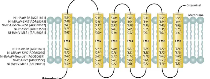
In NSP-1ab, the conserved aa motifs of Pro (65-68) as well as the two coiled coils (CC) and the multiple transmembrane (TM) helices upstream of Pro described for the "classical" HAstVs are present in all Ni-AstVs (57)(Fig. 4). Interestingly, NSP-1ab of Ni-AstVs may contain seven TM domains (Fig. 4 and 5) upstream of Pro, as predicted by the MEMSAT software of the PSIPRED Protein Sequence Analysis Workbench (69, 70), compared to the five or sixTM helices of "classical" HAstVs (57), although no insertions were found in the corresponding genome regions of Ni-AstVs compared to those of "classical" AstVs.
FIG 5 Schematic representation of seven predicted transmembrane domains (yellow columns; TM1 to -7) identifiable in the N-terminal region of NSP-1ab of all known Ni-AstVs. Numbers in parentheses and italics indicate the first (numbers in the upper part) and last (numbers in the lower part) aa positions of the given TM domain. The predictions are based on the sequences listed on the left side of the figure and were determined using the MEMSAT software of the PSIPRED Protein Sequence Analysis Workbench (69, 70). All Ni-AstVs contain essentially the same number and topology of TM domains (data not shown).
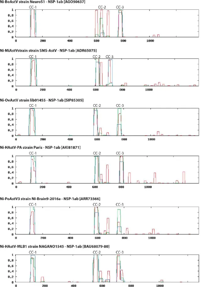
One of the well-conserved genome regions of Ni-AstVs is located downstream of the second CC and contains the conserved RExx(I/V)LRxE motif (x = variable aa) of unknown function (Fig. 4). Furthermore, besides the well-known two CCs located in the presumed Hel region and between the Pro and VPg regions of all AstVs, a third CC is predictable in the hypervariable region (HVR) of Ni-AstVs (Fig. 4 and 6)by using the coiled-coil prediction software of ExPASy (71, 72). The HVR of "classical" HAstVs may contain several insertions/deletions and is known to be associated with the adaptation of certain cell lines, different RNA replication and growth rates, and different viral RNA levels in feces (73, 74). The exact role of the HVR in the replication cycle or neuropathogenic potential of Ni-AstVs is currently unknown, although the presence of a CC structure—CC proteins are usually involved in important biological functions, such as transcriptional control (75)—and some highly conserved aa [Gxx(P/L)FxQR] (Fig. 4) may indicate a pivotal role of this region.
The identity graph for all capsids of the 26 Ni-AstVs shows the presence of two major parts, an N-terminal conserved part and a C-terminal variable part, which may correspond to the "conserved" and "hypervariable" capsid regions of "classical" HAstVs (57, 76, 77)(Fig. 4).
The N-terminal parts of the capsids of Ni-AstVs have a variable primary sequence (Fig. 7), although this region of all Ni-AstVs is equally rich in the basic aa lysine (K), arginine (R), and histidine (H), and therefore this region may act as an RNA-binding domain (RBD) required in the encapsidation process (57, 76, 77)(Fig. 4 and 7A). Interestingly, short glutamine-rich (Q-rich) motifs with unknown functions were identifiable in this region in the majority of Ni-AstVs of the VA/HMO clade (Fig. 7A). The conserved N-terminal part of the capsid may act as an assembly or core domain mainly responsible for the formation of the structural core of the virion, while the highly immunogenic hypervariable region is located on the surface of the viral particle and may be responsible for receptor interaction and the strain-specific tropism of the virus (76-78). The maturation process of AstV virions includes the trypsin-mediated cleavage of the capsid polyprotein between the conserved and hypervariable regions of "classical" HAstVs (79, 80). Trypsin is expressed predominantly in the gastroenteric tract. Currently, there is no experimental evidence about the role of trypsin in the maturation of Ni-AstV virions, although the aa alignment of the capsids of "classical" AstVs and Ni-AstVs revealed the potential presence of conserved arginine (R) and lysine (K) residues, in line with experimentally identified trypsin cleavage sites of "classical" HAstVs, in all Ni-AstVs (Fig. 7B). However, the first recent experimental investigation showed no significant effect of exogenous trypsin treatment on in vitro cultures of Ni-HAstV strain VA1 originated from human stool (55). The in vivo proteolytic processing
of Ni-AstVs is unclear. If this group of astroviruses does not absolutely require trypsin-dependent proteolytic activation, then Ni-AstVs may not depend on and be restricted to the gastrointestinal tract.
The potentially surface-exposed hypervariable region of the capsid of Ni-AstVs contains only nine, scattered identical aa identifiable in the MUSCLE alignment of capsid sequences (Fig. 4), which may be responsible for neuronal infectivity, although further sequences of Ni-AstVs and experimental evidence are needed to confirm this hypothesis.
The C-terminal parts of the capsid sequences of Ni-AstVs are rich in acidic aa, such as aspartic acid (D) and glutamic acid (E), similar to those in "classical" HAstVs. The acidic regions of the C-terminal end contain several presumed caspase cleavage sites (e.g., DXXD and XEXD), similar to those found in the "classical" HAstVs (81, 82). Interestingly, the conserved motif Q(I/L)QxR(F/Y), adjacent to the D/E-rich acidic section of unknown function, was identifiable in the majority of Ni-AstVs of the HMO clade (Fig. 7C).
In summary, the topology of the Ni-AstVs in the phylogenetic tree (Fig. 2) suggests a common lineage of neurovirulent astroviruses;
however, the determination of the specific nt and aa motifs that are involved in neuroinvasion warrants further experimental investigation.
FIG 6 Representative probability (y axis) diagrams for predicted coiled-coil structures and their localizations in the aa sequences of NSP-1ab (x axis). The diagrams were generated using Ni-AstV reference sequences (indicated above the diagrams) and screening windows of 14, 21, and 28 aa (blue, green, and red lines) by use of the coiled-coil prediction software of ExPASy (71, 72). All other Ni-AstV sequences showed essentially the same localization of coiled coils (data not shown).
FIG 7 (A) Amino acid alignment of the N-terminal ends (RNA-binding domain [RBD]) of representative capsid sequences of neurovirulent (Ni) AstVs. Conservative and basic amino acids (R K, and H) are shown with black and green backgrounds. Glutamine (Q) residues in Q-rich regions are marked with a deep yellow background and red boxes. Amino acid positions are shown according to the scale above the sequences. (B) Amino acid alignment and presumed trypsin cleavage sites (arrows) of the junctions of the conserved and hypervariable regions of representative neurovirulent (Ni) and "classical" AstV capsids. Red squares indicate N-terminal aa sequences of VP26 (HAstV2) and VP27/VP28 (HAstV8), experimentally identified by N-terminal sequencing (80, 89). Identical and highly conserved aa are marked with a black background in the alignment. Colored arrows indicate the conserved arginine (R) and lysine (K) residues which may act as potential trypsin cleavage sites (90), among which the conserved R/K residues marked with a green background and a red arrow are the most likely to be active. Numbers in parentheses indicate the first and last aa positions in the capsid polyprotein. (C) Representative amino acid alignment of the surroundings of the conserved acidic section (blue boxes), located in the C-terminal hypervariable regions, for all known neurovirulent (Ni; VA/HMO clade) and
"classical" AstV capsids. Conservative and acidic amino acids (inside blue boxes) are shown with black and yellow backgrounds, respectively.
Putative caspase cleavage sites of HAstV8 are marked in bold and underlined (81, 82). The conserved genome region that contains the Q(I/L)QxR(F/Y) motif, which is predominantly present among Ni-AstVs, is marked with a red box. Numbers in parentheses indicate the first and last aa positions in the capsid polyprotein.
POSSIBLE TRANSMISSION AND PATHOGENESIS OF NEUROVIRULENT ASTROVIRUSES
It is a general "dogma" that astroviruses are transmitted by the fecal-oral route, as they are associated with enteric infections and shedding in the feces. For humans, it has therefore also been postulated that invasion of the CNS occurs via the enteric tract as a consequence of immunodeficiency. This concept is supported by the finding that the VA1/HMO-C astroviruses have also been identified in the feces of a diseased patient as well as in healthy individuals (43). For humans, close contact with animals (mink) and young children and medical treatments (intravenous immunoglobulin and stem cell grafts) have all been suggested as possible—but not confirmed—sources of infection (9). While systematic investigations related to all potential sources and transmission modes of neurovirulent astrovirus are not available, there are some interesting novel findings which suggest the potential for extraintestinal virus shedding in different hosts. (i) The neurotropic astrovirus strains have not yet been identified in cattle feces (43). (ii) The human astrovirus VA1 was identified by NGS in a nasopharyngeal specimen from a
child with respiratory disease of unknown origin (83). (iii) Furthermore, Ni-HAstV was also detectable in throat swab and urine samples from an immunocompromised patient with HAstV-MLBI-associated acute encephalopathy (32). (iv) Neurovirulent PoAstV-3 (VA/HMO clade) was detected in high viral loads in nasal mucosa and tonsil samples from domestic pigs with encephalomyelitis, but no or consistently lower copy numbers were found in fecal samples from the affected animals by nested RT-PCR (15). (v) Overall, 33 (32%) nasal swabs and none of the 24 analyzed anal swabs, collected as sample pairs from 18- to 21-day-old healthy pigs on four different swine farms with no history of encephalitis in Hungary, were positive for PoAstV-3 by nested RT-PCR (Boros et al., unpublished data). These results indicate that certain astroviruses may be capable of being shed extraintestinally, and therefore investigations should be extended to the respiratory tract for determination of the possible respiratory source and/or route of infections.
The pathogenesis of and influential risk factors (host, virus, and environmental) for the neurovirulent astroviruses are unknown.
Immunosuppression and an immunocom-promised condition are common underlying diseases in neurovirulent astrovirus cases in humans and weaned pigs (15,19). Animals in highly prolific and intensive production management environments are often exposed to stressors which may also cause immunosuppression in cattle and pigs (15, 43).
LABORATORY DIAGNOSIS OF NEUROVIRULENT ASTROVIRUS INFECTIONS
There are several classical and modern laboratory methods for detection of classical (human) astroviruses, especially from feces (84).
However, these methods are limited for the detection of the newly discovered group of neurovirulent astroviruses. Neither the genetic diversity of the circulating astroviruses nor the specificity of the available tests is known for neurovirulent astroviruses. In addition, it seems that the optimal speci-men(s) for detection of neurovirulent astrovirus infection is probably something (e.g., CNS biopsy specimen or respiratory tract samples) other than feces (see Possible Transmission and Pathogenesis of Neurovirulent Astroviruses).
There is limited knowledge related to the in vitro tissue culture propagation of neurovirulent astroviruses. A recent study reported, for the first time, that the human astrovirus VA1 of stool origin can grow in cell cultures (without cytopathic effect), including cultures of human enteric Caco-2, human embryonic kidney HEK293T, and human respiratory A549 cells but not those of BHK-21 (baby hamster kidney) cells (55).
Interestingly, VA1 could be cultured equally well with and without the addition of exogenous trypsin (55). There is no report of the propagation of VA1/HMO-C strains directly from the affected CNS tissues.
At present, electron microscopy (EM), ISH, RT-PCR, real-time PCR (with strain-specific primer pairs), and viral metagenomics/NGS research-based methods are available for detection of neurovirulent astroviruses; however, there are no commercially available assays.
A hyperimmune antibody against the conserved N terminus of the viral capsid protein was recently developed for detection of BoAstV-CH13/BoAstV NeuroSI infections in cattle brain tissue samples (51). This immunohistochemistry (IHC) assay is sensitive and specific (51) for BoAstV-CH13/BoAstV NeuroSI; however, it is not available for commercial use.
Development and use of a sensitive routine laboratory test (e.g., using an enzyme-linked immunosorbent assay [ELISA], immunochromatography, or molecular method) for detection of neurovirulent astroviruses is necessary, especially for veterinary medicine.
THERAPY
Combinations of intravenous immunoglobulin (IVIG), methylprednisolone, ribavirin, and pegylated interferon (PEG-IFN) alpha-2b were applied as nonspecific antiviral treatments in a human case of neurovirulent astrovirus infection (30). In that study, progression of motor function and mild memory improvement were found at month 12 of follow-up (30). On the other hand, there was no response to ribavirin and IVIG therapy (after the patient previously received steroids and IVIG) in another fatal human case of neurovirulent astrovirus encephalitis (29). At present, there is no specific antiviral therapy for neurovirulent astrovirus infection of either humans or animals.
FUTURE PERSPECTIVE
Astrovirus was discovered in a fecal sample in 1975 (85). Until now, astrovirus has been thought to be an infectious agent only of the gastroenteric tract in humans and animals. The most recent and advanced high-throughput molecular techniques have changed our knowledge of astrovirus infection (84). The possibility of an extraintestinal infection or complication of astrovirus infection was first suggested in 2010 (9). Be- tween 2010 and 2017, significant numbers of studies reported an association between astrovirus and CNS infection. To date, this association has been reported for humans and four mammalian species. These results demonstrate a previously unrecognized neurotropism of neurovirulent astrovirus, a causative agent of neuroinfections, such as encephalitis, meningitis, myelitis, and ganglionitis. The first studies not only support the association but also suggest that certain astroviruses may be relatively common infectious agents of the CNS in cases of undifferentiated viral encephalitis. Retrospective and prospective systematic investigations of astrovirus should be performed, focusing on cases and outbreaks of unknown encephalitis—whose economic cost is profound for cases in both humans and domestic animals. It would also be interesting to determine if all astroviruses have an affinity for the CNS or if this affinity is restricted to certain neurovirulent astrovirus lineages.
Astrovirus should be considered in the differential diagnosis of CNS diseases, including encephalitis, especially for
nonimmunocompetent patients. In addition, we cannot exclude the possibility that a certain proportion of unknown encephalitis cases in domesticated mammals are also related to astrovirus. But timely diagnosis is hindered by the lack of available laboratory assays to rapidly survey the potential agents responsible for encephalitis. Development of a specific routine laboratory test is necessary for detection of astroviruses in encephalitis and other CNS infections. In particular, an astrovirus detection assay should be included in the diagnostic armory and routinely used in laboratories of veterinary medicine.
The optimal specimen type for laboratory diagnosis of neurovirulent astrovirus infection is also problematic. Based on the present data, no or very low levels of astrovirus RNA are present in the feces, serum, and CSF, which leads to misdiagnosis of neurological infections.
Invasive brain biopsy for correct diagnosis of astrovirus encephalitis clearly has related medical and ethical issues. A new report suggests that nasal and respiratory specimens are better and noninvasive samples for laboratory detection of neurovirulent astrovirus (15). According to the present research, a broad range of specimens should be collected for detection of astrovirus in CNS diseases.
The first reports indicated that the neurotropic astroviruses are geographically widespread in human, mink, bovine, and porcine species and that epidemic spread is also possible (12, 15). In addition, while neurovirulent astrovirus infections have been reported for humans and four mammalian species, it is possible that this type of infection occurs in other domestic and wild animal species as well. Extended investigations of known hosts and other related mammals (e.g., wild boar, roe deer, goat, etc.) with unknown meningoencephalitis causes should also be done.
The first reports have drawn our attention to neurovirulent astrovirus infections. More comprehensive epidemiological studies on the clinical presentation, prevalence, distribution, extraintestinal transmission, immunology, virology, and diversity of neurovirulent astrovirus infections, with determination of complete genome sequences of further neurovirulent astroviruses, should be performed. The detailed pathogenesis of neurovirulent astrovirus infections is also lacking, and exogenous and endogenous factors determining the tropism and neuroinvasion of these viruses are still unknown. In vitro cell culture and in vivo animal models (e.g., pig and mink models) would facilitate studies of the neurotropism, replication sites, neuropathogenesis, and immune control of astroviruses.
In the future, more attention should be paid to the specific prevention of CNS infections, especially among domesticated animals, and to the pathogenesis and epidemiological investigation of neurovirulent astroviruses. Development and use of a specific vaccine against neurovirulent astroviruses would be an important, logical, and probably cost-effective step, especially for domesticated animals (e.g., pigs).
Astroviruses show typical features suitable for a "one health" approach, in which human and animal health can be discussed together. Future research into astrovirus infections should include collaborative, multidisciplinary efforts in both veterinary and human medicine. Human and veterinary physicians should be aware of astrovirus infection as a potential differential diagnosis with corresponding signs of neurologic disease.
ACKNOWLEDGMENTS
This work was supported by a grant from the Hungarian Scientific Research Fund (grant OTKA/NKFIH K111615). P.P. and A.B. were supported by the János Bolyai Research Scholarship of the Hungarian Academy of Sciences.
The funders had no role in study design, data collection and interpretation, or the decision to submit the work for publication.
REFERENCES
1. Glaser CA, Honarmand S, Anderson LJ, Schnurr DP, Forghani B, Cossen CK, Schuster FL, Christie LJ, Tureen JH. 2006. Beyond viruses: clinical profiles and etiologies associated with encephalitis. Clin Infect Dis 43: 1565-1577. https://doi.org/10.1086/509330.
2. Long MT. Overview of meningitis, encephalitis, and encephalomyelitis. MSD veterinary manual.
http://www.msdvetmanual.com/nervous-system/ meningitis,-encephalitis,-and-encephalomyelitis/overview-of-meningitis, -encephalitis,-and-encephalomyelitis. Accessed 2 November 2017.
3. World Organization for Animal Health. 2004. Manual of diagnostic tests and vaccines for terrestrial animals, 5th ed. World Organization for Animal Health, Paris, France.
4. Saylor D, Thakur K, Venkatesan A. 2015. Acute encephalitis in the im-munocompromised individual. Curr Opin 28:330-336.
https://doi.org/10 .1097/YCO.0000000000000169.
5. Glaser CA, Gilliam S, Schnurr D, Forghani B, Honarmand S, Khetsuriani N, Fischer M, Cossen CK, Anderson LJ. 2003. In search of encephalitis etiologies: diagnostic challenges in the California Encephalitis Project, 1998-2000. Clin Infect Dis 36:731-742.
https://doi.org/10.1086/367841.
6. Kennedy PGE, Quan PL, Lipkin WI. 2017. Viral encephalitis of unknown cause: current perspective and recent advances. Viruses 9:138.
https:// doi.org/10.3390/v9060138.
7. Vora NM, Holman RC, Mehal JM, Steiner CA, Blanton J, Sejvar J. 2014. Burden of encephalitis-associated hospitalizations in the United States, 1998-2010. Neurology 82:443-451. https://doi.org/10.1212/WNL.0000000000000086.
8. Selimovic-Hamza S, Boujon CL, Hilbe M, Oevermann A, Seuberlich T. 2017. Frequency and pathological phenotype of bovine astrovirus CH13/ NeuroSI infection in neurologically-diseased cattle: towards assessment of causality. Viruses 9:12.
![FIG 7 (A) Amino acid alignment of the N-terminal ends (RNA-binding domain [RBD]) of representative capsid sequences of neurovirulent (Ni) AstVs](https://thumb-eu.123doks.com/thumbv2/9dokorg/1420084.120183/13.1262.107.756.97.868/amino-alignment-terminal-binding-domain-representative-sequences-neurovirulent.webp)
