Circadian rhythm and fetal programming
Ph.D. Thesis
Krisztina Mészáros, M.D.
Doctoral School of Clinical Medicine, Semmelweis University
Supervisor: Prof. Attila J. Szabó, M.D., Ph.D., D.Sc., (Habil.) Consultant: Prof. Franz Schaefer, M.D.
Official reviewers: Attila Patócs, MD, Ph.D.
Tibor Ertl, MD, Ph.D., D.Sc., (Habil.)
Head of the Final Examination Committee: Dr. László Szabó M.D., D.Sc.
Members of the Committee: Krisztina Káldi, M.D., Ph.D.
Tibor Kovács, M.D., Ph.D., (Habil.) Budapest
2017
1
“Nothing is as powerful as an idea whose time has come.”
(Victor Hugo)
2
CONTENTS
ABBREVIATIONS ... 5
1. INTRODUCTION ... 7
1.1. Circadian rhythm in mammals: an endogenous, self-sustained system ... 7
1.1.1. Dysfunction of the system: what happens when the clock goes wrong? ... 8
1.2. Molecular network of the circadian rhythms: what is ticking? ... 9
1.3. Circadian organization: the central and peripheral circadian clocks ... 11
1.3.1. The master clock: SCN ... 11
1.3.2. Input to the SCN ... 12
1.3.3. Output from the SCN ... 13
1.3.4. Other ins and outs: feeding the clock ... 14
1.3.5. Other ins and outs: glucocorticoids as potential synchronizers ... 15
1.3.6. Other ins and outs: melatonin, the night manager ... 16
1.4. Circadian rhythms during fetal development ... 19
1.5. Fatal consequences of the fetal programming ... 21
1.6. Circadian control of the kidney functions ... 22
1.7. Renal phenotypes in clock mutant models ... 25
2. THE AIMS OF THIS STUDY ... 27
3. MATERIALS AND METHODS ... 28
3.1. Time definition ... 28
3.2. Animals and experimental protocols ... 28
3.2.1. Disruption of maternal feeding regime ... 29
3.3. EXPERIMENT 1: Early effects of prenatal maternal circadian disruption ... 30
3.3.1. Effect on the fetal intrarenal clockworks ... 30
3
3.3.2. Telemetric measurements of the dams over a 24h period... 30
3.3.3. Circadian variation in renal excretion function during pregnancy ... 30
3.3.3.1. Urinary aldosterone excretion over the pregnancy ... 31
3.3.3.2. Urinary melatonin excretion... 31
3.4. EXPERIMENT 2: postnatal changes ... 32
3.4.1. Circadian gene variation after maternal circadian disruption ... 32
3.4.2. Postnatal behavioral measurements of offspring ... 33
3.4.3. Effect of timed maternal melatonin treatment in the early postnatal period.... 33
3.4.4. Periodic maternal withdrawal at the first postnatal week period ... 33
3.5. EXPERIMENT 3: long lasting effects ... 35
3.5.1. Renal function ... 35
3.5.2. Telemetric measurement ... 35
3.5.3. Echocardiography ... 36
3.6. Radiotelemetric measurement of blood pressure... 37
3.7. RNA extraction and quantitative real-time PCR ... 38
3.8. Statistical analysis ... 39
4. RESULTS... 40
4.1. Maternal outcomes (Experiment 1) ... 40
4.1.1. Effects of maternal circadian disturbance during pregnancy on the dams ... 40
4.1.2. Telemetric measurements of dams... 40
4.1.3. Circadian variation in renal excretory function during pregnancy ... 42
4.1.4. Daily urinary aldosterone over the pregnancy ... 44
4.1.5. Urinary melatonin excretion ... 48
4.2. Effects of prenatal maternal circadian disturbance (Experiment 1) ... 49
4.2.1. Intrauterine growth ... 49
4
4.2.2. Renal circadian gene variation at birth ... 51
4.3. Postnatal changes (Experiment 2) ... 56
4.3.1. Expression pattern circadian genes under LD photoperiod ... 56
4.3.2. Postnatal behavioral changes ... 63
4.3.3. The effects of timed maternal melatonin treatment ... 65
4.3.4. Periodic maternal withdrawal at the first postnatal week period ... 66
4.3.5. Postnatal changes of the gene expression pattern in offspring (LL and DD) .. 68
4.4. Follow-up experiment (Experiment 3) ... 73
4.4.1. Long-lasting effects in offspring ... 73
4.4.2. Renal function ... 78
4.4.3. Telemetric measurements ... 82
4.4.4. Echocardiography ... 84
5. DISCUSSION ... 85
6. CONCLUSIONS ... 97
7. SUMMARY ... 98
8. ÖSSZEFOGLALÁS ... 99
9. REFERENCES ... 100
10. OWN PUBLICATIONS ... 131
11. ACKNOWLEDGMENTS ... 132
5
ABBREVIATIONS
AAD aromatic amino acid decarboxylase AANAT arylalkylamine N-acetyltransferase ACTH adrenocorticotrop hormon
AMPK AMP (adenosine monophosphate) activated protein kinase ARNT Arylhydrocarbon Receptor Nuclear Translocator
AVPR2 arginine vasopressin type 2 receptor
bHLH basic-helix-loop-helix protein structural motif
BMAL brain and muscle Arnt-like protein, also known as ARNT-Like Protein cAMP cyclic adenosine monophosphate
Circadian from Latin: circa = approximately, diem = day CCGs clock controlled genes
CKI casein kinase 1
CLOCK Circadian Locomotor Output Cycles Kaput, its paralog NPAS2 CRE cAMP response element
CRY Cryptochrome (Cry1/2) DMH dorsomedial hypothalamus DRN dorsal raphe nucleus
E-box enhancer box
ELISA enzyme-linked immunosorbent assay
ENaC epithelial sodium channel (also known as amiloride-sensitive sodium channel)
E20 embryonic day 20
GHT geniculohypothalamic tract GC Glucocorticoid
HIOMT hydroxyindole-O-methyl transferase enzyme
6 HPA hypothalamic–pituitary–adrenal axis.
IGL intergeniculate leaflet LD light-dark cycle
LT long-term
miRNA small non-coding RNA molecule (ca. 22 nucleotides)
MPO medial preoptic region/area (abbreviation also known as mPOA) MRN median raphe nucleus
MT melatonin receptor (MT1 and MT2 subtypes)
NHE3 sodium-hydrogen antiporter 3 protein encoded by the scl9a3 genes.
NPAS2 Neuronal PAS domain containing protein 2 NPY neuropeptide Y
PAS Per/Arnt/Sim protein domain
PER period gene (per1 and its homologes per2 and per3) PVN paraventricular nucleus of the thalamus
RHT retinohypothalamic tract
ROR/RZR retinoic acid receptor-related orphan nuclear receptor subfamily
Rev-erbα encoded on the opposite strand of the alpha-thyroid hormone receptor (c- erbα); member of nuclear receptor subfamily
SCN paired suprachiasmatic nuclei
Sgk1 serum- and glucocorticoid-inducible kinase TRPH tryptophan hydroxylase
VIP vasoactive intestinal peptide Zeitgeber from German, means “time giver”
3-ß-HSD 3β-hydroxysteroid dehydrogenase 6-SMT 6-sulfatoxymelatonin
7
1. INTRODUCTION
1.1. Circadian rhythm in mammals: an endogenous, self-sustained system
The rhythmical environmental cues related to e.g. the Earth rotation forced life forms to synchronize their physiology via “predictive” rather than “reactive” mechanisms.1,2 The temporal organization of behavior into circadian cycles of rest- and activity periods is a fundamental feature of the organisms’ adaptation. Most organisms including mammals operate with a self-sustaining, endogenous, time-keeping cellular machinery, called the circadian clock, that allows them to facilitate their anticipatory adaptation to the rhythmic environmental changes, e.g. to the light-dark cycles.3-7 Because of its molecular regulation, the outputs of the circadian clock machinery, the circadian rhythms persist even in constant conditions (i.e. deprived solar or social contacts) with an endogenous (i.e. free running) period close to 24 h.8-11 Evidence exists that many physiological processes, e.g. sleep-wake cycle, the body temperature fluctuations, timing of hormones release as well as several aspects of cardiovascular and renal functions are governed by the circadian clock.12,13
Although the internal circadian clock is not the most accurate system: its free running period is only approximate that of the environment (24 h).14 However, under natural conditions the phase and the period of the circadian rhythms are being entrained (synchronized /adjusted /reset) by time cues (Zeitgebers) to maintain harmony with the external, environmental light-dark cycle.15 It is well established that the solar light is the major Zeitgeber, but other cyclic cues e.g. rhythmic feeding regime, behavioral activity are also dominant Zeitgebers entraining the clock.16
So far, three essential components of the circadian timing system have been described and intensively investigated: input pathways that mediating entrainment (i.e.
Zeitgebers), the circadian oscillators (i.e. cellular circadian clock machinery) and the output pathways (i.e. circadian rhythms) that express the circadian function.4
8
1.1.1. Dysfunction of the system: what happens when the clock goes wrong?
In the recent years, it has been recognized that the misalignment between the behavioral pattern driven even by social cues (social jet lag) and the endogenous rhythms causes surprisingly broad unwanted effects. Ignoring the internal circadian clock in our “24/7”
society (e.g. due to rotating shift work, jet lag) leads to reduced sleep efficiency which is associated with cognitive impairment and impaired social functions caused by mood disturbance.17 Above that, the long-term misalignment between the daily behavioral pattern (e.g. sleep-wake, or fasting-feeding cycles) and the internal circadian rhythms is known to influence essential physiological processes that are relevant to human diseases.18-20 For example, the disruption of the temporal endocrine regulation, as results of physiologic maladaptation to sleeping and eating at abnormal circadian time chronically, has a severe impact on metabolic health.20,21 For example, recent study from Harvard showed that a rearrangement of the day-night rhythm in healthy volunteers can cause insulin resistance.18
Furthermore, adverse effects of the circadian disruption have been linked to e.g.
cardiovascular dysfunctions and compromised pregnancy.22-25 An increased mortality from coronary heart disease among shift workers has been demonstrated in a historical cohort of Swedish pulp and paper industry workers.23 A correlation between shift work and prematurity or intrauterine growth retardation has been confirmed in large cohort study from China.26 Another study from Denmark also confirmed the adverse effect of the circadian disturbance by shift work on the fetal development and birth weight.25 More focused involvement of the circadian dysfunction has been observed in tumor development and progression.27 The disturbed internal clock has been linked to increased risk of e.g. breast cancer in women who work irregular shift patterns.28 Animal experiments have shown conclusively that the circadian disturbance accelerates diverse tumor development and play a significant role in the progression.29,30
9
1.2. Molecular network of the circadian rhythms: what is ticking?
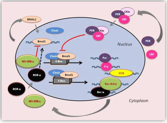
Understanding how the circadian system is controlled, first we need to understand the molecular-genetic processes at the cellular level. While, current evidence suggests that the circadian system itself is composed of as many tiny autonomously ticking clocks as there are cells of the body.31-34 On the molecular level, the fundamental circadian network which is essential for generating and sustaining circadian oscillation is based on series of transcriptional-translational feedback loops.19,35-42 To our knowledge, the mammalian genome encodes 4 core circadian genes directly involved in the regulation of the core feedback loop: Clock (its paralog protein: Npas2), Bmal1 (also known Arntl- 2), Period (Per homologs 1, 2 and 3) and Cryptochrome (Cry homologs 1 and 2). See Figure 1.
It should be noted that this scheme is oversimplified, but to put it briefly: the transcription factors that positively regulate the feedback loop are the CLOCK and BMAL1. These proteins have basic-helix-loop-helix (bHLH) PAS domains, so they can form heterodimer and bind to E-box elements in the promoter of their target genes, thus induce the expression of other clock components including Period 1-3 and Cryptochrome 1-2.38,43-45 The activation of the target gene expression is associated with chromatin remodeling by histone acetylation.46-49 The CLOCK protein itself has an intrinsic histone acetyltransferase activity.46
Then, closing the negative limb of the feedback loop, the homologues of PER and CRY proteins form complexes, accumulate rhythmically in the nucleus and once these complexes reached a critical concentration, inhibit their own transcription interacting with the BMAL1-CLOCK heterodimers.35,40,50,51 CLOCK-BMAL1 also drives the expression of nuclear hormone receptors i.e. orphan nuclear receptors: Rev-erbα and RORα which are also involved in the negative feedback loop. REV-ERBα represses, while RORα activates the Bmal1 expression.35,50,52-54 Thus, the mRNA expression and protein level of the core genes (except Clock in the many tissues) oscillate with a period close to 24h.52,55
Diverse ranges of transcripts (involved in multiple physiological functions) have been identified as targets, directly controlled by the core circadian component, e.g. CLOCK- BMAL1 heterodimer. These target genes called clock-controlled genes (CCGs) exhibit
10
rhythmic expression.44,56,57 Current estimations indicate that the clock-controlled genes constitute circa 10% of the expressed transcripts.43
Evidence suggests that the direct post-transcriptional mechanisms might have potent role in the circadian control.58,59 Recent studies have implicated that the micro-RNAs (miRNAs) - which are small non-coding RNAs regulate stability and/or translation of their target-mRNA - are involved in the precise regulation of the circadian network.60-62
Figure 1. The mammalian circadian clockwork. The circadian clock network involves transcription-translation feedback loops comprised of set core clock genes, i.e. Clock, Bmal1, Period (Per1, Per2 and Per3), Cryptochrome (Cry1 and Cry2). CLOCK and BMAL1 circadian proteins form heterodimer complexes and activate the transcription of the Per and Cry genes.
PER and CRY proteins heterodimerize, translocate back to the nucleus and interact with the CLOCK–BMAL1 complex to inhibit their own transcription. Retinoic acid-related orphan receptors, RORα and REV-ERBα which are also targets of the CLOCK–BMAL1 complex activates or represses Bmal1 transcription respectively, functioning as a secondary feedback loop. In addition, as post-translational modification of the clock proteins by phosphorylation i.e.
casein kinases (CKIɛ, CKIδ) are crucial for the circadian control. Clock-controlled genes (CCGs) directly driven by the CLOCK–BMAL1 complex show circadian expression.
11
Post-translational modifications of the clock proteins are also involved in the fine control by e.g. phosphorylation modulating the stability, heterodimer formation, subcellular localization (nuclear entry) and the degradation of the circadian clock proteins.51,63,64 The key kinases, CKIɛ, CKIδ, which are involved in the PER CRY and BMAL1 proteins phosphorylation for accelerated degradation and clearance from the nuclei are essential component of the clock machinery network.65,66 The mutations of these kinases could modify the stability of the regulated proteins, thus the period length or the amplitude of the circadian cycle.67,68 Interestingly, the first mammalian circadian mutation was documented in “tau mutant” hamster characterized by an abnormally short free running period (20 h) in behavior and the secretion pattern of melatonin as well as glucocorticoids in temporal isolation.69 It took ca. 10 years till Takahashi and colleagues revealed that the ground of abnormal circadian “phenotype” was a point mutation in the CK1ɛ.67 In humans, the same mutation leads to advance sleep phase disorders with a ca.
4-hour advance of the sleep onset in individuals, who are also called “morning larks”.68
1.3. Circadian organization: the central and peripheral circadian clocks
The mammalian endogenous timing system is divided into a central pacemaker which generate and synchronize the endogenous circadian rhythms and peripheral circadian oscillators sharing the same molecular clockwork at the cellular level. (Figure 1) In the current model of its regulation the central pacemaker is described as a conductor in an orchestra, in which the peripheral clocks can play music autonomously.70
1.3.1. The master clock: SCN
The central pacemaker of the circadian clock is located in the paired suprachiasmatic nuclei (SCN) of the anterior hypothalamus.35,71,72 The SCN cells (ca. 2x 10.000) generate and maintain self-sustained and cell-autonomous, strongly coupled (possibly mediated by GABA and VIP) circadian oscillations of the firing rhythms, metabolism as well as the expression of clock genes and their proteins at the tissue level.73,74 Recent studies have shown that SCN neurons are not identical in intrinsic properties and
12
function: the dorsal “shell” region (characterized by vasopressin expression) has been appeared to be the primary pacemaker, while the ventral “core” region receiving direct innervations from the retina (i.e. input pathways) is responsible for the (re)entrainment of the intrinsic oscillations.19 The circadian rhythm of the entire SCN is tightly synchronized, but the phase between the two regions could be different.74
The role of the SCN as the master clock in the circadian control has been extensively investigated. Accumulation of evidence includes lesion studies which are demonstrating the loss of circadian rhythmicity of the behavior pattern following ablation of the SCN in animal experiments. More importantly, the transplantation of an intact SCN grafts is restoring the rhythmicity.7275
1.3.2. Input to the SCN
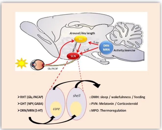
In order to keep harmony with the environmental time, SCN requires rhythmic exogenous input. The SCN receives photic and nonphotic information. The most important are the light cues detected by a subset of melanopsin expressing non-image- forming retinal ganglion cells which are directly projected to the SCN via the retinohypothalamic tract (RHT).70,76,77 Glutamatergic terminals from the retina activate multiple pathways in the “core” region of the SCN leading to light dependent phase advances or delays of the daily oscillation in clock gene expression.55 This stimulus drives a trans-synaptic activation of the “shell” region.19 In that way, continuous light could desynchronize the molecular rhythms of the SCN.78
On the other hand, RHT transfers photic information indirectly to the SCN via the geniculohypothalamic tract (GHT) pathway associated with the reward system by releasing NPY and GABA on the SCN neuron. However, its role remains controversial, serotonergic (5-HT) tracts end directly on the SCN mediating non-photic signals from the dorsal and median raphe nuclei.70,79-81 (Figure 2) These connections are made during the early postnatal period in rats and probably in the late gestation period in humans.82
13
Figure 2. Main input and output pathways of the SCN in rat. Photic input to the SCN through the retinohypothalamic tract (RHT) via glutaminergic pathway (Glu), non-photic input from the dorsal and medial raphe nucleus (DRN and MRN) via serotoninerg pathway (5-HT) and from the intergeniculate leaflet (IGL) as indirect photic input through geniculohypothalamic tract (GHT) via GABA and neuropeptide Y (NPY) pathways.
1.3.3. Output from the SCN
SCN neurons innervate diverse target regions. The main output pathway ends on the dorsomedial hypothalamus (DMH) contributing to the circadian regulation of the orexinergic system, thus to the timing of sleep and wakefulness.11,83 Through their neuronal connections SCN projects to the paraventricular nucleus of the thalamus (PVN) influencing behavior related to feeding and reward processing and to the medial preoptic region (MPO) controlling proper timing of diverse physiological functions.3
14
(Figure 2) Furthermore, SCN modulates the neuroendocrine and autonomic nervous system setting the sympathetic/parasympathetic balance.84
Beyond the sleep dependence hormonal secretion, e.g. growth hormone and prolactin, the glucocorticoid synthesis and release, as well as the nocturnal secretion of melatonin are controlled by SCN.11,85,86 Thus, the master clock, the SCN is able to synchronize circadian clocks on the periphery with the day-night rhythm due to its neuronal-, endocrine signals and the regulation of the behavior driven by the rest-activity cycle, as well as the body temperature rhythms.9,77
1.3.4. Other ins and outs: feeding the clock
The synchronization of the peripheral clocks by the SCN depends on the SCN’s control governing the rest-activity, thus the fasting-feeding cycles via autonomic and hormonal pathways.87,88 E.g. hormones which are driven by feeding or fasting might be able to adjust the phase of circadian oscillators in the periphery.89,90 However, the central pacemaker, the SCN itself has been shown to be unaffected by feeding-depend signaling pathways.91-93
The feeding time appears to be a potent Zeitgeber in some peripheral tissue i.e. in the liver, kidney and heart. 91,94,95 Under certain conditions, e.g. sudden disruption of the regular feeding regime, the peripheral oscillators can be uncoupled from the SCN’s synchronization. When the food ability is experimentally restricted to the inactive phase of nocturnal rodents (to the light period) the circadian oscillations in the periphery have been shown to be phase shifted accordingly.91,92,96,97 Furthermore, for example in the liver, the restricted feeding regime can entrain the phase of the clock gene expression in SCN-ablated mice which are otherwise completely arrhythmic.98
In mammals, most of the gene-expression patterns which are involved in the metabolic regulation show daily variation govern by the circadian clock.43,99,100 The circadian rhythms at the cellular level result from the rhythmic expression of 2 types of transcript groups: the central core circadian genes and the clock controlled genes (CCG), which are the downstream output driven by the core component. In the absence of feeding
15
cues, however, there is only a small part of transcripts including the core clock genes which maintain circadian oscillation in the peripheral tissues.101,102
Otherwise, in liver specific clock gene mutation experiments, in which the mice sustained rhythmic feeding behavior, revealed a subset of hepatic circadian clock controlled genes which maintain oscillation.103
It is becoming clear that the temporal metabolic status of a tissue might affect the circadian clock machinery of individual cells.64,104 It has been reported for example that the ratio of NADH to NAD+ is affecting the dimerization of Bmal1/Clock and their binding to their DNA sequences (E-boxes). Higher ratio of the reduced forms, NADH (NADPH) enhance, while higher ratio of the oxidized forms, NAD+, (NADP+) diminish the transcriptional activation by BMAL1/CLOCK heterodimer.64
Furthermore, it has been proposed that the adenosine monophosphate activated protein kinase (AMPK) acting as a food sensor directly phosphorylates the Cry1 protein, thereby marking it for degradation.105 Thus, Cry clock protein is functioning as a sensor of cellular energy rate and might have a pivotal role in the synchronization of the metabolism, e.g. glucose homeostasis at cellular level.106 Thus, signals related to food intake have major roles in the regulation of the daily expression pattern of clock- and clock controlled genes at the cellular level.107,108 In addition to the time of feeding, food content e.g. high salt diet can alter the circadian gene expression in the liver and kidney as well.109,110
1.3.5. Other ins and outs: glucocorticoids as potential synchronizers
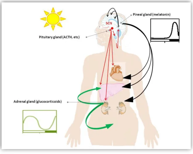
A group of steroid hormones involved in diverse functions including stress response, anti- inflammations, cardiovascular and metabolic functions are synthesized and secreted in circadian manner.86,111-113 Son et al. have reported that the rhythmic release of glucocorticoids (GCs) from the adrenal cortex is controlled by the peripheral clock in the adrenal gland.114,115 Otherwise it is known that the synthesis is controlled by the SCN via direct neuronal pathways.85,116 (Figure 4) Data are suggesting that the sensitivity of adrenal gland to ACTH depends on the daytime and the daily GCs peak is driven by the SCN.83,116 The ablation of SCN leads to loss of rhythmic secretion of
16
glucocorticoids in rats.71 Intracellular glucocorticoid receptors are expressed in almost all cells in the body (except the SCN, it has no GC receptors) which suggests their pivotal role in the synchronization of the peripheral clocks with the central pacemaker.89,108,117,118 Interestingly, the intracellular GC receptors, as well as numerous cytochrome P450 enzymes that are important in the steroids metabolism show robust circadian oscillation at the transcript level.119-122
The generally accepted mechanism for GC’s effects is the transcriptional activations of selective target genes, however, nongenomic mechanisms of the GC are also intensively studied.123,124 Indeed, a direct interaction between CRY1/2 and glucocorticoids has been recently reported.44,106 Furthermore, because of the CRE sequences of Period (1/2), it is strongly affected by glucocorticoid via cAMP/CRE signaling pathways.125 In animal studies, the synthetic glucocorticoid, dexamethasone can induce a phase shift of circadian rhythmicity in different peripheral tissue, while the SCN remains unaffected.89,126-128 However, in adrenalectomized rats most of the clock genes except Per1/2 remain rhythmic in the periphery.129 Furthermore, the circadian rhythm of GC in rodents might influence the daily rhythm of serotonin synthesis in the raphe nuclei.130 In adrenalectomized rats, serotonin is expressed at constant level which might indicate the circadian control of the behavioral and emotional function by the SCN through the GC rhythms.130
1.3.6. Other ins and outs: melatonin, the night manager
It has become evident for decades that the mammalian pineal gland (has been called as a
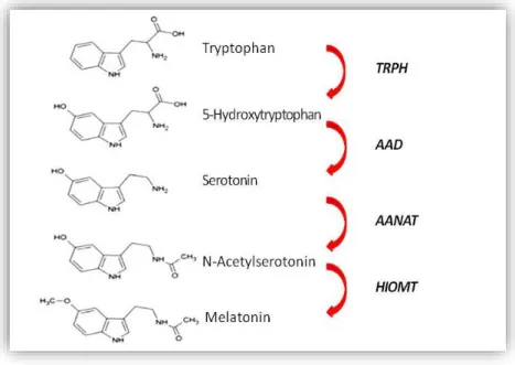
“third eye”) is involved in the synchronization of the circadian clock of the body to the external light-dark cycles.131-134 Melatonin is synthesized by the pinealocytes from the essential amino acid tryptophan with circadian manner (Figure 3).
Its synthesis is driven by the SCN via multi-synaptic neural pathway and inhibited by light exposure.135,136 During the light period serotonin, as an intermediate metabolite tends to be stored in the pinealocytes. With the onset of darkness begins the final step mediated by arylalkylamine N-acetyl-transferase (AANAT) of the melatonin production which is driven by the SCN via noradrenergic sympathetic fibers.137-139 The sympathetic
17
Figure 3. The melatonin synthesis pathway. Melatonin is synthesized from tryptophan. The rate limiting steps mediated by arylalkylamine N-acetyl-transferase (AANAT) and hydroxyindole-O-methyl transferase (HIOMT). TRPH: tryptophan hydroxylase, AAD: aromatic amino acid decarboxylase.
control is necessary in this regulation: it is known that patients with a cervical cord injury have a loss of circadian melatonin synthesis.140,141 Melatonin is released rapidly into the blood stream and the nocturnal plasma melatonin concentration is ascending more than 10-fold of the daytime level.141 (Figure 4) The circulated melatonin is then rapidly inactivated in the liver, thus its plasma level precisely reflects its pineal synthesis essentially. However, there is new evidence of its extrapineal synthesis (i.e.
gut, lymphocytes and placenta), the extrapineal melatonin production might not contribute significantly to its plasma level concentration.142-144
It has been suggested that melatonin has diverse roles in the physiology, the most important to see that diurnal melatonin release synchronizes the entire body with the nighttime.145,146 It is known that the production of melatonin declines with age. This decline may reflect the progressive age-related calcification of the pineal gland.147
However, the exact effects and side effect of the exogenous melatonin are not fully explored, melatonin supplementations have been used for decades as dietary supplements to treat “sleep problems” especially in the older population. Interestingly,
18
symptoms related to endogen overproduction of melatonin, i.e. Shapiro’s syndrome including periodic syncope, hypothermia and hyperhidrosis.148 On the other hand, the destruction of the functioning pineal gland is associated with reproductive disorder, e.g.
precocious puberty.149
Figure 4. Melatonin, the night manager and glucocorticoid signaling are master clock outputs and internal Zeitgebers. (The figure has been redrawn according to Pevet et al.
2011132) The master circadian clock is located in the SCN. Numerous peripheral oscillators are found in the brain and peripheral tissues expressing melatonin and glucocorticoidreceptor. Red arrows are presenting the neuronal fibers targets different tissue as outputs of the SCN.
Melatonin synthesis is also driven by the SCN via noradrenergic sympathetic fibers. Black arrows are examples of melatonin signaling synthesized by the pineal gland and secreted at night. The daily rhythm of plasma glucocorticoids (signaling pathway marked as green) are synthesized by the adrenal glands and secreted mainly around activity onset is controlled by the SCN.
19
In mammals, its effects are mediated in part through its G-protein-coupled membrane receptors known as melatonin receptor 1 and 2 (MT1, MT2).150,151 Furthermore, due to its lipophilic nature, melatonin may mediate intracellular signaling through the nuclear receptors that belong to the RORα/RZR family, or the quinone reductase II enzyme which has been described as intracellular melatonin receptor (MT3).150It has been recently reported that the expression of the melatonin receptors in different tissue varies with the time of the day and may vary with the seasons and developmental stage, or age.152-155 Thus, the effects of the melatonin depend on its plasma concentration and the concentration of its receptors in the target organs i.e. the brain, cardiovascular and immune system, testes, ovary, skin, liver, kidney, adrenal cortex, placenta, breast, retina, pancreas and spleen. The presence of melatonin receptors in the SCN indicates that melatonin has a feedback effect on the central pacemaker.151 E.g. melatonin signal mediated through the MT1 receptor in the SCN inhibits the firing of the SCN neurons during night.133
As an example of its effects in inflammation processes: it has been documented that MT2 receptor inhibits leukocyte rolling while MT3 regulates the adhesion of the leukocytes to the endothelium.156
1.4. Circadian rhythms during fetal development
During gestation, the optimal maternofetal milieu ensures adequate intrauterine development and appropriate transition to the extra-uterine life. In humans, prenatal circadian rhythms e.g. daily oscillations of fetal movements and heart rate have been observed.157,158 The central clock (SCN) in the fetus and the in- and output pathways of the SCN develop gradually and become sensitive to the external light-dark cues during late prenatal and early postnatal period.159 In rodents the neurogenesis of the SCN starts at embryonic day 13-14, the innervations by the RHT and morphological maturation are completed between the postnatal day 4 and 10 (P4-10).160,161 In humans, these processes are completed already by midgestation.162,163 It is considered that the fetal organs involving the fetal SCN are functioning as peripheral oscillators governed by maternal signals during the early and mid-gestation.164 The core components of the circadian
20
machinery that generate oscillations are already expressed in several fetal tissues, e.g.
the fetal brain, liver, kidney and adrenal gland.165-167 Interestingly, no circadian variation has been observed in embryonic tissue for BMAL1 and Per2 in vivo, however cultured cells from this tissue have capacity to express active circadian oscillation.168 A plausible explanation is that the individual cells which have the functioning circadian clockwork might not be synchronized until later development stage.
Before the light entrainment is established, the fetus is exposed to the maternal rhythms e.g. related to the sleep-wake status, thus melatonin signals.169 (Figure 5) Another potent Zeitgeber during pregnancy is the time of feeding via hormonal and metabolic cues.170
The melatonin production by the pineal cells starts postnatal in rats and humans.171 Thus, during the intrauterine development the maternal melatonin provides the photoperiodic information crossing the placenta and binding to the fetal e.g. SCN neurons, kidney and adrenal gland.172-176 Interestingly, the expression of the melatonin receptor (MT1) in the fetal SCN shows circadian manner which might reflect the maternal regulation.177,178
Studies in rats demonstrated that during the developmental stage the clockwork machinery undergoes developmental changes in tissue specific manner.163,165-167,179,180 In all studies increase in the amplitude of clock gene mRNA expression and phase shift of the circadian oscillation have been reported. In parallel with that human and animal experiments documented that the amplitude of the maternal nocturnal melatonin peak and the concentration of the maternal glucocorticoid level are elevated in the late pregnancy.173 One of the underlying molecular mechanisms is the alteration of the placental expression of 11-ß-hydroxysteroid-dehydrogenase (11ßHSD) enzyme throughout the pregnancy which regulates the passage of maternal glucocorticoids to the fetus.181
21
1.5. Fatal consequences of the fetal programming
It became evident that suboptimal intrauterine environment altering the organogenesis has potential long-lasting effects on diverse organ function.182-184 However, early postnatal development and adult behavioral factors modify the progression of these diseases; many factors have been described to have deleterious effects on fetal development e.g. inappropriate nutrient supply, high maternal salt intake, alcohol and smoking.185-190 Epidemiological studies reported that shift work during pregnancy is associated with an increased risk of preterm birth and low birth weight.24,25 A series of studies reported increased risk of late onset of cardiovascular, renal and metabolic diseases associated to low birth weight.191-194 Evidence is accumulating that suggests that circadian disruption as a suboptimal condition during pregnancy leads to adverse programming late-onset e.g. cardiovascular and metabolic diseases.195-197
The developing central circadian clock, the SCN could be vulnerable to adverse maternal exposure during the neuronal development which might have a long-lasting impact on the circadian timing system later in life.170,198 It has been reported that Figure 5. Schematic representations of the proposed entrainment pathways of the fetal SCN and fetal peripheral oscillators (adrenal gland, liver, heart, pineal gland and kidney)
The maternal SCN is entrained by the light-dark cycle, whereas peripheral clocks, thus, the fetal SCN, as well as the other fetal peripheral clocks are entrained by the maternal SCN through humoral and neuronal signals. E.g.
maternal melatonin, feeding and metabolic signals that able to cross the placenta might contribute to the entrainment of the peripheral clocks in the fetus.
22
intrauterine malnutrition as well as maternal stress during the last week of pregnancy can alter the sleep-wake cycle in the offspring.199,200
Maternal withdrawal in neonatal period can alter the circadian rhythm, basal- and stress- induced corticosterone plasma level and growth rate in the offspring.201,202 Prenatal overexposures to glucocorticoids, depending on the timing of exposure, has many adverse effects altering fetal development influencing the blood pressure control later in life.203-207 It is known that growth restricted infants have small kidneys with fewer nephron number.208 And the congenital nephron deficit caused by adverse intrauterine effects is strongly correlated with increased risk of hypertension and renal diseases in adulthood.194,209,210 It should be noted that the nephron deficit (e.g. children with only one kidney) do not necessarily lead to hypertension.211
1.6. Circadian control of the kidney functions
The circadian rhythm of various renal homeostatic functions e.g. daily fluctuations in urine volume, renal blood flow, glomerular filtration rate, sodium and water excretion, as well as the blood pressure has been described for decades and became a well-known phenomenon.212-215 The molecular clockwork which governs the circadian fluctuations of the renal function has been recently established and since then intensively investigated.56,216-222 It became evident that the nocturnal dipping of the normal blood pressure (10-20% decrease in nighttime) is regulated by the circadian clock.12,223,224 Although the underlying mechanisms are not fully understood, sympathetic vascular tone, renal sodium handling and NO signaling are involved in the circadian control.215,225-227 Hormones which are critical for the blood pressure regulation, e.g.
plasma renin, ACE activity, angiotensin II and aldosterone exhibit daily oscillation.
228,229 It is not surprising that the hypothalamic-pituitary-adrenal (HPA) axis has been revealed being under circadian control as well.230,231 Several reports link aldosterone signaling and the inappropriate sodium transport to the disruption of the circadian pattern of the blood pressure: e.g. patients suffering hyperaldosteronism exhibit the non- dipper pattern.232 Furthermore, the dietary sodium restriction in subjects with hyperaldosteronism can restore the nocturnal dipping blood pressure pattern.232
23
Individuals with chronic kidney disease frequently loose the night time dipping of the blood pressure which is associating with a faster decline in renal function and increased risk for cardiovascular event.233,234 And vice versa, the non-dipping pattern itself seems to be a preclinical marker for cardiovascular and renal diseases.235-238
Functional studies explored that the local renal circadian clock is involved in renal functions, including e.g. the maintenance of water, calcium, magnesium and acid-base balance. For example, growing number of genes, which encode products including sodium and water transporters along the nephron segments appear to be under the control of the circadian clockwork.239-245 (See Figure 6.) The gene of the Na+/H+ exchanger isoform 3 (NHE3) was the first identified clock-controlled gene (CCG) directly regulated by the CLOCK/BMAL1 heterodimers in the kidney.246,247 The circadian expression of the NHE3 is well documented. Its function is the absorption of NaCl in the proximal tubule and NaHCO3 in both the proximal tubule and the thick ascending limb.248 It has been observed that the rhythmic expression of mRNA encoding NHE3 is blunted in Cry1/Cry2-null mice.246
The epithelial sodium channel (ENaC) plays a crucial role for the sodium reabsorption in the aldosterone-sensitive distal nephron, thus in the long-term blood pressure control.
It is regulated by hormones such as aldosterone.249,250 The channel consists of three subunits, α, β and γ.251 It has recently been shown that αENaC is regulated by the circadian protein PER1, as well on a basal, as on an aldosterone-mediated level.252,253 The transcription of αENaC is induced by the interaction of Per1 with its E-box.240 Sgk1, a well-known serin-threonine kinase that activate variety of sodium transporters including ENaC appeared to be also directly regulated by the circadian clock.56 It is regulated by different hormones such as glucocorticoids on the genomic level. In humans, the overexpression of SGK1 is associated with variety of pathophysiological functions including high blood pressure.254 Furthermore, microarray studies (using microdissected nephron segments) revealed that many genes express daily variation in the kidney.239
24
Figure 6. The investigated clock controlled target genes in the kidney (CCGs )
Arginin-vasopressin which displays a circadian variation has a crucial role in the salt and water handling greatly affecting urine production and storage. However, vasopressin is synthesized in different region of the hypothalamus, significant circadian changes in mRNA and protein level have been only detected in the SCN.255,256 The SCN-derived vasopressin is probably one of the major rhythmic hormonal outputs of the central pacemaker. Daily expression level of vasopressin type 2 receptor (AVPR2) linked to its diuretic function has been shown to follow temporarily synchronized circadian oscillations.257 Interestingly, the suppression of the Clock gene leads to significant changes in the expression levels of this transcript. Furthermore, the phenotype analysis of Clock-deficient mice revealed an impaired capacity of the kidney to concentrate urine, a condition called as partial diabetes insipidus.216,239
25 1.7. Renal phenotypes in clock mutant models
Significant milestones in our understanding the effects of the single core circadian component have reached by knockout models. However, in animal models in which a single clock gene mutation causes behavioral misalignment, e.g. sleeping, feeding activity, it is difficult to evaluate the principal effect of peripheral disruption of the circadian network. Thus, tissue specific deletions of clock components might lead us to better understand the underlying mechanism.257 It should be considered additionally, that many genes which have no circadian oscillation are up- or down regulated by the core components.43
Bmal1 (Brain and muscle arnt like protein-1)
Bmal1 KO models display a wide range of organ pathologies e.g. loss of circadian rhythm of blood pressure, increased endothelial dysfunction, severe arteriosclerotic disease258,259, development of cardiomyopathy with age260, modified glucose homeostasis261-263, altered sleep pattern and infertility.264,265 Specific deletion of BMAL1 in renin-producing cell leads to e.g. decrease of plasma aldosterone and lower blood pressure.257 Mice with a specific deletion of BMAL1 in the central nervous system associated with reduced food intake and loss of body weight are not capable for re-entrainment by restricted feeding.266 In the conventional global BMAL1 KO mice display progressive muscle atrophy, behavior arrhythmic with a markedly reduced mobility, premature aging and shortened life span.267,268 However, when Bmal1 deletion is induced at an adult age, or at early developmental stages but selectively e.g. in the skeletal muscle, unaltered body weight, muscle mass and life span could be observed.257,269,270 Thus, the emerging role of the BMAL1 during development is supported by these findings.271
Clock (Locomotor Output Cycles Kaput)
Clock KO mice have NPAS2 as paralog within the SCN which can be the partner of Bmal1 driving the circadian rhythmicity of the gene expression.272 Thus, Clock KO mice sustain the circadian rhythm of behavior, which makes this mutation a suitable
26
model exploring the peripheral circadian dysfunction.273,274 However, clock delta19 mutation which compromises its transcriptional activation causes a longer free-running period (ca. 28 h) associated with metabolic syndrome in mice.261,274 Clock KO mice are hypotensive, displaying mild polyuria due to altered water and sodium handling and altered aldosterone plasma level.216,239 This mice display a slight degree of nephrogenic diabetes insipidus. This model also displays decreased fertility.275
Cryptochrome(s)
It has been observed that Cry-null mice exhibit no circadian pattern of the locomotor activity.276 Furthermore, the circadian oscillation of blood glucocorticoid levels is disturbed with significantly increased plasma aldosterone level which leads to increased kidney damage.11,277 A fascinating study revealed the molecular background: the Cry null mice (Cry1 and Cry2) exhibit enhanced aldosterone production due to marked increase in type VI 3ß-hydroxyl-steroid dehydrogenase (Hsd3b6) mRNA expression and enzyme activity by the adrenal gland.278 As previously mentioned, in this model the NHE3 expression seems to be severely blunted.246
Period(s)
The reduced level of Per1 in mice leads to sodium wasting.279 Per1 KO mice express significantly lower blood pressure, reduced insulin secretion, higher melatonin level during the active phase and reduced daily expression level of αENaC.242,252,280
Furthermore, Per1 KO mice exhibit increased level of corticosterone.281 Per2 KO mice are losing the circadian rhythmicity of behavioral activity, blood pressure and heart rate under constant darkness.282-284 Furthermore, they exhibit impaired endothelium- dependent relaxation.285,286
Rev-erbα
Rev-erbα KO mice exhibit altered period length and phase shifting in the molecular clock oscillatory properties and altered circadian wheel running behavior.52 KO mice display disturbed lipid metabolism and increased adiposity.287
27
2. THE AIMS OF THIS STUDY
Based on the presented, largely unexplored background we addressed the role of the prenatal maternal circadian misalignment in the fetal programming of chronic diseases in adulthood. We studied circadian entrainment during intrauterine period. We investigated the long-term effects of the maternal circadian disruption in early postnatal period and in adulthood.
While the kidney is one target organ of specific interest for us, our working hypothesis was the following: a disturbed maternal circadian rhythm due to altering fetal programming potentially addressing clock genes has adverse effects on the renal function of the offspring resulting i.e. hypertension later in life.
The following questions were intended to be answered:
➢ Whether the maternal circadian disruption during the intrauterine period influences the peripheral circadian clock machinery in the offspring’s kidney at the birth (more precisely at embryonic day 20 = E20), or later in life: i.e. in the postnatal period (at 1-week-old = 1W), at the weaning time (at 4-week-old = 4W) and in adult age (at 12-week-old = 12W)?
➢ Whether the prenatal circadian disruption adversely affects the intrauterine growth and/ or the kidney development?
➢ Whether the maternal circadian disruption during the intrauterine development period has any impact on the kidney functions and the blood pressure regulation in their offspring later in life?
28
3. MATERIALS AND METHODS
3.1. Time definition
Zeitgeber time (ZT) is a standard time based on the period of normal light-dark cycles (12:12h light - dark) in animal laboratories. Light onset defines Zeitgeber time ZT0.
Thus, ZT0-12 represents the light and ZT12-24 the dark period under normal laboratory condition. We chose 06:00 a.m. as ZT0.
3.2. Animals and experimental protocols
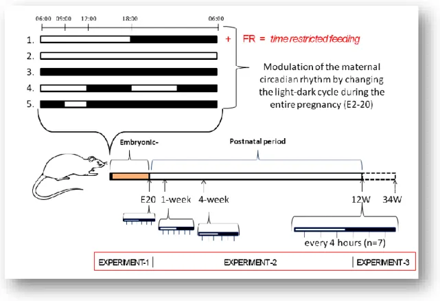
All animals were handled according to written approval from the local authority for animal experiments (Regierungspräsidium Karlsruhe, 35-9185.81/G-29/11). Pregnant Sprague Dawley rats (n = 240, pregnancy rate 93,5 %) were obtained from Charles River Co. (Sulzfeld, Germany) two days after conception. At the second day of gestation (considered the fetuses, in the following mentioned as embryonic day 2, E2) rats were randomly allocated to the following light-dark cycles with food and water supplied ad libitum under controlled temperature (21 ± 2°C):
1. 12 h:12 h light-dark (LD) cycle, normal photoperiod, ZT0 = 06:00 h 2. constant illumination, 24 h light (LL)
3. constant darkness (DD)
4. shortened, ultradian, 6 h:6 h light - dark cycle (6:6-LD), ZT0 = 06:00 h 5. prolonged dark phase condition, 3 h:21 h light - dark cycle (3:21-LD), ZT0
9:00h
Dams were housed (3-5 dams / 1800 cm², in Typ IV cages) under the same photoperiod conditions during the entire pregnancy. At the day before the expected delivery, i.e. at embryonic day 20 (E20) dams were housed separately (1 dam / Typ IV cages). Cohorts
29
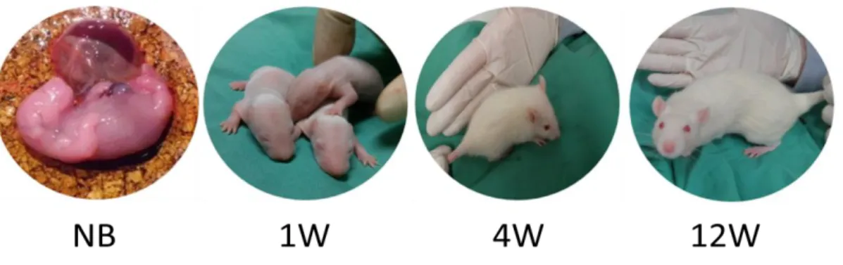
of animals at age E20, as well as at 1. (1W), 4. (4W) and 12. (W) and 34. week of age (34W, long term= LT) postnatally were used in the following experiments:
Experiment1, 2 and 3. (Figure 7)
3.2.1. Disruption of maternal feeding regime
To alter the normal maternal feeding regime, a subgroup of pregnant rats (n = 16) under normal 12:12 light-dark (LD) cycle were randomly assigned to be fed only during the light phase throughout the entire pregnancy (E2 - E21). Food was restricted to the 06:00-18:00 hour (ZT0-12) interval with free access to water. (FR-LD) (Figure 7)
Figure 7. Experimental protocols (1-5) were used to modulate the maternal circadian rhythm during the entire pregnancy (details see in the text). At embryonic day 20 (E20), groups of pregnant rats (n = 6-10 / from each group) were sacrificed with their fetuses (n = 12 ± 2 mothers) every 4 hours to cover one entire circadian period (ZT4-24 h, ZT0 = 06:00 a.m.) Postnatal, at 1, 4 and 12 weeks after birth (1W, 4W, 12W) 14 rats (7 male, 7 female) were sacrificed in 4 hours interval in the following photoperiod conditions: LD, LL, DD, 6:6-LD and 3:21-LD. Offspring (n=20/group) were investigated at long term (LT = 34W) period in each groups.
30
3.3. EXPERIMENT 1: Early effects of prenatal maternal circadian disruption At embryonic day 20 (E20), groups of pregnant rats (n = 6-10 / group) from each photoperiod conditions (LD, LL, DD, 6-6 LD and 3-21 LD) and from FR-LD were anesthetized (100 mg/kg ketamine and 3 mg/kg xylazine, which very rapidly passes the placenta) and sacrificed with their fetuses (n = 12 ± 2 mothers) every 4 hours to cover one entire circadian cycle (ZT 2-24 h, ZT0 = 06:00 h). Dams’ body and placentas’
weight were measured. Fetal tissues (kidney, heart and liver) were dissected and stored.
3.3.1. Effect on the fetal intrarenal clockworks
In the kidney, gene expression patterns of core clock genes (Clock, Bmal1, Rev-erbα, Per1, Per2, Cry1, Cry2) and renal clock controlled genes (αENaC, Sgk11, ENH3, AVPR2) were further analyzed in males.
3.3.2. Telemetric measurements of the dams over a 24h period
A subgroup of pregnant rats at embryonic day 2 in group LD, LL, DD and 3:21-LD (n
= 4-6 / group) underwent implantation of telemetrical device (model PA-C40; Data Science International) to monitor BP, HF and motor activity. After a 7-10 days recovery period daily measurement was recorded. The measurements at the third gestational week (3.GW) were further analyzed. A detailed description of the implantation and measurements can be seen below. (Page 37)
3.3.3. Circadian variation in renal excretion function during pregnancy
Urine samples were collected at 4-hour intervals over a 24 h period in metabolic cages (TECNIPLAST S.p.A., Buguggiate, Italy) at 1., 2. and 3. gestational week (1.GW, 2.GW and 3.GW) in LD, LL and DD groups (n = 8-12 / 4h over 24 h period in each group). Immediately after collection urine volumes were measured and fractionated into 2-3 equal parts. Aliquots were stored in a freezer at -20 °C until further analysis.
Urinary parameters, i.e. sodium, potassium, phosphate, calcium, glucose and albumin
31
excretion were analyzed using standard laboratory methods. (Central-labor, University of Heidelberg)
3.3.3.1. Urinary aldosterone excretion over the pregnancy
In group LD (n = 8 / 4h over 24 h period) and LL (n = 12 / 4h over 24 h period) the daily pattern of urinary aldosterone excretion rate with a 4-hour interval over a 24 h at 1. and 3. gestational week (1.GW and 3.GW) was measured using ELISA Kit. (The measurement was performed at the University of Silesia in Katowice, Poland)
3.3.3.2. Urinary melatonin excretion
At the second week of gestation (2.GW) in LD (n = 5 / 4h over 24 h period) and DD (n
= 7 / 4h over 24 h period) urinary melatonin concentration was measured with a 4-hour period over a 24 h. Urine samples were subjected in duplicate to Melatonin-Sulfat Urine ELISA Kit (IBL, Hamburg, Germany) to determine the immunoreactivity of urinary melatonin metabolite (6-SMT). The absorbance of the immunoreactions was recorded spectrophotometrically at 450 nm in an automated ELISA reader and hormone concentration was calculated using an automated method with standard curve. The intra- and inter-assay coefficients were 5.8–204 ng/ml (5.2–12.2%) and 12.4–220 ng/ml (5.1–
14.9%).
32 3.4. EXPERIMENT 2: postnatal changes
After the delivery, all dams along with their offspring (n = 12 ± 2 / liter) were housed with free access to food and water under constant temperature (21 ± 2°C) exposed to standard 12-12h light-dark cycles (light on at 06:00 h) with illumination ca. 200 lx at the cage level. The offspring were weaned at 4 weeks of age and housed in standard Typ IV cages (2-3 males or 4-5 female per cage). The following studies were performed in offspring from different mothers, in order to avoid litter effect.
At 1, 4 and 12 weeks after birth (1W, 4W, 12W) 14 rats (7 male, 7 female) were sacrificed in 4 hours intervals to cover one entire circadian period (ZT4-24 h, ZT0 = 06:00 a.m.) in the following photoperiod conditions: LD, LL, DD, 6:6-LD and 3:21-LD.
(Figure 8) Under global anesthesia (100 mg/kg ketamine and 3 mg/kg xylazine) the abdominal aorta was catheterized and retrograde pressure-controlled perfusion fixation was performed using 4% phosphate-buffered formaldehyde for morphological and 0,9%
NaCl for molecular investigation, respectively. Body and organ (kidney, heart and liver) weight were recorded.
3.4.1. Circadian gene variation after maternal circadian disruption
The gene expression pattern of core clock (Clock, Bmal1, Per1, Per2, Cry1, Cry2, Rev- erbα) genes and clock controlled genes (αENaC, Sgk11, ENH3, AVPR2) in the male kidney were studied in LD, LL and DD groups. Other tissue samples were stored for further analysis.
Figure 8. Clockwork of offspring after prenatal circadian disruption were investigated at embryonic day 20 (E20), as well as 1 (1W), 4 (4W) and 12 week (12W) of age.
33
3.4.2. Postnatal behavioral measurements of offspring
Mothers with their pups were maintained in cages equipped with infrared video camera (Sygonix, CCD-Camera and Digitalrecorder). The Hosttech video capture program was used to record maternal nursing- and feeding behavior, daily activity, feeding and drinking pattern of the offspring at age 4 and 12 weeks. Feeding times of the mothers, as well as the locomotor activity, the frequency of food and water intake of the pups and the mothers were evaluated by video analysis.
3.4.3. Effect of timed maternal melatonin treatment in the early postnatal period 6 dams with their pups were randomly allocated in a subgroup under LD condition (body weight at 3. GW 359,5 ± 23,5 g, 11,8 ± 3,8 pups / mothers). From the day of the delivery dams were treated with intraperitoneal injection of melatonin (Me: 4,8 mg/kg ip. at ZT3) or its vehicles (Vech: 1,4 g/kg, 6% ethanol in 0.9% NaCl at ZT3) over a week period. Pups (female n = 5-7 per 4 h over 24 h) were anesthetized (100 mg/kg ketamine and 3 mg/kg xylazine) and sacrificed at 4 h intervals for 24h at 1 week of age.
As a second control, females from LD at 1 week were sampled (n = 7 / 4 h over a 24 h period). Body weights were measured. Fetal tissues (kidney, heart and liver) were dissected, measured and stored. In the kidney, gene expression patterns of core clock genes (Clock, Bmal1, Rev-erbα, Per1, Per2, Cry1, Cry2) and clock controlled genes (αENaC, Sgk11, ENH3, AVPR2) were further analyzed.
3.4.4. Periodic maternal withdrawal at the first postnatal week period
Pregnant rats (n = 5) exposed to 12-12h light-dark cycles under the above described conditions were randomly allocated in a subgroup. After the delivery (Body weight at 3.GW 376 ± 14 g, 12,2 ± 2,5 pups / mothers), the pups were separated from their mothers for a 4- hour (Maternal withdrawal, MW: ZT3-7) period starting on the first postnatal day. The control group remained with their mother all the time. Five pups (male) per time point were anesthetized (100 mg/kg ketamine and 3 mg/kg xylazine) and sacrificed at 4 h intervals for 24h at 1 week of age. Body weights were measured.
Fetal tissues (kidney, heart and liver) were dissected, measured and stored. In the
34
kidney, gene expression patterns of core clock genes (Clock, Bmal1, Rev-erbα, Per1, Per2, Cry1, Cry2) and clock controlled genes (αENaC, Sgk11, ENH3, AVPR2) were further analyzed.
35 3.5. EXPERIMENT 3: long lasting effects
From each photoperiod group (LD, LL, DD, 6:6-LD, 3:21-LD) and FR-LD mothers were returned to LD condition after delivery. Offspring (n = 8-12 / group) weaned at 4- weeks-old were kept for longer period (34 ± 2 Week) under standard condition with free access to food and water under constant temperature (21 ± 2°C) exposed to standard 12- 12h light-dark cycles (ZT0 at 06:00 a.m.) with illumination between 50 and 200 lx at the cage level.
3.5.1. Renal function
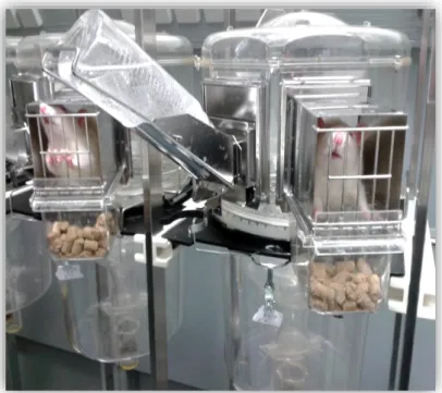
Urine was sampled in 24-hour intervals in metabolic cages at 34. week of age in all investigated groups (n = 8-12 / 4 h interval over a 24 period in each group). Urinary parameters, i.e. sodium, potassium, phosphate and calcium excretion, as well as albumin- and glucosuria were analyzed using standard laboratory methods. (Figure 9)
3.5.2. Telemetric measurement
A subgroup of female rats at 34 weeks of age in group LD, LL, DD, 6:6-LD, 3:21-LD and FR-LD (n = 5-11 / group) underwent implantation of telemetrical device (model PA-C40; Data Science International) to monitor BP, HF and motor activity. After a 7-10 days recovery period daily measurement was recorded. See below detailed description of the implantation and measurements. (Page 37)
Figure 9. Metabolic cages (TECNIPLAST)
36 3.5.3. Echocardiography
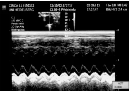
At age 34 week in LD, LL, 6:6-LD, 3:21-LD and FR-LD group the left ventricular function was measured under isoflurane anesthesia, after the anterior chest was shaved with transthoracic echocardiography. A parasternal short-axis view was performed for M-mode imaging at the papillary muscle level (See Figure 10.) using a 13-MHz linear transducer (GE 12L-RS, GE Healthcare), connected to an echocardiographic imaging unit (Vivid i, GE Healthcare). End-diastolic left ventricular internal diameter (EED) and end-systolic left ventricular internal diameter (ESD) were measured. The percentage of fractional shortening (FS%) was calculated ([(EDD-ESD)/EDD) x100], n = 6-10 / group)
At the end of the observation, under global anesthesia (100 mg/kg ketamine and 3 mg/kg xylazine) tissues (kidney, heart) were sampled for morphological and molecular investigation. Body and organ (kidney, heart and liver) weight were recorded. Blood samples were collected from the abdominal aorta in each group for further studies.
Figure 10. A representative picture of echocardiography at 34-week-old female from LL group.
37
3.6. Radiotelemetric measurement of blood pressure
Pregnant rats at embryonic day 2 in group LD (n = 4), LL (n = 4), DD (n = 6) and 3:21- LD (n = 7) as well as offspring (female rats) at 34 ± 2 weeks of age in group LD (n = 9), LL (n = 11), DD (n = 8), 6:6-LD (n = 5) and 3:21-LD (n = 7) and FR-LD (n = 6) underwent implantation of telemetrically device to monitor BP, HF and motor activity.
Under isoflurane (1-3%, 5% for induction) anesthesia and controlled temperature (37 ± 0,5 °C) in ventral abdominal incision the catheter of the telemetry sensor (model PA- C40; Data Science International) was implanted into the abdominal aorta at the level of the bifurcation, below the renal arteries. (Figure 10-11) Sensor was fixed using a small amount of tissue adhesive and small fiber patches, the transmitter itself was fixed intraperitoneally with non-absorbable suture with a simple interrupted pattern. After
a 7-14 days recovery, measurements were taken under normal feeding regime (except FR-LD) under ambient temperature. Signals were sent to a telemetry receiver that was placed under the cage (Typ IV) every 5-10 min (ca. 10.000 readings/rat) and transmitted in a 10- minute average from 3 consecutive measurements with use of Dataquest system for further analysis.
Figure 11. Drawing of a rat implanted with a transmitter (red arrow) capable of monitoring blood pressure and heart rate via the abdominal aorta, as well as a locomotor activity.
Figure 10. Implantation of the telemetric device (HD)