SEMMELWEIS EGYETEM DOKTORI ISKOLA
Ph.D. értekezések
2434.
JAKAB GÉZA
A gyógyszerészeti tudományok korszerű kutatási irányai című program
Programvezető: Dr. Antal István, egyetemi tanár Témavezető: Dr. Antal István, egyetemi tanár
Development and Formulation of Baicalin-loaded Drug Delivery Systems
PhD Thesis
Dr. Géza Jakab
Doctoral School of Pharmaceutical Sciences Semmelweis University
Supervisor: István Antal, D.Sc.
Official reviewers:
Angéla Jedlovszky-Hajdú, Ph.D.
Ildikó Bácskay, D.Sc.
Head of the Complex Examination Committee:
Romána Zelkó, D.Sc.
Members of the Complex Examination Committee:
Éva Szökő, D.Sc.
Miklós Vecsernyés, D.Sc.
Budapest, 2020
1 Table of contents
List of abbreviations ... 5
1. Introduction ... 8
1.1. Scutellaria baicalensis Georgi (Baical skullcap) ... 10
1.2. Baicalin ... 11
1.2.1. General notes ... 11
1.2.2. Physicochemical Profiling ... 12
1.2.3. Biopharmaceutical properties ... 15
1.2.4. Pharmacological effects ... 16
1.2.5. Baicalin-drug interactions ... 17
1.2.6. Bioavailability enhancement ... 19
1.3. Lipid-based formulations ... 20
1.4. Self-emulsifying Drug Delivery Systems ... 23
1.4.1. Microemulsions and nanoemulsions: similarities and differences ... 23
1.4.2. Role of excipients in the formulation of SEDDS ... 28
1.4.2.1. Oils ... 29
1.4.2.2. Surfactants ... 30
1.4.2.3. Co-surfactants/Co-solvents ... 31
1.5. Transformation of liquid SEDDS/SMEDDS/SNEDDS into solid dosage forms 31 1.6. Cyclodextrin complexation... 34
1.6.1. Short history of Cyclodextrins ... 35
1.6.2. Physicochemical characteristics of Cyclodextrins ... 37
1.6.3. Formation of drug-cyclodextrin inclusion complex ... 39
1.6.4. Toxicological assessment and regulatory status of Cyclodextrins ... 41
1.6.5. Methods of preparation of inclusion complexes ... 43
1.6.6. Pharmaceutical application of cyclodextrins ... 46
2. Objectives ... 48
3. Methods ... 49
3.1. Materials ... 49
3.2. Preparation of compendial and biorelevant media ... 50
3.3. Preformulation studies ... 50
2
3.3.1. Determination of Thermodynamic Solubility by Saturation Shake-Flask
Method ... 50
3.3.2. Acid-base properties ... 51
3.3.3. Distribution Coefficient Measurements by the Stir-Flask Method ... 52
3.3.4. Crystal habit ... 52
3.3.5. Particle size analysis ... 53
3.4. Baicalin-Cyclodextrin inclusion complexation ... 53
3.4.1. Phase solubility studies ... 53
3.4.2. Signal Assignment of Baicalin and Characterization of Baicalin-Cyclodextrin Inclusion Complexes Using 1H NMR and 2D ROESY Experiments ... 54
3.4.3. Molecular Modelling of the Binding into Cyclodextrin ... 55
3.5. Liquid Self-nanoemulsifying Drug Delivery Systems ... 55
3.5.1. Solubility Studies ... 55
3.5.2. Screening of Surfactants for Emulsifying Ability ... 56
3.5.3. Construction of Ternary Phase Diagram... 56
3.5.4. Preparation of Self-Nanoemulsifying Formulations without (SNEDDS) and with Baicalin (BSNEDDS) ... 57
3.5.5. Optimization of SNEDDS Preconcentrates ... 57
3.5.6. Characterization of Optimized BSNEDDS ... 58
3.5.6.1. Droplet size, Transmittance, PDI, and Zeta-Potential Measurements ... 58
3.5.6.2. Determination of the Thermodynamic Solubility of Baicalin in Optimized SNEDDS... ... 58
3.5.6.3. Cloudpoint Measurement ... 58
3.5.6.4. Effect of Dilution on Droplet Size and PDI ... 59
3.5.6.5. Long-Term Physical Stability of Nanoemulsions ... 59
3.5.6.6. Atomic Force Microscopy ... 59
3.6. Preparation of self-nanoemulsifying matrix pellets (SNEMPs) ... 60
3.7. Physical characterization of SNEMPs ... 61
3.7.1. Flow properties ... 61
3.7.2. Friability ... 61
3.7.3. Shape analysis ... 62
3.7.4. Residual water content ... 62
3.8. Solid state characterization of SNEMPs ... 62
3.8.1. FT-IR ... 62
3.8.2. Raman spectroscopy ... 63
3
3.9. In vitro dissolution study and reconstitution properties of SNEMPs ... 63
4. Results ... 64
4.1. Preformulation studies ... 64
4.1.1. Determination of Thermodynamic Solubility by Saturation Shake-Flask Method... 64
4.1.2. Acid-base properties ... 65
4.1.3. Distribution Coefficient Measurements by the Stir-Flask Method ... 69
4.1.4. Crystal habit ... 71
4.1.5. Particle size analysis ... 72
4.2. Baicalin-cyclodextrin inclusion complexation ... 73
4.2.1. Phase solubility studies ... 73
4.2.2. Signal Assignment of Baicalin and Characterization of Baicalin-Cyclodextrin Inclusion Complexes Using 1H NMR and 2D ROESY Experiments ... 75
4.2.3. Molecular Modelling of the Binding into Cyclodextrin ... 79
4.3. Liquid Self-nanoemulsifying Drug Delivery Systems ... 82
4.3.1. Solubility Studies ... 82
4.3.2. Screening of Surfactants for Emulsifying Ability ... 84
4.3.3. Construction of Ternary Phase Diagram... 86
4.3.4. Optimization of SNEDDS Preconcentrates ... 86
4.3.5. Characterization of Optimized BSNEDDS ... 92
4.3.5.1. Droplet size, Transmittance, PDI, and Zeta-Potential Measurements ... 92
4.3.5.2. Determination of the Thermodynamic Solubility of Baicalin in Optimized SNEDDS ... 92
4.3.5.3. Cloudpoint Measurement ... 93
4.3.5.4. Effect of Dilution on Droplet Size and PDI ... 93
4.3.5.5. Long-Term Physical Stability of Nanoemulsions ... 93
4.3.5.6. Atomic Force Microscopy ... 94
4.4. Physical characterization of SNEMPs ... 96
4.4.1. Flow properties ... 96
4.4.2. Friability ... 96
4.4.3. Shape analysis ... 97
4.4.4. Residual water content ... 98
4.5. Solid state characterization of SNEMPs ... 98
4.5.1. FT-IR ... 98
4
4.5.2. Raman spectroscopy ... 99
4.6. In vitro dissolution study and reconstitution properties of SNEMPs ... 100
5. Discussion ... 103
5.1. Preformulation studies ... 103
5.2. Baicalin-cyclodextrin inclusion complexation ... 103
5.3. Liquid Self-nanoemulsifying Drug Delivery Systems ... 104
5.4. Self-nanoemulsifying matrix pellets ... 104
5.5. Comparison of baicalin-CD inclusion complexes and baicalin-loaded SNEDDS ... 105
5.6. Comparison the two formulations presented in the thesis with the formulations already published in scientific literature ... 106
6. Conclusions ... 107
7. Summary ... 109
8. Összegzés ... 110
9. References ... 111
10. Publications ... 127
10.1. Publications pertaining to the doctoral thesis ... 127
10.2. Publications pertaining to different subjects ... 127
11. Acknowledgement ... 129
5 List of abbreviations
ABC ATP-binding Cassette
ADMET Absorption-Distribution-Metabolism-Excretion-Toxicity
AFM Atomic Force Microscopy
ANOVA Analysis of Variance
API Active Pharmaceutical Ingredient
BBB Blood Brain Barrier
BCRP Breast Cancer Resistance Protein
BCS Biopharmaceutical Classification System
BRM Biorelevant Media
BSNEDDS Baicalin-loaded Self-nanoemulsifying Drug Delivery System
CD Cyclodextrin
CYP 450 Cytochrome P450 Enzymes
DDS Drug Delivery System
DLS Dynamic Light Scattering
DSC Differential Scanning Calorimetry
EC European Commission
EMA European Medicines Agency
FaSSGF Fasted State Simulated Gastric Fluid FaSSIF Fasted State Simulated Intestinal Fluid
FDA Food and Drug Administration
FeSSIF Fed State Simulated Intestinal Fluid
GAL GalenIQ™ 800
6
GIT Gastro-intestinal Tract
GRAS Generally Regarded as Safe
HLB Hydrophilic-Lipophilic Balance
HME Hot-melt Extrusion
HPMC Hydroxypropyl Methylcellulose
HP-β-CD 2-hydroxypropyl-β-CD
HRE Heat Reflux Extraction
ICH International Conference on Harmonization
JPC Japan Pharmaceutical Codex
LBDD Lipid-based Drug Delivery
LD Laser Diffraction
LFCS Lipid Formulation Classification System
MCC Microcrystalline Cellulose
MRP Multidrug Resistance-associated Protein
MWI Microwave Irradiation
NCE New Chemical Entity
NIBS None-Invasive-Back-Scattering
NMR Nuclear Magnetic Resonance
OATP1B1 Organic Anion-transporting Polypeptide 1B1
PDI Polydispersity Index
Ph. Eur. European Pharmacopoeia
PVP Polyvinylpyrrolidone
RAMEB-CD Random Methylated β-CD
7
ROESY Rotating Frame Nuclear Overhauser Effect Spectroscopy
SBE-β-CD Sulfobutylether-β-CD
SEDDS Self-emulsifying Drug Delivery System
SEM Scanning Electron Microscopy
SFE Supercritical Fluid Extraction
SMEDDS Self-microemulsifying Drug Delivery System SNEDDS Self-nanoemulsifying Drug Delivery System
UAE Ultrasound-assisted Extraction
USP United States Pharmacopoeia
8
1. Introduction
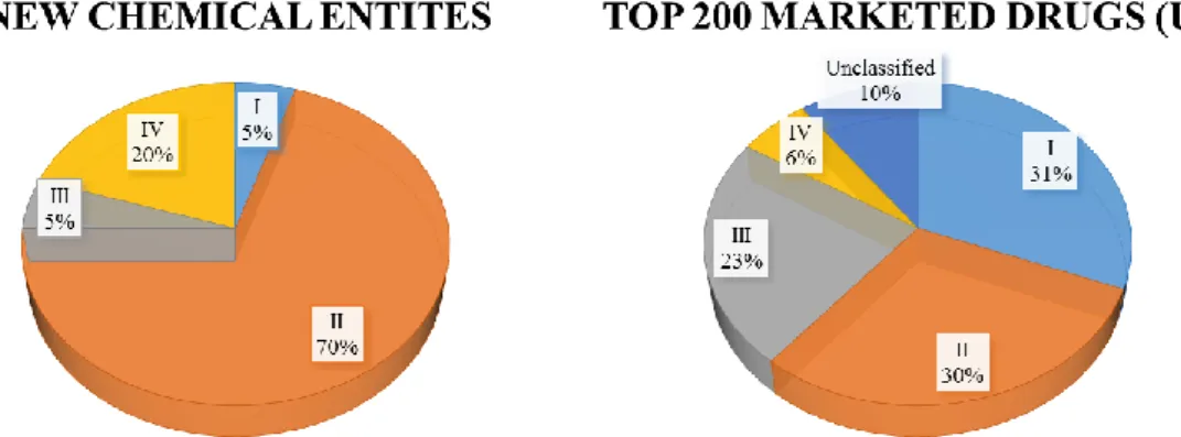
One of the most tremendous challenges faced by pharmaceutical scientists is poor solubility and/or permeability and the concomitant low bioavailability of new chemical entities (NCE). Researchers often find promising candidates during drug screening;
however, if the molecule exhibits unsuitable physico-chemical properties, the chance of delivering the adequate dose to the site of action is diminished. In the last years a negative tendency has appeared in the drug discovery pipeline: approx. 70% of NCEs demonstrate low solubility and high permeability, therefore belongs to Biopharmaceutical Classification System (BCS) II. Conversely, about only 30% of drugs previously brought to the market resides to BCS II (1). This challenging shift is also expressed in case of BCS IV (low solubility and low permeability) compounds: there is an increase of 3-4 times in favour of NCEs compared to marketed drugs (Fig.1.) (2). Baicalin -the subject of this PhD Thesis- is a bioactive phytopharmacon and can be classified as Class IV (3). In the scientific literature numerous approaches came up to improve the low bioavailability of different active pharmaceutical ingredients (APIs). With no claim of being exhaustive I would like to notice some of them: cyclodextrin complexation (4), solid dispersions (5), liposomes (6), lipid-based formulations (7), self-emulsifying systems (8). In the following sections a detailed explanation can be found related to lipid- based formulations focused on self-emulsifying systems and cyclodextrin complexes.
Figure 1. Trending in Biopharmaceutical Classification System (BCS) (http://www.samedanltd.com/magazine/11/issue/158/article/3039; 01/10/2019) (BCS I – high solubility and permeability, BCS II – low solubility, high permeability, BCS III –
high solubility, low permeability, BCS IV- high solubility and permeability of drug substance)
9
In the last decade the pharmacological and formulation aspects of phytopharmacons captured the attention of scientist worldwide. This is largely a result of the higher tendency among the general population to use complementary and alternative medicine and live a health-conscious lifestyle. The motivation behind the topic selection was the renaissance of natural products and the challenging physico-chemical and biological properties of baicalin.
10
1.1.Scutellaria baicalensis Georgi (Baical skullcap)
Scutellaria baicalensis G. (S. baicalensis G.) is one of the fundamental herbs in traditional Chinese herbal medicine known as Huang Qin (mandarin: 黄芩) (9). It is indigenous to several East Asian countries (Mongolia, China, Korea) and the Russian Federation and has been cultivated in many European countries (Fig.2.) (10). Baical skullcap is a species of flowering plant in the family Lamiaceae. The root is officially listed in the Chinese Pharmacopoeia and was assumed in European Pharmacopoeia (Ph. Eur.) 9th Edition in 2018 (11). The major components in the dried root of the herb are baicalin (D-Glucuronic acid-5,6-dihydroxyflavone) and its aglycone, baicalein (5,6,7-trihydroxyflavone) (12).
As minor component wogonin (5,7-dihydroxy-8-methoxyflavone) and wogonoside (5,7- Dihydroxy 8-methoxyflavone 7-glucuronide) can be extracted from the dried root. Heat reflux extraction (HRE), ultrasound-assisted extraction (UAE), and supercritical fluid extraction (SFE) are common extraction methods to isolate and purify baicalin (13).
Figure 2. The medical plant of Scutellaria baicalensis (A) and its dried root (B).
Extracted and purified powder of baicalin (C). 3D structure of baicalin (D).
(https://en.wikipedia.org/wiki/Scutellaria_baicalensis; 01/10/2019) C and D are self- made
11 1.2.Baicalin
1.2.1. General notes
Baicalin is a flavone glycoside extracted from the root of Scutellaria baicalensis. In traditional Chinese medicine it had been used for the treatment of diarrhoea, dysentery, haemorrhaging, insomnia, inflammation and different respiratory infections (10).
Nowadays baicalin extracts are easily accessible over-the-counter herbal remedies, purchasable online and in numerous stores in liquid, bulk powder, capsule or tablet dosage form. Recommended daily dosage of powder is 60-500 mg. Baicalin enjoys fairly high popularity in ointments, lipsticks and skin care preparations. As a flavonoid derivate, its anti-oxidant and anti-aging property can contribute to the skin’s overall quality and appearance. Food industry utilizes baicalin as a nutritional adjuvant for improving production performance and nourishing feed intake in farm animals (14).
Figure 3. Growing interest in baicalin as a bioactive phytopharmacon 1982-2019 Source: PubMed, keyword: baicalin [all]
Baicalin is easily extractable and it has a wide range of therapeutical and pharmacological effects (see detailed in section: 1.2.4.), which have brought it into focus as a safe and natural phytopharmacon. A PubMed search with the key word Baicalin [all] returns approx. 1700 articles from ‘80s to date with 50% of them published in the last five years (Fig.3.). Since 2000 the interest in the subject has been increasing gradually. On the one hand, in the US and Europe baicalin-loaded formulations have no marketing authorization
0 20 40 60 80 100 120 140 160
1982 1988 1994 2000 2006 2012 2018
Number of publications
Year
12
as of December 2019. On the other hand, in 2005 the State Food and Drug Administration of China approved baicalin (approval No. H20158009) for the adjuvant therapy of hepatitis (1500 mg/day, 2 times 3 capsules).
1.2.2. Physicochemical Profiling
The most important physicochemical properties influencing the pharmacokinetic behavior of drugs and biomolecules are the acid-base properties, lipophilicity,
permeability and
solubility (15). Physicochemical profiling is an integral part of preformulation studies, which lays down foundation for transforming a new drug entity into a pharmaceutical dosage form in such a way that it can be administered in a right way, in right amount, and on the right target (16). Possessing these kinds of information, the optimization of an active molecular entity is also possible.
The acid-base character determines the ionization state of a molecule in solution having a particular pH. Consequently, all pharmacokinetic properties, namely absorption, distribution, metabolism, excretion and toxicity (ADMET) are influenced by the ionization state under varying pH conditions (17). Molecular structure of baicalin can be derived from its aglycone, baicalein by linking a glucuronic acid to the aglycone via a glycosidic bond in position 7 (Fig.4.). It has three acidic functional groups, namely one carboxyl (ring: D) and two phenolic hydroxyl groups (ring: A).
Figure 4. Chemical structure of Baicalin and Baicalein (self-made)
13
Proton transfer processes can be regarded either from the point of view of dissociation or association. Because of the acidic nature of baicalin, these processes will be characterized by acid dissociation constants (Ka). The determination of pKa value is important in understanding the in vivo behaviour of a drug, which influences not only its solubility and dissolution, but also the membrane-penetration capacity (18). The acid-base properties of baicalin have been investigated before, resulting in pKa values of 5.05, 7.6 and 10.1 (19). However, internal Nuclear Magnetic Resonance (NMR) investigations showed the molecule rapidly decomposes above pH 9, making the reported values in alkaline solutions highly dubious.
The determination of the octanol/water partition coefficient (log P) is essential during preformulation studies, which is in close correlation with membrane-penetration capability and is the most frequently determined lipophilicity descriptor (20). There have been attempts in characterizing the lipophilicity of baicalin ((log P=1.27 (pH=7)), but the applied methods and study design can induce doubts (19). It is not exactly clear what the authors meant by this, probably they calculated the log P of baicalin from log D measured at pH 7. Therefore, the data provided is to be considered as indicative only. However, if we accept with restrictions the above-mentioned value, the lipophilicity of baicalin must be considered as low. Calculation of log P value provides data regarded to passive diffusion through biological membranes, but it does not describe properly the carrier- mediated active transport mechanisms. The Caco-2 monolayer is isolated from human colorectal adenocarcinoma and widely used across the pharmaceutical industry as an in vitro model of the human intestinal barrier to predict the active and passive absorption mechanisms of orally administered drugs (21). The permeability potential of baicalin was evaluated by Caco-2 assay (Papp 9.2×10⁻⁸ cm/s), which revealed low permeability (22). The low lipophilicity and permeability value of baicalin presumes low absorption from the gastro-intestinal tract (GIT).
Aqueous solubility is a fundamental attribute of an active substance and its examination is a mandatory step during the drug discovery process. To achieve adequate plasma drug levels and clinical response, dissolution of the active ingredient in physiological environment is a leading precondition. New drug molecules as well as phytopharmacons tend to have larger molecular weights, resulting often in decreased solubility and dissolution, which may lead to limited absorption and poor pharmacokinetics. Like pKa
14
and log P values of baicalin, its aqueous solubility is also marked by discrepancies in the scientific literature; 0.18 mg/ml, 0.08 mg/ml and 0.052 mg/ml can be found (23–25).
Furthermore, the factors affecting solubility (e.g. temperature, pH, ionic strength, surface tension) were not investigated yet in a comprehensive study. It can be seen from the chemical structure that both the glucuronide and the flavone part forms intramolecular H- bonds, which can be partly responsible for the poor water solubility and high melting point (26). If an oral drug is under development, particularly one with low solubility, biorelevant measurements are extremely useful because through simple in vitro tests they can predict how it's likely to dissolve in vivo in the GIT. Over the past 15–20 years, biorelevant media simulating conditions in the stomach and small intestine before and after meals have been developed (27). Simulated intestinal fluid in fasted state (FaSSIF) and in fed state (FeSSIF) along with fasted state gastric fluid (FaSSGF) have been suggested first by Dressman et al (28). In addition, the application of these media can be used to predict food effects (29). In case of baicalin these kind of physiologically relevant solubility data have not been published yet.
The chemical stability of baicalin was evaluated in buffered aqueous solutions at different pH (2.0, 3.0, 4.5, 6.8, 7.4 and 9.0) and temperatures (4, 25 and 40 °C). Acidic environment and low temperature were protective factors for the long term stability of this compound (30).
15 1.2.3. Biopharmaceutical properties
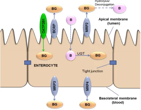
Several studies described the pharmacokinetics of baicalin according to the ADME concept. The absorption mechanism was evaluated in rats by Liu et al. and found that baicalin was moderately absorbed in the stomach but poorly in the small intestine and colon (31). More research pointed out that baicalin undergoes extensive hydrolysis by intestinal β-glucuronidase or intestinal microbiome to form its aglycone, baicalein (Fig.5.).
Figure 5. Role of transporters in transport of baicalin in enterocytes (Interaction of baicalin with transporters, Bernadett Kalaposné Kovács, Semmelweis University, 2016)
Since the aglycone is absorbed much more better compared to baicalin, this cleavage reaction has a potent role in the absorption process (32, 33). Baicalin is substrate of uptake transporter organic anion-transporting polypeptide 1B1 (OATP1B1) (33). In the intestinal enterocyte baicalein is transformed to baicalin, and effluxed to the blood mainly by multidrug resistance associated protein 3 (MRP3) and MRP4, located on the basolateral side of the cell (33). Accordingly, after administration of a single ascending dose of baicalein (100-2800 mg) chewable tablets to healthy subjects, the Cmax values of
16
baicalin were about ten-fold higher than Cmax values of baicalein (34). Part of baicalin is pumped back to the intestinal lumen by apically located MRP2 and breast cancer resistance protein (BCRP) (35).
In the distribution mechanisms of baicalin the high plasma protein-binding capacity (86- 92%) plays a prominent role (36). Wei et al. revealed the tissue distribution of baicalin after intravenous administration of liposomal and injectable formulations to rabbits.
Liposomal drug delivery indicated a significantly increased lung accumulation compared to injectable solution (37). Baicalin penetrates moderately through the blood-brain- barrier (BBB) (38).
Validated method was applied to analyse and screen the in vivo metabolism of baicalin in rats. No less than 32 metabolites were identified in the rat plasma and urine. The results demonstrated that the rat liver and kidney are the most important organs for the distribution of baicalin metabolites. Methylation, hydrolysis, hydroxylation, methoxylation, glucuronide conjugation, sulphate conjugation, and their composite reactions were all identified (39).
Baicalin is primarily excreted in bile in the form of glucuronides and undergoes a prominent enterohepatic recycling through different ABC transporters (33, 40). As an alternative excretion pathway, the fraction of baicalin excreted in urine appears to be negligible compared to the biliary route (41).
1.2.4. Pharmacological effects
Baicalin has numerous pharmacological activities demonstrated by in vitro and in vivo studies among others anti-inflammatoric, anti-allergic, anti-depressant, anti-microbial, anti-oxidant as well as anti-psoriatic effects (42–47). Ding et al. showed that baicalin relaxes vascular smooth muscle and lowers blood pressure in spontaneously hypertensive rats by regulating KATP channels and the intracellular Ca2+ level (48). It was revealed that long-term baicalin administration ameliorates metabolic disorders and hepatic steatosis in rats given a high-fat diet (49). Baicalin might serve in the future as a novel compound in the treatment of the most common neurodegenerative diseases in elderly patients, the Parkinson’s (50). The preliminary results suggest that the mechanism of action is a decreased iron accumulation in the substantia nigra. Wang et al. investigated the
17
hyperglycaemia-induced cardiovascular malformation of embryos in mice. The cardiovascular abnormality can be attenuated by baicalin administration. This compound is a promising candidate for women suffering from gestational diabetes mellitus (51). Cancer is one of the leading causes of death worldwide and is very likely to overtake heart diseases, which hastraditionally topped the list as the leading cause of death in higher income countries. Scientific projects reveal an ever-increasing number of evidence that baicalin exhibits its antitumor function in a wide range of cancers such as breast cancer, colon cancer, gallbladder carcinoma, haematological malignancies, hepatic cancer, lung cancer, prostate cancer (52–57). The chemical modification of the baicalin aglycone pointed out promising development in preclinical studies (58). Therefore, the chemical and pharmacological optimization and the production of semisynthetic derivates could be an interesting research direction in the future. It is important to note that extensive clinical studies are needed for the confirmation of the above mentioned in vitro and in vivo results.
1.2.5. Baicalin-drug interactions
Several studies have been carried out on the effects of baicalin and co-administered drugs.
Remarkable baicalin-drug interactions can be observed, when both compounds share the same cytochrome P450 (CYP) enzyme or exhibiting high plasma protein binding. The relevant preclinical and clinical research findings and the mechanism of herb-drug interactions are summarized in Table I.
Two clinical trials can be found in the literature regarding baicalin-drug interactions. Liu et al. executed the first clinical study to investigate the effect of baicalin on cyclosporine A pharmacokinetics in humans (n=16) (59). The applied dosage was 500 and 200 mg in case of baicalin and cyclosporine A, respectively. The combination was well-tolerated and the pharmacokinetic behaviour of cyclosporine A was not changed to a clinically relevant extent. The reported adverse events were mild and the data didn’t lay down evidence that baicalin co-administration with cyclosporine A would cause an additional risk. Fan et al. analysed the pharmacokinetics of rosuvastatin, an antihyperlipidemic drug co-administered with baicalin. In the study 18 healthy adults were enrolled, possessing various organic anion-transporting polypeptide 1B1 (OATP1B1) haplotypes (60).
OATP1B1 is considered the main uptake mechanism for rosuvastatin into the liver (61).
18
The volunteers received placebo or 50 mg baicalin 3 times a day for 14 days than on the 15th day, all subjects were given a single oral dose of 20 mg rosuvastatin. It was revealed that baicalin reduces systemic plasma exposure of rosuvastatin in an OATP1B1 haplotype–dependent manner. Baicalin treatment was well-tolerated in the administered dose and can be considered as safe.
Table I. Interactions between baicalin and prescribed drugs Co-administered
drug
Pharmacokinetic parameter
Mechanism of herb-drug
interaction Ref.
Caffeine CYP1A, CYP2B ↓ Conc. of baicalin was not
enough to inhibit CYP enzymes (62) Chlorzoxazone Cmax↓, t1/2↑, V↑, CL
Ø AUC Ø
Competition for plasma protein
binding and CYP2E1 inhibition (63)
Cyclosporine A Ø No relevant interaction (59)
Dextromethorphan Cmax↑, t1/2 Ø, V↓, CL↓, AUC↑
Competition for plasma protein
binding and CYP3A inhibition (64) Midazolam Cmax↑, t1/2↑, V↓,
CL↓, AUC↑, CYP3A inhibition (65)
Nifedipine Cmax↓, V↑, CL↑, AUC↓
Competition for plasma protein
binding and CYP3A inhibition (66) Phenacetin Cmax↓, t1/2↑, V↑,
CL↓, AUC↑
Competition for plasma protein
binding and CYP1A2 inhibition (67) Rosuvastatin t1/2↓, CL↑, AUC↓ OATP1B1 induction (60)
Theophylline
Cmax↓, t1/2↑, V↑, CL AUC depends on
regime
Competition for plasma protein
binding and CYP1A2 inhibition (68, 69)
Abbreviations: Cmax: peak plasma concentration, t1/2: terminal half-life, V: apparent volume, CL:
clearance, AUC: area under the curve, OATP1B1: organic anion-transporting polypeptide B1,
↑: increase, ↓: decrease, Ø: no change
19 1.2.6. Bioavailability enhancement
Baicalin has diversified pharmacological effects, however its low solubility and permeability along with the concomitant poor bioavailability precludes sufficient oral administration. It was pointed out that the absolute bioavailability of baicalin is 2.2 ± 0.2% in rats (40). In order to overcome the physico-chemical and pharmacokinetic limitations of baicalin, the development of novel drug delivery systems (DDS) and formulations has attracted an increasing attention in the pharmaceutical field. Summary of innovative formulations are listed in Table II.
Table II. Innovative methods for the solubility and bioavailability enhancement of baicalin
Drug Delivery System
AUC ↑
(fold) Excipients used Ref.
Liposome 2.81 Tween 80, Phospholipon 90H, citric acid (70) Surfactant-free
nanosuspension 2.01 Co-processed nanocrystalline cellulose-
carboxymethyl starch (71) PEGylated lipid
nanoparticles 7.2 Glycerol monostearate, oleic acid,
polyethylene glycol monostearate (72) Nanoemulsion 7.0 Soy-lecithin, Tween® 80, PEG 400,
Isopropyl myristate, Distilled water (73) Solid self(nano)-
emulsifying system - Peceol™, Kolliphor® EL, Transcutol® P,
Microcrystalline Cellulose, Isomalt (74) Cyclodextrin
complex - α-, β-, γ-, HP-β-, SBE-β-, RAMEB-CD (3) Solid dispersion 3.38, 1.83 Polyvinyl-pyrrolidone, Mesoporous
carbon nanopowder (75, 76) Mixed micelle
system 1.54 Pluronic P123, Sodium taurocholate (77) Thermosensitive
hydrogel 3.3 Chitosan, Glycerophosphate, PEG 6000,
Hydroxypropyl methyl cellulose (78)
20 1.3. Lipid-based formulations
Lipid-based drug delivery (LBDD) have gained much importance in the recent years due to their ability to improve the solubility and bioavailability of drugs with poor water solubility. Lipid formulations generally consist of a drug dissolved in a blend of two or more excipients, which may be triglyceride oils, partial glycerides, surfactants and/or co- surfactants. The absorption of drug from these formulations depend on numerous factors, e.g. particle size, degree of emulsification, rate of dispersion and precipitation of drug upon dispersion (79). These systems increase absorption from the gastrointestinal tract by accelerating the dissolution process (rate-limiting liberation step is excluded), facilitating the formation of solubilized phases by reduction of particle size to the molecular level, changing drug uptake, efflux and disposition by altering enterocyte- based transport, and enhancing drug transport to the systemic circulation via intestinal lymphatic system (7). Pouton et al. introduced the Lipid Formulation Classification System (LFCS) in 2000 (Tbl. III.). The foundation of LFCS rests on the principle of the polarity of the blend and differentiates 4 types. Preparations which comprise drug dissolved in triglycerides or mixed glycerides, require digestion in GIT are classified as Type I. Adding water insoluble surfactants to the oily phase may improve the solvent capacity of the formulation. Self-emulsifying systems are classified as Type II, which emulsifies into crude oil in water emulsion in aqueous solutions under gentle agitation.
There is a threshold in surfactant content at approx. 25% (w/w); passing this value the progress of emulsification is compromised by viscous liquid crystalline gels. Preparations which include water-soluble components are classified as Type III formulations, and have been referred as self-microemulsifying systems (droplet size <200 nm), due to the optical clarity which can be achieved with relatively high hydrophilic emulgent and co-solvent content.
21
Table III. The Lipid Formulation Classification System
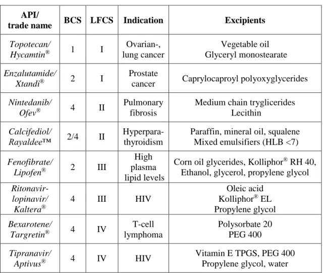
Co-administration of emulgents and co-emulgents induces significant higher solubilisation capacity in vivo compared to co-emulgent free preparations (80). Type III systems can be split into Type III/A and Type III/B depending on the oil: hydrophilic content ratio. One of the first Type III products on the market was Sandimmune Neoral®, a supergeneric reformulation of immunosuppressant cyclosporine A (81). Some years later, Pouton and his research group integrated in the classification system a new oily phase free category (Type IV), which disperses to form a micellar solution and remains relevant in case of highly hydrophobic compounds (82). However, the high surfactant and co-solvent content should not be forgotten, because it may be poorly tolerated in chronic use. The choice of formulation requires detailed analysis and depends on several factors: dose, type and molecular weight of drug, stability of drug in various excipients, significance of emulsified droplet size, risk of precipitation due to high surfactant content, solubilisation capacity, digestion by intestinal enzymes (7). Table IV indicates some commercially available FDA approved drugs of each lipid-based classes.
Excipient Type I Type II Type III/A Type III/B Type IV
Oil 100 40–80 40–80 <20 –
Hydrophobic surface-active
agent (HLB
<12)
– 20–60 – – 0–20
Hydrophilic surface-active
agent (HLB
>12)
– – 20–40 20–50 30–80
Hydrophilic
co-solvent – – 0–40 20–50 0–50
22
Table IV. FDA approved products formulated by lipid-based systems API/
trade name BCS LFCS Indication Excipients Topotecan/
Hycamtin® 1 I Ovarian-, lung cancer
Vegetable oil Glyceryl monostearate Enzalutamide/
Xtandi® 2 I Prostate
cancer Caprylocaproyl polyoxyglycerides Nintedanib/
Ofev® 4 II Pulmonary
fibrosis
Medium chain tryglicerides Lecithin
Calcifediol/
Rayaldee™ 2/4 II Hyperpara- thyroidism
Paraffin, mineral oil, squalene Mixed emulsifiers (HLB <7) Fenofibrate/
Lipofen® 2 III
High plasma lipid levels
Corn oil glycerides, Kolliphor® RH 40, Ethanol, glycerol, propylene glycol Ritonavir-
lopinavir/
Kaltera®
4 III HIV
Oleic acid Kolliphor® EL Propylene glycol Bexarotene/
Targretin® 4 IV T-cell lymphoma
Polysorbate 20 PEG 400 Tipranavir/
Aptivus® 4 IV HIV Vitamin E TPGS, PEG 400
Propylene glycol, water
23 1.4.Self-emulsifying Drug Delivery Systems
Self-emulsifying Drug Delivery Systems (SEDDS) are isotropic mixtures (preconcentrates) of drugs, natural/synthetic oils, hydrophilic/lyophilic emulgents and optionally hydrophilic co-emulgents. Following their oral intake and dilution in gastric juice, the gentle agitation of GIT creates a fine oil in water (o/w) type emulsion (Fig.6.).
SEDDS present the drug in a dissolved form, excluding the rate-limiting dissolution step.
The onset of action is rapid but it can be sustained by application of polymers. Effective solubilisation is a key attribute of emulsified systems in order to avoid precipitation of drug in GIT. Important aspect the small droplet size in case of micro-, and nanoemulsions provide a large interfacial area for drug absorption. Further advantages of SEDDS are high drug loading capacity, ease of manufacture and scale-up, protection of sensitive drugs and decreased food effects (83). A distinction is made between SEDDS, Self- Nanoemulsifying Drug Delivery Systems (SNEDDS) and Self-Microemulsifying Drug Delivery Systems (SMEDDS) on the basis of emulsified droplet size, thermodynamic stability of droplets and preconcentrate composition.
Figure 6. Composition and conception of Self-emulsifying Drug Delivery Systems (self-made)
1.4.1. Microemulsions and nanoemulsions: similarities and differences
After dilution in the GIT, SMEDDS or SNEDDS form the corresponsive microemulsions and nanoemulsions. Because of their biopharmaceutical and therapeutical advantages, these are the two most common types of colloidal dispersions (84). Furthermore, they exhibit better stability against aggregation and sedimentation along with the potential for industrial scaling-up (85). These superior properties materialize in colloidal systems possessing a droplet size <200 nm in diameter (86). It is usually easy task to define a macroemulsion (e.g. droplet size, composition, optical properties), but how to distinguish between microemulsions and nanoemulsions? Currently, there is a considerable confusion about the precise use of these terms. The reason is for the misconception that
Oil Emulgent Co-emulgent
SEDDS/SMEDDS/
SNEDDS
Macro/micro/
nanoemulsion
+ API Dilution
in GIT
24
there are many structural similarities between these two kinds of colloidal dispersion, but there are also basic differences (87).
The terminology commonly used to refer micro-, and nanoemulsions is misleading. Micro is a unit prefix denoting a factor of 10-6, nano means 10-9. At first glance one could believe that there is a three order of magnitude disparity in size between them, which is far from the truth. In practice the opposite is usually the case: the particles in a microemulsion are smaller than those in a nanoemulsion. The answer for this discrepancy lies within the development of colloidal chemistry. The first scientific article using the term
“microemulsion” was published in 1961, whereas first article related to “nanoemulsion”
appeared in 1996. The term “microemulsion” spread among researchers well before the introduction of term “nanoemulsion”, and before they were clearly defined or distinguished from one another (87).
25
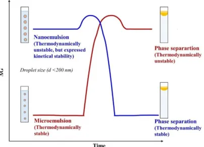
The most meaningful difference between the systems in question is the thermodynamic standpoint: microemulsion forms spontaneously as a thermodynamically stable dispersion, contrary to nanoemulsion which requires minimal energy input (88). The free energy of a microemulsion (droplets in water, ΔGcoll.disp.) is lower than the free energy of separate phases (oil and water, ΔGow), so the self-emulsification of these formulations are thermodynamically favourable (ΔGcoll.disp. <ΔGow). The situation is reversed for nanoemulsions: the free energy of the colloid dispersion is higher compared with the separated phases (ΔGcoll.disp.> ΔGow), so the creation of nanoemulsion is energetically unfavourable (unstable thermodynamics) (Fig.7.).
Figure 7. Demonstration of the free energy (ΔG) of nanoemulsion and microemulsion compared to the phase separated state. Microemulsions have a lower free energy than the phase separated state, whereas nanoemulsions have a higher free energy. (self-made)
In spite of their thermodynamic instability, nanoemulsions are able to put up aggregation resistance for months during storage; the system will persist in a metastable state. If there are sufficient energy barriers separating the two phases, the coalescence of nanoemulsified dispersion will be slow and sustained. The height of the needed energy barrier is mainly determined by physicochemical phenomena that prevent the droplets from coming into close proximity, such as repulsive hydrodynamic and colloidal (e.g., steric, electrostatic) interactions operating between droplets (89) . In general, it can be
26
concluded that the higher the energy barrier is, the more expressed the kinetic stability.
There are a lot of various physicochemical phenomena which can lead to the breakdown of a nanoemulsion (e.g. flocculation, creaming, Ostwald-ripening, coalescence) (90). The role of stabilizer excipients such as weighting agents, surface active agents, texture modifiers, polymers is essential in the long-term stability of colloidal dispersions (91).
Table V. Comparison of microemulsion and nanoemulsion
From the point of view optical properties, the droplet size is decisive: the system begins to clear up around 200 nm and turns transparent under 60 nm (92). Nanoemulsions tend to have spherical structure because of the large Laplace pressure. Within that size range the surface tension (γ: N/m) is high, while particle radius (r: m) is low, therefore - according to Law of Laplace (∆𝑃 =2𝛾
𝑟)- significant Laplace pressure can be detected. A sphere has the lowest interfacial area and concomitant surface tension for a given volume of material, that is the reason behind nanoemulsions favoured character (86).
Character Microemulsion Nanoemulsion
Thermodynamic stability Stable Unstable
Kinetic stability Unstable, but compensated
Metastable, steric and electrostatic repulsive
forces Morphology Disparate (e.g. spherical,
lamellar, cylinder) Spherical
Composition
Higher emulgent/oil ratio Emulgent with low
molecular mass
Lower emulgent/oil ratio Proteins, polymers, emulgents with high
molecular mass Optical properties Clears up around 200 nm, turns transparent under 60 nm
Particle-size distribution Single, narrow peak, low polydispersity
Multiple, broad peaks, relative high polydispersity
27
Microemulsions exhibit a wide range of shapes (e.g. worm-like, bicontinuous sponge- like, liquid crystalline, or hexagonal, spherical swollen micelles). Influencing factors are type and quantity of incorporated oil and surfactant, and the ideal curvature of utilized surfactant(s) (Tbl. V.) (93). In this thesis I am primarily focus on microemulsions and nanoemulsions that can be used to encapsulate lipophilic components, that consist of small spheroid particles comprised of oil and surfactant molecules dispersed within water.
Other kinds of microemulsion systems (e.g. liquid crystalline, worm-like) are out of the scope of this thesis.
It is important to distinguish between microemulsions and nanoemulsions in practice since this predispose their long-term stability, functionality, robustness against altered environmental conditions (temperature, relative humidity, dilution) and bioavailability.
Some practical methods provide data which category a colloidal dispersion belongs to.
Composition, particle size distribution studies, long-term storage measurements supplemented with altering environmental conditions, particle shape analysis might be constructive for determining the type of system.
28
1.4.2. Role of excipients in the formulation of SEDDS
The utilized excipients have a definitive influence on self-emulsification, stability and drug delivery (Fig.8.). Chemical structure and concentration of oil and emulgent, oil/emulgent ratio, quality and quantity of co-emulgent, emulgent/co-emulgent ratio, weigh-in order, temperature, ionic strength were shown to have significant effect on the quality of SEDDS (94). It is clear that a lot of parameters must be considered to fulfil the critical quality attributes of SEDDS:
• Solubility of API in the oily phase must be maximal
• Achieve in the various physiological conditions of GIT constant droplet size and stability
• Toxicity, purity, stability, price, acquisition of excipients are important criteria
• Improved bioavailability by reduced droplet size and enhanced absorption via lymphatic transport and solubilisation
Figure 8. Schematic diagram of o/w nanoemulsified droplet solubilizing lipophilic substance (self-made)
29 1.4.2.1.Oils
Oils are the most significant components of SEDDS, which promote self-emulsification, facilitate drug absorption through the mucosal membrane and allow the dissolution of large amounts of lipophilic substances. The solubilisation capacity of GIT is also enhanced by using lipid components because they stimulate pancreatic and bile juice excretion (95). Oils can be classified as natural, semi-synthetic and synthetic derivates.
According to their chemical structure we can distinguish between triglycerides and mixed glycerides (mixture of mono-, di-, and triglycerides) esterificated by medium or long- chained fatty acids (saturated/unsaturated). Natural oils are derived primarily from plant sources comprised of mixtures of triglycerides which contain fatty acids of varying chain lengths and degrees of unsaturation (e.g. soy oil, sunflower oil, coconut oil, olive oil).
Their main advantage is the fully digestion and absorption in the GIT mediated by physiological enzymes and therefore they are generally regarded as safe (GRAS). The acceptance of patients is also higher in case of natural medicines and excipients. The low resistance to oxidation and decreased solvent capacity compared to semi-synthetic and synthetic derivates are challenging formulation issues (7).
Semi-synthetic and synthetic oils are partial glycerides, prepared by glycerolises, a transesterification reaction of triglycerides in order to increase the hydrophilic character of natural oils. There are several modified oils on the market: glyceryl- monocaprylocaprate (Capmul® MCM); glyceryl-monostearate (Geleol™, Imwitor® 191, Cutina™ GMS, Tegin™); glyceryl-distearate (Precirol™ ATO 5); glyceryl-monooleate (Peceol™); glyceryl-monolinolate (Maisine™ 35-1); glyceryl-dibehenate (Compritol®888 ATO). PEGylated polyoxylglycerides are also available: oleoyl macrogol-6 glycerides (Labrafil® M 1944CS), lauroyl Macrogol-32 glycerides (Gelucire® 44/14). Numerous research groups investigated the relationship between solubility and bioavailability of API and structure of oil (96–98). The components, process parameters and conclusions were different and therefore does not enable unambiguous interpretation. They pointed out as a collective and general experience that the incorporated active substance has a significant impact on the in vitro/in vivo fate of lipid-based DDS.
30 1.4.2.2.Surfactants
Surfactants are amphiphilic molecules that lower the surface tension, meaning they contain both hydrophilic and hydrophobic groups. Emulsifiers are indispensable components of SEDDS as they strengthen drug absorption by altering the lipid bilayer organization, enhance the lipid-intestinal membrane interactions, dissolve hydrophobic substances between their hydrocarbon chains and moreover kinetically stabilize the droplet size distribution (99). Application of higher surfactant concentrations don’t always reduce droplet size of colloidal dispersion, formed after dilution of SEDDS preconcentrate in GIT, but may even increase it. The phenomenon can be attributed to the increased water penetration, and concomitant disruption of interfacial film, which causes oil droplets to be expelled into external water phase (82). Surfactants are classified into cationic, anionic, zwitterionic and non-ionic types. It was revealed that non-ionic surfactants demonstrating lower toxicity and have better tolerability in case of chronic use compared to ionic ones (100). Hydrophilic-lipophilic balance (HLB) is an important indicator for the characterization of surfactants that quantifies the oil and water attracting capacity of the surface-active molecule. Using 30-70 % (w/w) emulgent provides colloidal SNEDDS/SMEDDS dispersions with long-term physical stability and narrow particle-size distributions (101). From the point of view self-emulsification, the preferred HLB value should be higher than 12 (102). The HLB for a mixture of emulsifiers is calculated in proportion to the concentrations. Droplet size analysis was demonstrated as a suitable method for the determination of required HLB value in lemon oil emulsions (103). A special property of non-ionic surfactants is cloud point, the temperature above which the surfactant phase separates and precipitates out of solution. The cloud point is higher than 37 °C in ideal case because of the risk of irreversible phase separation in the body (104). Furthermore, it was demonstrated that several emulgents have an inhibitory impact on different CYP-enzymes and on intestinal P-glycoprotein, which phenomena can be used to enhance the bioavailability in special cases (105). Some of the typically used surface active agents in SEDDS formulations are macrogolglycerol-ricinoleate (Kolliphor® EL), macrogolglycerol-oleate (Labrafil® M 1944 CS), caprylocaproyl macrogol-8 glycerides (Labrasol®), propylene glycol monocaprylate type II (Capryol® 90) polysorbate 20 (Tween® 20).
31 1.4.2.3.Co-surfactants/Co-solvents
Co-solvents are integral part of SEDDS. They initiate self-emulsification, increase the elasticity of interfacial film, lower interfacial tension, allow the dissolution of remarkable amount of API and prevent formation of liquid crystals (106). Co-surfactants can also be used to fine-tune the formulation phase behaviour, for example, by expanding the temperature or salinity range of microemulsion formation. In general, the optimal applied concentration lies between 20 and 50 %(w/w). Application of co-surfactant is not mandatory at all, several studies focused on cosolvent-free formulations (107, 108).
Ethanol, propylene glycol, Transcutol® P and PEG400 are some of the widely used co- surfactants.
1.5.Transformation of liquid SEDDS/SMEDDS/SNEDDS into solid dosage forms
Transforming liquid self-emulsifying preconcentrates to solid carriers is a strategy in lipid-based formulation design, which besides solubility improvement, offers further advantages over liquid systems; increased long-term physicochemical stability, ease to scale up, processability, precise dosing and improved patient compliance (109).
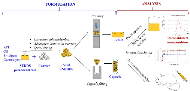
However, it is important that the solidification procedure must preserve self-emulsifying properties and small droplet size which have to be demonstrated by reconstitution studies during formulation development. The most relevant solidification methodologies are spray drying, extrusion-spheronization and adsorption onto porous solid carriers (110).
Adequate drug loading, compatibility, suitable drug release profile, processability are critical quality attributes of carriers used for converting liquid SEDDS to solid dosage forms (111). Oral delivery of pH-, and enzyme-sensitive biologics may be achieved by solid SEDDS formulated with special environment responsive excipients. The basic methodologies are reviewed briefly below (Fig.9.).
32
Figure 9. Formulation and analysis possibilities of solid SEDDS (self-made)
Extrusion-spheronization
Extrusion-spheronization is an agglomeration technique involving multiple processes for the preparation of matrix pellets. A compromise between the smallest amount of adsorbent material needed and the largest amount of liquid SEDDS required is essential to produce pellets with good physical characteristics and highest possible drug loading (112). Sticking of extrudate, friable and soft pellets, challenging spheronization process and decreased porosity are the main consequences of high liquid SEDDS content (113).
Reconstitutability and self-emulsifying properties of matrix pellets were confirmed in several studies (114, 115). The order of components mixing was identified as a critical parameter for a successful extrusion/spheronization process and for the roundness value of pellets (116). Colloidal silicon dioxide, microcrystalline cellulose and lactose were successfully utilized for the preparation of self-emulsifying matrix pellets. HLB value of emulgent in liquid phase influences the processability and mechanical properties of pellets. Matsaridou et al. pointed out that the required water quantity for pelletization rose linearly with increasing HLB values (117). Furthermore, the higher the HLB, the faster the disintegration time. Extrusion-spheronization method is suitable for the production of modified release dosage forms. It can either be carried out by formation of a slowly dissolving matrix structure (e.g. HPMC, PVP) that delay release of the drug or by coating the surface of pellets with gastroretentive polymeric dispersion (118).
33 Adsorption onto porous solid carriers
The simplest and most intensively investigated method to formulate solid SEDDS is adsorption to highly porous solid carriers. Generally used carriers for adsorption of liquid SEDDS are:
• silicon dioxide; fumed silica with different grades of specific surface (Aerosil®) or micronized amorphous silica with different grades of pore volume (Sylysia®);
• magnesium aluminometasilicate with different surface properties (alkaline or neutral) and particle size (Neusilin®)
• porous dibasic calcium phosphate anhydrous (Fujicalin®) and
• microcrystalline cellulose
Combined application of these carriers have been reported as effective approach for producing solid SEDDS with adsorption method (119). Relative simplicity, absence of organic solvents, easy scale up, high drug loading and small number of solid excipients needed are advantages of adsorption method (120). Drug loading is in close correlation with carrier’s oil adsorbing capacity; even loadings up to 80 % have been reported;
nevertheless, general value is about 50% (121). Beg at al. evaluated the oil adsorbing capacity of various porous carriers, which declines in the following order:
Sylysia® 350 > Neusilin® US2 > Sylysia® 550 > Sylysia® 730 > Aerosil® 200 (122). High drug loading and high oil adsorbing capacity are not associated always with superior bioavailability. Yeom et al. pointed out that SEDDS formulated with low oil adsorbing capacity mannitol had better bioavailability compared to other carriers (123). Particle size, porosity (length and diameter of pores), type and amount of adsorbent along with the specific carrier-SEDDS interactions like wettability play a crucial role in dissolution kinetics and drug release. The effect of wettability, the affinity between lipid components and surface of solid carriers was highlighted by hydrophilic (Aerosil® 200) and hydrophobic porous silica particles (Aerosil® R972) with similar nanostructures (124).
The surface interactions were significantly greater between hydrophobic particles and lipid components, which was confirmed by only 33% nifedipine release, compared to 80% of hydrophilic silica particles. Thus, the remarkable hydrophobic forces can be utilized to sustain the solubilisation of lipophilic drugs. However, these interactions have been reported as contraproductive in some cases; the incomplete drug release had to
34
counterbalance using superdisintegrant in the formulations (125). Complete drug release can be also hampered due to gel formation and clogging of pores (126).
Spray drying
Spray drying is a well-known gentle, rapid, cost-effective and scalable process in pharmaceutical industry, where the dry powder is made from a fluid material by atomization it into a hot drying gas, usually air. For the production of solid SEDDS sufficient carriers are needed that can influence drug release and oral bioavailability of incorporated drug (127). Low yield and the stability issue of volatile cosurfactants (e.g.
Transcutol®, ethanol) due to relative high drying temperature must be kept in mind during formulation development (128). Further limiting factor for this technique is the viscosity of SEDDS; it must be low enough to enable spraying onto the carrier. Several hydrophobic (colloidal silica, magnesium stearate) and hydrophilic (e.g. maltodextrin, dextran, HPMC, lactose, poly vinyl alcohol) carriers were investigated for spray dried solid SEDDS. Controlled drug release from solid SEDDS can be obtained by water- soluble carriers, which exert erosion and swelling-dependent drug release mechanisms.
Yi et al. prepared nimodipine-loaded solid SEDDS stabilised with HPMC of various viscosities. Formulation with higher HPMC viscosity demonstrated a reduced release rate due to the inhibition of drug diffusion through dense polymer matrix and sustained polymer erosion (129). Prevention of in vivo drug precipitation can be achieved by adding polymers like gelatine and Soluplus® to the dispersion to be spray dried (130).
1.6.Cyclodextrin complexation
Cyclodextrins (CDs) are cyclic oligosaccharides composed of α-1,4-linked D- glucopyranose units possessing a hydrophilic exterior and hydrophobic cavity, where lipophilic molecules can form a non-covalently bonded inclusion complex (131).
Although they have been discovered in the early 1890s, the attention was drown to CDs in the 1980s with the first application in the pharmaceutical and food industries (132).
These molecules can operate and become widespread as excipients throughout the whole industrial and agricultural sector. In pharma industry CDs are used for the improvement of water-solubility and bioavailability of medicinal products. They are used for example in tablets, aqueous parenteral solutions, nasal sprays and eye drop solutions (4). In food
35
industry their taste masking and flavour stabilizing along with food preservative effect is utilized (133). In agriculture CDs are applied for improvement of the physico-chemical characteristics of pesticides, extension of shelf-life and reduction of environmental pollution (134).
1.6.1. Short history of Cyclodextrins
Antoine Villiers French pharmacist described in 1891 the formation of unexpected crystals with particular properties during a potato starch fermentation reaction by Bacillus amylobacter. Villiers concluded that the properties of these special dextrins were very clearly different from those of the various saccharides known at that time (132). The next exciting scientific result of CD research was linked to the work of Austrian Franz Schardinger. In two publications (1903, 1911) he described the preparation, separation and purification of Dextrin A and B, which he classified as cyclic polysaccharides (135).
His hypothesis regarded to the cyclic structure of Dextrin A and B was confirmed in 1948 by Freudenberg et al. Friedrich Cramer introduced first the term “cyclodextrin” (1956) and provided numerous data on cyclodextrin host-guest complexes and basic physicochemical characteristics (136). The “CD bomb” was exploded in the 1980’s: the nontoxicity of CDs became increasingly accepted, and several manufacturers started to produce and to market CDs. The first CD-containing product, a prostaglandin E2/β-CD (Prostarmon E™) sublingual tablet was launched in Japan, 1976. Since then a great number of patents, publications and products were the outcome of CD inclusion complexes to enhance solubility, improve stability and increase bioavailability of drugs.
Today the annual CD production is over 10000 tonnes and CDs can currently be found in over 60 marketed pharmaceutical products (Tbl. VI.) (137). Traditionally, CDs are used as excipients, however several research groups all over the world are investigating their potential as active pharmaceutical ingredients. Bridion® (modified γ-CD by placing eight carboxyl thio ether groups at the sixth carbon positions) and HP-β-CD, which are used for reversal of neuromuscular block in anaesthesia and for the treatment of fatal genetic Niemann Pick Type C disease, a cholesterol metabolism disorder, respectively. The latter one has received an orphan drug designation (EU/3/11/895) (138).
36
Table VI. FDA approved pharmaceutical products with cyclodextrins
Drug CD Trade name Dosage form
Alprostadil α-CD Edex® Injection
Piroxicam β-CD Brexin® Tablet
Omeprazol β-CD Omebeta® Enteric capsule
Metronidazole β-CD Flagyl® Vaginal gel
Aripiprazole SBE-β-CD Abilify® I.m. solution
Ziprazidone SBE-β-CD Zeldox® Capsule
Itraconazole HP-β-CD Sporanox® Oral solution
Mytomicin HP-β-CD MitoExtra® I.v. infusion
Chloramphenicol RAMEB Clorocil® Nasal spray
Minoxidil γ-CD Alopexy® Hair solution
Abbreviations: HP-β-CD ((2-hydroxypropyl)-β-CD), RAMEB-CD (random methylated β-CD) (SBE-β-CD (sulfobutylether-β-CD), I.m. (intramuscular), I.v. (intravenous)
37
1.6.2. Physicochemical characteristics of Cyclodextrins
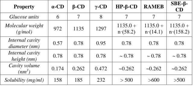
Naturally occurring parent CDs are crystalline, homogeneous, nonhygroscopic substances, which are toroidal macro-rings built up from glucopyranose units. α, β, and γ CDs comprise of 6, 7, and 8 glucopyranose units, respectively, and differ in their molecular weight, cavity size, and solubility (Fig.10/A., Tbl. VII.). There are bigger CDs composed of 9 (δ-CD), 10 (ε-CD), and 11 (ζ-CD) glucose units, but their practical suitability is negligible in the field of pharmaceutical sciences (139). The glucopyranose units adopt a 4C1 conformation and orient themselves so that the molecule forms the aforementioned torus-like geometry. The consequence of this geometry is that the CD molecule has a hydrophilic (exterior) surface and a considerably less hydrophilic (interior) microenvironment in its cavity (140).
Figure 10. Chemical structure of α-, β-, and γ-CD (A). Schematic demonstration of CD and drug inclusion mechanism (B) (self-made)
38
The polarity of the cavity has been estimated to be similar to that of aqueous ethanolic solution. This attribute gives CDs the opportunity to host molecules possessing low water solubility (Fig.10/B.). All secondary hydroxyl groups are situated on one of the two rims (“wider”) of the ring, whereas all the primary ones are placed on the other rim (“narrower”) (141). These hydrophilic groups are outside of the molecular cavity, whereas the inner surface has a less hydrophilic character due to the ether-like anomeric oxygen atoms and the skeletal carbon atoms.
Table VII. Physicochemical properties of parent and derivate cyclodextrins (self-made) Property α-CD β-CD γ-CD HP-β-CD RAMEB SBE-β-
CD
Glucose units 6 7 8 7 7 7
Molecular weight
(g/mol) 972 1135 1297 1135.0 + n·(58.2)
1135.0 + n·(14.1)
1135.0 + n·(158.2) Internal cavity
diameter (nm) 0.57 0.78 0.95 0.78 0.78 0.78
Internal cavity
height (nm) 0.78 0.78 0.78 ~ 0.78 ~ 0.78 ~ 0.78 Cavity volume
(nm3) 0.174 0.262 0.472 ~0.262 ~0.262 ~0.262 Solubility (mg/ml) 158 185 232 > 500 >600 >500
Substitution of any of the hydrogen bond-forming hydroxyl groups of native CDs, even by lipophilic functions, results in a dramatic improvement in their aqueous solubility, referred as CD derivates (141). The modification of natural (parent) CDs generally aims at converting them into amorphous, non-crystallisable derivatives, to provide high CD concentration in aqueous solutions. Number of CD derivates exceeds 11.000, but new types are permanently developed (142). At present, α-CD, β-CD, γ-CD, HP-β-CD, RAMEB and SBE-β-CD are used in medicines on the European market (143). According to regulatory requirements, the extent of substitution must be defined to avoid any misunderstandings. Conventionally, the so-called average degree of substitution (DS) is used, which gives the average number of substituted hydroxyls of a glucose unit (142).
One of the largest developer, producer and supplier of CD derivates and a pioneer in CD- based innovation is CycloLab, headquartered in Budapest, Hungary.
![Figure 3. Growing interest in baicalin as a bioactive phytopharmacon 1982-2019 Source: PubMed, keyword: baicalin [all]](https://thumb-eu.123doks.com/thumbv2/9dokorg/1362787.111096/13.892.221.678.519.796/figure-growing-baicalin-bioactive-phytopharmacon-source-pubmed-baicalin.webp)
