SEMMELWEIS EGYETEM DOKTORI ISKOLA
Ph.D. értekezések
2300.
LÁSZLÓ ANDREA
Experimentális és klinikai farmakológia című program
Programvezető: Dr. Szökő Éva, egyetemi tanár Témavezető: Dr. Nemcsik János, családorvos
Associations of affective temperaments with arterial stiffness and Brain-derived Neurotrophic Factor in
hypertension
Ph.D. Thesis
Andrea László M.D.
Semmelweis University School of Ph.D.
Studies Pharmaceutical Science
Tutor: János Nemcsik, M.D., Ph.D.
Opponents:
László József Wágner, M.D., Ph.D.
Tibor Kovács, M.D., Ph.D.
Head of the Complex Examination Committee:
Szabolcs Várbíró, M.D., Ph.D.
Members of the Complex Examination Committee:
Krisztián Szabolcs Vörös, M.D., Ph.D.
Péter Légrády, M.D., Ph.D.
Budapest 2019
2
ABBREVIATIONS ... 5
1. BACKGROUND ... 8
1.1. Cardiovascular diseases, hypertension and mood disorders: incidence and connections ... 8
1.1.1. Cardiovascular diseases in the XXIst century ... 8
1.1.2. The importance of hypertension ... 8
1.1.3. Mood disorders: a common public health problem in the Western world . 9 1.1.4. Well-known connections between CVDs and depression ... 9
1.2. Affective temperaments: risk factors for psychiatric disorders and CVDs ... 10
1.2.1. Background, definition and assessing of affective temperaments ... 10
1.2.2. Five types of affective temperaments ... 12
1.2.3. Dominant affective temperaments and their relation to affective disorders – a spectrum concept ... 13
1.2.4. Affective temperaments: molecular genetic backgrounds... 14
1.2.5. Affective temperaments are, in healthy populations, part of the bipolar and unipolar mood disorder spectrum ... 15
1.2.6. The role of affective temperaments in the psychopathology: bipolar and unipolar mood disorders ... 15
1.2.7. Affective temperaments and cardiovascular risk... 16
1.3. Arterial stiffening as a risk factor for CVDs ... 17
1.3.1. Definition and pathophysiology of arterial stiffness ... 17
1.3.2. Methods for measuring arterial stiffness, focused on arterial tonometry . 18 1.3.3. Parameters of arterial stiffness ... 19
1.3.4. Pulse wave velocity ... 19
1.3.5. Arterial stiffness parameters and CV risk estimation, according to actual guidelines ... 23
3
1.4. New psychosomatic connections: the possible role of the brain-derived
neurotrophic factor (BDNF) in psychopathology and cardiovascular diseases ... 26
1.4.1. Brain-derived neurotrophic factor (BDNF) ... 26
1.4.2. BDNF in mood disorders ... 26
1.4.3. BDNF and CV pathology ... 27
1.4.4. BDNF polymorphism ... 27
2. OBJECTIVES ... 29
3. PATIENTS AND METHODS ... 30
3.1. Study 1: patients and methods in the study measuring arterial stiffness and serum BDNF level in patients with and without dominant affective temperaments .. 30
3.1.1. Questionnaires ... 31
3.1.2. Clinical measurements ... 32
3.1.3. Statistical analysis ... 33
3.2. Study 2: patients and methods in the study exploring the association of affective temperaments with blood pressure and arterial stiffness in hypertensive patients ... 33
3.2.1. Statistical analysis ... 34
3.3. Study 3: patients and methods in the study measuring serum BDNF levels in a hypertensive and a control population to discover the associations of BDNF with affective temperaments, depression, anxiety and arterial stiffness ... 34
3.3.1. Statistical analysis ... 35
4. RESULTS... 36
4.1. Study 1: results of hemodynamic, arterial stiffness and serum BDNF levels in hypertensive patients with or without dominant affective temperaments ... 36
4.2. Study 2: results of affective temperaments’ association with blood pressure and arterial stiffness in hypertensive patients ... 39
4
4.3. Study 3: results of serum BDNF levels in hypertensive patients and healthy controls; associations of serum BDNF with affective temperaments, depression,
anxiety and arterial stiffness ... 47
5. DISCUSSION ... 53
5.1. Study 1: arterial stiffness and serum BDNF levels in hypertensive patients with or without dominant affective temperaments ... 53
5.2. Study 2: association of affective temperaments with blood pressure and arterial stiffness in hypertensive patients ... 56
5.3. Study 3: serum BDNF levels in a hypertensive and a control population; associations of serum BDNF with affective temperaments, depression, anxiety and arterial stiffness. ... 60
5.4. Limitation of the studies ... 67
5.4.1. Limitations of Study 1 ... 67
5.4.2. Limitations of Study 2 ... 68
5.4.3. Limitations of Study 3 ... 68
5.5. Future perspectives ... 68
6. CONCLUSIONS ... 69
7. SUMMARY ... 70
ÖSSZEFOGLALÁS ... 71
8. REFERENCES ... 72
9. PUBLICATION SUMMARY... 106
9.1. Publications related to the thesis ... 106
9.2. Publications not directly related to the thesis ... 106
10. ACKNOWLEDGEMENTS ... 109
5 ABBREVIATIONS
CCTA Coronary computed tomography angiography 5-HTTLPR Serotonin-transporter-linked polymorphic region
AC Abdominal circumference
ACE Angiotensin-converting enzyme
AIx Augmentation index
Alp Patients regularly using alprazolam;
AP Augmentation pressure
ARB Angiotensin II receptor blocker
AT Angiotensin
BDNF Brain-derived neurotrophic factor
BMI Body mass index
cDBP Central diastolic blood pressure CHD Coronary heart disease
cMBP Central mean blood pressure
CONT Controls
cPP Central pulse pressure
CREB cAMP response element binding protein CRH Corticotropin-releasing hormone
CRP C-reactive protein
cSBP Central systolic blood pressure
CV Cardiovascular
CVD Cardiovascular disease DBP Diastolic blood pressure
DBPB Diastolic brachial blood pressure
DOM Patients with dominant affective temperaments
DT Diastolic time
ECG Electrocardiography
ESBP End-systolic blood pressure ESC European Society of Cardiology ESH European Society of Hypertension
6 ESRD End-stage renal disease GABA Gamma-Aminobutyric acid
GFR-EPI Glomerular filtration rate assessed by the chronic kidney disease epidemiology collaboration glomerular filtration rate equation HDL High-density lipoprotein
HP Heart periode
HPA Hypothalamic-pituitary-adrenal
HR Heart rate
HT Hypertensive patients
ICAM-1 Intercellular Adhesion Molecule 1
IL Interleukin
IMT Intima media thickness LDL Low-density lipoprotein LPR LDL receptor-related protein
LV Left ventricular
LVET Left ventricular ejection time MAP Mean arterial pressure
MDBP Mean diastolic blood pressure MDD Major depressive disorder MRI Magnetic resonance imaging MSBP Mean systolic blood pressure
NF-κB Nuclear factor kappa-light-chain-enhancer of activated B NGF Nerve growth factor
NO Nitric oxide
NT Neurotrophin
NT-3 Neurotrophin 3
NT-4 Neurotrophin 4
p75Ntr Neurotrophin receptor p75 Partial Ra Partial correlation coefficient
PP Pulse pressure
PPAmp Pulse pressure amplification PPB Brachial pulse pressure
7
PWV Pulse Wave Velocity
QOL Quality of life
RAAS Renin-angiotensin-aldosterone system RAS Renin-angiotensin system
Rpm Revolutions per minute SBP Systolic blood pressure
SBPAmp Systolic blood pressure amplification SBPB Systolic brachial blood pressure
SD Standard deviation
SE Standard error
seBDNF Serum Brain-derived neurotrophic factor SEM Standard error of the mean
TEMPS-A Temperament Evaluation of Memphis, Pisa, Paris and San Diego Autoquestionnaire
TIA Transient ischemic attack TNF Tumour necrosis factor Trk Tropomyosin receptor kinase VEGF Vascular endothelial growth factor WHO World Health Organisation
8 1. BACKGROUND
1.1. Cardiovascular diseases, hypertension and mood disorders: incidence and connections
1.1.1. Cardiovascular diseases in the XXIst century
Cardiovascular diseases (CVDs) are leading causes of death, major causes of health loss worldwide and therefore major barriers to sustainable human development (1). In 2011, the United Nations formally recognized noncommunicable diseases, including CVDs – among cancer, chronic respiratory disease, and diabetes – as major concerns for global health. According to the World Health Statistics, an estimated 17.9 million deaths occurred due to CVDs, accounting for 31.4% of the overall total of 57 million deaths in the year 2016 (2). Ischemic heart disease was the leading cause of CVD health lost globally and also in each world region, followed by stroke. Even though over the past 25 years sociodemographic changes have been associated with dramatic declines in CVDs, at present there is only a gradual decrease or no change in most regions. So, the trends in CVD mortality have plateaued and are no longer declining in high-income regions (3).
1.1.2. The importance of hypertension
Among the risk factors of CVDs hypertension has a major role affecting 1 billion adults globally (4): it is the leading cause of death and disability-adjusted life years (5) having an excessive impact on mortality worldwide. In the United States hypertension accounted for more cardiovascular (CV) deaths than any other modifiable CVD risk factor and was second only to cigarette smoking as a preventable cause of death for any reason (6, 7). In Europe the prevalence of high blood pressure among adults is between 30 and 45%, rising with aging (8). In Hungary, the number of subjects with hypertension is approximately 3.5 million and this high prevalence contributes markedly to the poor Hungarian CV morbidity and mortality figures (9): in 2014, 51.7%
of all-cause mortality was due to CVDs (10).
There are modifiable (e.g. smoking, obesity, unhealthy diet) and non-modifiable (e.g.
positive family history, age, male sex) risk factors of CVD which have a complex and interdependent relationship with hypertension. CVD risk factors can influence those
9
pathophysiological pathways which are also involved in the development of hypertension, such as the renin-angiotensin-aldosterone system (RAAS), sympathetic nervous system, cardiac natriuretic peptide system or the endothelial function (11-13).
Consequently, hypertension can also be considered as an outcome of different harmful processes, not only a simple disease. On the other hand, the development of high blood pressure can lead in longer term to adverse CV outcomes (14).
Hypertension is the most prevalent modifiable risk factor for global disease burden in both sexes, regardless of ancestry, geographic region or income (5), thus early diagnosis and treatment of this condition is essential. Among the healthy population it would be required to find subpopulations, in which the risk of the onset of this disease is higher.
Furthermore, among the hypertensive subjects the recognition of patients with elevated CV risk would help reach more effective primary and secondary prevention.
Hypertension prevention receives a great attention, but the role of personality in the development of high blood pressure has not been fully clarified yet.
1.1.3. Mood disorders: a common public health problem in the Western world According to the World Health Organisation (WHO) depression is a common disease worldwide,involving up to 15% of the general population, with more than 350 million people affected in 2018 (15). According to WHO’s estimation, by 2020 depression will be already the second leading cause of world disability (16) and by 2030 it is expected to be the largest contributor to disease burden (17). Major depressive disorder (MDD) accounts for 12.3% of the global burden of disease (18). Based on the fact that depressive disorders often start at a young age – reducing the person’s functioning and oftentimes recurring – it is not surprising, that depression is the leading cause of disability for both men and women worldwide in terms of total years lost due to disability (19). While in 1990 depressive disorders were listed as the fourth leading cause of burden in the Global Burden of Disease (20), in 2000 they reached the third place (21).
1.1.4. Well-known connections between CVDs and depression
There is growing evidence, that physical and psychological diseases are not independent from each other. Between 9.3% and 23% of individuals with one or more chronic physical disease suffer from co-morbid depression (22). It is also known, that there is a
10
complex reciprocal interrelationship between depression and coronary heart disease (CHD) (23, 24). Depression is associated with a 3- to 4-fold increase in the risk of recurrent cardiac events and death (25). In addition, among people with CHD, depression increases the risk of future cardiac mortality and morbidity (26, 27). On the other hand, patients with CHD have a depression prevalence of about 300% than healthy subjects (28). In patients after an acute myocardial infarction, major depression is three times more common than in the general population (29).
There are multiple mechanisms by which depression could increase the probability of vascular diseases. These include increased platelet activation (30), endothelial dysfunction (31), elevation of inflammatory markers (32), as well as reduced heart rate variability (33). Dysregulation of metabolic, immune-inflammatory and autonomic systems, as well as the hypothalamic-pituitary axis, also seem to build a link between MDD and CVDs (34). Furthermore neurohormonal factors, and genetic linkages – such as with the serotonin transporter mechanism (35, 36) –, and also behaviour mechanisms are part of the pathophysiological background connecting both diseases (37).
1.2. Affective temperaments: risk factors for psychiatric disorders and CVDs
Parallel to depression, well-established biomarkers of inflammation were also found to be elevated in subjects in states of anger or hostility (38, 39), while a reduced function of the autonomic nervous system was observed in subjects in states of anxiety (40), and interpersonal antagonism was connected with carotid arterial intima-media thickening (41). These findings suggest that there are also psychological conditions in the range of normality which could influence CV risk. These data suggest that besides the evaluation of depression, analysing people’s characteristics based on their individual nature would lead us to a better understanding and prevention of CV risk.
1.2.1. Background, definition and assessing of affective temperaments
The concept of human personality dates back to Hippocrates (ca. 460 BC - ca. 370 BC) and Aristotle (384-322 BC). Four temperamental types were differentiated based on body fluids (called “humors”): melancholic, choleric, sanguine and phlegmatic (42).
Not only the ancient Greeks, but much later Luois XIV was delighted by the theory of the four temperament types, as clearly illustrated by his “grande commande” of statues
11
ordered to decorate the gardens of the Palace of Versailles in 1674. Among the 24 statues, the “The Four Humors of Man” were also included (Figure 1).
Figure 1. Charles Le Brun: The Four Humors of Man.
From left to right: choleric, melancholic, sanguine and phlegmatic.
Building on such classical theory and on Kraepelin's four distinct basic temperament types from 1913, professor Hagop S. Akiskal formulated the modern concept of affective temperaments and completed them with another one. Since then, we speak about five affective temperaments: depressive, irritable, hyperthymic, cyclothymic and anxious (43). Furthermore, Akiskal gave a new point of view on the definition of temperament and widened the term “temperament” to include healthy personality. Thus temperament – like personality – does not by itself necessarily constitute a pathology (44). All of us have our own characteristic temperamental profile determining our emotional response to environmental stimuli. In their natural mixture affective temperaments are responsible for cultural characteristics on a national level and can be associated with desirable outcomes such as adaptation and achievement (44-46).
The presence of these affective temperaments can be evaluated with the autoquestionnaire version of Temperament Evaluation of Memphis, Pisa, Paris and San
12
Diego (TEMPS-A). The questionnaire was developed by Akiskal and his co-workers (47, 48), and since 2005 it has been translated and/or validated in more than 30 languages, including such diverse cultures as the USA, Egypt, Japan, Lebanon, Portugal, Italy, Turkey, Germany, France, China, Korea, Russia, Tunis, Argentina, Brazil, Armenia, Hungary and many others (49). The questionnaire consists of 5 subscales – according to the 5 affective temperaments, requiring only simple “yes”
(score 1) or “no” (score 0) answers.
1.2.2. Five types of affective temperaments
As mentioned above, according to Akiskal's concept, 5 affective temperament types can be distinguished: depressive, cyclothymic, irritable, anxious and hyperthymic. A person with a depressive temperament can be described as pessimistic, highly self-critical, gloomy, excessively worrying, preoccupied with personal failure, lacking assertiveness, self-denying, and striving to please others (48). In cyclothymic temperament, a person shows abrupt biphasic alterations in mood between highs and lows in energy, self- confidence and agility of mind with a frequency of several days each (50). A person with an irritable temperament reacts to negative events with negative emotions, presents fluctuations in mood and complaints, and shows close linkages to the cyclothymic temperament (51). People with anxious temperaments show uncontrolled worry even about their routine, an unfounded sense of danger, a real fear of unhappiness and an overalertness. They are unable to relax, stressed, restless and anxious (52). The difference between the hyperthymic temperament and the other four is that it proves to be more adaptive and beneficial for the patient, since it involves potentially positive characteristics: inter alia higher scores in sociability, energy and mood. On the other hand, such characteristics come in excess, and the flip side of this temperament is that it can involve maladaptive behavior, e.g. overconfidence, over-socialization, irresponsibility and ruthlessness, which can all result in undesirable outcomes of a person’s actions (50).
It is important to note that sex differences are present in relation with affective temperaments. Vázquez et al. – and also other studies – found in the general population significantly higher irritable and hyperthymic temperament scores in men, and significantly higher anxious, depressive and cyclothymic temperament scores in women
13
(43, 53-55). On the other hand, each person scores differently in the different temperament directions and these are not fully independent form each other: positive associations were described between depressive and anxious, and cyclothymic and irritable temperaments (43).
1.2.3. Dominant affective temperaments and their relation to affective disorders – a spectrum concept
Dominant affective temperaments are defined with a cut-off point of z-scores above +2 standard deviation (SD) in the TEMPS-A questionnaire. Investigating the different countries, 13 to 20% of the population has some kind of dominant affective temperaments; in Hungary this value is 16.8 % (54). Temperaments can also be so extreme that they could be considered abnormal in a statistical sense – even with the absence of mental disorder (44).
It is unclear whether Aristotle discovered the concept of the bipolar spectrum, but he seems to have placed the query on exceptional greatness within the dimensional space between temperament and affective disease. At the beginning of the twentieth century, Kraepelin believed that the temperaments are subclinical forms and many times the precursors of major affective psychoses (56). According to Akiskal's theory of dominant affective temperaments – temperaments in their extreme forms, these are regarded as the subclinical/trait-related manifestations or phenotypes of bipolar or unipolar mood disorders (49). They often present an increased inclination towards these diseases and are located at the end of the continuum, close to affective illness (45, 49, 57, 58). Based on these facts, Akiskal visualized the spectrum concept of temperament and bipolar disease on the basis of Kretschmer’s work (1936) (42). In a Gaussian curve Figure 2 shows the normal population, the 13-20% of people having dominant affective temperaments and the remaining small part of the population with bipolar diseases – depending on the genetic loading.
14
Figure 2. Temperament and bipolar disease in a spectrum concept.
Adopted from: (42).
1.2.4. Affective temperaments: molecular genetic backgrounds
Since different affective temperaments are predominant in the population in different geographic regions and cultures, a connection to genetic causes appears logical.
Temperament in general (59) and affective temperaments in particular are biological in nature and are strongly determined by genetic factors (60-62). Molecular genetic studies show a strong connection to the central serotonergic regulation in depressive, cyclothymic, irritable and anxious temperaments, while the involvement of dopaminergic regulation seems to be significant in hyperthymic temperament (49).
Gonda et al. found a significant association between the “s” allele of serotonin- transporter-linked polymorphic region (5-HTTLPR) gene and the TEMPS-A scores of depressive, anxious, irritable, and particularly of cyclothymic temperaments – like in depression (63). While people with one or two short alleles of the 5-HTT serotonin transporter gene become more often depressed after stressful events, subjects with two long alleles of this gene are less prone to such mood changes. No such association emerged with respect to the hyperthymic temperament (64). However, the same findings could not be demonstrated in South Korea. This may be due to the very different genotype distribution of 5-HTTLPR in the populations of the two mentioned countries (64-66). On the other hand, Kang et al. found a significant association
15
between the dopamine receptor 4 polymorphism and cyclothymic and irritable temperaments in men (65).
1.2.5. Affective temperaments are, in healthy populations, part of the bipolar and unipolar mood disorder spectrum
Not only in cases of affective temperaments, but also in unipolar and bipolar major mood disorders, the role of genetic modulation related to the disturbance of central serotonin and dopamine/noradrenaline neurotransmission is proven. The “s” allele of the HTTLPR gene shows a strong association with major depression (67), as well as with subthreshold forms of depression (66). So, these findings show that the genetic potential of major mood disorder episodes is based on affective temperaments (49).
It is already known that there is a continuum between dysthymia, subsyndromal depression, minor depression and unipolar major depression (68-72). Such a continuum could be demonstrated between cyclothymic disorder, bipolar I, and bipolar II disorders (73-75). Other studies on affective temperaments in healthy controls without affective family history showed the lowest, and in recovered bipolar I patients the highest mean hyperthymic and cyclothymic scores (60-62). In line with these findings, a pronounced onset of hyperthymic and cyclothymic temperaments was demonstrated in nonaffected first-degree relatives of bipolar I disorder patients. However, this does not mean that cyclothymic temperament itself is necessarily a good genetic marker for bipolar disorders in general (61). It is possible however, that it is relevant for bipolar II disorder (76).
1.2.6. The role of affective temperaments in the psychopathology: bipolar and unipolar mood disorders
According to data in literature, the relationship between affective temperaments and major mood episodes is quite complex. It is already known that hyperthymic and – to a lesser degree of magnitude – cyclothymic temperament are characteristic for bipolar I disorders, while the depressive temperament dominates in unipolar major depression (47).
16
Among bipolar I patients with higher frequency of maniac episodes, hyperthymic temperament is more pronounced, whereas among those with predominant depressive polarity, the characteristic temperament type is the depressive one (77).
However, cyclothymic temperament is the most common affective temperament type among patients with bipolar II disorder (47, 76, 78, 79). It is the precursor of bipolar disorder often with earlier onset, atypical features, more relapses and worse prognosis (49, 80). This temperament is 88% sensitive in identifying bipolar II disorder (81) and it has high predictive power for bipolar, especially for bipolar II transformation (82).
Interestingly, in a smaller number of “unipolar” depressive patients – especially with a positive family history of bipolar disorder – the core feature of cyclothymic temperament can also be detected because it is typically accompanied by mood and energy alterations (83). On the other hand, cyclothymic temperament was also shown to be associated with atypical depression (49, 84).
1.2.7. Affective temperaments and cardiovascular risk
According to the European Guidelines on CVD prevention in clinical practice, personality can also affect vulnerability to and prognosis of CV disorders (85).
Hostility, anxiety, and type D personality promote the development, clinical course and prognosis of CVD. The reasons are behavioral risk factors, such as unhealthy lifestyle, low adherence to behavior-change recommendations or to cardiac medications, which are preponderant in CV disorders (85). Thus, personality patterns and possibly temperaments can also be considered moderators, and not causes of specific diseases (86, 87).
We had limited knowledge about the association between affective temperaments and hypertension. Eőry et al. demonstrated that dominant cyclothymic temperament is associated with the presence of chronic hypertension – independently of age, diabetes mellitus and obesity; suggesting an additional risk factor in CV morbidity (88).
Unfortunately, in this study, the patients‘ blood pressure was not measured, only the diagnosis of high blood pressure was present in their medical history. In another study of Eőry et al., cyclothymic temperament was also associated with the history of coronary events in chronic hypertensive patients, independently of depression or
17
depressive symptoms, age, gender and smoking, but in this cross sectional study only a very low number of patients (n=16) had CV events in their history (89).
Based on these results, it was reasonable to study in more depth the associations between affective temperaments and hypertension, including hemodynamic parameters, as well as to evaluate the differences between men and women.
1.3. Arterial stiffening as a risk factor for CVDs
1.3.1. Definition and pathophysiology of arterial stiffness
Arterial stiffness is a generic term describing the rigidity of the arterial wall. Vascular stiffening develops from a complex interaction between stable and dynamic changes including structural and cellular elements of the vessel wall (90). Interestingly, stiffness is not uniformly disseminated throughout the vascular tree (91-93) occurring in elastic arteries, while mostly sparing peripheral arteries (94, 95).
The pathophysiological background of arterial stiffening is complex. The chronic cyclical stress on the walls of large arteries leads to irreversible elastin fracturing and thinning (96). The activation of RAAS also enhances arterial stiffening through stimulating multiple inflammatory pathways such as tissue growth factor-β and nuclear factor kappa-light-chain-enhancer of activated B (NF-κB), promoting reactive oxygen species production with reduction in nitric oxide bioavailability (97-99). Inflammatory processes disturb the balance between the production of proteases and their inhibitors and promote the synthesis of advanced glycation end-products (97). Sodium also plays an important role in the process of arterial stiffening: a high sodium level causes vascular smooth muscle cell hypertrophy (100), while in sodium sensitive patients it seems to lead to alterations in the viscoelastic properties of arterial wall characteristics (101).
Arterial stiffness and blood pressure are closely related. Although arterial stiffness was for a long time considered to be one of the complications of hypertension, there is now growing evidence that arterial stiffening itself can lead to an increase in systolic blood pressure (SBP); and in this vicious circle, this blood pressure elevation causes further arterial stiffening (102-104), suggesting that arterial stiffening is both a cause and a consequence of hypertension (105).
18
The result of the pathophysiological changes described above is the increased arterial stiffness in the aorta and in the carotid arteries, but not in the large peripheral muscular arteries. Thus, the peripheral impedance to the forward component of the arterial pulse- wave is reduced and the pulsatile energy transmission to the microcirculation is increased (106). The increase of blood flow and pressure pulsatility can lead to damage of high flow, low impedance organs, especially the kidneys and the brain (106).
Additionally, aortic stiffening promotes left ventricular (LV) remodelling, hypertrophy and dysfunction (107, 108).
1.3.2. Methods for measuring arterial stiffness, focused on arterial tonometry In clinical practice, non-invasive measurements of arterial stiffness using numerous devices are becoming more and more relevant. Regional methods are based on direct measurements of parameters strongly linked to wall stiffness. The most commonly applied non-invasive, “gold standard” method of evaluating arterial stiffness involves the use of a tonometer or a mechanotransducer to measure carotid and femoral pulse waves (109). Arterial tonometry is based on the applanation tonometry principle. The sensor must be placed on the skin above the artery, applying a moderate pressure. In this way the artery becomes slightly compressed with a balance of the circumferential forces inside the vessel, enabling the sensor to record the pressure in the middle of the compressed artery. However, these devices require a skilled operator to produce measurements of acceptable quality. With this method, the carotid-femoral pulse wave velocity (PWV) can be determined, which means the velocity of pulse wave as it travels from the heart to the carotid and then to the femoral artery. The pulse waves are usually registered on the right common carotid and the right femoral arteries (105). There are also other possibilities for regional arterial stiffness measurements: new and less operator-dependent methods are available. Some of these oscillometric methods are accompanied by ambulatory blood pressure monitoring and are able to determine the 24-hour profile of arterial stiffness and central blood pressure. Although clinical data are accumulating, questions remain as to the real relevance of these new devices, which cannot replace the use of the “gold standard” direct measurement of PWV (110).
19 1.3.3. Parameters of arterial stiffness
Various parameters can be measured and calculated in order to evaluate arterial stiffness and wave reflections in a non-invasive manner. Central SBP (cSBP), central pulse pressure (cPP), augmentation index (AIx) and PWV all show an increase with age and in association with CV risk factors, like hypertension, diabetes mellitus, and hypercholesterolemia. Although all of them are associated with target organ damage – like LV hypertrophy, increased carotid intima media thickness (IMT), microalbuminuria, endothelial dysfunction – and clinical outcomes, these are not replaceable indexes of arterial stiffness.
1.3.4. Pulse wave velocity
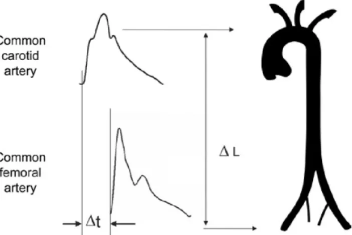
PWV is defined as the speed of the pulse pressure wave travelling along the arterial system. Arterial pressure waves are recorded on both proximal and distal arteries. The length of the arterial segment (ΔL) between the two measured points with surface tape must be determined, as well. The wave transit time (Δt) is obtained either by using the QRS complex of a simultaneously recorded electrocardiography (ECG) in order to have a reference frame, or by using two probes above the femoral and the carotid arteries at the same time (Figure 3) (109).
Figure 3. Measurement of carotid-femoral PWV with the foot-to-foot method.
Adopted from: (109).
Arterial wall properties are also important in determining PWV. These factors are described by the Moens-Kroteweg equation (105):
20
PWV= ∗ ∗ .
Here, PWV is the pulse wave velocity, Einc is the Young’s elastic modulus of arterial wall describing the intrinsic elastic properties of the vessel walls, h is the arterial wall thickness, ρ is the blood density and D is the vessel diameter. An increase of Einc, and of arterial wall thickness results in elevated PWV, while the arterial calibre is inversely proportional to PWV. Taken together, arterial stiffness can be modulated directly by arterial wall mechanical properties, thickness and changes in vascular tone, but also indirectly, by changes in blood pressure (97).
The “gold standard” PWV measurement is usually evaluated using the “foot-to-foot”
velocity method, as mentioned above, from a number of waveforms using the following formula expressed in the unit of meter/second (m/s):
= .
D is the distance between the two recording sites and Dt is the time delay between the
“foot” of the carotid and the femoral waveforms. The “foot” of the wave is defined at the end of the diastole, when the steep rise of the waveform begins (111). In order to reach a proper result, the correct measurement of the covered distance is essential:
according to a consensus document from 2012, 80 % of the direct carotid to femoral distance shall be used (112). The more elasticity the great arteries show, the lower is the PWV value; while a higher PWV value indicates increased arterial stiffness.
1.3.4.1. Central systolic blood pressure
Brachial blood pressure is only a surrogate marker of central, i.e., aortic blood pressure. Recalculation of the artery tonometer pressure wave that was measured on the carotid artery is based on the observation that diastolic and mean arterial pressures are relatively constant throughout the large artery tree. However, systolic blood pressure may be up to 40 mmHg higher in the brachial artery compared to the aorta due to an increase in arterial stiffness moving away from the heart (113-115). This phenomenon is called the systolic blood pressure amplification (SBPAmp). As the pressure wave travels from the elastic central arteries to the stiffer peripheral (brachial) artery, the upper portion of the wave becomes narrower, the systolic peak turns to be more
21
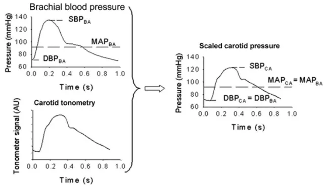
prominent, and systolic pressure increases (116). In practice, blood pressure is measured at the reference (brachial) artery with a validated blood pressure device. From the direct measured peripheral diastolic blood pressure (DBP) and SBP mean arterial pressure (MAP) can be calculated. Using brachial DBP and MAP carotid SBP can be determined (Figure 4).
Figure 4. Calibration method for central pulse pressure.
Adapted from Kelly and Fitchett and Verbekeet al. (117, 118). SBPBA: systolic brachial blood pressure; DBPBA: diastolic brachial blood pressure; MAPBA: mean brachial blood pressure;
SBPCA: systolic carotid blood pressure; DBPCA: diastolic carotid blood pressure; MAPCA:
mean carotid blood pressure.
1.3.4.2. Central pulse pressure
Arterial blood pressure and flow waves are generated by LV contraction and its interaction with the elastic arterial walls. In order to provide a steady flow distribution to the tissues and organs, arteries distend while accommodating the sudden increase in blood volume caused by cardiac contraction, and contract in the diastole, when the elastic energy generated during distension is released. As a result of this phenomenon, arteries present a regular beating, the pulse, which follows the heartbeat and propagates in the form of pulse waves (119). The arterial pressure waveform itself is a mixture of the forward wave (pressure wave spreading away from the heart) and the late arriving
22
reflected wave from the periphery, mainly at branch points or sites of impedance mismatch (pressure wave running towards the heart) (109). Therefore, pulse pressure and pulse wave shape are determined by the physical properties of the CV system, like the arterial geometry and distensibility, the LV ejection, and the impedance due to the smallest blood vessels (119). A schematic representation of central pulse pressure is shown in Figure 5.
1.3.4.3. Augmentation index
From the central diastolic to the systolic pressure the inflection point (Pi) can be determined, where the pressure indicates the beginning upstroke of the reflected wave.
The time (Ti) – belonging to the inflection point – defines the timing of the reflection.
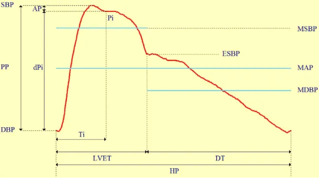
Augmentation pressure (AP) is the pressure above the inflection point – the point in time which coincides with the peak of the flow wave in the artery. AIx is defined as the ratio between the second and first systolic peaks, the AP and pulse pressure (PP), usually expressed in percentage (Figure 5) (120, 121).
Figure 5. Pulse wave of the carotid artery.
AP: augmentation pressure; HR: heart rate; SBP: systolic blood pressure; DBP: diastolic blood pressure, MAP: mean arterial blood pressure; MSBP: mean systolic blood pressure;
MDBP: diastolic blood pressure; ESBP: end-systolic blood pressure; PP: pulse pressure; Ti:
rise time of the reflected wave; Pi: arterial pressure at the Ti; dPi: pulse pressure at the Ti;
LVET: left ventricular ejection time; DT: diastolic time; HP: heart period.
23
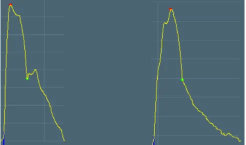
In elastic vessels the reflected wave tends to arrive back at the central arteries during diastole. If the arteries are stiff, the reflected wave arrives back at the aortic root earlier, adding to the forward wave and augmenting the systolic pressure (Figure 6) (109).
Thus, for young patients the AIx value tends to be negative, while it will almost certainly be positive in elderly patients (Figure 6, left graph AIx=-5%; right graph AIx=+10%).
Figure 6. Changes of the carotid pulse curve with ageing: on the left side of a young patient (29 years), on the right side of an aged patient (84 years).
The red points indicate the systolic peak, the green points the end-systolic blood pressure. (The curves are selected from our own patients.)
1.3.5. Arterial stiffness parameters and CV risk estimation, according to actual guidelines
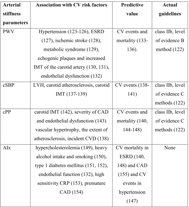
The association of the above mentioned arterial stiffness parameters with different CV risk factors as well as their role in the recent European Society of Cardiology (ESC) and the European Society of hypertension (ESH) (122) is summarized in Table 1.
24
Table 1. The association of pulse wave velocity, Augmentation index, central systolic and pulse pressure with different CV risk factors and their role in the actual guidelines.
PWV: pulse wave velocity; AIx: augmentation index; ESDR: end-stage renal disease; IMT:
intima-media thickness; CV: cardiovascular; cSBP: central systolic blood pressure; cPP:
central pulse pressure; CAD: coronary artery disease; LVH: left ventricular hypertrophy;
CVD: cardiovascular disease; CRP: C-reactive protein.
Arterial stiffness parameters
Association with CV risk factors Predictive value
Actual guidelines PWV Hypertension (123-126), ESRD
(127), ischemic stroke (128), metabolic syndrome (129), echogenic plaques and increased IMT of the carotid artery (130, 131),
endothelial dysfunction (132)
CV events and mortality (133-
136).
class IIb, level of evidence B method (122)
cSBP LVH, carotid atherosclerosis, carotid IMT (137-139)
CV events (138- 141)
class IIb, level of evidence C methods (122) cPP carotid IMT (142), severity of CAD
and endothelial dysfunction (143) vascular hypertrophy, the extent of atherosclerosis, incident CVD (138)
CV events and mortality (140,
144-148)
class IIb, level of evidence C methods (122)
AIx hypercholesterolemia (149), heavy alcohol intake and smoking (150), type 1 diabetes mellitus (151, 152),
endothelial function (132), high sensitivity CRP (153), premature
CAD (154)
CV mortality in ESRD (140, 148) and CAD
(155) and CV events in hypertension
(147)
None
25
Patients at intermediate risk could be reclassified into a lower or higher CV risk category based on PWV measurements (136, 156, 157). In the Framingham follow-up study of 7.8 years for individuals at intermediate CV risk, higher aortic PWV was associated with a 48% increase in CVD risk. After adding PWV to a standard risk factor model, a result of an integrated discrimination improvement of 0.7% was achieved.
Consequently, PWV resulted in upward reclassification of 14.3 % of subjects who suffered a CV event and downward reclassification of 1.4 % of participants who did not experience any CV events, meaning a reclassification altogether of 15.7 % (156). A recent meta-analysis demonstrated clearly the independent predictive value of PWV in CV risk. Ben-Shlomo et al. found that after the adjustment of additional risk factors an increase of 1 SD in log PWV is related to a 30% increase in CV events, a 28% increase in CV mortality and a 17% increase in all-cause mortality (133). In other words it means that for a 60-year-old non-smoker, non-diabetic, normotensive and normolipemic man, an increase of 1 m/s of PWV leads to a 7% growth of the hazard for CV events (133, 158). Reference values for PWV are available in healthy populations and also in patients with increased CV risk (159). The recent cut-off value of PWV>10 m/s is considered a conservative estimate of significant alterations of aortic function in middle-aged hypertensive patients (112). Whereas such cut-off value is set for PWV, indicating target organ damage and an increased CV risk, the other arterial stiffness parameters are showing less evidence.
In summary, the measurement of arterial stiffness can be considered to assess CV risk.
As depression is connected to CV risk, it is not surprising, that depression was found to be associated with elevated arterial stiffness as well. Tiermeier et al found in their population-based, cross-sectional study including 3704 elderly subjects that patients with increased arterial stiffness were more likely to have depressive symptoms. The association was stronger in cases with diagnosed depressive disorders. The authors concluded that arterial stiffness may partly cause the proposed relationship between vascular factors and depression (160). Since dominant affective temperaments can be the precursors of depression, a question arises: is it possible that increased blood pressure values and elevated arterial stiffness can be detected in hypertensive patients with dominant affective temperaments, indicating an increased CV risk compared with hypertensive patients without dominant temperaments? Additionally, as temperaments
26
can be described as continuous variables, it would be important to ascertain the continuous association between affective temperament scores and blood pressure or arterial stiffness parameters.
1.4. New psychosomatic connections: the possible role of the brain-derived neurotrophic factor (BDNF) in psychopathology and cardiovascular diseases
Neurortophic factors or neurotrophins (NTs) consist a family of trophic factors of secreted proteins that promote growth, survival and differentiation of neurons both in the central and peripheral nervous system (161). We summarized the role of NTs in psychopathology and CV diseases in a recent review paper (162).
1.4.1. Brain-derived neurotrophic factor (BDNF)
BDNF is the most frequently studied NT. It is initially synthesized in the endoplasmic reticulum as its precursor protein, preproBDNF. After the cleavage of the signal peptide this form becomes proBDNF, and proBDNF is converted by extracellular proteases to mature BDNF (163, 164).
Mature BDNF binds with higher-affinity Trk family receptors (165) – especially TrkB – increasing cell survival and differentiation, dendritic spine complexity, long-term potentiation (166, 167), synaptic plasticity (168), and the resculpting of neuronal networks (169). ProBDNF is also biologically active, it mediates its actions through binding to low-affinity p75Ntr, having antagonistic effect compared to matured BDNF:
reducing spine complexity and density (170) and promoting neuronal cell death (171).
1.4.2. BDNF in mood disorders
Although NTs themselves do not control mood directly, they are fundamental in the activity-dependent modulation of networks and changes in plasticity can affect mood as well (172). Numerous clinical studies confirm the involvement of BDNF in the pathophysiology of depression (173). Reductions in serum and plasma mature BDNF have been demonstrated in patients suffering from depression (164, 174) and in those who committed suicide (175, 176). Significantly lower levels of serum BDNF were found in antidepressant-free patients with major depressive disorder compared with healthy controls (177), which findings were confirmed by a large cohort study (178) and by three meta-analyses as well (179-181).
27 1.4.3. BDNF and CV pathology
BDNF plays an important role during development of the CV system: the activated TrkB receptor leads to the survival of endothelial cells and the formation of the cardiac vasculature (182). Embryonic BDNF deficiency impairs the development of intramyocardial vessels and can also lead to cardiac hypercontractility (183). This NT functions as an angiogenic regulator, promoting angiogenesis (184). It is expressed in a greater amount in the peripheral vessels, where it could influence vasoreactivity (185, 186). BDNF is able to enhance vascular flow and can regulate the revascularization of ischemic tissues (184). It was shown to be vasorelaxant on pulmonary arteries (187) and on rat aortic rings (185). Furthermore, it improves left-ventricular function in ischemic myocardium (188).
Numerous data are available about the association between BDNF and CV health. In the general population a significant positive correlation was observed between plasma BDNF and diastolic blood pressure and sex differences were demonstrated in relation with different serum lipids (189). As it was a cross-sectional study, it is unclear whether the associations observed are casual or elevated plasma BDNF represents a compensatory response for the disrupted lipid metabolism and hypertension.
Increased BDNF expression was found in atherosclerotic coronary arteries in humans (190), and decreased plasma BDNF level was observed in metabolic syndrome (191), in acute coronary syndrome (192, 193) and in type 2 diabetes mellitus (194). In patients with angina pectoris Jiang at al. found that plasma BDNF was inversely associated with triglyceride and low-density lipoprotein (LDL)-cholesterol, male sex and age, while it was correlated positively with (HDL)-cholesterol. In this cohort, during the 4-year follow-up period, low plasma BDNF was an independent predictor of future coronary events and mortality (195). The predictive role of BDNF for future CV events and mortality was confirmed by other studies as well: higher serum BDNF (seBDNF) was found to be associated with decreased risk of CV morbidity and mortality (196). On the contrary, decreased serum BDNF was found to be associated with increased risk of incident stroke/transient ischemic attack (TIA) (197).
1.4.4. BDNF polymorphism
A functional polymorphism Val66Met in the BDNF gene – an amino acid substitution valine to methionine at codon 66 in the precursor BDNF peptidesequence (198) – was
28
found to influence BDNF’s secretion and function. Moreover, it is also associated with mood and cognitive-related phenotypes. There is ample biological evidence that the BDNF gene is an attractive candidate gene for MDDs (199). Nevertheless, a recent meta-analysis of Li et al. showed that the Val66Met polymorphism was significantly associated with bipolar disorder in Europeans, but not with MDD (200). Regarding to affective temperaments, the variant of BDNF polymorphism has already been studied.
However, no significant difference in the frequency of alleles between subjects with and without affective temperaments was shown (201).
Taken together, the role of BDNF is already proven in psychiatric disorders, and it seems to have an influence on CV risk. In different animal models BDNF was shown to be vasorelaxant, also on aortic rings. Based on these data, a possible association between the seBDNF level and different arterial stiffness parameters in hypertension can also be supposed in humans. On the other hand, BDNF polymorphism has already been investigated in MDDs and in affective temperaments, but in hypertension the association between affective temperaments and the serum level of BDNF has not been studied yet.
29 2. OBJECTIVES
1. In the first study, our aim was to measure arterial stiffness and serum BDNF levels in hypertensive patients with and without dominant affective temperaments.
We hypothesised that hypertensive patients with dominant affective temperaments score higher depression and anxiety values and have impaired arterial stiffness, central blood pressure or serum BDNF compared with hypertensive patients without dominant affective temperaments, forming a high-risk subgroup patient population.
2. In the second study, our aim was to explore the associations of affective temperaments with blood pressure and arterial stiffness in hypertensive patients. We hypothesized that individual affective temperament scores may be related to brachial blood pressure as well as arterial stiffness in chronic hypertensive patients. We speculated a positive association in instances of depressive, cyclothymic, irritable or anxious temperaments and an inverse association in instances of hyperthymic temperament. We also hypothesized the presence of sex differences in relation to these studied associations.
3. In our third study, our aim was to measure BDNF serum levels in hypertensive patients and in healthy controls to discover the associations of BDNF with affective temperaments, depression, anxiety and arterial stiffness. We hypothesized that as hypertension is a risk factor for cardiovascular diseases and BDNF is protective in cardiovascular pathology, seBDNF can be altered in hypertension. We also presumed that seBDNF is associated with different affective temperaments, depression, anxiety, and arterial stiffness parameters providing a new bridge of psychosomatic processes.
30 3. PATIENTS AND METHODS
3.1. Study 1: patients and methods in the study measuring arterial stiffness and serum BDNF level in patients with and without dominant affective temperaments
Caucasian patients were selected from two primary care practices in Budapest, Hungary. Out of the 183 patients 175 completed the TEMPS-A, Beck Depression Inventory (BDI) and Hamilton Anxiety Scale (HAM-A) questionnaires in order to evaluate the presence of affective temperaments, or the depression and anxiety, respectively. Exclusion criteria were the history or ongoing treatment of depression or anxiety (as with arterial stiffening the associations are clarified (160)), bipolar disorders, schizophrenia, dementia posing an obstacle to completing questionnaires or denial of consent, the presence of atrial fibrillation or uncontrolled hypertension (>145/95 mmHg in repeated office measurements). In patients with an average blood pressure between 140/90 and 145/95 mmHg in repeated office blood pressure measurements, ambulatory 24-hour blood pressure monitoring or home blood pressure monitoring was performed and only well-controlled patients were admitted into the study. Prior to the participation, all patients gave their written informed consent. All the studies were approved by the Scientific and Research Ethics Committee of the Medical Research Council, Hungarian Ministry of Health and carried out in accordance with the tenets of the Declaration of Helsinki.
Following the initial screening, 29 hypertensive patients with dominant affective temperaments (reaching the mean+2 SD point scores or higher in each affective temperament subscale, DOM) were identified, and 24 were investigated in our study. 24 hypertensive controls without DOM, matched in age, gender and presence of diabetes, were selected from the initial hypertensive patient cohort and included in the arterial stiffness and seBDNF measurements. As blood pressure medication can highly influence arterial stiffness, it was further analyzed, but patients were not matched in this aspect.
During the initial visit patients completed the questionnaires. Physical examination (blood pressure, heart rate, height, weight and waist circumference) were completed and
31
data on medical history (with special attention to CV risk factors, complications and depression) as well as on current medication was collected.
3.1.1. Questionnaires
3.1.1.1. Temperament Evaluation of Memphis, Pisa, Paris and San Diego Autoquestionnaire
The TEMPS-A was used to assess affective temperaments on depressive, cyclothymic, hyperthymic, irritable and anxious subscales, requiring “yes” (score 1) or “no” (score 0) answers [17]. TEMPS-A contains 110 items (109 in the version for males) and the questions of the various temperament types are grouped together as follows:
depressive temperament: questions 1 to 21 (21 points)
cyclothymic temperament: questions 22 to 42 (21 points)
hyperthymic temperament: questions 23 to 63 (21 points)
irritable temperament: questions 64 to 84 (21 points in women, 20 in the men’s version)
anxious temperament: questions 85 to 110 (26 points).
TEMPS-A is used to assess the point scores of each subscale and also to measure the presence of the dominant form of affective temperaments by taking the mean of the subscale and adding up two standard deviations to it. Those reaching the mean+2 SD level or higher in each subscale are considered to have dominant affective temperaments.
3.1.1.2. The Beck Depression Inventory
The BDI, created by Aaron T. Beck, is a 21-question multiple-choice self-report questionnaire, one of the widely used instruments for measuring the severity of depression. This questionnaire is designed as a measure of severity of depressive symptoms, not as a diagnostic instrument. In the 21 items the most common depressive symptoms (e.g. sad mood, pessimism, failure, dissatisfaction, guilt, crying, irritability, social withdrawal, inability to resolve, insomnia, appetite loss, etc.) are evaluated.
Participants are asked to make ratings on a four-point scale, where a higher score correlates with more severe depression. The total value of the BDI can range from 0 to 63 points. The cut-off values might differ in various populations, but there are generally
32
accepted thresholds. Values below 11 points are considered normal. Values between 11 and 17 points are regarded as a mild to moderate expression of depressive symptoms.
Values of 18 and more are considered as a clinically relevant depression (202).
3.1.1.3. Hamilton Anxiety Scale
HAM-A was evaluated by the examiner. This questionnaire was one of the first rating scales developed to measure the severity of anxiety symptoms, and is still widely used today in both clinical and research settings. The scale consists of 14 items, each defined by a series of symptoms, and measures both psychic anxiety (mental agitation and psychological distress) and somatic anxiety (physical complaints related to anxiety).
Each item is scored on a scale of 0 (not present) to 4 (severe), with a total score range of 0–56, where <17 indicates mild severity, 18–24 mild to moderate severity and 25–30 moderate to severe (203).
3.1.2. Clinical measurements 3.1.2.1.Arterial stiffness recordings
Measurements were performed in a temperature-controlled room in supine position, on the day of blood sampling, prior to it, between 7:00 and 8:00 a.m. Patients were asked to refrain from eating, smoking, and caffeine containing drinks in the morning of the procedure, but to take their regular antihypertensive medication. Upon arrival after 5 minutes rest, two brachial blood pressure measurements were taken on each arm in the sitting position with a validated oscillometric blood pressure device (Omron M3). The mean value of the higher side was further taken into calculation as brachial systolic and diastolic blood pressures (SBPB and DBPB) and heart rate. Brachial pulse pressure (PPB) was also calculated from these data. Next, subjects were equipped with arterial stiffness measurement device and then rested in the supine position for approximately 15 minutes before being measured. Arterial stiffness parameters were evaluated with the gold-standard tonometric method (PulsePen, DiaTecne, Milan, Italy) (204). This method provides estimates of PWV and in which cSBP, cPP and pulse pressure amplification (PPAmp) can be calculated. Aix, a widely used wave reflection parameter, can also be measured by automatic identification of the “first shoulder” (inflection point) of the averaged carotid pulse signal by the PulsePen software. This index is provided by the pressure amplitude following this point divided by the pulse pressure
33
and calculated as a percentage. In these calculations, brachial blood pressure values measured in the supine position were used, which were required for calibration after each (carotid or femoral) pulse wave detection. In each subject, two sequences of arterial stiffness measurements were performed and their mean used for statistical analysis. In the PWV calculations, 80 % of the carotid-femoral distance was used, according the most recent recommendation guideline (112). The intra- and interobserver variability of PWV measurements obtained by the PulsePen device in hypertensive patients was 4.6 and 6.3 %, respectively. Since PulsePen calculates pressures based on brachial diastolic blood pressure calibration, the calculated central diastolic blood pressure is identical to the brachial diastolic blood pressure assessed in the supine position (204).
3.1.2.2. Measurement of serum BDNF concentrations
Peripheral blood samples of patients were collected in anticoagulant-free tubes, right after the measurement of arterial stiffness. After centrifugation at 3600 revolutions per minute (rpm) for 6 minutes, the serum was stored at -20 °C. SeBDNF was measured using commercially available sandwich enzyme-linked immunosorbent assay (R&D Systems, Minneapolis MN, USA) according to the manufacturer’s protocol, and seBDNF level was determined in pg/ml.
3.1.3. Statistical analysis
Differences in variables between controls and DOM patients were analyzed using unpaired Student’s t-tests or Mann-Whitney rank-sum tests for data failing tests of normality. Blood pressure medications were calculated and compared using equivalent doses, differences were analysed with unpaired Student’s t-tests or Mann-Whitney rank- sum tests. Data were expressed as mean±SEM and medians, significance was accepted at p<0.05. Statistical analysis was performed using the SigmaStat for Windows Version 3.5 (SPSS) program package.
3.2. Study 2: patients and methods in the study exploring the association of affective temperaments with blood pressure and arterial stiffness in hypertensive patients
The difference in patient selection compared to the first study was that in the second cross-sectional study patients with well-controlled or grade 1 chronic (on medication for
34
more than 3 months) hypertension were investigated from three primary care practices in Budapest. In this study a total of 173 subjects were included. Moderate use of the anxiolytic alprazolam (less than 0.5 mg/day) was not a restrictive criterion. All of the patients of Study 1 were also involved.
The methods regarding to the questionnaires and arterial stiffness measurement were the same like in the first study, but seBDNF was not investigated.
3.2.1. Statistical analysis
Normality of continuous parameters was tested with the Kolmogorov-Smirnov test.
Pearson correlation coefficients were calculated to study the relationship between affective temperament scores and demographic, hemodynamic or arterial stiffness parameters. Multiple linear regression analysis was used to study the determinants of these hemodynamic or arterial stiffness parameters which were associated in univariate analysis with affective temperaments. Based on literature data, sex differences in the association between affective temperaments and the studied hemodynamic or arterial stiffness parameters (43) were expected, and therefore sex and its interaction with the given affective parameter was included into all regression models and where an interaction was found, such interaction was further studied. Descriptive data were expressed as mean±SD or median with interquartile ranges or percentages. A two-sided p<0.05 was considered to be significant. SPSS 13.0 for Windows was used for all calculations.
3.3. Study 3: patients and methods in the study measuring serum BDNF levels in a hypertensive and a control population to discover the associations of BDNF with affective temperaments, depression, anxiety and arterial stiffness
The difference in patient selection compared to the first study was that in the third cross-sectional study chronic (>12 months medication) well-controlled or grade 1 consecutive hypertensive patients (HT) and age-matched healthy controls (CONT) were involved from three primary care practices in Budapest. In this study a total number of 183 patients were investigated: 151 HT and 32 CONT. Moderate use of the anxiolytic alprazolam (less than 0.5 mg/day) was not a restrictive criterion. All of the chronic hypertensive patients of Study 1 were involved as well. In the case of CONT, the denial
35
of consent was the only exclusion criterion. Data of the subjects were analysed for the relationship between the seBDNF level, routine laboratory parameters, affective temperaments, anxiety, depression, and arterial stiffness parameters.
The methods were the same like in Study 1.
3.3.1. Statistical analysis
Normality of the parameters was tested with the Kolmogorov–Smirnov test. Descriptive characteristics, laboratory, arterial stiffness parameters and TEMPS-A, BDI, HAM-A scores were compared between CONT and HT groups using unpaired Student’s t-tests or Mann-Whitney rank sum test for data failing tests of normality. The equality of variances was studied with Levene’s test. Pearson correlation coefficients were calculated to study the relationship between seBDNF and all other factors measured.
Hierarchic linear regression analysis was used to study the determinants of seBDNF in the whole population with a stepwise entry of variables with either previously described association with seBDNF or with a significant univariate correlation with seBDNF in the present data set. As a bidirectional association can be hypothesized between affective temperaments and hypertension (88), predetermined interaction analysis was performed to investigate moderation between hypertension and affective temperament scores on seBDNF level. Data were expressed as mean±SD or mean with interquartile ranges, and p<0.05 was considered to be significant. SPSS 13.0 for Windows was used in calculations.
36 4. RESULTS
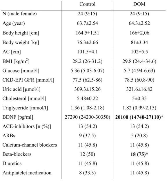
4.1. Study 1: results of hemodynamic, arterial stiffness and serum BDNF levels in hypertensive patients with or without dominant affective temperaments In this cross-sectional case-control study well-treated chronic (>12 months medication) hypertensive patients were investigated. Out of the 29 DOM four patients declined to further participate in our study and one died 3 days prior to the planned arterial stiffness measurement. The arterial stiffness and BDNF of altogether 48 hypertensive patients was evaluated: 24 DOM and 24 control subjects matched in age, gender and presence of diabetes. Among the DOM patients, six subjects were found to have a depressive, five an irritable and four an anxious dominant temperament. In the other patients, combinations of dominant temperaments were present: three patients had cyclothymic and depressive, two had cyclothymic and irritable, two had cyclothymic, depressive and anxious, one had cyclothymic, irritable and anxious temperaments and one patient was dominant for cyclothymic, irritable, anxious and depressive affective temperaments. No patient with a dominant hyperthymic temperament was found in our cohort.
Comparing the control and DOM patients for statistical differences baseline demographic, anthropometric and laboratory parameters and the used CV medications of the patients are presented in Table 2.