SEMMELWEIS EGYETEM DOKTORI ISKOLA
Ph.D. értekezések
2295.
GAMPE NÓRA
A gyógyszerészeti tudományok korszerű kutatási irányai című program
Programvezető: Dr. Antal István, egyetemi docens Témavezetők: Dr. Béni Szabolcs, egyetemi docens
Dr. Kursinszki László egyetemi docens
Phytochemical analysis of Ononis species
Ph.D. dissertation
Nóra Gampe
Semmelweis University
Doctoral School of Pharmaceutical Sciences
Supervisors: Szabolcs Béni, Ph.D.
László Kursinszki, Ph.D.
Reviewers: Márta Mazákné Kraszni, Ph.D.
Dezső Csupor, Ph.D.
Chair of final examination committee: Imre Klebovich, D.Sc.
Members of final examination committee:
Éva Ledniczkyné Lemberkovics, Ph.D.
Zsuzsanna Hajdú, Ph.D.
László Abrankó, Ph.D.
Budapest
2019
2 Table of contents
List of abbreviations ... 5
Introduction ... 7
1.I. Characterization of Ononis species ... 8
1.I.1. Botanical characterization of Ononis spinosa L. and Ononis arvensis L. .. 8
1.I.2. Chemical characterization of Ononis species ... 10
1.I.3. Biological activities of Ononis spinosa extracts... 12
1.I.4. Biological activities of Ononis arvensis ... 18
1.I.5. Biological activities of other Ononis species ... 19
1.I.6. Possibilities to produce isoflavonoids by in vitro cultures of Ononis species 19 1.II. Isoflavonoids ... 24
1.II.1. Structural features and occurrence of isoflavonoids... 24
1.II.2. Biosynthesis of isoflavonoids ... 27
1.II.3. Biological activities of selected isoflavonoids ... 32
1.II.4. Analytical methods aiming the qualitative and quantitative determination of isoflavonoids ... 43
Objectives ... 58
Materials and methods ... 59
3.I. Plant material ... 59
3.II. Solvents and chemicals ... 59
3.III. Extraction and sample preparation ... 60
3.III.1. Analytical samples ... 60
3.III.2. Isolation of aglycones ... 61
3.III.3. Isolation of licoagroside B ... 61
3.III.4. Isolation of but-2-enolide aglycones and calycosin D aglycone ... 62
3.III.5. Isolation of but-2-enolide glycosides and calycosin D glycosides ... 63
3
3.III.6. Isolation of homopipecolic acid isoflavonoid glucoside esters ... 63
3.IV. Chromatographic conditions ... 64
3.IV.1. HPLC-DAD conditions ... 64
3.IV.2. HPLC-DAD-ESI-MS/MS conditions ... 65
3.IV.3. UHPLC-DAD-ESI-Orbitrap-MS/MS conditions ... 66
3.V. Polarimetry and CD conditions ... 66
3.VI. NMR conditions ... 66
3.VII. Method validation ... 67
Results ... 68
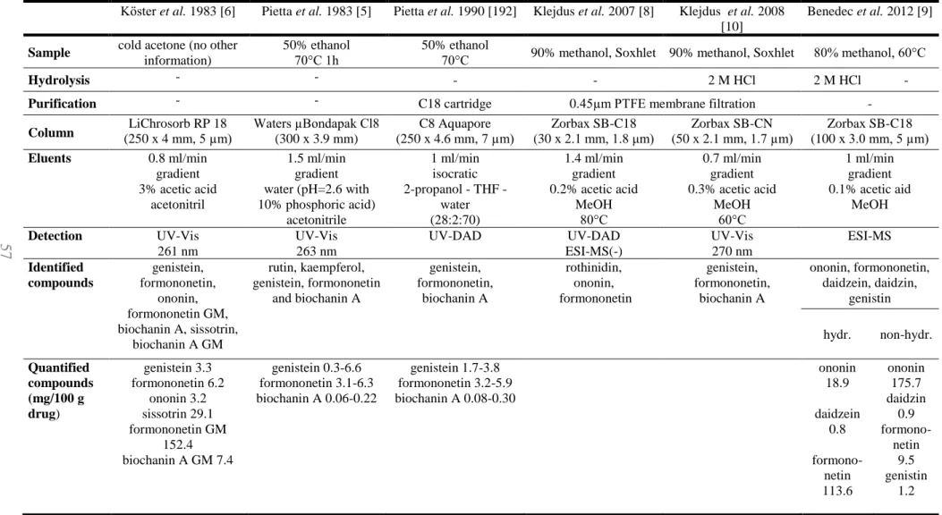
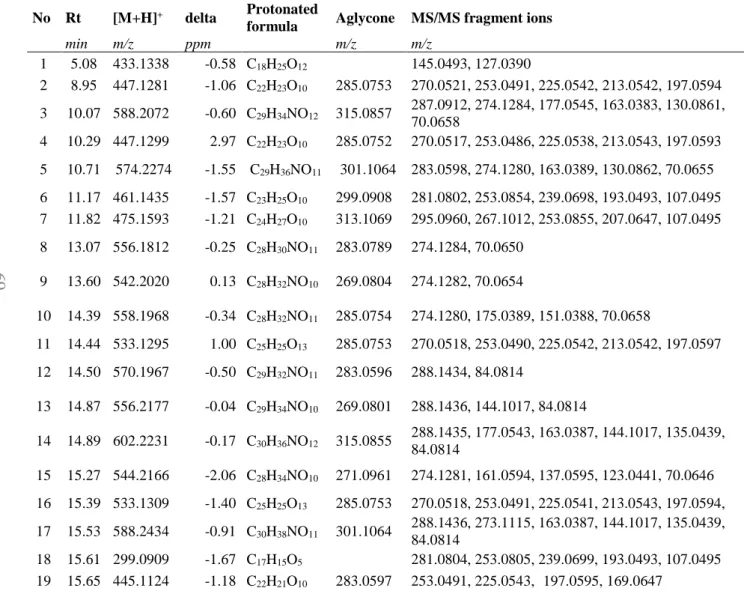
4.I. Qualitative phytochemical results ... 68
4.I.1. Chromatographic results ... 68
4.I.2. Mass spectrometry results ... 74
4.I.3. Detailed structural identification ... 75
4.II. Quantitative phytochemical results ... 89
4.II.1. Sample preparation ... 89
4.II.2. Method validation ... 91
4.II.3. Quantitative results ... 92
Discussion ... 94
5.I. Qualitative phytochemical results ... 94
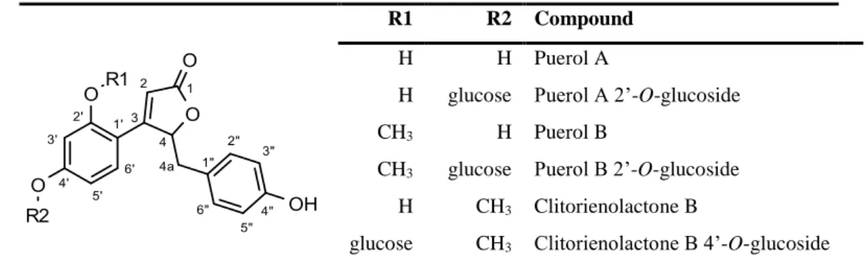
5.I.1. Structural identification of licoagroside B ... 94
5.I.2. Structural identification of but-2-enolides ... 94
5.I.3. Structural identification of isoflavones ... 96
5.I.4. Structural identification of isoflavanones ... 98
5.I.5. Structural identification of pterocarpans ... 100
5.I.6. Structural identification of 2’-methoxy isoflavones ... 101
5.I.7. Structural identification of nitrogen-containing derivatives ... 104
4
5.II. Quantitative phytochemical results ... 108
5.II.1. Sample preparation ... 108
5.II.2. Quantitative results ... 109
Conclusions ... 113
Summary ... 114
Összefoglalás ... 115
Bibliography ... 116
List of own publications ... 137
Acknowledgement ... 138
5 List of abbreviations
2-HIS 2-hydroxy isoflavone synthase
ABTS 2,2'-azino-bis(3-ethylbenzothiazoline-6-sulphonic acid) AChE acetylcholinesterase
Aβ amyloid beta
BChE butirylcholinesterase BPC base peak chromatogram CID collision induced dissociation COSY correlation spectroscopy
CUPRAC cupric reducing antioxidant capacity CZE capillary zone electrophoresis DAD diode-array detector
DMID 7,2’-dihydroxy-4’-methoxyisoflavanol dehydratase DMSO dimethyl sulfoxide
DPPH 2,2-diphenyl-1-picrylhydrazyl ED electrochemical detection EDTA Ethylenediaminetetraacetic acid ERα, β estrogen receptor α, β
ESI electrospray ionization
FRAP ferric reducing antioxidant capacity
HI4’OMT hydroxy isoflavone 4’-O-methyltransferase HM3OMT 6a-hydroxymaackiain 3-O-methyltransferase HMBC heteronuclear multiple bond correlation HPLC high pressure liquid chromatography HSQC heteronuclear single quantum coherence HUVEC human umbilical vein endothelial cell I2’H, I3’H isoflavone 2’/3’-hydroxylase
IC50 50 % inhibitory concentration IFR isoflavone reductase
IOMT isoflavone-O-methyltransferase LC liquid chromatography
LOD limit of detection
6 LOQ limit of quantitation
LPS lipopolysaccharide MAO monoamine oxidase
MEKC micellar electrokinetic capillary chromatography MIC minimal inhibitory concentration
MS mass spectrometry
MS/MS tandem mass spectrometry
NADPH nicotinamide adenine dinucleotide phosphate NF-κB nuclear factor κB
NMR nuclear magnetic resonance
NOESY nuclear Overhauser effect spectroscopy Ovx ovariectomized
P2CP, P4CP pterocarpan C-2/4 prenyltransferase P6aH pterocarpan 6a-hydroxylase
PPARα, , δ peroxisome proliferator-activated receptor α, , δ pRi T-DNA root infecting plasmid transfer DNA
rDA retro Diels-Alder
ROESY rotating frame Overhauser effect spectroscopy SAM S-adenosyl-L-methionine
SC50 50% radical scavenging concentration SFE supercritical fluid extraction
SIM selective ion monitoring SPE solid-phase extraction
TIC total ion-current chromatogram TOCSY total correlation spectroscopy
TRAIL tumor necrosis factor α-related apoptosis inducing ligand UHPLC ultrahigh pressure liquid chromatography
VEGF vascular endothelial growth factor VR vestitone reductase
7 Introduction
Natural products and plant derived remedies accompanied us since prehistoric times and they are still a popular choice of self-healing. Although, many herbal extracts are proved to have beneficial health effects, the responsible components are undefined in the majority of cases. With the help of phytochemical and phytoanalytical techniques, the aim of pinpointing the most potent molecules in the herbal drugs can be executed. The results of these studies are not only feeding the intrinsic curiosity – How and why does it work? – but can be the base of the quality control of herbal drugs and preparations. Taking advantage of the capacities of biotechnology, the metabolite profile of the plant can be fine-tuned, resulting in easier accessibility to the beneficial molecules. Furthermore, with the structural knowledge of the effective compounds, new semisynthetic, structurally optimized molecules can be created.
The aim of this work is to reveal the phytochemical and phytomedicinal values of O.
spinosa and O. arvensis, as these plants have been used in ethnomedicine since hundreds of years. The roots of this plants are a typical example of herbal drugs, where the ethnomedicinal use is confirmed by in vitro and in vivo tests, however, the exact compounds responsible for the effects are unknown.
In the literature overview, the as complete as possible description of the plants, their chemical constituents and their biological effects was carried out. As related species can possess similar phytochemical profile, their most interesting constituents and pharmacological effects are summarized, too. As isoflavonoids seemed to be the most promising compounds, their analytical possibilities are presented, as a base of the experimental work. During our research, we aimed to explore the isoflavonoid profile in details using various analytical techniques, to understand the operation of the plant as a miniature chemical factory. The chemical profile of O. spinosa and arvensis roots and their in vitro hairy root cultures were compared regarding qualitative and quantitative points of view.
8 1.I. Characterization of Ononis species
1.I.1. Botanical characterization of Ononis spinosa L. and Ononis arvensis L.
The members of the Ononis genus, which belongs to the family Leguminosae, are natively distributed in Europe, Central Asia and North-Africa. Ononis genus includes 86 species (see Figure 1) [1].
Figure 1: The phylogenic tree of Ononis species (and some other Leguminosae species) based on ITS DNA sequences [1].
Ononis spinosa L. (spiny restharrow) is a tall subshrub which grows to a height of 60 cm.
It has a short rhizome and a ligneous taproot which can be up to 50 cm long. The upright stems which grow out of this are lignified at the base and slightly hairy. Numerous short shoots have developed into prickly thorns, but thornless varieties also exist. The lower leaflets are trifoliate and pinnate, while the upper ones are small and unifoliate with a
9
long oval shape and dentate edges. The leaves are covered with very fine glandulous hairs.
The striking butterfly-shaped flowers are pink or purplish in color and grow in the upper angles between leaf and stem. The flowers are generally solitary but sometimes also grow in loose clusters (Figure 2). They produce soft-haired pods bearing rounded, lumpy seeds.
Spiny restharrow is to be found in dry meadows and pastures, by fields and waysides, but also in peat areas and sand dunes. It favors sunny locations with weak loamy or chalky soils. Fertilizing causes it to disappear [2].
Ononis arvensis L. (field restharrow) is a perennial shrub preferring humid fields and meadows overall in Europe. The 30–80 cm high erect stem is covered by long and thick trichomes, but thornless. The plant has an unpleasant smell due to the excrete of the glandular trichomes. It has alternating stalked leaves; the blade has three elliptic-quite round leaflets with serrated margins. The terminal leaflet is stalked. The large stipules are united with the stalks. The butterfly-like corolla is pinkish and fused at base. The flowers are axillary in pairs. Inflorescence is a leafy terminal raceme or flowers axillary in pairs (Figure 3). The 1-3 seeded opening pod is 6-9 mm. The synonym names are Ononis hircina Jacq. and Ononis spinosa subsp. hircina (Jacq.) Gams. It usually grows in dry places in different kinds of fields like inland meadows, grazing land and banks [3].
Figure 2: Ononis spinosa L.
Figure 2: Ononis spinosa L.
Figure 2: Ononis spinosa L. Figure 3: Ononis arvensis L.
10 1.I.2. Chemical characterization of Ononis species Ononis spinosa L.
The following chemical constituents were identified in Restharrow root:
Isoflavonoids [4]–[11]:
• formononetin, ononin (formononetin 7-O-glucoside), formononetin 7-O- glucoside 6”-malonate, rothinidin (pseudobaptigenin 7-O-glucoside), genistein, genistin (genistein 7-O-glucoside), biochanin A, sissotrin (biochanin A 7-O- glucoside), biochanin A 7-O-glucoside 6”-malonate, daidzein, daidzin (daidzein 7-O-glucoside), calycosin D (3’-methoxy daidzein), tectoridin,
• trifolirhizin (maackiain 3-O-glucoside), medicarpin 3-O-glucoside
• 2,3-dihydro-ononin, onogenin, onogenin-7-O-glucoside, sativanone, and sativanone-7-O-glucoside.
Phenolic lactones:
• spinonin [12], and clitorienolactone B [11] were also characterized.
Triterpenes:
• α-onocerin (onocol) [13]
• β-sitosterol, stigmasterol, campesterol, cholesterol, α-spinasterol [7], [14]
• saponin (3-O-[α-L-rhamnopyranosyl-(1→2)-β-D-xylopyranosyl-(1→2)-β-D- glucuronopyranosyl]-3β,22α-dihydroxyolean-13-en-11) [15]
Phenolic acids:
• p-hydroxybenzoic, vanillic acid, caffeic acid, syringic acid, p-coumaric acid, cinnamic acid, sinapin acid, salicylic acid, gentisic acid [7, 14]
Lectins:
• Lectins Index Nomenclature LECp.Ono.Spi.ro.Hga1 [16]
Essential oil (small amount):
• trans-anethole (major constituent), carvone, menthol, menthone, isomenthone, linalool, estragole, borneol and cis-anethole [17]
Ononis arvensis L.
Isoflavonoids (from roots) [18]–[21]:
• ononin
• onogenin
• maackiain, trifolirhizin
11 Phenolic acids (from aerial parts):
• hydroxycinnamic acids [22]
• hydroxybenzoic acid, ellagic acid [23]
Flavonoids (from aerial parts) [23]:
• dihydro-quercetin, rutin, quercetin, kaempferol, isorhamnetin
• catechin, epicatechin
• luteolin, apigenin, chrysin, eriodyctiol, naringenin
• phloridzin, phloretin Stilbenes (from aerial parts):
• piceol, trans-resveratrol [23]
Coumarins (from aerial parts):
• scopoletin, scopolin [24]
Triterpenes (from whole plant) [13]:
• α-onocerin (onocol)
• β-sitosterol, stigmasterol, campesterol, cholesterol, α-spinasterol Lectins [25]
The representative structures of isoflavonoid derivatives, special phenolics and terpenoids can be seen in Figure 4.
Figure 4: Representative structures of O. spinosa and O. arvensis
12 Other Ononis species
Isoflavonoids were also described for O. angustissima [26], O. speciosa [27], O. natrix [28], [29], O. viscosa [30], O. vaginalis [31], [32], O. serrata [33], O. pusilla [34], while flavonoids were found to be in O. angustissima [35], [36], O. speciosa [27], O. natrix [37], and O. pusilla [34]. Special phenolic lactones were isolated from O. angustissima [26] and O. speciosa [38]. The essential oil of O. angustissima [39], [40], O. reclinata [34], O. natrix [41], O. viscosa [42] were studied. Probably the most unique compounds in Ononis species are the alkyl derivatives of resorcinol, benzoic acid, isocoumarin and anthranilic acid (see Figure 5), which are present in O. speciosa [27], [43], O. natrix [28], [37], [44]–[49], O. viscosa [50], [51], O. pubescens [52] and O. pusilla [34].
Figure 5: Alkyl derivatives of resorcinol, benzoic acid, isocoumarin and anthranilic acid found in various Ononis species
1.I.3. Biological activities of Ononis spinosa extracts Ethnomedicinal use
O. spinosa root has been widely used since ancient times and it is mentioned in many old documents such as Dioskurides (increases diuresis and breaks stones), Plinius (the root expels bladder-stones), Matthiolus (stimulates diuresis, powerfully breaks the stones), Lonicerus (expels the stone and urine) or Schroder (stimulates diuresis and against kidney- and bladder-stones). It has been traditionally used for the treatment of the lower urinary tract disorders in the irrigation therapy as a diuretic medicine for inflammatory conditions of the lower urinary tract and for preventing and treating kidney and bladder disorders, gravel and small stones [53]. Ononis species have been used for centuries as
13
folk remedies in Turkey as diuretic, antiseptic and antimicrobial aids. In their folk medicine, restharrow root is also used for gout and rheumatic complaints and skin disorders [40], [54].
Diuretic effect
In vivo experimental studies on the diuretic effect of Ononidis radix were performed and described in a dissertation thesis of Wilhelm Bulow already in 1891 [55]. Fifty years later, Vollmer et al. investigated again the diuretic effects of Ononis extracts on various species.
The per os administration of aqueous extract of the roots to rats induced the most pronounced effect, although in a very high dose of 1 g/animal. Infusion of O. spinosa root was administrated orally to rabbits and resulted in an elevation of urinary output by 26%
[56], [57].
Hilp et al. confirmed a diuretic activity with in vivo studies. Oral administration of infusions of the O. spinosa root caused slight diuresis (average of 12% increase compared to the control) and decoctions an antidiuretic effect of 7-20% in rats. The essential oil extracted with steam distillation showed a diuretic effect as well, however, the aqueous residue of steam distillation exhibited a weak anti-diuretic effect [17].
Rebuelta et al. aimed to investigate the diuretic mechanism of O. spinosa extracts and investigated the urinary output as well as the alkali ion excretion. In order to specify the compounds responsible for the flushing effect, they tested the dried aqueous and methanol extracts and the ash of the roots on rats in a dose of 0.3 g. Theophylline (5 mg/kg) served as positive control, whereas the control group received distilled water. The urinary volume was registered in every hour (see Figure 6) and the cumulative amount of Na+ and K+ ions was measured by atom absorption spectrometry (see Figure 7). The results showed that the various extracts and ash of the root were slightly more effective than theophylline. The highest increase in volume was experienced with the aqueous extract and the ash. The Na+ and K+ excretion was elevated mostly by the administration of ash and dried methanol extract. Rebuelta et al. have drawn the conclusion that the diuretic effect is caused mainly by the flavonoid glycosides and the high potassium content of the root [58].
14
Figure 6: Effects of intragastrical administration of distilled water, teophyllin, dried aqueous and methanol extract and the mix of ash and methanol of O. spinosa root [58]
Figure 7: Cumulative sodium- and potassium ion content of urine collected from rats treated with distilled water, theophyllin, dried aqueous and methanol extract of O.
spinosa root, ash, and mix of dried methanolic extract and ash [58]
Bolle et al. administered 2 g/kg O. spinosa root extract to rats per os and intraperitoneally.
As a control, isotonic saline solution was used. After 2 hours the urinary volume increased in the per os group with 103%, which means a significant difference. However, according
31.9 47.25 56.85 67.2 75.55
51.95 63.1 72.65 81.55 89.6
54.7 70.4 85.8 98.7 107.55
42.1 61.55 74.65 87.73 99.35
44.7 56.7 73.75 84 93.39
60.9 72.25 83.3 93.15 104.25
1 h 2 h 3 h 4 h 5 h
URINE VOLUME (%)
Distilled water Thephyillin Aqueous extract
Methanol extract Ash Methanol extract and ash
6 16 23.55 20.31 32.69 20.97
43.87 60.95 62.2 95.5 78.89 65.87
D I S T I L L E D W A T E R
T H E O P H Y I L L I N A Q U E O U S E X T R . M E T H A N O L E X T R .
A S H M E T H A N O L
E X T R . + A S H
ION CONTENT OF URINE (MG)
Sodium ions Potassium ions
15
to their results, the excretion of Na+ and K+ did not change. Interestingly, in the intraperitoneal group difference in diuresis was not observable [59].
Anti-inflammatory and analgesic effect
Investigating the analgesic activity, nor the per os neither the intraperitoneally administered ethanol extract showed effectiveness during the murine hot plate test. The phenylquinone-induced writhing reaction was decreased by 80% if the rats had been pre- treated with 100 or 500 mg/kg dry ethanol extract. Administering 500 mg/kg ethanol extract intraperitoneally to rats decreased the carrageen induced paw edema by 46% after 3 hours and 34% after 5 hours [59].
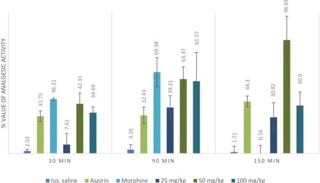
Yölmaz et al. evaluated the analgesic effect of aqueous O. spinosa root extract using the murine tail flick test. For positive controls, aspirin (100 mg/kg) and morphine (10 mg/kg) were used. The herbal extract was administered in 25 mg/kg, 50 mg/kg and 100 mg/kg intraperitoneally. Their results showed, that the O. spinosa extract significantly alleviated pain after 30, 90 and 150 minutes to administration in 50 mg/kg and 100 mg/kg doses.
The most outstanding results emerged in the 50 mg/kg group (see Figure 8) [60].
Öz et al. investigated the anti-inflammatory effect of fractions of O. spinosa root ethyl acetate extract obtained by column chromatography. In the carrageenan-induced paw edema in mice, the dried extract administered in 100 mg/kg dose showed significant inhibition of swelling thickness, which was similar to the effect of 10 mg/kg indomethacin. However, nor in the 12-O-tetradecanoylphorbol-13-acetate-induced ear edema test, neither in the adjuvant-induced chronic arthritis exhibited any extract significant inhibitory effect [61].
The cytosolic phospholipase A2α is one of the potential targets for anti-inflammatory drugs, since this enzyme plays a key role in the inflammation processes seen in health disorders, like asthma, allergic reactions, arthritis and neuronal diseases. In the study of Wink et al., inhibition of the enzyme by methanol extracts from medicinal plants rich in polyphenols was determined. O. spinosa exhibited an IC50 value of 39.4 µg/mL, resulting one of the most potent tested medicinal plant [62].
16
Figure 8: The standardized results of tail flick test using aspirin, morphine and 25, 50 and 100 mg of aqueaous extract of O. spinosa root [60]
Antibiotic effect
Petroleum ether, ethanol, butanol and aqueous crude extracts of the whole aerial parts of O. spinosa were tested against four bacterial and three fungal species by Mahasneh et al.
Using the disc diffusion method, methanol and hexane extracts did not show any activity and compared with standard antibiotics, the other extracts had low to moderate activity.
The butanol extracts at 4 mg/disc had high-moderate antifungal activity against Aspergillus flavus, Fusarium moniliforme and Candida albicans relative to miconazole nitrate at 40 μg/disc [63].
Çitoǧlu and Altanlar aimed to evaluate the antibiotic effect of the aerial parts of O.
spinosa using the same method. The discs were impregnated with O. spinosa extract made with 75% aqueous ethanol, ampicillin or fluconazole. The tested microorganisms and the results can be seen in Table 1, which suggest that the extract had a moderate antibiotic and antifungal effect [64]
2.03 3.26 1.21
31.75 32.43 44.3
46.31 69.38 0.16
7.61 39.21 30.8242.31 63.37 96.69
34.69 61.57 40.9
3 0 M I N 9 0 M I N 1 5 0 M I N
% VALUE OF ANALGESIC ACTIVITY
Iso. saline Aspirin Morphine 25 mg/kg 50 mg/kg 100 mg/kg
17
Table 1: Inhibition zones after the application of O. spinosa extract, ethanol, ampicillin or fluconazole
Inhibition zones (mm) E.
Coli P.
Aeruginosa B.
Subtilis S.
Aureus C.
Albicans C.
Glabrata C.
Krusei
O. spinosa 11 11 1 11 16 7 16
Ethanol - - - -
Ampicillin 12 n.d. 13 15 n.d. n.d. n.d.
Fluconazole n.d. n.d. n.d. n.d. 18 20 20
Antioxidant effect
Çoban et al. evaluated ethanol extracts from six species representing six different families, used in traditional medicine in Turkey for their antioxidant activities. In vitro tests included superoxide anion radical scavenging activity (determined spectrophotometrically on the basis of inhibition of cytochrome c reduction) and lipid peroxidation (investigated on rat liver homogenate induced with FeCl2-ascorbic acid).
The extract of O. spinosa aerial part showed concentration-dependent superoxide anion radical scavenging activity. The results of the superoxide anion formation assay showed that the ethanol extract of O. spinosa had moderate scavenging activity (IC50 1.35 mg/ml) and it had no significant effect on lipid peroxidation [65].
Wound healing effect
In order to establish the ethnopharmacological wound healing use of Ononis species, Öz et al. investigated the n-hexane, ethyl acetate and methanol extracts of O. spinosa roots and aerial parts. As the results showed that the most potent was the ethyl acetate extract of the roots [66], they continued the experiments with the silica gel fractions of this extract. The fractions were tested in linear incision and circular excision wound models and hydroxypyroline estimation assay on rats. The fifth fraction (Fr5) exhibited remarkable wound healing activity with the 33.4% tensile strength value on the linear incision wound model (madecassol as internal standard 53.8%) and 51.4% reduction of the wound area at the day 12 on the circular excision wound model (madecassol 100%).
Hydroxyproline content of the tissue treated by Fr5 was found to be 30.9 ± 0.72 μg/mg (madecassol 47.7 ± 0.57 μg/mg). Trifolirhizin, ononin, medicarpin 3-O-glucoside, onogenin 7-O-glucoside and sativanone 7-O-glucoside were isolated from Fr5 and tested for their wound healing activities by measuring their inhibition of hyaluronidase,
18
collagenase and elastase enzymes. Ononin and sativanone 7-O-glucoside inhibited hyaluronidase and elastase enzymes by 31.66% and 41.75%; 45.58% and 46.88%, respectively, at the dose of 100 μg/mL [61].
Addotey et al. executed bioactivity guided isolation from O. spinosa root extracts, based on hyaluronidase inhibition activity, too. Hot water and hydroalcoholic extracts showed moderate inhibiting effects (IC50 1.36 and 0.73 mg/ml) while dichloromethane extract exerted an IC50 of 190 μg/mL. Bioassay guided fractionation of the dichloromethane extract yielded four isoflavonoids with anti-Hyal-1 activity: onogenin, sativanone, medicarpin and calycosin-D with inhibition rates of 25.4, 61.2, 22.4 and 23.0%, respectively, at test concentration level of 250 μM. The IC50 value of sativanone, the most active compound was determined of 151 μM, which was better than that of the positive control glycyrrhizinic acid (177 μM) [11].
1.I.4. Biological activities of Ononis arvensis
In ethnomedicine, the aerial part has been applied for typhus and hernia, as aphrodisiac, and for stomach disorders as decoction. It is believed, that the plant maintains the optimal level of living water in the body therefore it can be used against diarrhea. The herb is traditionally applied for liver and stomach disorders as a decoction in the human and veterinary medicine [23].
Dénes et al. investigated the antimicrobial activity of different extract of O. arvensis aerial parts. Firstly, the methanolic extracts of separated leaves and stems were partitioned with n-hexane, chloroform, ethyl acetate and butanol, then these fractions were dried and re- dissolved in DMSO. The activity of the extracts was tested again Staphylococcus aureus (ATCC 25923), Escherichia coli (ATCC 25922), Pseudomonas aeruginosa (ATCC 27853), Salmonella typhimurium (ATCC 14028) and Candida albicans (ATCC 90028) using tube diluting and microdiluting method. The chloroform extract of the leaves exhibited strong antimicrobiotic activity against E. coli and C. albicans (MIC = 51 μg/ml and 12.75 μg/ml), interestingly, the n-hexane extract showed only antifungal activity against C. albicans (MIC = 8 μg/ml). The ethyl acetate extract of the leaves inhibited the growths of S. aureus and C. albicans, however, the same extracts of the stems proved to be more effective (MIC = 16 and 8 μg/ml). The chloroform extract of the stems could inhibit only the growth of P. aeruginosa (MIC = 43.5 μg/ml) and the butanol extract could
19
inhibit only S. typhimurium (MIC = 46.5 μg/ml). The methanol and aqueous extracts showed no significant antimicrobial effects and no extract could inhibit the growth of E.
coli.
1.I.5. Biological activities of other Ononis species
The results of the most important in vitro and in vivo tests of various other Ononis species are summarized in the Table 2.
1.I.6. Possibilities to produce isoflavonoids by in vitro cultures of Ononis species Plant cells are miniature reactors which produce a vast array of natural products. Most of these products are small molecules and are of keen interest, because they have shown potential as food additives, nutraceuticals, pharmaceuticals and cosmetic ingredients.
Some metabolites can be accumulated with a higher yield in in vitro cultures than those in parent plants, suggesting that the production of plant-specific secondary metabolites by plant in vitro cultures instead of whole plant cultivation possesses great potential.
Plants can also be genetically engineered to produce commercially interesting proteins including vaccines, antibodies, and other mainly therapeutic proteins [67], [68].
Secondary metabolites, including isoflavonoids, can be produced by using different biotechnological approaches, such as callus cultures, cell suspension cultures and/or organ cultures. Since it was observed that production of secondary metabolites is generally higher in differentiated plant tissues, there were attempts to cultivate whole plant organs, i.e. shoots or roots in in vitro conditions. As it was expected, such organ cultures produced similar patterns of secondary metabolites as intact plants.
20
Table 2: Summary of the biological activities, the type of experiments and used extracts of various Ononis species Species Investigated effect Type of
substance
Experiment Result Source
O. angustissima
α-amylase and α- glucosidase inhibition
MeOH-H2O extract
n-BuOH extract
in vitro enzymatic test IC50 = 0.94 mg/ml IC50 = 0.99 mg/ml
[69]
antioxidant isolated trifolirhizin
DPPH, ABTS,
reducing power assays
IC50 = 19.53 µg/ml IC50 = 28.29 µg/ml IC50 = 38.53 µg/ml
[26]
neuroprotective isolated maackiain and medicarpin (30 / 75 µM)
protective effect on Aβ25–35 peptide induced cytotoxicity in PC12 cells
significant increase in cell viability
O. macrosperma
wound healing effect
aqueous and EtOH extracts of aerial parts (5 mg extract topically)
in vivo linear incision model significant increase in tension strength (40.7% and 45.3%)
[54]
wound healing effect
aqueous extract of aerial parts (5 mg extract topically)
in vivo circular excision model significant wound contraction after 8, 10 and 12 days
anti-inflammatory activity
aqueous (200 mg/kg) and EtOH extracts of aerial parts (100 and 200 mg/kg)
in vivo acetic acid-induced
increase in capillary permeability
inhibition of 35.7%
and inhibition of 30.3% and 37.6%
20
21
O. vaginalis
anti-inflammatory activity
isolated
maackiain (2.8 mg/kg)
in vivo carregeenan induced rat paw edema
65.7% [32]
hepatoprotective activity
isolated trifolirhizin (7.5 mg/kg)
in vivo CCl4 induced liver damage
SGOT 28.79% decrease, SGPT 34.57% decrease, ALP 31.20% decrease, bilirubin 27.48% decrease
estrogenic activity isolated trifolirhizin
in vivo increase in uterine weight 93%
O. natrix
cholinesterase inhibition
EtAc extract of aerial parts
in vitro enzyme inhibitory assay
(Elmann’s method) 1.46 mg galanthamine equivalent/g AChE 0.93 mg galanthamine equivalent/g BChE
[29]
tyrosinase inhibition aqueous extract of aerial parts
in vitro enzyme inhibitory assay (dopachrome method)
52.81 mg kojic acid equivalent/g
α-glucosidase inhibition
MeOH extract of aerial parts
in vitro enzyme inhibitory assay (chromogenic PNPG method)
17.52 mg acarbose equivalent/g
DNA protective effect
aqueous extract of aerial parts (5 and 10 mg/ml)
in vitro plasmid stability 70 and 78% inhibition
cytotoxicity EtAc extract of aerial parts
in vitro test on MDA MB-231 cell line
IC50 = 28.75 µg/ml [70]
anti-inflammatory activity
EtAc extract of aerial parts
in vitro test on LPS induced HeLa cell line
10 µg/ml 80-100% inhibition of production of TNFα
[71]
21
22
The advantage of using organ cultures is that they are relatively more stable in production of secondary metabolites than cultures of undifferentiated cells, such callus or suspension cultures. For the objective of production of plant secondary products, generally two types of organ cultures are considered, i.e. root cultures and shoot cultures. Root systems of higher plants, however, generally exhibit slower growth than cultures of undifferentiated plant cells and are difficult to harvest. Compared to the cell suspension cultures, organ cultures generally display a lower sensitivity to shear stress, but they show a high degree of spatial heterogeneity in biomass production [72].
Callus culture is the culture of dedifferentiated plant cells induced on media usually containing relatively high auxin concentrations or a combination of auxin and cytokinin in in vitro conditions. Fedoreyev et al. established callus cultures from Maackia amurensis and analyzed them for isoflavonoids. The isoflavones daidzein, retuzin, genistein and formononetin and the pterocarpans maackiain and medicarpin were found to be produced by these cultures in approximately four times higher concentration than the content of the heartwood of M. amurensis plants [73]. Moreover, Li et al. established six callus cultures of Genista species with the objective to produce isoflavones with phytoestrogenic activity. Callus cultures of all species produced more isoflavones than the parent herbs. In vitro cultures had lower contents of genistein esters than the herbs [74]. Stable and optimized callus cultures are the first step of preparing the inoculum for liquid suspension cultures. Production of secondary metabolites in cell suspension cultures have been widely published and it was proposed as a technology to overcome problems of variable product quantity and quality from whole plants due to the effects of different environmental factors, such as climate, diseases and pests. The approach of using plant cell suspension cultures for secondary metabolite production is based on the concept of biosynthetic totipotency of plant cells, which means that each cell in the cultures retains the complete genetic information for production of the range of compounds found in the whole plant. The effect of the potential elicitors (killed cells of P. aeruginosa and E. coli, linoleic acid, arachidonic acid, chitosan, CrCl3, AgNO3, CoCl2, NiCl2, CdCl2, CuSO4, jasmonic acid, salicylic acid, iodoacetic acid and substituted anilides as pyrazine-2-carboxylic acid) on the production of flavonoids in callus culture and cell suspension of Ononis arvensis L. was examined by Tůmová and coworkers. All the tested elicitors markedly increased the production of flavonoids in comparison to the
23
control, however, only spectrophotometric methods were used to characterize the flavonoid content [77–86].
Genetic transformation of plants mediated by root inducing (Ri) plasmid of Agrobacterium rhizogenes bacterium occupies a special place in plant cell engineering, since this technique is based on a natural phenomenon that allows cultivation of isolated growing plant roots on hormone-free media. Application of wild-type unmodified agrobacterial strains allows to obtain root cultures capable of long-term growth in vitro due to an increased sensitivity of the cells to auxins while other biochemical properties remain unaltered. The in vitro cultivated roots could synthesize root-specific metabolites, which makes their application possible for large-scale biotechnological production of ecologically pure crude drugs. A collection of pRi T-DNA transformed roots of certain dicotyledons was made by Kuzovkina et al; including O. arvensis transformed by R 1601, A4 plasmids and O. spinosa transformed by R 1601 plasmid [89]. Although, the qualitative and quantitative relations of isoflavonoid content of these root cultures are unknown.
24 1.II. Isoflavonoids
1.II.1. Structural features and occurrence of isoflavonoids
The Leguminosae, with 19500 estimated species, is the main source of naturally occurring isoflavonoids. With only a few exceptions, Leguminosae isoflavonoids are restricted to the Papilionoideae, the largest of the three subfamilies currently recognized by legume taxonomists (Figure 9). For example, no reports of isoflavonoids from either the Caesalpinioideae or the Mimosoideae appeared [90].
Figure 9: Distribution of isoflavonoids in legumes. Clades that produce isoflavonoids are highlighted with pink [91]
Isoflavones are the most frequently reported of all the isoflavonoid subclasses, whereas isoflavanones are considerably rarer and can be found usually in racemic form (Figure 10). Reduction of isoflavanones leads to isoflavans, many of which act as phytoalexins in legumes. Laevorotatory, dextrorotatory, and racemic isoflavans are known in nature.
Pterocarpans contain a tetracyclic ring system derived from the basic isoflavonoid skeleton by an ether linkage between the C-4 and C-2´positions. Pterocarpans act as phytoalexins in leguminous plants and are produced following either fungal infection or
25
abiotic elicitor treatment. Dextrorotatory, levorotatory and racemic pterocarpans are known in nature with either a 6aR,11aR or 6aS,11aS configuration at the two chiral centers. Coumestans represent the fully oxidized version of pterocarpans. Rotenoids are a class of isoflavonoids characterized by the inclusion of an extra carbon atom into a heterocyclic ring. Almost all the known rotenoids contain an isoprenoid substituent and are noted for their insecticidal, piscicidal and antiviral activities. They can be conveniently subdivided into three major types, rotenoids, 12a-hydroxyrotenoids and dehydro-rotenoids depending on the oxidation level. Some rarities amongst isoflavonoids are coumaronochromones, isoflav-3-enes, isoflav-3-en-2-ones (3-arylcoumarins) and 2- arylbenzofurans (see Figure 10). In contrast to flavonoid oligomers which are a major group of flavonoid derivatives, similar oligomers of isoflavonoids are sporadic in nature [92].
Figure 10: Structure and numbering of isoflavonoid skeletons
26
The structural diversity of isoflavonoids is a result of the metabolic engineering of the aglycone skeleton and glycosylation. The number and complexity of possible substituents on the basic structural skeleton (hydroxyl, methoxy, methylenedioxy, prenyl or isoprenyl, etc.), the different oxidation levels and the frequent presence of extra heterocyclic rings (formed by cyclization between vicinal hydroxy and methoxy or monoprenyl groups) account for the multiplicity of subgroups among isoflavonoids (see Figure 11). Some isoflavonoids are amino-substituted; these are called “isoflavonoid alkaloids”,whereas others may be chlorinated [93]
Figure 11: Examples for the structural modification of isoflavonoid skeletons
Figure 12: Examples for O- and C-glycosides
In plants, isoflavonoids may be encountered as aglycones or as glycosides with generally glucose, rhamnose or apiose as the sugar component [93]. The number of known isoflavonoid glycosides is extremelysmall compared with the vast range of flavonoid glycosides [92]. Although the majority of glycosides are O-linked glycosides, several C- linked glycosides are also known together with a few compounds characterized by both C- and O-glycosylation (see Figure 12). One of the most important sources of C-linked isoflavones is the genus Pueraria. Through an enzymatic step, the 7-O-glucosides of
27
isoflavones and isoflavanones, and the 3-O-glucosides of pterocarpans could be malonylated to their 6”-O-malonyl derivatives [90].
Although isoflavonoids could be observed with vast majority in Leguminosae species, there are some examples in other families, as well. In monocots, few families (only 6 have been reported) seem able to produce isoflavonoids. The Iridaceae are the major source of isoflavonoids in this group, with more than 50 different compounds described, mainly in the genus Iris where they are present in the rhizomes of approximately 20 species. Among the dicots, five non-leguminous families are particularly outstanding because of their relative abundance in isoflavonoids: the Asteraceae (21 molecules), the Chenopodiaceae and the Nyctaginaceae (19 molecules each), the Moraceae (18 molecules), the Ochnaceae (17 molecules). However, the presence of isoflavonoids has been reported in only few species among these five families. Isoflavonoids have also been mentioned in various commercial beers, bourbons, teas and coffees but no study has documented their occurrence in the plants themselves [93].
1.II.2. Biosynthesis of isoflavonoids
Both flavonoids and isoflavonoids are synthesized through the central phenyl-propanoid pathway [94]. The factor differentiating isoflavonoids from other flavonoids is the linking of the B-ring to the C-3 rather than the C-2 position of the C-ring. The initial steps of isoflavonoid biosynthesis are now well characterized at the molecular level, but there is limited progress on the later enzymatic steps that produce the wide range of complex derivatives found in different legume species [95].
2-Hydroxyisoflavon synthase
2-HIS is a key cytochrome P450 enzyme that catalyzes the entry-point reaction into isoflavonoid biosynthesis, converting flavanones to isoflavones by an unusual aryl migration reaction (Figure 13). 2-HIS only present in legumes for the synthesis of isoflavonoids. Arabidopsis possesses 273 putative P450 genes, but none of them has 2- HIS activity for synthesizing isoflavone. Introducing soybean 2-HIS into Arabidopsis thaliana resulted in production of genistein at a high level, indicating its central role in the biosynthesis of isoflavonoids and extremely high substrate specificity [94].
28
Figure 13: The biosynthesis of isoflavonoids from flavonoids catalyzed by 2-HIS [95]
Methylation
Methylation is one of the key modifications of natural products including (iso)flavonoids to modulate their in vivo activity by limiting the number of reactive hydroxyl groups, thus altering their solubility and functions. Isoflavone-O-methyltransferase (IOMT) has been identified in various leguminous plants. It is an S-adenosyl-L-methionine (SAM)- dependent methyltransferase transferring a methyl group from SAM to isoflavones yielding the methyl ether derivatives (see Figure 14). 4′-O-methyltransferase (HI4′OMT) is a dual function enzyme with both 4′-O- and 3-O-methylation activity [94].
Isoflavone 2’- and 3’-hydroxylase
Two P450s of the CYP81E subfamily, isoflavone 2’-hydroxylase (I2’H) and isoflavone 3’-hydroxylase (I3’H), catalyze key steps in the formation of the more complex isoflavonoids. Hydroxylation of formononetin at the C-3’ position produces calycosin, a precursor for subsequent methylenedioxy bridge formation (yielding pseudobaptigenin) as part of the branches leading to maackiain- and pisatin-type phytoalexin end products, as well as to the rotenoids and other complex derivatives. Hydroxylation at the C-2’
position of daidzein, formononetin, or pseudobaptigenin provides the hydroxyl required for C–O–C bridge formation that defines the pterocarpans (e.g., glycinol) (Figure 14) [96].
29 Isoflavone reductase
The 2’-hydroxyisoflavones are reduced to the corresponding isoflavanones by a NADPH- dependent isoflavone reductase (IFR). IFR catalyzes enantiospecific conversions with high specificity for isoflavones [94]. The isoflavanones are the final isoflavonoid intermediates of pterocarpan biosynthesis (see Figure 14). Variant IFR activities between species are thought to contribute to the stereochemistry of the pterocarpans produced, in particular, (+)-maackiain in Pisum sativum, (-)-maackiain in Cicer arietinum, (-)-3,9- dihydroxypterocarpan in G. max, and (-)-medicarpin in M. sativa [95].
Figure 14: Steps of isoflavonoid biosynthesis resulting pterocarpans [95]
30
Vestitone reductase and 7,2’-dihydroxy-4’-methoxyisoflavonol dehydratase
Based on analysis of enzyme preparations, the conversion of isoflavanones to pterocarpans was thought initially to be catalyzed by a single NADPH-dependent enzyme, termed the pterocarpan synthase. However, it was subsequently shown that in M. sativa the conversion of vestitone to medicarpin involves two enzymes, vestitone reductase (VR) and 7,2’-dihydroxy-4’-methoxyisoflavanol dehydratase (DMID) (Figure 14). The reaction series from vestitone to the pterocarpan is thought to proceed by the VR-catalyzed reduction of vestitone to DMI, followed by the loss of water and formation of the C–O–C bridge between the heterocycle and the B-ring, catalyzed by DMID. In some species, the products of VR and DMID (maackiain and medicarpin) are the main pterocarpan phytoalexins. They are typically glucosylated and malonylated and stored in the vacuole. In species such as G. max, P. sativum and P. vulgaris, the pterocarpans are further converted by a series of reactions to species-specific compounds. For G. max and P. sativum, the initial reaction is a hydroxylation catalyzed by pterocarpan 6a- hydroxylase (P6aH) (Figure 15) [95].
SAM:6a-hydroxymaackiain 3-O-methyltransferase
Methylation of the 3-hydroxyl of (+)-6a-hydroxymaackiain by hydroxymaackiain 3-O- methyltransferase (HM3OMT) produces the major phytoalexin of P. sativum, (+)-pisatin (Figure 15) [95].
Prenyltransferases
The formation of phytoalexins such as glyceollins and phaseollins requires C-prenylation by a range of pterocarpan prenyltransferase activities, with dimethylallyl pyrophosphate as the prenyl donor. For glyceollins and phaseollins, prenylation occurs at position C-2 (pterocarpan C-2 prenyltransferase, P2CP) or C-4 (pterocarpan C-4 prenyltransferase, P4CP) of glycinol or C-10 of 3,9-dihydroxypterocarpan (Figure 15) [95].
Prenylpterocarpan cyclases
The final step of glyceollin and phaseollin formation is the cyclization of the prenyl residues of glyceollidins and phaseollidins, carried out by P450 prenylcyclases. These activities have been studied in detail for the formation of three glyceollins (I, II, and III) from glyceollidin I and II, and it is thought that specific activities are involved in each reaction (Figure 15) [95].
31
Figure 15: Biosynthesis of pterocarpane based phytoalexins [95]
Isoflavonoid glucosyltransferase and malonyltransferase
Glycosylation is a key modification to decorate plant natural products with various sugars, enhancing their solubility and stability and facilitating their storage and accumulation in plant cells. It is also a major factor in determining the bioactivity and bioavailability of natural products. Uridine diphosphate glycosyltransferases are the central players in glycosylation of plant natural products. Isoflavonoids are often modified by glycosylation, for example, the predominant soy isoflavones are genistein 7- O-glucoside and daidzein 7-O-glucoside and some malonyl conjugates [94]. In various isoflavonoid O-glucoside containing plants the main isoflavone constituents are actually isoflavone 7-O-glucoside 6”-O-malonates, however, these labile esters could be overlooked due to inadequate sample preparation methods [97].
32 Flavonoid 6-hydroxylase
The hydroxyl groups at C-5 and C-7 of the A-ring are introduced during the formation of chalcones by chalcone synthase. However, isoflavonoids also occur with C-6 and C-8 hydroxylation. The cDNA of the possible enzyme represented an elicitor induced P450 (CYP71D9) with flavonoid 6-hydroxylase activity, which may be involved in the biosynthesis of isoflavonoids with 6,7-dihydroxylation of the A-ring. The recombinant protein did not act on the isoflavonoids or pterocarpans directly, but rather accepted flavanone and dihydroflavonol substrates, including liquiritigenin, suggesting hydroxylation occurs prior to aryl migration of the B-ring (see Figure 13) [95].
1.II.3. Biological activities of selected isoflavonoids
In the following section, the biological activities of the eight isoflavonoids characterized in this thesis are highlighted in order to emphasize their importance.
Biological activities of formononetin
As formononetin is one of the most basic isoflavonoids regarding their biosynthesis (see page 27), it can be identified in many Leguminosae plants (175 hits for “Isolated as Natural Product” in Reaxys database) which possesses beneficial biological activities. As a consequence, there is magnitude orders higher amount of publications investigating the biological effects of formononetin.
• Neuroprotective effect
Amyloid beta (Aβ) is the main component of the amyloid plaques that accumulate in the brains of Alzheimer patients. Chen et al. reported the protective role of formononetin against Aβ25-35-induced neurotoxicity as formononetin (1-5 μM) significantly increased the viability of the cells and decreased the cell apoptosis. Formononetin also accelerated the non-amyloidogenic process of amyloid β precursor protein by enhancing α-secretase activity and soluble amyloid β precursor protein α release [98]. The study of Sun et al.
also shows that formononetin increases soluble amyloid β precursor protein α secretion and thus protects cells from hypoxia-induced apoptosis [99]. In the study of Fei et al., it is established that formononetin in a 15 mg/kg/day dose significantly improved learning and memory ability by suppressing Aβ production from amyloid β precursor protein processing. Moreover, these results did not differed significantly from that of achieved with 1 mg/kg/day donepezil [100].
33
The work of Li et al. concluded that formononetin mediates promising anti-traumatic brain injury effects against neurocytes [101], [102]. In addition, the work of El-Bakoush et al. established that formononetin inhibits neuroinflammation by targeting nuclear factor κB (NF-κB) [103]. In the study of Jia et al., formononetin reduced H2O2-induced apoptosis of retinal ganglion cells in a dose of 0.5-10 μM [104].
• Angiogenic effect
The study of Huh et al. investigated the influence of formononetin on the expression of growth factors contributing to wound healing in human umbilical vein endothelial cells (HUVECs). Formononetin (10 and 50 μM) in in vitro and ex vivo tests resulted to be more potent than recombinant vascular endothelial growth factor (VEGF125), but in the in vivo test of wound closure it did not differed from VEGF125 significantly [105]. Based on in vivo tests, formononetin promotes early fracture healing through angiogenesis activation in the early stage of fracture repair, and osteogenesis acceleration in the later stages used in a 200 μg/kg dose [106]. On the contrary to the previous results, Auyeung et al. aimed to examine the potential of formononetin in controlling angiogenesis and tumor cell invasiveness in human colon cancer cells and tumor xenografts in mice. The results showed that 200 μM formononetin downregulated the expression of the key pro- angiogenic factors. The tumor size and the number of proliferating cells were reduced in the tumor tissues obtained from the formononetin-treated group [107]. In the study of Wu et al., formononetin could inhibit the VEGF secretion of human retinal pigment endothelial cells under hypoxia in 0.2-5.0 μM concentration. Furthermore, formononetin could prevent hypoxia-induced retinal neovascularization in vivo (5-10 mg/kg), however, it did not reach the effectiveness of conbercept (1 mg/kg) [108].
• Vasorelaxive activity
Wu et al. and Sun et al. evaluated the vasorelaxation effects of formononetin, on rat isolated aorta and in vivo, the underlying mechanisms involved. Their results suggest that formononetin caused vascular relaxation via endothelium/NO-dependent mechanism and endothelium-independent mechanism which involves the activation of BKCa and KATP
channels. The antihypertensive mechanism in vivo may be associated with the down- regulation of α1-adrenoceptors and 5-HT2A/1B receptors, and the up-regulation of endothelial NO synthase expression in arteries [109]–[111]. The NO-dependent mechanism was also supported by the study of Bai et al. [112].
34
• Osteogenic activity
In the study of Ha et al., therapeutic potencies of phytoestrogens were investigated. In ovariectomized (Ovx) rats, formononetin-treated groups given 1 and 10 mg/kg/day displayed increased trabecular bone areas within the tibia [113]. Tyagi et al. achieved similar results. Formononetin treatment (10 mg/kg/day for 12 weeks) significantly restored the lost trabecular microarchitecture in the femurs and tibia of osteopenic Ovx rats and promoted new bone formation [114], [115]. Microcomputed tomography analysis showed that formononetin promoted bone healing and this effect observed was equal to parathyroid hormone treatment in many aspects [116]. In the study of Huh et al., formononetin led to a dramatic increase in normal osteoblast progenitor proliferation and differentiation, while decrease in osteoarthritic osteoblasts [117]. According to the results of Gautam et al. formononetin maximally stimulated osteoblast differentiation at 100 nM but had no effect on osteoblast proliferation. It neither activated estrogen receptor in osteoblasts nor had any effect on osteoclast differentiation and did not exhibit estrogen agonistic effect in uteri. Daily oral administration at 10.0 mg/kg/day dose to recently weaned female Sprague-Dawley rats for 30 consecutive days increased bone mineral density [118].
• Phytoestrogenic activity
To investigate a possible mechanism by which phytoestrogens might influence mammary carcinogenesis, Wang et al. examined the capacity of formononetin to stimulate mammary gland proliferation. Among animals treated with formononetin at 40 mg/kg/day for five days, mammary gland proliferation was enhanced 3.3-fold over saline- treated controls and was comparable to that of animals treated with 17β-estradiol at 1 μg/kg/day for five days. In subsequent in vitro binding studies, formononetin competitively bound murine mammary estrogen receptors, but with a relative binding affinity 15,000 times less potent than that of 17β-estradiol [119]. Ji et al. established a highly sensitive bioassay system by placing estrogen-responsive elements upstream of the luciferase reporter gene and used this assay to determine the estrogenic activity of formononetin. Formononetin activated expression of the estrogen-responsive reporter gene in human breast cell line MCF-7 in a concentration-dependent manner (0.5-500 μM), and this activation was inhibited by estrogen antagonist (ICI 182780 at 100 nM) [120]. Umehara et al. investigated the concentrations, in which isoflavones can show
35
similar results to 10 and 100 pM 17β-estradiol. On on T47D cell line 30 nm formononetin had equivalent effect to 10 pm 17β-estradiol. Formononetin activated expression of the estrogen-responsive reporter gene in cell line MCF-7 and T47D equivalent to 10 pM 17β- estradiol at 10 nM concentration [121]. Chen et al. aimed to further investigate the potential effect of formononetin in promoting cell proliferation in ER-positive cells and used in vivo and in vitro studies to elucidate the possible mechanism. Compared with the control, low formononetin concentrations (2-6 μM) stimulated ERα-positive cell proliferation. Additionally, in the in vivo studies, uterine weight in Ovx mice treated with formononetin increased significantly (in contrast to page 36) [122]. The growth of uterine tissues was confirmed by other researchers, as well [123], [124].
• Antigiardial activity
Khan et al. isolated several isoflavones from Dalbergia frutescens and determined their antiprotozoal activities against Giardia intestinalis. Among the isolated compounds, formononetin was the most potent antigiardial agent, with an IC50 value of 0.03 μg/ml, as compared to the value for metronidazole, the current drug of choice, of 0.1 μg/ml. The in vivo results indicated that, although formononetin is active, relatively large doses are required [125]. According to Lauwaet et al. the antigiardial activity of formononetin is at least partially due to its capacity to rapidly detach trophozoite [126].
• α-glucosidase activity
The heartwoods extract of Dalbergia odorifera demonstrated a potent inhibition on yeast α-glucosidase in vitro. Thus, bioassay-guided purification of EtAc soluble fraction was conducted to purify the active principles responsible for the inhibition. All active components isolated demonstrated a significant inhibition on yeast α-glucosidase in a dose-dependent manner, but formononetin showed an outstanding activity (IC50 = 0.51 mM) [127]. As the methanol extracts of all the parts of Dalbergia tonkinensis were found to be potential sources of α-glucosidase inhibitors, as well (showing more efficient inhibitory activity than that of acarbose), Van Bon et al. executed a bioassay-guided fractionation and isolated the most active compounds, sativanone and formononetin.
Formononetin showed weak inhibition against rice α-glucosidase, good inhibition against rat α-glucosidase, and a very efficient effect on α-glucosidases from yeast and bacterium (on yeast, its potency was approximately the twice as in the report of Choi et al.) (see Figure 16) [128].
36
Figure 16: α-glucosidase inhibitory effect of formononetin and acarbose in different enzyme systems [128]
• Pro-apoptotic effect
Many researchers aimed to define the pro-apoptotic characteristics of formononetin, however, no outstanding results have been published yet. On the other hand, these studies are a great help for the understanding of the underlying molecular biological pathways which contribute to the beneficial health effects of formononetin [129]–[144].
Biological activities of pseudobaptigenin
• Peroxysome proliferator-activated receptor agonism
Salam et al. executed a structure-based virtual screening of the peroxisome proliferator- activated receptor γ (PPARγ) ligand binding domain against a natural product library which revealed 29 potential agonists. In vitro testing of this list identified six flavonoids to have stimulated PPARγ transcriptional activity in a transcriptional factor assay. Of these, isoflavonoid pseudobaptigenin was classed as the most potent PPARγ agonist, possessing low micromolar affinity (EC50 = 2.9 μM) [145]. Matin et al. aimed to develop novel, efficacious, and yet safer pan agonists of PPAR receptors. Based on the aformentioned results of Salam et al., pseudobaptigenin and its derivatives were screened beside numerous other natural product-based derivatives. 3’,5’-dimethoxy-7- hydroxyisoflavone, pseudobaptigenin, 4’-fluoro-7-hydroxyisoflavone and 3’-methoxy-7- hydroxyisoflavone exhibited a substantially higher PPARγ fold activation compared to rosiglitazone at 5 μM and 25 μM. In the PPARα activity tests they all demonstrated higher
0.98mg/ml 0.03mg/ml 0.06mg/ml 0.23mg/ml
0.031mg/ml 0.001mg/ml 1.321mg/ml 0.121mg/ml
R I C E B A C T E R I A L Y E A S T R A T
IC50 VALUES
formononetin acarbose
37
fold activity than the positive control fenofibrate (1.3-fold activation). Pseudobaptigenin exhibited more potent PPARα activation than PPARγ activation. These four molecules were therefore identified as the most potent dual PPARα and γ agonists evaluated. The highest PPARδ agonist activity in the isoflavone series was observed for the same compounds as mentioned previously. Moreover, these isoflavones proved to be almost twice as potent activators of PPARδ than bezafibrate. To ensure that these active compounds were nontoxic to HEK 293 cell line, cytotoxicity profiles were investigated.
Pseudobaptigenin and the other three compounds exhibited excellent tolerability [149, 150].
Biological activities of calycosin
• Antioxidant activity
Promden et al. evaluated the antioxidant activities of 24 isoflavonoids that were previously isolated as pure compounds from D. parviflora using three different in vitro antioxidant-based assay systems: xanthine/xanthine oxidase, oxygen radical absorbing capacity, and 2,2-diphenyl-1-picrylhydrazyl (DPPH). The subgroup of isoflavones, and particularly calycosin showed the highest activities with 50% radical scavenging concentration (SC50) value of 0.25 μM. A further SAR analysis revealed that these active isoflavonoids from all three subgroups have the following common substituent pattern:
the presence of R7-OH in ring A and the R4′-OMe in ring B with either R3′-OH or R5′- OH. However, the presence of R7-OH and R4′-OMe in the molecule with no other key substituents (i.e., formononetin, SC50 = 117 µM), showed considerably lower activity, suggesting that these two substituents are not as important. In the other two assays, calycosin displayed only intermediate activity [148].
• NO production inhibition
Morikawa et al. isolated constituents from Erycibe expansa (Convulvaceae) originating from Thailand and examined their inhibitory activities on lipopolysaccharide-activated nitric oxide production in mouse peritoneal macrophages. The most active component was calycosin showing an IC50 value of 13 μM. This result is nearly equivalent to that of caffeic acid phenetyl ester, an inhibitor of NF-κB activation (IC50 15 μM) [149].
• Alleviating the cellular effects of hypoxia
38
Fan et al. intended to investigate the effect of calycosin on impairment of barrier function induced by hypoxia in HUVECS. The in vitro results indicated that calycosin could inhibit hypoxia induced hyperpermeability [150].
• Antiprotozoal activity
Araujo et al. isolated calycosin from Centrolobium sclerophyllum (Leguminosae) and tested its activity against Leishmania amazonensis promastigotes. Calycosin showed good antileishmanial activity, with calculated IC50 = 140 nM [151]. ElSohly et al. isolated calycosin, based on bioassay-guided fractionation, as the most active antigiardial compound of Machaenum aristulatum. Calycosin was tested against G. intestinalis and the antigiardial IC50 was determined as 1.9 μg/ml, which is twice as high as the IC50 of metronidazole (1.1 μg/ml) and ten times higher that of formononetin (0.28 μg/ml) [152].
The extracts of Psorothamnus arborescens, a desert shrub common in Mojave Desert, exhibited significant activity against Leishmania donovani axenic amastigotes and bloodstream form Trypanosoma brucei brucei. Bioassay-guided fractionation led to the isolation of calycosin, which showed very weak activity against L. donovani axenic amastigotes, but it displayed selective toxicity (about 15-fold) toward T. b. brucei bloodstream forms (IC50 = 12.7 µM) over Vero cells [153].
Biological activities of calycosin D (3’-methoxydaidzein)
• Antiprotozoal activity
Beldjoudi et al. isolated 4 new flavonoids along with 13 known compounds from the heartwood of Dalbergia louvelii by following their potential to inhibit the growth of Plasmodium falciparum in vitro. One of the most active compounds 3’-methoxydaidzein showed antiplasmodial activity with IC50 value at 6.8 µM, however, this result is not comparable with the activity of chloroquine (IC50 = 0.13 µM). Since the antiplasmodial effect of the extracts of the plant and their fractions showed higher activity, than the isolated compounds itself, a synergistic play of the constituents is supposed [154].
![Figure 9: Distribution of isoflavonoids in legumes. Clades that produce isoflavonoids are highlighted with pink [91]](https://thumb-eu.123doks.com/thumbv2/9dokorg/1377067.113266/25.892.136.803.360.799/figure-distribution-isoflavonoids-legumes-clades-produce-isoflavonoids-highlighted.webp)
![Figure 13: The biosynthesis of isoflavonoids from flavonoids catalyzed by 2-HIS [95]](https://thumb-eu.123doks.com/thumbv2/9dokorg/1377067.113266/29.892.121.809.122.436/figure-biosynthesis-isoflavonoids-flavonoids-catalyzed.webp)
![Figure 16: α-glucosidase inhibitory effect of formononetin and acarbose in different enzyme systems [128]](https://thumb-eu.123doks.com/thumbv2/9dokorg/1377067.113266/37.892.148.749.142.408/figure-glucosidase-inhibitory-effect-formononetin-acarbose-different-systems.webp)
![Figure 17: α-glucosidase inhibitory effect of sativanone and acarbose in different enzyme sytems [128]](https://thumb-eu.123doks.com/thumbv2/9dokorg/1377067.113266/41.892.136.759.131.410/figure-glucosidase-inhibitory-effect-sativanone-acarbose-different-enzyme.webp)
![Table 3: Various isoflavone glucoside derivatives in the extract made of O. spinosa roots [6]](https://thumb-eu.123doks.com/thumbv2/9dokorg/1377067.113266/52.892.126.770.651.840/table-various-isoflavone-glucoside-derivatives-extract-spinosa-roots.webp)
![Table 5: The method of determination and the quantity of various isoflavones before and after hydrolysis [9]](https://thumb-eu.123doks.com/thumbv2/9dokorg/1377067.113266/54.892.128.765.783.1082/table-method-determination-quantity-various-isoflavones-hydrolysis.webp)