Taste reactivity alterations after streptozotocin microinjection into the mediodorsal prefrontal cortex
Bernadett Nagy
Gábor Takács,
István Szabó,
László Lénárd,
Zoltán Karádi
Pécs University, Medical School, Institute of Physiology, Pécs, Hungary
Abstract
The mediodorsal prefrontal cortex (mdPFC), as an integrant part of the forebrain glucose- monitoring neural network, plays important roles in neural control of feeding. Previous studies suggested that streptozotocin (STZ) causes selective destruction of forebrain glucose- monitoring (GM) neurons leading to development of feeding disturbances. The goal of this research was to evaluate gustatory consequences of bilateral streptozotocin microinjection into the mediodorsal prefrontal cortex of male Wistar rats during conditioned taste avoidance (CTA) acquisition, as well as during taste reactivity tests. Bilateral streptozotocin microinjection failed to impair CTA learning, tested in a saccharin CTA paradigm. However, taste reactivity deficit was found by a modified version of the protocol introduced by Grill and Norgren. The streptozotocin treated animals displayed significantly poorer ingestive reactions to pleasant taste stimuli than did rats of the control group. The unpleasant taste stimuli elicited ingestive and rejective taste reactivity patterns in a comparable manner in rats of the STZ vs.
vehicle microinjected groups. The glucose-monitoring neurons of the mdPFC and their distinct role in the gustatory perception may have particular significance in the adaptive control of feeding.
Highlights
► Streptozotocin (STZ) microinjection into the mediodorsal prefrontal cortex was performed.
► Bilateral destruction of glucose-monitoring neurons elicited gustatory alterations. ► STZ microinjection caused taste reactivity deficit to pleasant taste stimuli. ► Significantly poorer ingestive patterns were found in STZ treated animals to pleasant tastants. ► STZ treatment failed to impair the acquisition of saccharin conditioned taste avoidance.
Keywords
Glucose-monitoring neurons;
Streptozotocin;
Mediodorsal prefrontal cortex;
Conditioned taste avoidance;
Taste reactivity
The diet of an organism is largely determined by the perceived palatability of foods.
Nutritional chemoreception is followed by adaptive feeding behaviors and other homeostatic changes.
The prefrontal cortex (PFC) is one of the key structures to monitor the internal state of the organism and initiate behavioral outputs accordingly. The PFC is implicated in many regulatory processes, including cognitive functions, attention, drive and motivation, decision making and working memory [1], [2] and [3]. Food and fluid intakes are also regulated by PFC. Bilateral lesions of the medial PFC result in finickiness, but failed to induce aphagia, while ventral-lateral PFC damages lead to development of aphagia [3].
Anatomically, the PFC is situated in the anterior pole of the mammalian brain and it has reciprocal connections with the mediodorsal thalamic nucleus. The term of mediodorsal prefrontal cortex (mdPFC) is used for neuroanatomical subdivisions of the PFC such as the prelimbic and cingulate cortices. Among other structures, mdPFC has direct connections with brain regions involved in the central regulation of feeding, such as the amygdala (AMY), the lateral hypothalamic area (LHA) and the nucleus accumbens (NAcc) [4], [5], [6] and [7].
In previous studies, special chemosensory neurons, the so-called glucose-monitoring (GM) neurons, have been discovered in the above interconnected regions [8], [9], [10] and [11] as well as in mdPFC [12]. GM neurons display firing rate changes in response to elevation of blood glucose level or to local microelectrophoretic administration of d-glucose. A majority of the GM neurons also display responses to intraorally delivered taste stimuli [9], [13], [14] and [15]. These homeostatically relevant, taste-sensitive GM neurons are suggested to form a complex hierarchically organized neural network integrating endogenous and exogenous information to control food and fluid intake and metabolic processes [9], [13] and [15].
Streptozotocin (STZ) is known to selectively destroy β-cells of the Langerhans-islets so it is widely used to induce experimental type 1 diabetes mellitus in animals [16] and [17]. STZ enters the cell via glucose transporter type 2 (GLUT2) and causes alkylation of DNA. The cytotoxic action of STZ is mediated by reactive oxygen species [17]. Furthermore, previous studies have also reported that intracerebral microinjection of streptozotocin (STZ) can specifically destroy the GM neurons of various brain areas (e.g. ventromedial hypothalamus, orbitofrontal prefrontal cortex and globus pallidus) causing severe deficits of feeding and metabolism [13], [15], [18], [19] and [20].
Considering the above, it can be supposed that selective destruction of glucose-monitoring cells of mdPFC may lead to disturbances of taste sensation and ingestive behavior. The present experiments were, therefore, designed to evaluate taste associated behavioral consequences of bilateral streptozotocin microinjection into the mdPFC of rats during conditioned taste avoidance (CTA) acquisition, as well as during taste reactivity tests.
Sixty-five male Wistar rats initially weighing 265–330 g, were used in the experiments. Rats were housed individually in a temperature and light controlled room (21 ± 2 °C; 12–12 h light–dark cycle, starting at 6:00 a.m.) where constant humidity (60 ± 5%) was also assured.
Standard laboratory chow food and tap water were available ad libitum except where
indicated. Rats were handled in daily regularity during the experiments. Animals were divided into two evenly formed, counterbalanced and randomized groups. Rats were kept and cared for in accordance with institutional (Pécs University, Medical School), national (Law XXVIII, 1998, Hungary) and international regulations (European Community Council Directive 86/609/EEC; 1986, 2006; European Directive 2010/63/EU of the European Parliament and of the Council; National Institutes of Health Guidelines).
Animals were anesthetized with intraperitoneal injection of ketamine (Calypsol, 50 mg/ml;
Richter Gedeon, Inc., Hungary; 0.3 ml/100 g body weight). The head of the rat was fixed in a stereotaxic device and an incision was made in the scalp. A small (3–4 mm in diameter) hole was drilled through the skull under microscopic control.
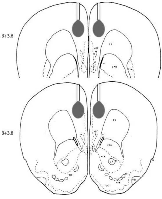
Stainless steal guide cannulas (23 G) were carefully lowered and placed on the surface of the dura above the mdPFC by a mechanical microdrive (MN-33 Narishige, Japan). After positioning, the guide cannulas were fixed to the cranium using dental acrilyc and anchoring screws. Microinjection cannulas (30 G) were passed through these guide cannulas to deliver the chemicals directly to the mdPFC. Stereotaxic coordinates for mdPFC according to the stereotaxic rat brain atlas of Pellegrino et al. [21] were: AP: +3.7 mm anterior to bregma (B), ML: 1 mm, and V: 1.5 mm (from dura).
Animals taking part in the taste reactivity test were also implanted with chronic intraoral taste cannula made from polyethylene (PE) tube (o.d. 1.33 mm). Each taste cannula was placed lateral to the first maxillary molar and was tunneled subcutaneously to ascend lateral to the skull. Local antibiotic profilaxis was applied after the operation (Tetran, Richter Gedeon, Inc., Hungary). The taste cannulas did not interfere with the normal eating behavior of the rats and allowed the direct infusion of taste solutions into the mouth. The animals were allowed to recover for 1 week before the STZ microinjection.
Alert rats were microinjected bilaterally with 7.5 μg STZ (Sigma S-0130, 10 μg/μl; dissolved in sterile physiological saline) or sterile physiological saline alone in a volume of 0.75 μl into both sides of the brain for 1 min. Solutions were microinjected by a microinfusion pump (Cole Parmer 789200C) into the mdPFC (V: 1.5 mm from the brain surface) through a stainless steal injection cannula (o.d. 0.3 mm) extending 1.5 mm below the tips of the guide cannula fixed on the skull with dental acrylic. A 25 μl Hamilton syringe was attached to an injection cannula via a short polyethylene tube. Microinjection cannulas were left in place for an additional 1 min after infusion to allow diffusion into the target area. Cannula arrangement and the microinjection are schematically shown in Fig. 1.
Fig. 1. Representative drawing of the delivery cannula and the microinjection target area in the mdPFC at two antero-posterior levels (B+3.6, B+3.8) according to the rat brain atlas of Pellegrino et al. [21].
Figure options
In the CTA test, animals were put on a 30-min drinking schedule and learned to consume the daily amount of their water intake from 10:00 to 10:30 a.m. every day. On the day of the operation and also on the following 2 days of recovery, water was offered them ad libitum.
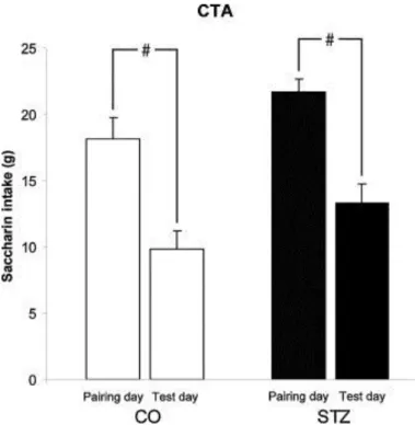
After the recovery period, animals were allowed to drink water in the 30-min schedule. Four days after the microinjection of STZ or physiological saline, on the pairing day, animals had access to 0.1% Na-saccharin for 30 min, and 30 min later they were injected i.p. with lithium chloride (0.15 M, 20 ml/kg b.w.), a malaise inducing drug. After this conditioning procedure, animals had water available for 3 days in the 30-min schedule. On the 4th (test) day, water was replaced by saccharin in the drinking period. The consumption of saccharin solution measured in the STZ treated and control groups on the pairing and test days were statistically compared.
In the taste reactivity test, after the surgery, rats with intraoral taste cannulas were given a 7- day habituation period during which they were placed into a Plexiglass cylinder of 30 cm in diameter and 30 cm height, followed by intraoral infusion of water. The taste reactivity test was performed 7 days after the STZ microinjection.
The animals were given two concentrations of taste solutions representing the five basic tastes: sweet, sucrose (0.05 and 0.5 M); salty, NaCl (0.05 and 0.5 M); sour, HCl (0.03 and 0.3 M); bitter, QHCl (0.03 and 3.0 mM); and umami, monosodium-l-glutamate (MSG, 0.05 and 0.5 M). With the help of a microinfusion pump (Cole Parmer 789200C), 0.5 ml of taste solution was infused into the mouth of the animal at a constant rate (0.5 ml/min) for 1 min.
After the infusion of a taste solution, the taste cannula was rinsed with distilled water and blown through by air. Based on previous experiments [22] and [23], both concentrations of sucrose and the lower concentrations of NaCl and MSG were regarded as pleasant tastes, while both concentrations of HCl and QHCl and the higher concentrations of NaCl and MSG were considered as unpleasant taste stimuli.
The behavior of rats was recorded by video camera (Panasonic SDR-H85) and later analyzed frame by frame. A mirror in a 45° tilted angle was mounted on a wooden frame enabling observation of the rat's mouth during the test. Mimical and postural-locomotor responses were scored using an adapted and modified version of the protocol introduced by Grill and Norgren [24] and [25]. Ingestive actions were characterized by rhythmic mouth movements, rhythmic tongue protrusions along the midline, lateral tongue movements and paw licking. Aversive behaviors were gaping, chin rubbing, head shaking, forelimb flailing and rapid locomotion around the chamber. The species specific response patterns were analyzed and compared in the STZ treated and control animals.
At the end of the tests, animals were i.p. overdosed with 20% urethane solution (8 ml/kg b.w.) and transcardially perfused with 0.9% NaCl followed by 4% formalin. The brains were removed, fixed in 4% formalin, frozen, cut in 40 μm sections and stained with cresyl violet to examine the microinjection sites. Four animals with inappropriate cannula positions were excluded from further analysis.
In the CTA experiment, saccharin intakes of animals on the pairing and test days were compared and statistically analyzed. In the taste reactivity test, the mimical and locomotor responses were scored according to the character, number, order and intensity of their elements. The patterns were evaluated and scored by minimum 3 independent, well-trained examiners, who were blind to treatment. Taste reactivity indices were calculated by dividing averaged sum of scores with the value of maximum possible scores [24] and [25].
The results are reported as means ± SEM. One-way analysis of variance (ANOVA) and the Tukey's test for post hoc comparisons were used for statistical analysis. Differences were considered significant at p < 0.05.
Bilateral STZ microinjection into the mdPFC did not impair the acquisition of LiCl induced saccharin CTA. ANOVA test showed that CTA developed in the control and also in the STZ treated group (F3,85 = 14.161; p < 0.001). There was no significant difference in the testing day in saccharin consumptions of the two groups by the post hoc test. Results of the CTA experiment are shown in Fig. 2.
Fig. 2. Bilateral STZ microinjection into the mediodorsal prefrontal cortex (mdPFC) fails to impair the aquisition of conditioned taste avoidance. CTA developed in the control and also in the STZ treated group (#p < 0.001). The saccharin intake of streptozotocin treated (STZ; black columns, n = 19) and control (CO; open columns, n = 24) animals did not differ significantly on the testing day. Columns represent mean saccharin consumptions + SEM.
Figure options
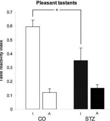
Bilateral microinjection of STZ induced taste reactivity alteration. Taste reactivity test revealed characteristic gustatory deficit in the case of pleasant taste stimuli (F3,35 = 19.451;
p < 0.001). The ingestive responses of STZ treated animals to pleasant taste stimuli were significantly poorer than those of control animals (p < 0.05). Considering the aversive patterns to pleasant tastes, the STZ treated animals and also the control rats gave similar amount of aversive reactions to pleasant tastes. These results are demonstrated in Fig. 3.
Fig. 3. Results of taste reactivity test to pleasant taste stimulation. Impaired ingestive response of the STZ treated animals (STZ; n = 8) compared to control ones (CO;
n = 10) to pleasant taste stimuli. Columns represent taste reactivity indices (mean + SEM). I and A, ingestive and aversive patterns, respectively. *p < 0.05.
Figure options
Taste reactivity deficit of STZ treated animals to pleasant taste stimuli was the most obvious in the case of higher concentration of sucrose and the lower concentration NaCl solutions.
Detailed result of the various pleasant taste stimulations is shown in Fig. 4.
Fig. 4. Ingestive taste reactivity responses to pleasant taste stimulation in the case of sucrose (0.05 M and 0.5 M), NaCl (0.05 M) and MSG (0.05 M). Significant
differences between STZ treated (STZ) and control (CO) rats are indicated (*p < 0.05;
#p < 0.001).
Figure options
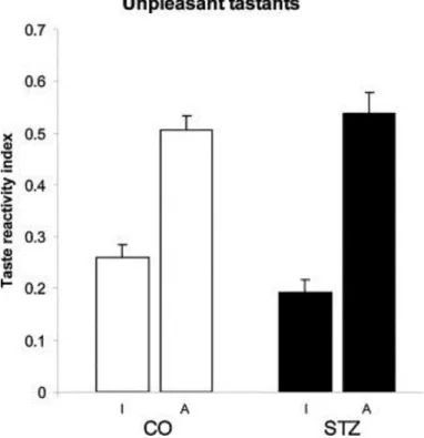
As far as the unpleasant gustatory stimuli are concerned, there was no significant difference in the taste elicited responses between the STZ treated and control animals (ingestive response:
p = 0.388; aversive response: p = 0.877). Mostly aversive reactions were detected in both groups to these tastants (Fig. 5).
Fig. 5. Results of taste reactivity test to unpleasant taste stimulation. There is no significant difference between STZ treated (STZ) and control (CO) rats. I and A, ingestive and aversive patterns, respectively.
Figure options
It has been proven in the present study that bilateral STZ microinjection fail to impair taste avoidance learning in a saccharin CTA paradigm. Results of the present experiments are in agreement with previous data, where animals with mediodorsal or dorsolateral PFC ablations were not impaired in CTA learning [26] and [27]. There are also data available, however, that microiontophoretically applied neurotoxins (kainic acid and 6-hydroxydopamine) in the medial prefrontal cortex cause significant deficits in CTA acquisition and retention [28].
Discrepancies among the findings probably rise from the different cell specificity of the microdamages, because STZ causes selective destruction of the GM neurons. Previous experiments provided evidence for that STZ microinjection into the NAcc causes CTA deficit in rats [13]. In the present experiment, we failed to detect similar changes after STZ microinjection of mdPFC indicating that not all parts of the GM neural network are involved in CTA aquisition. Consequently, our results suggest that the GM neurons of the mdPFC are not essential to associate the taste of ingested food with delayed noxious consequences of ingestion tested in a CTA paradigm.
The present study revealed taste reactivity deficit of rats after bilateral STZ microinjection into the mdPFC. The STZ treated animals displayed significantly poorer ingestive reactions to pleasant taste stimuli than did rats of the control group. The taste reactivity changes were the most pronounced in the case of sweet and salty taste stimulations. The underlying neural mechanism of these symptoms is supposed to be the selective cytotoxic effect of STZ on GM neurons. Our previous single neuron recording studies provided evidence for the existence of these special chemosensory neurons in the mdPFC [12]. Almost 25% of the mdPFC neurons tested changed in firing rate in response to microelectrophoretically administered glucose, thus, proved to be elements of the forebrain GM neural network. The central glucose- monitoring neural network has already been shown to be a major protector of the adaptive homeostatic balance integrating endogenous and exogenous chemical information and participating in sensory-motor and perceptual-motivational integration, as well as in reinforcement, learning and memory processes of feeding behavior [8], [9], [10], [15] and [29]. A single bilateral microinjection of the STZ into either the ventromedial hypothalamic nucleus (VMH) or orbitofrontal cortex (OBF) induced specific destruction of the forebrain GM neurons resulting in development of complex metabolic and feeding disturbances [15] and [18] and also characteristic taste reactivity alterations [13]. Selective damage of GM neurons in the OBF caused significantly stronger aversive reaction to pleasant tastants and more ingestive patterns to unpleasant tastes. Since the mdPFC is an integrant part of the forebrain glucose-monitoring system, it is reasonable to suppose that GM neurons of the mdPFC have similar functional roles in the adaptive feeding actions.
Although it has been shown that chronic decerebrate rats executed both ingestion and rejection response sequences similar to those observed in controls [30], we suppose that the lack of inputs of certain taste-responsive cortical neurons may cause palatability shift. Our hypothesis is in agreement with previous findings proving that rats with gustatory cortex lesions failed to display aversive reactivity to LiCl-paired tastants in a taste reactivity test [31]. A possible neural background of our findings might be the lack of appropriate mdPFC input to thalamus, since the thalamus appears to be essential in mimetic responses associated with ingestion [30]. In our experiment, deficit of the ingestive patterns might be due to a GM cell destruction induced imbalance of complex homeostatic and hedonic processes of food and fluid intake behaviors.
In the present study, evaluating the gustatory consequences of bilateral streptozotocin microinjection into the mdPFC, taste reactivity deficit to pleasant tastes was found. To elucidate the exact neural as well as the perceptual-motivational background of this deficit, further experiments are needed.
Acknowledgements
The authors thank Ildikó Fuchs, Dr. Dóra Keresztes, Bence Faragó, Barnabás Hideg, Tímea Csulak, Dr. Csaba Szalay and Márk Bajnok Góré for their valuable technical help. This work was supported by the Health Care Scientific Council (ETT 315/2006), National Research Fund of Hungary (OTKA K 68431), Ajinomoto51064/2009, SROP-4.2.1.B-10/2/KONV- 2010-0002, SROP-4.2.2/B-10/1-2010-0029 and the Hungarian Academy of Sciences.