Physiology & Behavior 236 (2021) 113410
Available online 2 April 2021
0031-9384/© 2021 The Authors. Published by Elsevier Inc. This is an open access article under the CC BY-NC-ND license
(http://creativecommons.org/licenses/by-nc-nd/4.0/).
Caffeine – treat or trigger? Disparate behavioral and long-term dopaminergic changes in control and schizophrenia-like Wisket rats
G Horvath
a,*, G Adam
e, G Tuboly
f, G Kekesi
a, A Büki
a, E Ducza
b, E Sz ˝ ucs
c,d, S Benyhe
c, G Benedek
aaDepartment of Physiology, Faculty of Medicine, University of Szeged, H-6720 Szeged, Hungary
bDepartment of Pharmacodynamics and Biopharmacy, Faculty of Pharmacy, University of Szeged, H-6720 Szeged, Hungary
cInstitute of Biochemistry, Biological Research Center, Hungarian Academy of Sciences, H-6726 Szeged, Hungary
dDoctoral School of Theoretical Medicine, Faculty of Medicine, University of Szeged, H-6720 Szeged, Hungary
eDepartment of Pulmonology, Faculty of Medicine, University of Szeged, H-6772 Szeged, Hungary
fDepartment of Neurology, Faculty of Medicine, University of Szeged, H-6725 Szeged, Hungary
A R T I C L E I N F O Keywords:
Behavior Caffeine Cognition
Dopamine D2 receptor Rat Schizophrenia
A B S T R A C T
The influence of caffeine on behavioral functions in both healthy and schizophrenic subjects is controversial.
Here we aimed to reveal the effects of repeated caffeine pre- and post-training treatments on motor and exploratory activities and cognitive functions in a reward-based test (Ambitus) along with a brain region-specific dopamine D2 receptor profile in control and schizophrenia-like WISKET model rats. In the control animals, pre- treatment caused temporary enhancement in motor activity, while permanent improvement in learning function was detected in the WISKET animals. Post-treatment produced significant impairments in both groups. Caffeine caused short-lasting hyperactivity followed by a rebound in the inactive phase determined in undisturbed circumstance. Caffeine treatment substantially enhanced the dopamine D2 receptor mediated G-protein activa- tion in the prefrontal cortex and olfactory bulb of both groups, while it increased in the dorsal striatum and cerebral cortex only in the WISKET animals. Caffeine enhanced the maximal binding capacity in the hippo- campus and cerebral cortex of WISKET animals, but it decreased in the prefrontal cortex of the control animals.
Regarding the dopamine D2 receptor mRNA expression, caffeine treatment caused significant enhancement in the prefrontal cortex of WISKET animals, while it increased the hippocampal dopamine D2 receptor protein amount in both groups. This study highlights the disparate effects of caffeine pre- versus post-training treatments on behavioral parameters in both control and schizophrenia-like animals and the prolonged changes in the dopa- minergic system. It is supposed that the delayed depressive effects of caffeine might be compensated by frequent coffee intake, as observed in schizophrenic patients.
1. Introduction
Disrupted behavioral activity and cognitive function are hallmarks of schizophrenia [1–3], and the antipsychotic drugs can partially relieve these deficits. Caffeine (CAFF) exhibits a variety of stimulant effects in the central nervous system as a non-selective adenosine A1/A2A receptor antagonist, leading to reduced drowsiness and enhanced locomotor ac- tivity [4,5]. However, reports on its influence on the learning processes are conflicting [6–11] and may also depend on the time of its adminis- tration — i.e., before or after cognitive tests [9,10,12,13]. Besides dis- rupting dopaminergic and glutamatergic neurotransmissions, adenosine
dysfunction may also contribute to the features of schizophrenia [14].
Increased coffee intake among patients is well documented, but its ef- fects seem to be controversial [6,15]. Thus, CAFF may evoke psychosis, however, it may also improve negative symptoms and/or compensate the antipsychotic medication-induced side effects [6,16]. Despite these reports, only one study provides evidence for its beneficial effects on schizophrenia-related cognitive impairments [17].
While several symptoms of schizophrenia in humans (e.g. halluci- nations and delusions) cannot be modeled in animals, other signs can reliably be reproduced [18]. To provide high constructive validity of this disease, a “multiple hit” WISKET rat model was generated by combined
* Corresponding author: Department of Physiology, Faculty of Medicine, University of Szeged, ´om t´er 10, H-6720 Szeged, Hungary, Phone: +36-62-544-971, Fax:
+36-62-545-842.
E-mail address: horvath.gyongyi@med.u-szeged.hu (G. Horvath).
Contents lists available at ScienceDirect
Physiology & Behavior
journal homepage: www.elsevier.com/locate/physbeh
https://doi.org/10.1016/j.physbeh.2021.113410
Received 15 January 2021; Received in revised form 12 March 2021; Accepted 26 March 2021
developmental (post-weaning social isolation), pharmacological (keta- mine, treatment) and genetic (selective breeding based on behavioral phenotype) manipulations. WISKET animals, besides behavioral dis- turbances, have altered opioid, cannabinoid and dopamine D2 receptor signaling, glutamate decarboxylase 67, oxytocin and dopamine D2 re- ceptor mRNA and protein expressions [19–28].
Few schizophrenia animal models investigated the effects of CAFF administration, with conflicting results [5,15,29–31]. Therefore, our first goal was to reveal the pre- and post-training CAFF treatments-related alterations in locomotor and exploratory activities and cognitive functions of the control and WISKET animals in the reward-based Ambitus test. As CAFF may cause rebound hypoactivity [32], the study was extended by the analysis of the rats’ activity in a large home cage, as an undisturbed condition.
CAFF alters the function of various neurotransmitters, including the dopaminergic system, which is disturbed in schizophrenic patients and its preclinical animal models [30,33–35]. Therefore, the second goal was to detect the potential long-term effects of repeated CAFF admin- istration on brain region-specific dopamine D2 receptor signaling, binding, and expression.
2. Materials and methods 2.1. Animals
The Hungarian Ethical Committee for Animal Research (RN: XIV/
1248/2018 in accordance with EU Directive 2010/63EU) approved all experiments. Male Wistar (control) and WISKET rats were used and kept with a 12 h light/dark cycle under controlled temperature (22±1 ◦C) and humidity (55±10%). Before the cognitive tests, the animals were food-deprived for two days, but water was freely available. Moderate food restriction (10–15 g/day/animal) was maintained throughout the experiments (for 5 days) with body weight control.
2.2. Interventions in Wisket rats and baseline behavioral tests for all groups
Based on earlier studies, at 3 weeks of age both control and WISKET rats were tested in the tail-flick test (TF; 48 ◦C hot water) to assess basal pain sensitivity [19,36]. Then, WISKET animals were housed individu- ally for 28 days and treated with ketamine (30 mg/kg/day intraperito- neally (ip); Calypsol, Gedeon Richter Plc., Hungary) from 5 to 7 weeks of age. Subsequently, the animals were re-socialized, and 1 week of re- covery followed. Control animals were socially reared with no
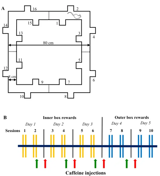
Fig. 1. A: Ground plan of the Ambitus with corridors and side boxes (1–16) equipped with photo beams (dashed lines) and showing a rat at the starting point. The height of the apparatus is 50 cm. B: Time scale of the 5-day-long Ambitus test paradigm. Yellow lines indicate Task 2 (inner box rewards); blue lines indicate Task 3 (outer box rewards). The red and green arrows indicate the time points of caffeine injection in the case of pre- and post-training treatments, respectively.
treatment. At the age of 9 weeks, all of the animals were involved in TF and sensory gating (pre-pulse inhibition: PPI) tests. The PPI was measured as described previously [19]. Briefly, after 10 min habituation in plexiglas startle chambers rats were exposed to two different trial types: pulse alone (PA: 40 ms 95 dB white noise burst); and pre- pulse–pulse pair (PP), in which a prepulse stimulus (20 ms, 76 dB) was followed by a startle stimulus with a latency of 150 ms. Both types of stimuli were applied 20 times in a random pattern. The interstimulus intervals ranged from 7 to 13 s. PPI was calculated as PPI(%)=[1− (startle response for PP)/(startle response for PA)] ×100. Furthermore, the locomotor and exploratory activities along with cognitive functions were assessed in the Ambitus with Task1 and Task2 at the age of 10 weeks (see below).
2.3. The Ambitus apparatus
The Ambitus apparatus is a rectangular corridor constructed of clear plexiglas on a black floor (Fig. 1A; www.deakdelta.hu) [37]. Each corridor has four side boxes (altogether 16) containing a food reward (puffed rice: 20 mg). Infrared beams detect the exploratory activity at each side box and the locomotor activity in midway of each corridor with 1 ms time resolution.
After inserting the food rewards, trials commenced by placing the rats at the starting point (Fig. 1A); thereafter, the experimenter imme- diately left the room. The animals were allowed to explore the corridor and collect food rewards for 300 s. The number of food rewards eaten was recorded and the apparatus was cleaned with 70% alcohol between trials. Experiments were recorded by an infrared video device (WCM- 21VF, CNB, China). If an animal had eaten all the rewards, its video recording was analyzed to determine the time required to complete the task.
Three different tasks were applied during the study. In Task 1 (Trials 1–2 during baseline measurements) all boxes; in Task 2 (Trials 3–4 during baseline measurements and on Days 1–3 during the 5-day-long experiment) only the inside boxes and in Task 3 (on Days 4–5) only the outside boxes were baited. All of the rats performed two sessions (two trials/session 1 min apart) of tasks per day — one in the morning and another 3 h later (Fig. 1B).
2.3.1. The 5-day-long experimental paradigm in the Ambitus system Four groups of both control and WISKET rats were involved in a 5- day-long experiment at the age of 13 weeks (Fig. 1B). The animals were daily injected with saline or CAFF (20 mg/kg; 4 ml/kg ip; Sigma- Aldrich Ltd, Hungary) [8,10], as follows: 30 min before Session 1 on Days 2–5 (pre-treatment) or promptly after the end of Session 2 on Days 1–4 (post-treatment). Since the pre- and post-training saline-treated animals showed similar behavior in the Ambitus system, their data were pooled into a 1–1 group. Therefore, data of 6 groups (saline-control [n = 13], saline-WISKET [n = 11], pre-CAFF-control [n = 9], pre-CAFF-WISKET [n = 9], post-CAFF-control [n = 9], and post-CAFF-WISKET [n =11]) were analyzed.
2.4. Video track analysis of activity in home cage
A separate series of experiment (6–6 control and WISKET rats) was performed to determine the activity pattern of the animals after a single CAFF injection in a large cage with 3 floors (60 ×60 ×60 cm with environmental enrichment) in undisturbed circumstances. The animals were single-housed and undertaken side-view motion detection by infrared video device.
After accommodation (2 days), saline (4 ml/kg ip) was administered to all of the animals on Day 1, and CAFF (20 mg/kg, ip) on Day 2. The animals’ behavior was recorded till the end of Day 3 to determine the prolonged effects of CAFF administration. The durations of the hourly active and inactive phases were determined, except between 10h00 and 11h00 and between 17h00 and18h00, when animal care was performed.
Because the preliminary analysis showed no significant differences in the activity pattern between the control and WISKET animals, we pooled the data of the two groups. Since the video records could be satisfactorily completed for only 4 control and 5 WISKET animals, therefore, data of 9 animals were analyzed.
2.5. Brain extraction
Control and WISKET rats with or without CAFF treatments aged 15 weeks were terminated and their brains were quickly removed, dissected on dry ice (prefrontal cortex [PFC], cerebral cortex, hippo- campus, dorsal striatum [STR] and olfactory bulb [OB]), frozen in liquid nitrogen, and stored at − 80 ◦C until further analysis. Since the brain extraction was performed after a long washout period (11 days) of the CAFF treatment, and the preliminary analysis did not show significant differences between the pre- vs post-CAFF-treated subgroups, they were pooled and saline-control, saline-WISKET, CAFF-control, and CAFF- WISKET groups were analyzed.
2.6. Preparation of brain samples for receptor binding assays
Neuronal membrane fractions were prepared from frozen brain specimens for in vitro receptor binding (RBA) [40] and the functional [35S]GTPγS binding experiments [25].
The protein content of the membrane preparations was determined as described in [38]. [35S]GTPγS (1000 Ci/mmol) was from Hartmann Analytic, [3H]spiperone (selective dopamine D1 receptor antagonist;
80.2 Ci/mmol) and UltimaGold™ MV scintillation reagent were from PerkinElmer.
2.6.1. Functional [35S]GTPγS binding experiments
[35S]GTPγS binding assays were performed basically as written in [39,40]. Agonist induced receptor mediated G protein activation was measured with 0.05 nM [35S]GTPγS in the presence of (10−10–10−5 M) sumanirole maleate (dopamine D2 receptor full agonist, Tocris Biosci- ence). Specifically bound radioactivities were separated by vacuum filtration method (Brandel M24R Cell Harvester, Whatman GF/B glass fiber filters).
2.6.2. Saturation binding experiments
Equilibrium saturation binding assays were performed in the pres- ence of [3H]spiperone in increasing concentrations (0.08 nM–4.98 nM) at 25 ◦C for 120 min. Ketanserin (1 µM) was used to block radioligand binding to 5-HT2 serotoninergic receptors [41]. Non-specific binding was determined with 10 µM unlabeled spiperone.
2.7. Dopamine D2 receptor expression studies
These investigations were performed in the PFC and hippocampus, both important structures for cognitive functions. All of the products were obtained from (ThermoFisher Scientific (Hungary)
2.7.1. Total RNA preparation from brain tissue
Total RNA was isolated by extraction with guanidinium thiocyanate- acid-phenol-chloroform method [42]. RNA purity was controlled at an optical density of 260/280 nm with BioSpec Nano (Shimadzu, Japan);
all samples exhibited an absorbance ratio in the range of 1.6–2.0. RNA quality and integrity were assessed by agarose gel electrophoresis.
2.7.2. Real-time quantitative reverse transcription-PCR (RT-PCR) Reverse transcription and amplification of the PCR products were performed by using the TaqMan RNA-to-CT-Step One Kit and an ABI Step One Real-Time cycler. RT-PCR amplifications were performed as follows: at 48 ◦C for 15 min and at 95 ◦C for 10 min, followed by 40 cycles at 95 ◦C for 15 s and at 60 ◦C for 1 min. The following primers were used: assay ID Rn00561126_m1 for the dopamine D2 receptor and
Rn00667869_m1 for β-actin as endogenous control. All samples were run in triplicate. The fluorescence intensities of the probes were plotted against PCR cycle number. The amplification cycle displaying the first significant increase of the fluorescence signal was defined as the threshold cycle (CT).
2.7.3. Western blot analysis
25 µg of protein per well was subjected to electrophoresis on 4–12%
NuPAGE Bis-Tris Gel in XCell SureLock Mini-Cell Units. Proteins were transferred from gels to nitrocellulose membranes, using the iBlot Gel Transfer System. The antibody binding was detected with the West- ernBreeze Chromogenic Western blot immunodetection kit. The blots were incubated with dopamine D2 receptor and β-actin antibody (1:200) in the blocking buffer. Images were captured with the EDAS290 imaging system (Csertex Ltd., Hungary), and the optical density of each immu- noreactive band was determined with Kodak 1D Images analysis software.
2.8. Statistical analysis
All data are expressed as means ± S.E.M., and significance was accepted at the p <.05 level. For the statistical analyses STATISTICA 13.4.0.14 (TIBCO Software Inc., USA) and GraphPad Prism (Inc., San Diego, CA) softwares were used.
2.8.1. In vivo experiments
Regarding the Ambitus test, data were evaluated by repeated mea- surements ANOVA, where the repeated measurements were sessions (10), and the factors were group (control, WISKET) and treatment (sa- line, pre-CAFF, post-CAFF). Table 1. gives definitions and denotes sig- nificances of the analyzed behavioral parameters.
For the statistical analysis in the large home cage, one-way ANOVA or t-test for dependent samples were performed.
Post hoc comparisons were performed by using the Fisher LSD test.
2.8.2. In vitro experiments
Radioreceptor binding data (saturation curves, one binding site model) and [35S]GTPγS binding data (sigmoid dose response stimula- tion) were processed by the professional curve-fitting program (Graph- Pad Prism 5.0.) using non-linear regression analysis. In the [35S]GTPγS binding assays the maximal stimulation or efficacy (Emax) of the re- ceptors G-protein and the ligand potency (EC50) were determined. In saturation binding assays, the concentration of the radioligand that produced 50% of the maximal binding capacity (dissociation constant [Kd]) and the maximum binding capacity of the receptor (Bmax) were established.
Factorial variance analysis was performed to determine the signifi- cance level of groups (control, WISKET) and treatments (saline, CAFF) for all the obtained in vitro parameters. The post hoc comparisons were performed by using the Newman-Keuls test.
3. Results
3.1. Baseline behavioral results
In agreement with our recent studies [19,37], the WISKET model rats showed impaired pain sensitivity, sensory gating, exploratory activity and learning capacity. These parameters did not differ significantly between the groups (saline, pre-CAFF, post-CAFF; data are not shown).
3.2. Behavioral results during the 5-day-long training
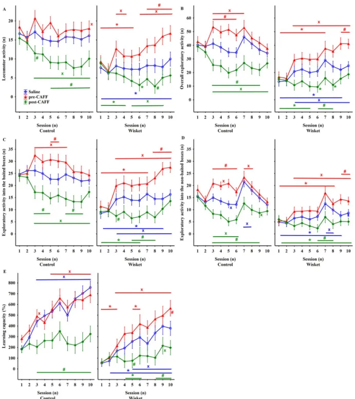
For all of the obtained behavioral parameters (Table 1), ANOVA revealed significant effects of group, treatment and session. Thus, saline- WISKET group differed significantly from their control counterparts during the whole investigated period (Fig. 2). The locomotor activity
was not changed in the saline-treated groups during the investigated period (Fig. 2A). Temporary hyperactivity was observed in pre-CAFF- control group during Session 3 and 5. In contrast, the hyperlocomotor effect of pre-CAFF treatment increased steadily in the WISKET animals, thus the significant differences between the two groups disappeared from Session 6. The post-CAFF treatment decreased the locomotion primarily in the control group, thereby reducing the significant differ- ences between the control and WISKET animals at the later sessions.
When Task 3 (Session 7) was introduced, the saline-control group showed significant enhancement in their overall exploratory activity (Fig. 2B) and the exploration into the baited side boxes, specifically (Fig. 2C), compared to the baseline values. In contrast, the saline- WISKET animals showed enhanced exploration from Session 3 compared to the baseline values, but at a significantly lower level than the control saline-treated animals. The control group responded with significant, but temporary hyperactivity to pre-CAFF treatment, during Sessions 3–7. While, exploratory activity into the baited side boxes enhanced persistently in the pre-CAFF-WISKET group, the post-CAFF treatment decreased the exploratory activity primarily in the control Table 1
Definitions and significances of the different parameters detected in the AMBI- TUS system, and shown in Fig. 2.
Parameter/Definition Significance F;
(df);p Locomotion: number of entries into
corridors up to 5 min Group 25.96;
(1,56);<0.001 Treatment 10.38;
(2,56);<0.001 Session 4.94;
(9504);<0.001 Group/session 3.83;
(9504);<0.001 Treatment/
Session 5.79;
(18,504);<0.001 Exploration: number of visits in all side-
boxes up to 5 min Group 37.88;
(1,56);<0.001 Treatment 17.11;
(2,56);<0.001 Session 4.84;
(9504);<0.001 Group/session 8.74;
(9504);<0.001 Treatment/
Session 5.35;
(18,504);<0.001 Baited Exploration: number of visits in
baited side-boxes up to 5 min Group 32.10;
(1,56);<0.001 Treatment 16.08;
(2,56);<0.001 Session 2.57;(9504);<0.01 Group/session 10.78;
(9504);<0.001 Treatment/
Session 5.29;
(18,504);<0.001 Non-baited Exploration: number of visits in
non-baited side-boxes up to 5 min Group 41.90;
(1,56);<0.001 Treatment 16.43;
(2,56);<0.001
Session 14.47;
(9504);<0.001 Group/session 5.01;
(9504);<0.001 Treatment/
Session 4.10;
(18,504);<0.001 Learning capacity (%): [(number of rewards
eaten)x(300)x100] / [(number of rewards)x(time to complete the task)]
Group 26.16;
(1,56);<0.001 Treatment 13.88;
(2,56);<0.001
Session 35.56;
(9504);<0.001 Treatment/
Session 4.56;
(18,504);<0.001 Abbreviations: df: degree of freedom.
groups (Fig. 2C). Thus, the differences between the control and WISKET animals regarding these parameters vanished during the later sessions.
Regarding the exploratory activity into the non-baited boxes (Fig. 2D), both the hypo- and hyper-exploratory effects of post- vs pre- CAFF treatments, respectively, were more pronounced in the control groups during Task 2; furthermore, the introduction of Task 3 caused a temporary enhancement, especially in the control saline-treated animals.
The learning capacity significantly increased by pre-CAFF treatment only in the WISKET animals, but it showed impairments in both post- CAFF groups (Fig. 2E; Table 1).
3.3. The effects of CAFF treatment on the activity in undisturbed condition
The saline injection on Day 1 did not significantly modify the activity of the animals, but a short-lasting (for 2 h), acute stimulating effect of CAFF treatment on Day 2 was indicated by the increased activity (Fig. 3A). To ensure the correct comparison of the results obtained in the Ambitus test in post-CAFF-treated groups (where the different behav- ioral parameters were evaluated 21–24 h after CAFF administration), the means of 3 h were analyzed. A rebound increase in inactive phases was detected between 12 and 18 h after CAFF administration (Fig. 3B).
Fig. 2.Time-course curves of locomotor (A), and exploratory (B–D) activities and learning capacity (E) in the control and WISKET groups depending on the different treatments. Data are presented as means ±S.E.M. Symbols denote significant differences between the two groups (*), treatments (#), and comparison to the baseline values obtained in the morning or afternoon of Day 1 (x).
3.4. In vitro studies
As it was presented recently, WISKET rats showed diverse changes in G-protein activation, binding, and dopamine D2 receptor properties in several brain regions compared to those in control animals [27].
3.4.1. Binding assays
Regarding the results related to G-protein activation, the factorial analysis of EC50 values did not show significant changes due to the CAFF administration (data are not shown). In contrast, CAFF enhanced the Emax values in the PFC and OB in both groups, but in the STR and ce- rebral cortex only in the WISKET animals (Fig. 4A). No any treatment effects could be detected in the hippocampus.
According to the saturation binding experiments, the Kd values were not influenced significantly by CAFF (data are not shown). However, CAFF treatment caused greater Bmax values in the cerebral cortex and hippocampus of the WISKET group, but significantly lower Bmax values in the PFC of control samples (Fig. 4B).
3.4.2. Dopamine D2 receptor expression in the PFC and hippocampus In the PFC of the WISKET animals, CAFF caused a significantly greater level of dopamine D2 receptor mRNA expression, and in the hippocampus of both groups a tendency to enhancement was observed (Fig. 4C). Regarding dopamine D2 receptor protein expression, CAFF treatment had a significantly different effect in the two groups in the PFC, i.e. we detected a significantly higher level of dopamine D2 re- ceptor protein in the WISKET animals. CAFF caused increased protein content in the hippocampus of both groups, and this enhancement was higher in the control animals; the significant differences between the control and WISKET animals, therefore, disappeared (Fig. 4D).
4. Discussion
This study characterized the effects of 5-day long pre- vs post-CAFF treatments on behavioral parameters and the dopamine D2 receptor system. In the control group, pre-CAFF treatment caused temporary hyperactivity without changes in cognitive function, while the post- CAFF treatment evoked significant impairments in all of the obtained Fig. 3.The acute (A) and prolonged (B) time-course effects of caffeine treatment on the duration of activity. Data are presented as means ±S.E.M. Symbols indicate significant differences compared to Day 1 (*), Day 2 (o), and to Day 3 (#). The arrows show the time of saline (on Day 1) or caffeine (on Day 2) injections. Black and white boxes represent the dark and light phases, respectively.
behavioral parameters. Regarding the WISKET animals, pre-CAFF treatment improved, while post-CAFF treatment further impaired these parameters. The home cage analysis revealed that the stimulant effect of CAFF was followed by rebound inactivity, suggesting that the post-CAFF treatment produced delayed changes in the behavioral ac- tivity in undisturbed circumstance, too. Furthermore, CAFF caused prolonged changes in the dopamine D2 receptor functions in most of the investigated brain structures, especially in WISKET animals.
The hippocampus, and the PFC play central roles in memory for- mation, whereas the STR and OB are involved in storing and retrieving learned motor behavior and olfaction-related learning [43–46]. The impaired cognitive functions in schizophrenia patients might reflect disruption of encoding, storage and/or retrieval capability [47,48].
Most schizophrenia symptoms are due primarily to disturbed dopami- nergic and glutamatergic neurotransmissions, but alterations in adeno- sinergic neurotransmission have also been shown [14,49]. The adenosinergic system is linked to motivation and cognitive processes, thus, the influence of adenosine on both the dopaminergic and gluta- matergic neurotransmissions might be relevant [4,49–52]. Adenosine A1
and A2A receptor agonists reverse both dopaminergic hyper- and NMDA receptor hypofunction, while CAFF may worsen the positive symptoms of schizophrenia patients [6]. Many data suggest an improving effect of CAFF in learning and memory [29,49,53–55], however, the results are controverting [8,9,56,57]. It is supposed that while CAFF did not modify the normal memory process in control animals, it could restore the memory disruption in different models of memory deficits [7,58,59].
Neurocognitive performance is affected by several psychological factors, including attention, motivation and/or exposure to stress [43, 60–62]. Increased stress reactivity has been reported in schizophrenia
patients [60,62]. The frequently used punishment in cognitive tests of animals have high emotional impact, and may be relatively resistant to disruption [8–10,57]. The reward-based learning paradigms are less stressful; thus, the control animals acquired the tasks in the Ambitus by few repetitions, and it was easily influenced by CAFF treatment.
Interpretation of the learning behavior in the pre-CAFF treated groups must take into consideration its concomitant motor-stimulant effect [4,52,63]. In agreement with earlier data, the CAFF-induced hy- peractivity in control animals was short-lasting, and tolerance devel- oped within a few days [5,15,63,64], however, their cognitive performance was not influenced, suggesting that control animals would not obtain beneficial effects from CAFF [7,56]. However, pre-CAFF treatment enhanced the exploratory activity and the learning ability of the WISKET animals during the whole investigated period. These results suggest that, in agreement with earlier studies, CAFF sensitivity is maintained for parameters related to attention and/or motivation in the schizophrenia-like animal model, which was accompanied with enhancement of their cognitive functions [48,56].
CAFF has generally been administered before cognitive tests; thus, its effects on memory are impossible to dissociate from increased activity.
Few studies investigated the effects of post-training CAFF treatment.
Two human studies suggested the task-dependent effects of post-training CAFF treatment on cognitive function [11,12]. Animal studies investi- gating cognitive behaviors 24 or 48 h after CAFF administration indi- cated either augmentation or no alteration of this parameter [9,10,13, 57,65]. Surprisingly, in our study, for all of the investigated parameters in both the control and WISKET groups, the impairing effects of post-CAFF treatment were evident. We surmise that the rebound hypo- activity accompanied with decreased motivation masked any possible Fig. 4. Results of signaling, binding, and dopamine D2 receptor expression assays. Data are presented as means ±S.E.M. The changes of Emax (A) and Bmax (B) values of dopamine D2 receptor in the different rat brain structures. The changes of mRNA (C) and protein (D) expression of dopamine D2 receptor (55 kDa) in the PFC and hippocampal regions. Symbols indicate significant differences between groups (*) and treatments (#).
positive effects of post-CAFF treatment on memory consolidation [66].
The more prolonged depressive effects obtained in the Ambitus test compared to the home cage circumstance might be due to the testing- related higher stress level.
In contrast to the pre-CAFF treatment, no tolerance developed to the behavioral inhibitory effects of post-CAFF treatment in either control or WISKET animals, which may reflect the differences between the acute stimulant and prolonged depressive effects of CAFF. We cannot exclude that the moderate changes in the pre-CAFF-control and post-CAFF- WISKET animals might be due, at least partially, to the ceiling vs.
floor effects, respectively. Few schizophrenia models have investigated the behavioral effects of CAFF treatment with controverting results, suggesting that the type of tests and administration route may signifi- cantly influence the results [5,15,29–31].
It is well-known that tolerance to the stimulant effects of CAFF could be developed shortly after its repeated administration, which might be due to the changes in different transmitters and their receptors, including adenosine, dopamine or acetylcholine [5,15,63,64,67–69].
Surprisingly, this phenomenon was observed only in the control ani- mals, but not in the Wisket ones. Since D2 and adenosine receptors are co-localized in the brain [34], it cannot be excluded that the altered D2R system in the Wisket animals, as was detected in our recent study [27], could lead to altered responses to repeated CAFF. Furthermore, while it is well-known that CAFF withdrawal is accompanied by depressive behavior [70], we did not observe tolerance on this sign in both groups.
Based on our results and earlier data, no exact explanation could be provided for these signs, but it might be suggested that altered adenosine receptor function might be similar during the post-CAFF state in both groups [71].
In spite of the common consumption of CAFF, the long-term biochemical effects of its administration have not been comprehen- sively investigated [72]. A few studies reported the effects of adenosi- nergic drugs on the dopamine D2 receptor system, but the signaling changes after CAFF administration have not been explored [33,34,69, 73]. One possible confounding bias might be that the investigation of the brain was performed over a long period. However, we wanted to allow for sufficient time without behavioral testing and for washout of CAFF.
Even after this long period CAFF enhanced G-protein signaling in the STR, PFC, cerebral cortex, and OB of the WISKET animals, but only in the PFC and OB of the control group. The binding assays showed increased Bmax values only in the cerebral cortex and hippocampus in the WISKET animals. Surprisingly, we found decreased or non-altered the Bmax values in the PFC of the control and WISKET animals, respec- tively, accompanied by enhanced Emax values, suggesting enhancement in the high-activity dopamine D2 receptor [74,75]. dopamine D2 re- ceptor mRNA expression in the PFC was enhanced in the CAFF-treated WISKET animals, while the dopamine D2 receptor protein expression increased in the hippocampus of both groups.
An in vitro study found that adenosine A2A receptor stimulation reduced the affinity of dopamine D2 receptor agonist binding sites of striatal neurons, but neither the affinity of dopamine D2 receptor antagonist binding nor the number of dopamine D2 receptors were affected [34]. In agreement with our results obtained in control animals, chronic CAFF administration had no apparent effect on the number and affinity to dopamine D2 receptor binding sites in the STR [33,73]. CAFF stimulated transcription of the dopamine D2 receptor gene and protein expression in the striatal cell culture [76]. The only in vivo study found that a single CAFF injection decreased the striatal dopamine D2 receptor mRNA, but did not change the dopamine D2 receptor protein expression [76]. However, all of these studies performed the experiments a few hours after CAFF administration. Only one study investigated the effects of chronic CAFF administration 7 days after the cessation of drug administration, and detected no changes in the striatal dopamine D2
receptor mRNA expression [72].
It is justified that the correlation analysis between the behavioral and the D2R system-related alterations would be useful in the interpretation
of our findings. While during the behavioral studies the experimental animals were tested individually, in vitro measurements we had to pool the tissue samples by brain region and treatment group, especially during radioligand binding tests. The main reasons were: in one hand the large number of samples to be tested in determining the concen- tration dependence of the ligand binding parameters over a wide range;
in the other hand the protein content of the cell membrane fractions used in the receptor binding experiments had to be at least 100 µg in each reaction tube. Many biomedical experiments are carried out by pooling individual biological samples [77]. The applied tissue prepa- ration procedure allows the relatively accurate determination of each measurement parameter. However, pooling samples can potentially hide biological variance, due to the fulfillment of the two necessary condi- tions, we were forced to apply this practice. Our data are therefore suitable for comparing receptor function or mRNA expression in various brain areas, but in our opinion, they cannot be directly correlated with in vivo behavioral data.
While data suggest short-lasting behavioral alterations after CAFF withdrawal [70], long-term effects for the D2R system, obtained pri- marily in Wisket animals, might suggest subtle changes in their behavior, or it might be supposed that these animals have enhanced stress sensitivity leading to positive signs. However, the enhanced function of the D2R in the cortical areas might be beneficial for the cognitive functions. Further preclinical and clinical studies could answer these suggestions/questions.
In conclusion, our findings demonstrated that CAFF pre-treatment blunted the cognitive impairments without tolerance in a schizophrenia-like rat model. In contrast, post-CAFF treatment led to significant behavioral deterioration in both groups. Therefore, the delayed depressive effects of CAFF might be unbeneficial, which can be compensated by frequent coffee intake, as observed in schizophrenic patients [6,15]. Furthermore, CAFF produced prolonged region-specific effects on the dopamine D2 receptor system, especially in the WISKET animals. This study provides novel insight into the adenosinergic system and its potential relevance in managing conditions of schizophrenia.
Funding and disclosure
This work was supported by GINOP 2.3.3–15–2016–00,031. The authors declare no competing interests.
Author contributions
All persons listed as authors made important contributions in one or more of the following areas: conception and design, analysis and inter- pretation of data, drafting of the manuscript, or making intellectual contributions to its content; and no person or persons other than the authors listed have contributed significantly to this manuscript.
Acknowledgements
The skilled technical assistance of Agnes Tandari is gratefully ´ acknowledged.
References
[1] T.A. Lesh, T.A. Niendam, M.J. Minzenberg, C.S. Carter, Cognitive control deficits in schizophrenia: mechanisms and meaning, Neuropsychopharmacology 36 (2011) 316–338.
[2] D.A. Lewis, Cortical circuit dysfunction and cognitive deficits in schizophrenia - implications for preemptive interventions, Eur. J. Neurosci. 35 (2012) 1871–1878.
[3] K.H. Nuechterlein, D.M. Barch, J.M. Gold, T.E. Goldberg, M.F. Green, R.K. Heaton, Identification of separable cognitive factors in schizophrenia, Schizophr. Res. 72 (2004) 29–39.
[4] B.B. Fredholm, K. B¨attig, J. Holm´en, A. Nehlig, E.E. Zvartau, Actions of caffeine in the brain with special reference to factors that contribute to its widespread use, Pharmacol. Rev. 51 (1999) 83–133. https://pubmed.ncbi.nlm.nih.gov/10049999 /?from_term=Fredholm+bb+caffeine+1999&from_pos=1 (accessed April 7, 2020).
[5] O.P. Dall’Igna, A.L. Da Silva, M.O. Dietrich, A. Hoffmann, R.V. De Oliveira, D.
O. Souza, D.R. Lara, Chronic treatment with caffeine blunts the hyperlocomotor but not cognitive effects of the N-methyl-D-aspartate receptor antagonist MK-801 in mice, Psychopharmacology (Berl) 166 (2003) 258–263, https://doi.org/10.1007/
s00213-002-1362-1.
[6] J.R. Hughes, P. McHugh, S. Holtzman, R.J. Frances, Caffeine and schizophrenia, Psychiatr. Serv. 49 (1998) 1415–1417, https://doi.org/10.1176/ps.49.11.1415.
[7] K.H. Alzoubi, K.K. Abdul-Razzak, O.F. Khabour, G.M. Al-Tuweiq, M.A. Alzubi, K.
A. Alkadhi, Caffeine prevents cognitive impairment induced by chronic psychosocial stress and/or high fat-high carbohydrate diet, Behav. Brain Res. 237 (2013) 7–14, https://doi.org/10.1016/j.bbr.2012.09.018.
[8] L.K. Pedraza, R.O. Sierra, F.N. Lotz, L.D.O. Alvares, Periodical reactivation under the effect of caffeine attenuates fear memory expression in rats, Sci. Rep. 8 (2018), https://doi.org/10.1038/s41598-018-25648-6.
[9] S. Dubroqua, S.R.L. Low, B.K. Yee, P. Singer, Caffeine impairs the acquisition and retention, but not the consolidation of Pavlovian conditioned freezing in mice, Psychopharmacology (Berl) 232 (2015) 721–731.
[10] M.E.M. Angelucci, M.A.B.F. Vital, C. Ces´ario, C.R. Zadusky, P.L. Rosalen, C. Da Cunha, The effect of caffeine in animal models of learning and memory, Eur. J.
Pharmacol. 373 (1999) 135–140, 10.1016/S0014-2999(99)00225-3.
[11] D. Borota, E. Murray, G. Keceli, A. Chang, J.M. Watabe, M. Ly, J.P. Toscano, M.
A. Yassa, Post-study caffeine administration enhances memory consolidation in humans, Nat. Neurosci. 17 (2014) 201–203.
[12] S.J. Hussain, K.J. Cole, No enhancement of 24-hour visuomotor skill retention by post-practice caffeine administration, PLoS ONE 10 (2015), https://doi.org/
10.1371/journal.pone.0129543.
[13] S.R. Kopf, A. Melani, F. Pedata, G. Pepeu, Adenosine and memory storage: effect of A1 and A2 receptor antagonists, Psychopharmacology (Berl) 146 (1999) 214–219, https://doi.org/10.1007/s002130051109.
[14] D.R. Lara, D.O. Souza, Schizophrenia: a purinergic hypothesis, Med. Hypotheses.
54 (2000) 157–166.
[15] R.V. De Oliveira, O.P. Dall’Igna, A.B.L. Tort, J.F. Schuh, P.F. Neto, M.W.S. Gomes, D.O. Souza, D.R. Lara, Effect of subchronic caffeine treatment on MK-801-induced changes in locomotion, cognition and ataxia in mice, Behav. Pharmacol. 16 (2005) 79–84, https://doi.org/10.1097/00008877-200503000-00002.
[16] K.K. Gandhi, J.M. Williams, M. Menza, M. Galazyn, N.L. Benowitz, Higher serum caffeine in smokers with schizophrenia compared to smoking controls, Drug Alcohol Depend 110 (2010) 151–155, https://doi.org/10.1016/j.
drugalcdep.2010.01.021.
[17] C. Núnez, C. Stephan-Otto, J. Cuevas-Esteban, J.Maria Haro, E. Huerta-Ramos, ˜ S. Ochoa, J. Usall, G. Br´ebion, Effects of caffeine intake and smoking on neurocognition in schizophrenia, Psychiatry Res 230 (2015) 924–931, 10.1016/j.
psychres.2015.11.022.
[18] J. Pratt, C. Winchester, N. Dawson, B. Morris, Advancing schizophrenia drug discovery: optimizing rodent models to bridge the translational gap, Nature 11 (2012) 560–579.
[19] Z. Petrovszki, G. Adam, G. Tuboly, G. Kekesi, G. Benedek, S. Keri, G. Horvath, Characterization of gene-environment interactions by behavioral profiling of selectively bred rats: the effect of NMDA receptor inhibition and social isolation, Behav. Brain Res. (2013) 240, https://doi.org/10.1016/j.bbr.2012.11.022.
[20] G. Horvath, G. Kekesi, Z. Petrovszki, G. Benedek, Abnormal motor activity and thermoregulation in a schizophrenia rat model for translational science, PLoS ONE 10 (2015), https://doi.org/10.1371/journal.pone.0143751.
[21] G. Horvath, Z. Petrovszki, G. Kekesi, G. Tuboly, B. Bodosi, J. Horvath, P. Gombk¨oto, G. Benedek, A. Nagy, Electrophysiological alterations in a complex rat model of schizophrenia, Behav. Brain Res. (2016) 307, https://doi.org/
10.1016/j.bbr.2016.03.051.
[22] A. Büki, G. Kalm´ar, G. Kekesi, G. Benedek, L.G. Nyúl, G. Horvath, Impaired pupillary control in “schizophrenia-like” WISKET rats, Auton. Neurosci. Basic Clin.
213 (2018) 34–42, 10.1016/j.autneu.2018.05.007.
[23] A. Büki, G. Horvath, G. Benedek, E. Ducza, G. Kekesi, Impaired GAD1 expression in schizophrenia-related WISKET rat model with sex-dependent aggressive behavior and motivational deficit, Genes, Brain Behav 18 (2019), https://doi.org/10.1111/
gbb.12507.
[24] G. Horvath, P. Liszli, G. Kekesi, A. Büki, G. Benedek, Cognitive training improves the disturbed behavioral architecture of schizophrenia-like rats, “Wisket,”, Physiol.
Behav. 201 (2019) 70–82, https://doi.org/10.1016/j.physbeh.2018.12.011.
[25] E. Szucs, A. Büki, G. K´ekesi, G. Horv´ath, S. Benyhe, Mu-Opioid (MOP) receptor mediated G-protein signaling is impaired in specific brain regions in a rat model of schizophrenia, Neurosci. Lett. 619 (2016) 29–33, https://doi.org/10.1016/j.
neulet.2016.02.060.
[26] E. Sz˝ucs, S. Dvor´acsko, C. T´ ¨omb¨oly, A. Büki, G. K´ekesi, G. Horv´ath, S. Benyhe, Decreased CB receptor binding and cannabinoid signaling in three brain regions of a rat model of schizophrenia, Neurosci. Lett. 633 (2016) 87–93, https://doi.org/
10.1016/j.neulet.2016.09.020.
[27] E. Sz˝ucs, E. Ducza, A. Büki, G. Kekesi, S. Benyhe, G. Horvath, Characterization of dopamine D2 receptor binding, expression and signaling in different brain regions of control and schizophrenia-model Wisket rats, Brain Res (2020), 147074, 10.1016/j.brainres.2020.147074.
[28] L. Banki, A. Büki, G. Horvath, G. Kekesi, G. Kis, F. Somogyv´ari, G. Jancs´o, L. V´ecsei, E. Varga, G. Tuboly, Distinct changes in chronic pain sensitivity and oxytocin receptor expression in a new rat model (Wisket) of schizophrenia, Neurosci. Lett.
714 (2020), 134561, 10.1016/j.neulet.2019.134561.
[29] R.D.S. Prediger, F.A. Pamplona, D. Fernandes, R.N. Takahashi, Caffeine improves spatial learning deficits in an animal model of attention deficit hyperactivity
disorder (ADHD) – the spontaneously hypertensive rat (SHR), Int. J.
Neuropsychopharmacol. 8 (2005) 583–594.
[30] P. Pandolfo, N.J. Machado, A. Kofalvi, R.N. Takahashia, R.A. Cunhab, Caffeine regulates frontocorticostriatal dopamine transporter density and improves attention and cognitive deficits in an animal model of attention deficit hyperactivity disorder, Eur. Neuropsychopharmacol. 23 (2013) 317–328, https://
doi.org/10.1016/j.euroneuro.2012.04.011.
[31] G. Sandner, M.J. Angst, T. Guiberteau, B. Guignard, A. Nehlig, Effects of caffeine or RX821002 in rats with a neonatal ventral hippocampal lesion, Front. Behav.
Neurosci. 8 (2014), https://doi.org/10.3389/fnbeh.2014.00015.
[32] T. Roehrs, T. Roth, Caffeine: sleep and daytime sleepiness, Sleep Med. Rev. 12 (2008) 153–162, 10.1016/j.smrv.2007.07.004.
[33] K.R. Powell, P.M. Iuvone, S.G. Holtzman, The role of dopamine in the locomotor stimulant effects and tolerance to these effects of caffeine, Pharmacol. Biochem.
Behav. 69 (2001) 59–70, 10.1016/S0091-3057(01)00497-X.
[34] S. Ferre, G. Von Euler, B. Johansson, B.B. Fredholm, K. Fuxe, Stimulation of high- affinity adenosine A2 receptors decreases the affinity of dopamine D2 receptors in rat striatal membranes, Proc. Natl. Acad. Sci. U. S. A. 88 (1991) 7238–7241, 10.1073/pnas.88.16.7238.
[35] G.R. Stoner, L.R. Skirboll, S. Werkman, D.W. Hommer, Preferential effects of caffeine on limbic and cortical dopamine systems, Biol. Psychiatry. 23 (1988) 761–768, https://doi.org/10.1016/0006-3223(88)90064-9.
[36] G. Kekesi, Z. Petrovszki, G. Benedek, G. Horvath, Sex-specific alterations in behavioral and cognitive functions in a “three hit” animal model of schizophrenia, Behav. Brain Res. (2015) 284, https://doi.org/10.1016/j.bbr.2015.02.015.
[37] G. Horvath, P. Liszli, G. Kekesi, A. Büki, G. Benedek, Characterization of exploratory activity and learning ability of healthy and “schizophrenia-like” rats in a square corridor system (AMBITUS), Physiol. Behav. 169 (2017) 155–164, 10.1016/j.physbeh.2016.11.039.
[38] M.M. Bradford, A rapid and sensitive method for quantitation of microgram quantities of protein utilizing principle of protein-dye binding, Anal. Biochem. 72 (1976) 248–254.
[39] L.J. Sim, D.E. Selley, S.R. Childers, In vitro autoradiography of receptor-activated G proteins in rat brain by agonist-stimulated guanylyl 5’-[gamma-[35S]thio]- triphosphate binding. Proc Natl Acad Sci USA. 92 (1995) 7242–7246.
[40] J.R. Traynor, S.R. Nahorski, Modulation by mu-opioid agonists of guanosine-5’-O- (3-[35S]thio)triphosphate binding to membranes from human neuroblastoma SH- SY5Y cells, Mol. Pharmacol. 47 (1995) 848–854.
[41] A. Janowsky, K.A. Neve, J.M. Kinzie, B. Taylor, T. de Paulis, J.K. Belknap, Extrastriatal dopamine D2 receptors: distribution, pharmacological
characterization and region-specific regulation by clozapine, J. Pharmacol. Exp.
Ther. 261 (1992) 1282–1290.
[42] P. Chomczynski, N. Sacchi, Single-step method of RNA isolation by acid guanidinium thiocyanate-phenol-chloroform extraction, Anal. Biochem. 162 (1987) 156–159.
[43] C.V. Vorhees, M.T. Williams, Assessing spatial learning and memory in rodents, ILAR J 55 (2014) 310–332.
[44] G. Buzsaki, Time, space and memory, Nature 497 (2013) 568–569.
[45] Z. Nusser, L.M. Kay, G. Laurent, G.E. Homanics, I. Mody, Disruption of GABAA receptors on GABAergic interneurons leads to increased oscillatory power in the olfactory bulb network, J. Neurophysiol. 86 (2001) 2823–2833, https://doi.org/
10.1152/jn.2001.86.6.2823.
[46] J. Beshel, N. Kopell, L.M. Kay, Olfactory bulb gamma oscillations are enhanced with task demands, J. Neurosci. 27 (2007) 8358–8365.
[47] A.A. Moustafa, J.K. Garami, J. Mahlberg, J. Golembieski, S. Keri, D. Frydecka, Cognitive function in schizophrenia: conflicting findings and future directions, Rev. Neurosci. 27 (2016) 435–448.
[48] K. Takeda, T. Sumiyoshi, M. Matsumoto, K. Murayama, S. Ikezawa, K. Matsumoto, K. Nakagome, Neural correlates for intrinsic motivational deficits of schizophrenia;
implications for therapeutics of cognitive impairment, Front. Psychiatry. 9 (2018).
10.3389/fpsyt.2018.00178.
[49] D. Boison, P. Singer, H.Y. Shen, J. Feldon, B.K. Yee, Adenosine hypothesis of schizophrenia - Opportunities for pharmacotherapy, Neuropharmacology 62 (2012) 1527–1543.
[50] E. Ongini, B.B. Fredholm, Pharmacology of adenosine A2A receptors, Trends Pharmacol. Sci. 17 (1996) 364–372.
[51] D. Quarta, S. Ferre, M. Solinas, Z.B. You, J. Hockemeyer, P. Popoli, S.R. Goldberg, Opposite modulatory roles for adenosine A1 and A2A receptors on glutamate and dopamine release in the shell of the nucleus accumbens. Effects of chronic caffeine exposure, J. Neurochem. 88 (2004) 1151–1158.
[52] T.M. McLellan, J.A. Caldwell, H.R. Lieberman, A review of caffeine’s effects on cognitive, physical and occupational performance. Neurosci. Biobehav. Rev. 71 (2016) 294–312, https://doi.org/10.1016/j.neubiorev.2016.09.001.
[53] V.A. Pires, F.A. Pamplona, P. Pandolfo, R.D.S. Prediger, Chronic caffeine treatment during prepubertal period confers long-term cognitive benefits in adult spontaneously hypertensive rats (SHR), an animal model of attention deficit hyperactivity disorder (ADHD), Behav. Brain Res. 215 (2010) 39–44.
[54] R.D.S. Prediger, L.C. Batista, R.N. Takahashi, Caffeine reverses age-related deficits in olfactory discrimination and social recognition memory in rats. Involvement of adenosine A1 and A2A receptors, Neurobiol. Aging. 26 (2005) 957–964.
[55] P.B. Lucas, D. Pickar, J. Kelsoe, M. Rapaport, C. Pato, D. Hommer, Effects of the acute administration of caffeine in patients with schizophrenia, Biol. Psychiatry. 28 (1990) 35–40, 10.1016/0006-3223(90)90429-6.
[56] N. SanMiguel, M. Pardo, C. Carratal´a-Ros, L. L´opez-Cruz, J.D. Salamone, M. Correa, Individual differences in the energizing effects of caffeine on effort-based decision-
making tests in rats, Pharmacol. Biochem. Behav. 169 (2018) 27–34, 10.1016/j.
pbb.2018.04.004.
[57] M.E.M. Angelucci, C. Ces´ario, R.H. Hiroi, P.L. Rosalen, C. Da Cunha, Effects of caffeine on learning and memory in rats tested in the Morris water maze, Brazilian J. Med. Biol. Res. 35 (2002) 1201–1208, 10.1590/S0100-879X2002001000013.
[58] R.A. Cunha, P.M. Agostinho, Chronic caffeine consumption prevents memory disturbance in different animal models of memory decline, in: J. Alzheimer’s Dis.
IOS Press (2010) https://doi.org/10.3233/JAD-2010-1408.
[59] O.K. Çakır, N. Ellek, N. Salehin, R. Hamamcı, H. Keles¸, D.G. Kayalı, D. Akakın, ¨ M. Yüksel, D. Ozbeyli, Protective effect of low dose caffeine on psychological stress ¨ and cognitive function, Physiol. Behav. 168 (2017) 1–10, 10.1016/j.
physbeh.2016.10.010.
[60] A. Knapman, J.M. Heinzmann, F. Holsboer, R. Landgraf, C. Touma, Modeling psychotic and cognitive symptoms of affective disorders: disrupted latent inhibition and reversal learning deficits in highly stress reactive mice, Neurobiol.
Learn. Mem. 94 (2010) 145–152.
[61] H. Temmingh, D.J. Stein, Anxiety in patients with schizophrenia: epidemiology and management, CNS Drugs 29 (2015) 819–832.
[62] K. Krkovic, S. Moritz, T.M. Lincoln, Neurocognitive deficits or stress overload: why do individuals with schizophrenia show poor performance in neurocognitive tests?
Schizophr. Res. 183 (2017) 151–156.
[63] O. Nikodijevi´c, K.A. Jacobson, J.W. Daly, Locomotor activity in mice during chronic treatment with caffeine and withdrawal, Pharmacol. Biochem. Behav. 44 (1993) 199–216, 10.1016/0091-3057(93)90299-9.
[64] S.G. Holtzman, I.B. Finn, Tolerance to behavioral effects of caffeine in rats, Pharmacol. Biochem. Behav. 29 (1988) 411–418, https://doi.org/10.1016/0091- 3057(88)90179-7.
[65] L. Molinengo, M. Orsetti, B. Pastorello, I. Scordo, P. Ghi, Habituation of exploratory activity in rats: action of N6phenylisopropyladenosine, caffeine and their combination, Prog. Neuropsychopharmacol. Biol. Psychiatry. 19 (1995) 1189–1200, 10.1016/0278-5846(95)00236-7.
[66] I.A. Sukhotina, E.E. Zvartau, W. Danysz, A.Y. Bespalov, Caffeine withdrawal syndrome in social interaction test in mice: effects of the NMDA receptor channel blockers, memantine and neramexane, Behav. Pharmacol. 15 (2004) 207–214. http ://www.ncbi.nlm.nih.gov/pubmed/15187578 (accessed April 15, 2020).
[67] B.E. Garrett, S.G. Holtzman, Caffeine cross-tolerance to selective dopamine D1 and D2 receptor agonists but not to their synergistic interaction, Eur. J. Pharmacol. 262 (1994) 65–75, https://doi.org/10.1016/0014-2999(94)90029-9.
[68] E. Acquas, G. Tanda, G. Di Chiara, Differential effects of caffeine on dopamine and acetylcholine transmission in brain areas of Drug-naive and Caffeine-pretreated Rats, Neuropsychopharmacology 27 (2002) 182–193, https://doi.org/10.1016/
S0893-133X(02)00290-7.
[69] D. Shi, O. Nikodijevic, K.A. Jacobson, J.W. Daly, Effects of chronic caffeine on adenosine, dopamine and acetylcholine systems in mice, Arch. Int. Pharmacodyn.
Ther. 328 (1994) 261–287.
[70] L.M. Juliano, R.R. Griffiths, A critical review of caffeine withdrawal: empirical validation of symptoms and signs, incidence, severity, and associated features, Psychopharmacology (Berl) 176 (2004) 1–29, https://doi.org/10.1007/s00213- 004-2000-x.
[71] G.B. Kaplan, D.J. Greenblatt, M.A. Kent, M.M. Cotreau-Bibbo, Caffeine treatment and withdrawal in mice: relationships between dosage, concentrations, locomotor activity and A1 adenosine receptor binding, J. Pharmacol. Exp. Ther. 266 (1993).
[72] J.A. Mu˜niz, G. Gomez, B. Gonz´alez, M.C. Rivero-Echeto, J.L. Cadet, E. García-Rill, F.J. Urbano, V. Bisagno, Combined effects of simultaneous exposure to caffeine and cocaine in the mouse striatum, Neurotox. Res. 29 (2016) 525–538, https://doi.org/
10.1007/s12640-016-9601-0.
[73] K.A. Jacobson, D.K.J.E. von Lubitz, J.W. Daly, B.B. Fredholm, Adenosine receptor ligands: differences with acute versus chronic treatment, Trends Pharmacol. Sci. 17 (1996) 108–113.
[74] M. Seeman, P. Seeman, Is schizophrenia a dopamine supersensitivity psychotic reaction? Prog. Neuro-Psychopharmacology Biol. Psychiatry. 48 (2014) 155–160, 10.1016/j.pnpbp.2013.10.003.
[75] P. Seeman, Schizophrenia and dopamine receptors, Eur. Neuropsychopharmacol.
23 (2013) 999–1009, https://doi.org/10.1016/j.euroneuro.2013.06.005.
[76] A.H. Stonehouse, M. Adachi, E.C. Walcott, F.S. Jones, Caffeine regulates neuronal expression of the dopamine 2 receptor gene, Mol. Pharmacol. 64 (2003) 1463–1473, https://doi.org/10.1124/mol.64.6.1463.
[77] E.C. Hulme, M.A. Trevethick, Ligand binding assays at equilibrium: validation and interpretation, Br. J. Pharmacol. 161 (2010) 1219–1237, 10.1111/j.1476- 5381.2009.00604.x.