Hungarian Academy of Sciences Doctoral Dissertation
TWO NOVEL NEUROPEPTIDES WITH MATERNAL FUNCTIONS
Dr. Árpád Dobolyi, PhD
Semmelweis University
Department of Anatomy, Histology and Embryology Laboratory of Neuromorphology
Budapest, 2013
2 TABLE OF CONTENTS
1. ABSTRACT………...8
2. LIST OF ABBREVIATIONS………...9
3. INTRODUCTION………13
3.1. Significance of the topic ... 13
3.2. Maternal alterations in brain physiology ... 13
3.3. Neuropeptides ... 16
3.4. Neuropeptides in maternal regulations ... 17
3.5. Tuberoinfundibular peptide of 39 residues ... 19
3.6. Amylin ... 19
4. OBJECTIVES………..22
4.1. The specific objectives of the study ... 22
5. MATERIALS AND METHODS………24
5.1. Experimental subjects and tissues ... 24
5.1.1. Rodents ... 24
5.1.2. Knock-in mice expressing -galactosidase driven by the PTH2 receptor promoter ... 24
5.1.3. PTH2 receptor knock-out mice ... 25
5.1.4. Macaque tissue ... 25
5.1.5. Human tissue ... 25
5.2. Microdissection of brain tissue ... 27
5.2.1. Microdissection of human brain tissue samples ... 27
5.2.2. Microdissection of rat brain tissue samples ... 27
5.3. Microarray ... 28
5.4. RT-PCR ... 29
5.4.1. RT-PCR of the PTH2 receptor from human ... 29
3
5.4.2. Real-time RT-PCR for TIP39 and amylin measurement in rat samples ... 30
5.5. In situ hybridization histochemistry ... 31
5.5.1. Production of rat in situ hybridization probes for amylin, TIP39, and PTH2 receptor ... 31
5.5.2. Macaque probe preparation for in situ hybridization ... 31
5.5.3. In situ hybridization protocol ... 32
5.5.4. Quantitation of in situ hybridization data by counting autoradiography grains ... 32
5.5.5. Densitometric analysis of in situ hybridization histochemistry ... 33
5.6. Histology ... 33
5.6.1. Tissue collection ... 33
5.6.2. Cresyl-violet staining ... 33
5.6.3. Luxol fast blue staining ... 33
5.6.4. X-gal labeling ... 34
5.6.5. Immunohistochemistry ... 34
5.6.6. Microscopy and image processing ... 37
5.7. Fos activation study ... 38
5.7.1. Pup exposure of mother rats ... 38
5.7.2. Fos immunohistochemistry ... 38
5.7.3. Double immunolabeling of Fos and TIP39 ... 38
5.7.4. Analysis of double immunolabeling for TIP39 and Fos ... 39
5.7.5. Triple-immunolabeling of Fos, TIP39, and Kv2.1 ... 39
5.7.6. The analysis of TIP39 innervation of preoptic neurons ... 39
5.7.7. Double labeling of Fos immunoreactivity and amylin mRNA ... 39
5.7.8. Analysis of amylin and Fos double labeling ... 40
5.8. Electrolytic lesions ... 40
5.9. Transection of the supraoptic decussations ... 40
5.10.Retrograde tracer experiments ... 41
5.10.1. Iontophoretic targeting of cholera toxin B subunit (CTB) ... 41
5.10.2. CTB immunolabeling ... 41
5.10.3. Double labeling TIP39 with CTB ... 41
4
5.11.Experiments using PTH2 receptor-expressing viruses ... 42
5.11.1. Virus preparation ... 42
5.11.2. Virus injection ... 43
5.11.3. Validation of the virus injection ... 43
5.12.Measurement of suckling-induced prolactin release ... 43
5.12.1. Implantation of jugular cannulae ... 43
5.12.2. Blood sampling ... 43
5.12.3. Prolactin assay ... 44
5.12.4. Statistical analysis of the prolactin assay data ... 44
5.13.Conditioned place preference test ... 44
5.13.1. Conditioning ... 45
5.13.2. Testing of preference ... 45
5.13.3. Control conditioned place preference test with non-maternal virgin females ... 46
5.13.4. Statistical analysis of the conditioned place preference test data ... 46
6. RESULTS……….47
6.1. The gene encoding TIP39 ... 47
6.1.1. Identification of the gene encoding TIP39 ... 47
6.1.2. The distribution of TIP39 expression in different organs ... 48
6.2. The distribution of TIP39 neurons in the brain... 48
6.2.1. TIP39-expressing neurons in young adult male rat ... 48
6.2.2. The distribution of TIP39 neurons in the periventricular gray of the thalamus (PVG) ... 50
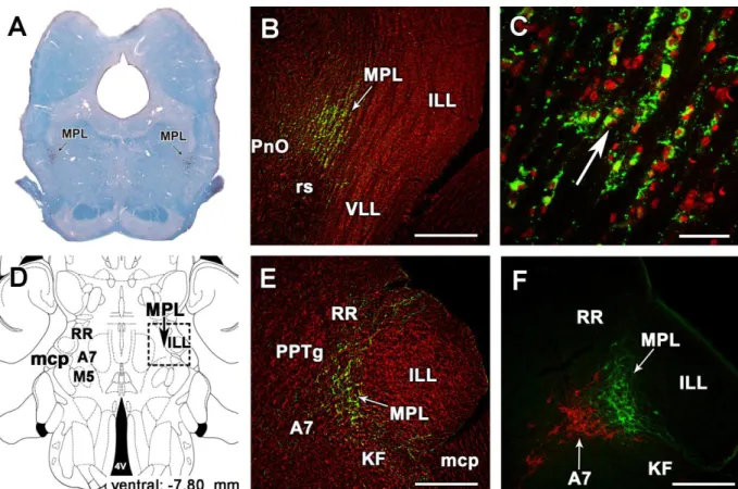
6.2.3. The distribution of TIP39 neurons in the medial paralemniscal nucleus (MPL) .. 51
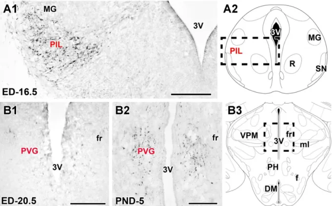
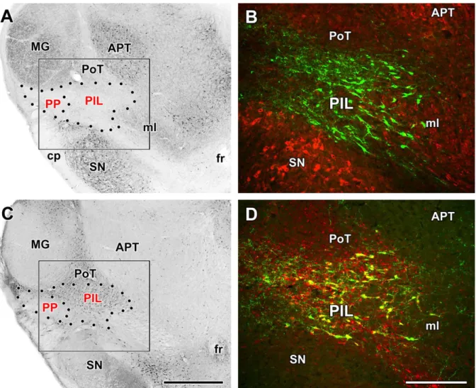
6.2.4. Identification of a third group of TIP39 neurons, the posterior intralaminar complex of the thalamus (PIL) ... 53
6.3. TIP39- and PTH2 receptor-containing neuronal networks and fibers ... 56
6.3.1. TIP39- and PTH2 receptor-containing neuronal networks and fibers in rodents . 56 6.3.2. Distribution of PTH2 receptor-expressing cell bodies ... 60 6.3.3. Mapping of the expression of the PTH2 receptor in the human brain by
5
RT-PCR ... 62
6.3.4. Distribution of PTH2 receptor immunoreactivity in the human brain ... 63
6.4. Neuronal connections of TIP39 neurons ... 68
6.4.1. Mapping of the disappearance of TIP39 fibers following selective lesion of TIP39 cell groups ... 68
6.4.2. Retrograde labeling of TIP39 neurons following injections into the arcuate nucleus and the medial preoptic area ... 75
6.4.3. Afferent neuronal connections of the PIL ... 77
6.4.4. Afferent neuronal connections of the MPL ... 79
6.5. Assessment of c-Fos activation in TIP39 neurons of lactating dams ... 82
6.5.1. Fos activation in the brain in response to suckling ... 82
6.5.2. Suckling-induced Fos expression in TIP39 neurons of the PIL ... 82
6.5.3. Suckling-induced Fos expression in TIP39 neurons of the MPL ... 84
6.6. The effect of the PTH2 receptor block on the plasma prolactin level ... 86
6.6.1. Effect of intracerebroventricular injection of a PTH2 receptor antagonist on suckling-induced prolactin secretion ... 86
6.6.2. Effect of local viral production of a PTH2 receptor antagonist on suckling-induced prolactin secretion ... 87
6.7. Preoptic actions of TIP39 ... 89
6.7.1. Evaluation of maternal motivation after preoptic antagonism of the PTH2 receptor ... 89
6.7.2. Association of PIL TIP39 neurons with suckling-activated preoptic neurons ... 91
6.8. Induction of TIP39 in mother rats ... 93
6.8.1. Alterations of TIP39 mRNA expression in mother rats ... 93
6.8.2. Increased TIP39-immunoreactivity in the brain of rat dams ... 95
6.8.3. Measurement of TIP39 mRNA level in mother rats with real-time RT-PCR ... 97
6.8.4. Levels of TIP39 mRNA in the PIL of pregnant females and postpartum rat dams.99 6.9. Amylin as the gene with the most salient expressional change in the preoptic area ……….101
6
6.9.1. Genes with altered mRNA expression in the preoptic area of rat dams ... 101
6.9.2. RT-PCR validation of the induction of amylin in the preoptic area of rat dams 102 6.10. Amylin neurons in the preoptic area ... 103
6.10.1. The distribution of amylin mRNA in the female rat brain ... 103
6.10.2. Time course and distribution of amylin mRNA expression in the peri- and postpartum periods in preoptic amylin neurons ... 105
6.10.3. Amylin-immunoreactivity in the preoptic area of the female rat brain ... 107
6.11. Pup exposure-induced activation of amylin-neurons in the preoptic area of rat dams ... 108
6.11.1. Fos-labeled amylin mRNA-expressing neurons in the preoptic area in response to suckling ... 108
6.11.2. Fos-labeled amylin-immunoreactive neurons in the preoptic area in response to suckling ... 109
6.12.The connection between TIP39 and amylin neurons ... 111
6.12.1. Innervation of amylin-immunoreactive neurons by TIP39 fibers ... 111
6.12.2. The maternal induction of amylin in mice lacking the PTH2 receptor ... 112
7. DISCUSSION……….114
7.1. TIP39 in the central nervous system ... 114
7.1.1. TIP39 neurons in the PIL ... 114
7.1.2. TIP39 neurons in the PVG ... 115
7.1.2. TIP39 neurons in the MPL ... 116
7.2. The TIP39-PTH2 receptor neuromodulator system ... 118
7.2.1. Comparison of the distribution of TIP39 to that of the PTH2 receptor provides anatomical evidence for a TIP39-PTH2 receptor neuromodulator system ... 119
7.2.2. PTH2 receptor distribution in the human brain ... 120
7.3. TIP39 functions in the maternal brain ... 120
7.3.1. Maternal induction of TIP39 ... 120
7.3.2. Activation of TIP39 neurons in reponse to pup exposure ... 121
7
7.3.3. The involvement of the TIP39-PTH2 receptor system in the regulation of prolactin
release ... 122
7.3.4. The involvement of the TIP39-PTH2 receptor system in the regulation of maternal motivation ... 123
7.3.5. TIP39 neurons in the PIL as relay stations of suckling information towards the hypothalamus ... 124
7.3.6. Additional potential functions of thalamic TIP39 neurons ... 126
7.3.7. Potential role of TIP39 neurons of the MPL in auditory information transfer between rat mothers and pups ... 127
7.4. Amylin as a novel neuropeptide ... 127
7.4.1. Induction of amylin in the preoptic area ... 128
7.4.2. Maternal activation of preoptic amylin neurons ... 128
7.4.3. Amylin as a novel neuropeptide potentially involved in maternal control ... 129
7.4.4. The maternal functions of amylin ... 130
8. SUMMARY………132
9. GRANT SUPPORT………...133
10. ADDITIONAL ACKNOWLEDGEMENTS………...134
11. REFERENCES………..135
8 1. ABSTRACT
The discovery and functional characterization of 2 neuropeptides are reported.
Tuberoinfundibular peptide of 39 residues (TIP39) was previously purified as the endogenous ligand of the so far orphan parathyroid hormone 2 receptor (PTH2 receptor) but we first identified the gene that encodes TIP39, and discovered its maternal functions. The other neuropeptide is amylin, whose gene had been described previously and shown to be expressed in the pancreas. We first determined its expression in the brain, therefore, the status of amylin as a neuropeptide and provided strong evidence for its involvement in maternal regulations.
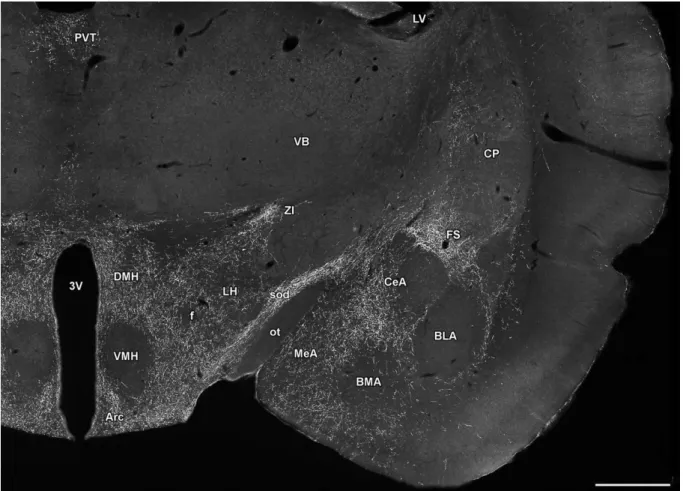
TIP39 is predominantly expressed in the brain, in 3 different sites. While the expression level of TIP39 is diminished postnatally, its expression is induced in mother rats in 2 of the 3 sites of its expression, the posterior intralaminar complex of the thalamus (PIL), and the medial paralemniscal nucleus (MPL). The distribution of TIP39 fibers is similar to that of PTH2 receptor in the brain. The PIL projects mostly to hypothalamic brain regions such as the preoptic area and the arcuate nucleus while the MPL provides TIP39 predominantly to brainstem auditory regions. We also showed that the PIL receives ascending input from the spinal cord while the MPL has afferent neuronal connections with auditory brain regions.
TIP39 neurons in both the PIL and MPL were activated by pup-exposure in mother rats.
Suckling-induced elevation of serum prolactin was reduced by a PTH2 receptor antagonist applied into the lateral ventricle or locally into the arcuate nucleus but not the preoptic area by means of focal virus infection. In turn, local PTH2 receptor antagonist in the preoptic area reduced maternal motivation without affecting prolactin levels. These findings are in agreement with literature data that the preoptic area is the central regulator of maternal behaviors while prolactin secretion required for lactation is regulated by neurons in the arcuate nucleus. The results also provide the long sought-after reflex arch how suckling information reaches the hypothalamus to affect maternal behaviors and hormonal changes. To find targets of TIP39 in the preoptic area, a microarray study was performed, which identified amylin as a novel neuropeptide expressed only in the maternal preoptic area. Amylin neurons are also activated by suckling. They have a distribution similar to that of TIP39 fibers within the preoptic area, and are closely apposed by TIP39 terminals. Furthermore, the maternal induction of amylin is reduced in mice lacking the PTH2 receptor.
The 2 newly identified maternal peptides in the brain are unique among neuropeptides in the high degree of induction in mothers. Together with their receptor, they represent important new mechanisms in the control of central maternal adaptation and future targets for the development of drugs affecting specific maternal disorders such as postpartum depression.
9 2. LIST OF ABBREVIATIONS
ac anterior commissure AcN accumbens nucleus
AH anterior hypothalamic area
AHi amygdalo-hippocampal transition zone al ansa lenticularis
AM anteromedial thalamic nucleus
AN accumbens nucleus
AON anterior olfactory nucleus APT anterior pretectal nucleus aq cerebral aqueduct
Arc arcuate nucleus
Au1 primary auditory cortex
AuV secondary auditory cortex, ventral area AVPe anteroventral periventricular nucleus A7 A7 noradrenergic cell group
A11, A13 A11, A13 dopaminergic cell group BDA biotinylated dextran amine
BL basolateral amygdaloid nucleus
BLA basolateral amygdaloid nucleus, anterior part BMA basomedial amygdaloid nucleus, anterior part BNST bed nucleus of the stria terminalis
BNSTv ventral subdivision of the BNST
BSTMPM posteromedial part of the medial subdivision of the BNST cAMP cyclic adenosine 3 5'-monophosphate
ca cerebral aqueduct
cc corpus callosum
CeA central amygdaloid nucleus CG periaqueductal central gray CGRP calcitonin gene-related peptide
CIC central nucleus of the inferior colliculus CM central medial thalamic nucleus
CP caudate putamen
Cu cuneate nucleus
CuF cuneiform nucleus
CoA cortical amygdaloid nucleus cp cerebral peduncle
CRH corticotropin-releasing hormone CTB cholera toxin β subunit
cu cuneate fasciculus
DA dopamine
10
DG dentate gyrus
DH dorsal horn
Dk nucleus of Darkschewitsch dlf dorsolateral fasciculus
DM(H) dorsomedial hypothalamic nucleus DpMe deep mesencephalic nucleus DR dorsal raphe nucleus
DTg dorsal tegmental nucleus
ECIC external cortex of the inferior colliculus EGP external globus pallidus
f fornix
fr fasciculus retroflexus FS fundus striati
GABA γ-amino-butyric acid
GAD glutamic acid decarboxylase
GAPDH glyceraldehyde 3-phosphate dehydrogenase GFP green fluorescent protein
GnRH gonadotropin-releasing hormone Gr gracile nucleus
Hipp hippocampus
IC inferior collicle
IGP internal globus pallidus
ILL intermediate nucleus of the lateral lemnsicus Inf infundibular nucleus
IPF interpeduncular fossa KF Kölliker-Fuse nucleus L2 lamina 2 of the dorsal horn LG lateral geniculate body LH lateral hypothalamic area ll lateral lemniscus
LPAG periaqueductal gray, lateral subdivision LS lateral septal nucleus
LSv ventral subdivision of the lateral septal nucleus LV lateral ventricle
mcp middle cerebellar peduncle MD mediodorsal thalamic nucleus
me median eminence
MeA medial amgydaloid nucleus mfb medial forebrain bundle MG medial geniculate body MGN medial geniculate nucleus
ml medial lemniscus
11 mlf medial longitudinal fasciculus
MMG medial nucleus of the medial geniculate body MMN medial mamillary nucleus
MPA medial preoptic area
MPL medial paralemniscal nucleus MPN medial preoptic nucleus mt mamillothalamic tract M5 motor trigeminal nucleus NAcc nucleus accumbens och, ox optic chiasm OT olfactory tubercle
ot optic tract
PAG periaqueductal gray PB phosphate buffer PBG parabigeminal nucleus PBL lateral parabrachial nucleus pc posterior commissure
Pe hypothalamic periventricular nucleus PF parafascicular thalamic nucleus PH posterior hypothalamic nucleus
PIL posterior intralaminal complex of the thalamus Pir piriform cortex
Pn pontine nuclei
PnO pontine reticular nuclei Po posterior thalamic nucleus POA preoptic area
PoT triangular subdivision of the posterior thalamic nucleus PP(N) peripeduncular nucleus
PPTg pedunculopontine tegmental nucleus PrC precommissural nucleus
Prh perirhinal cortex
PRL prolactin
PTH parathyroid hormone
PTHrP parathyroid hormone-related peptide
Pu putamen
Pul pulvinar thalami
PVG periventricular gray of thalamus PVN hypothalamic paraventricular nucleus PVT paraventricular thalamic nucleus py pyramidal tract
R red nucleus
Re reuninens thalamic nucleus
12 Reth retroethmoid nucleus rs rubrospinal tract RM nucleus raphe magnus RR retrorubral nucleus Rt reticular thalamic nucleus Sag sagulum nucleus
SC superior colliculus SCh suprachiasmatic nucleus scp superior cerebellar peduncle SI susbstantia innominata sm stria medullaris
SN substantia nigra SO supraoptic nucleus SOC superior olivary complex sod supraoptic decussations SPA subparafascicular area SPF subparafascicular nucleus
SPFm subparafascicular nucleus, magnocellular part SPFp subparafascicular nucleus, parvicellular part Sp5 spinal trigeminal nucleus
STh subthalamic nucleus
Sub subiculum
TIP39 tuberoinfundibular peptide of 39 residues TH tyrosine hydroxylase
TIDA tuberoinfundibular dopaminerg neurons TRH thyrotropin releasing hormone
VA ventroanterior thalamic nucleus VB ventrobasal complex of the thalamus VLL ventral nucleus of the lateral lemiscus
VMG ventral nucleus of the medial geniculate body VMH hypothalamic ventromedial nucleus
VPL ventral posterolateral thalamic nucleus VPM ventral posteromedial thalamic nucleus
VPPC ventral parvicellular posteromedial thalamic nucleus VTA ventral tegmental area
ZI zona incerta
3V third ventricle 4n trochlear nerve 4V fourth ventricle 5n trigeminal nerve
5th root of the trigeminal nerve
7n facial nerve
13 3. INTRODUCTION
3.1. Significance of the topic
There are several reasons why studying the maternal nervous system is important.
First, motherhood is an outstanding element of humanity, one that has attracted artists for centuries and has crucial significance in society. However, its scientific investigation lags behind its importance. Second, the maternal brain is a model of changes of the adult nervous system. The most remarkable adult physiological and behavioral changes take place during motherhood. Finally, a dam (rodent mother) represents an easily reproducible subject to model emotional motivation and social interactions. Maternal instinct to take care of the pups could also be considered as a model of love and self-sacrifice, feelings that are difficult to approach experimentally. Furthermore, the dysfunctions of maternal adaptations could also be studied in animal models, which could lead to the understanding of postpartum depression, the most frequent psychiatric disorder after childbirth with a prevalence rate of 10% to 15%
(Abou-Samra et al., 1992; Mallikarjun and Oyebode, 2005). The high percentage of depression in the postpartum period and the deteriorated mother-infant communication as an early sign of maternal depression (Breese McCoy, 2011) suggest a causal relationship between dysfunctions of maternal adaptation and the onset of depression.
Neuropeptides are signaling molecules in the nervous system and typically have slow actions but play important neuromodulatory functions during adaptive processes. Therefore, they represent a group of regulatory molecules, which could have important maternal functions in the female brain. Indeed, this aspect of some peptides has been intensively investigated in the past but none of them has been shown to be selectively expressed in the maternal brain. Therefore, we focused our attention to peptides that – at least in some brain sites – demonstrate markedly enhanced expression in the postpartum period as they can reveal important mechanisms of regulations in the maternal brain.
3.2. Maternal alterations in brain physiology
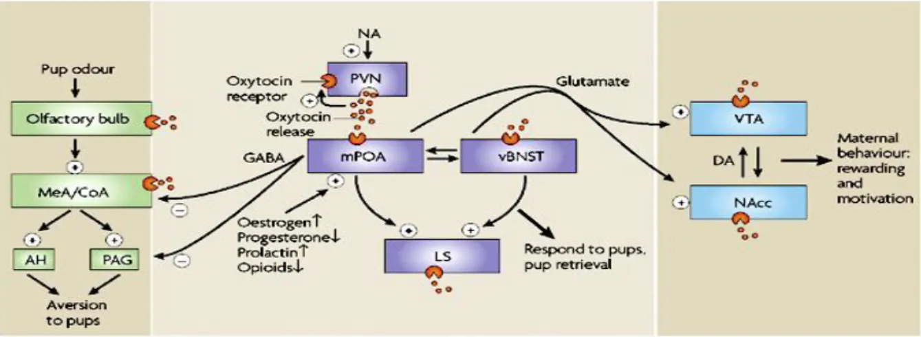
Behavioral, endocrine, and psychological changes in mothers represent one of the most profound physiological alterations in the adult central nervous system (Fig.1). Experimental models of maternal behaviors are well established in rodents as control females avoid or even hurt pups while mothers take care of them: retrieve them in the nest, nurse them, perform anogenital licking of pups etc (Numan and Insel, 2003). Additional emotional changes include maternal aggression towards intruders, decreased anxiety in general, and reduced
14
responsiveness of the hypothalamo-pituitary-adrenal axis in stress situations (Carter et al., 2001; Neumann, 2003). Some other behavioral changes support the increased food and fluid intake required for lactation (Knobil and Neill, 2006). Major endocrine alterations include prolactin and oxytocin release to support lactation, and the suppression of gonadotropin- releasing hormone (GnRH) secretion leading to lactational anoestrous.
Fig. 1. The summary of adaptive responses to motherhood (Russell et al., 2001).
The mechanisms of maternal adaptations are not well established but are not likely to be simply mediated by hormonal changes. Although the behavioural changes are initiated by hormonal alterations in the last days of pregnancy (Bridges, 1996; Siegel, 1986) decreased levels of these hormones are detected during lactation (Lamming, 1994). In addition, the full range of maternal behaviors can be induced by prolonged pup exposure even in ovariectomized virgin females. These maternally sensitized rats do not lactate and provide a model to separate metabolic regulations from regulations of maternal behaviors (Rosenblatt,
15
1967). Somatosensory inputs derived from the pups play the most important role in sensitization (Stern, 1989). The same somatosensory inputs are thought to maintain maternal behaviours in dams after parturition (Febo et al., 2008). Lesion and microstimulation studies suggested that the reflex arch ascends conveying information from the nipples travels through the lateral mesencephalic tegmentum and enter the zona incerta ventromedial to the medial geniculate body (Dubois-Dauphin et al., 1985; Juss and Wakerley, 1981; Tindal and Knaggs, 1977; Wakerley et al., 1978). Excitotoxic lesions of this area blocked the milk-ejection reflex (Hansen and Kohler, 1984) and c-fos expression was detected here in lactating mothers (Lin et al., 1998) suggesting relay of the pathway in this position. It remained to be established if this pathway participates only in the neuroendocrine responses of prolactin and oxytocin release or also in more general maternal influences on brain adaptations.
The regulation and coordination of maternal adaptations take place in the central nervous system. A number of different approaches support that the preoptic area plays a crucial role in the initiation and maintenance of maternal behaviours. Large electrical as well as axon-sparing excitotoxic lesions of the MPOA eliminate all maternal behaviours without affecting other, e.g. feeding behaviours (Numan and Woodside, 2010) and similar effects were found following temporal pharmacological inactivation of MPOA (Arrati et al., 2006;
Pereira and Morrell, 2009). In contrast to lesions, electrical stimulation of the MPOA increased maternal responsiveness (Morgan et al., 1997). In addition, brain activity is elevated in the MPOA in response to pup exposure based on c-fos (Li et al., 1999b; Lonstein et al., 1998b), 2-deoxyglucose (Del Cerro et al., 1995) and fMRI techniques (Febo et al., 2005).
Subsequent lesion and activation studies suggest that other brain regions may also be involved in specific aspects of maternal behaviors including the lateral septum, the bed nucleus of the stria terminalis, the medial and cortical amygdaloid nuclei, the anteroventral periventricular nucleus, hypothalamic paraventricular nucleus, parts of the periaqueductal gray, the accumbens nucleus and the ventral tegmental area (Fig. 2).
16
Fig. 2. Brain centers involved in maternal behaviors and motivation (Brunton and Russell, 2008). Abbreviations: AH – anterior hypothalamic area; CoA – cortical amygdaloid nucleus;
DA – dopamine; MeA - medial amygdaloid nucleus; mPOA – medial preoptic area; NAcc – accumbens nucleus; PAG – periaqeductal gray; PVN – hypothalamic paraventricular nucleus;
vBNST – ventral subdivision of the bed nucleus of the stria terminalis; VTA – ventral tegmental area.
3.3. Neuropeptides
Neuropeptides consist of 3-50 amino acids (aa) encoded by the genome.
Neuropeptides are always cleaved enzymatically by prehormone convertases in the endoplasmic reticulum and the Golgi apparatus, and often undergo additional posttranslational modifications such as amidation, pyroglutamate formation, etc. (Hökfelt et al., 2000).
Neuropeptides are then packed into so-called dense core vesicles and transported to the presynaptic terminals (Bean et al., 1994). Importantly, these vesicles are not released by single action potentials or low-frequency stimulation even though small vesicles containing neurotransmitters are released by this stimulus at the same synapse (Hökfelt et al., 1992).
Thus, neuropeptides typically have no effect on the basal function of the network (Hökfelt et al., 1987). In turn, high-frequency stimulation leads to the secretion of a large amount of neuropeptides, which can act locally or more distantly by diffusion. Neuropeptides always act via high-affinity G-protein coupled receptors (Baraban and Tallent, 2004). There is not a single neuropeptide the receptor of which is a ligand-gated ion channel in vertebrates. Some proteins considered neuropeptides by some researchers, e.g. prolactin and leptin, have single transmembrane domain tyrosine-kinase receptors. However, it is more logical to not consider
17
these signal proteins as neuropeptides. Thus, all neuropeptides possess 7 transmembrane domain receptors, which belong to 2 subgroups (Fredholm et al., 2007). Typically, the receptors of small neuropeptides (3-30 aa) are class I, rhodopsin-like G-protein coupled receptors while larger neuropeptides (25-50 aa) possess class II, secretin-like G-proteins coupled receptors (Jacoby et al., 2006). Neuropeptides are eliminated from the extracellular space by proteolytic degradation. After neuropeptides are released from the presynaptic terminal, they are not taken back by the terminals and the large dense core vesicles cannot be replenished (Bean et al., 1994). Instead, newly synthesized neuropeptide packages arrive from the cell body, a process, which takes a significant amount of time, several hours or even days (van den Pol, 2012). These characteristics require a significant induction of neuropeptide expression during recruitment, e.g. in the postpartum period for maternal neuropeptides.
3.4. Neuropeptides in maternal regulations
A number of neuropeptides play important roles in different aspects of maternal regulations:
Prolactin secretion into the circulation is essential to the production of milk in the mammary gland of lactating mothers (Neville, 2006). Prolactin also stimulates maternal behaviors, reduces the stress response and anxiety, participates in the suppression of fertility, and the regulation of appetite and body weight (Grattan and Kokay, 2008). Prolactin release from anterior pituitary lactotrop cells is primarily regulated by inhibitory dopamine neurons in the hypothalamus (Freeman et al., 2000) but is also affected by several neuropeptides (Bodnar et al., 2009). The regulatory roles of thyrotropin releasing hormone (Lyons et al., 2010;
Yamada et al., 2006) and opioid peptides (Arbogast and Voogt, 1998; Callahan et al., 2000;
Selmanoff and Gregerson, 1986; Tavakoli-Nezhad and Arbogast, 2010) are best established.
The expression level of thyrotropin releasing hormone is increased (Fjeldheim et al., 2005) while that of the opioid peptides are decreased during lactation (Kim et al., 1997).
Suckling also evokes the secretion of oxytocin synthesized in the paraventricular hypothalamic nucleus and secreted from the neural lobe of the pituitary to induce milk ejection (Burbach et al., 2006; Wakerley et al., 1978). However, oxytocin produced and released in different brain sites including the medial and central amygdaloid nuclei is an established regulator of emotional and social behaviors and also plays a role in the regulation of maternal behaviors and reduced responsiveness of the HPA axis (Neumann, 2008; Slattery and Neumann, 2008; Terenzi and Ingram, 2005).
18
The demand of milk production creates negative energy balance, which, however, does not account for the entire hyperphagia during lactation (Smith and Grove, 2002; Xu et al., 2009). Increased prolactin levels and the suckling stimulus may also play a role in the increased food intake (Chen and Smith, 2003; Woodside, 2007). Peptide products of proopiomelanocortin and other peptides involved in food intake regulation (Meister, 2007;
Palkovits, 2003) may play a role in the elevated food intake during pregnancy and lactation (Crowley et al., 2007; Ladyman et al., 2010). Neuropeptide Y induced in the dorsomedial hypothalamic nucleus (Chen and Smith, 2003) may have a specific role in sustaining the chronic hyperphagia of lactation (Xu et al., 2009) and it might also participate in the suppression of GnRH secretion during lactation (Pralong, 2010; Wojcik-Gladysz and Polkowska, 2006).
The suppression of GnRH secretion leading to reduced estrogen serum level and lactational anoestrous is characteristic in mothers (McNeilly, 2006). The central common pathways for the regulation of GnRH neurons are kisspeptin neurons (Oakley et al., 2009) located in the anteroventral periventricular and arcuate nuclei (Lehman et al., 2010). All factors influencing estrogen levels, such as stress, suckling, body weight, estrogen itself, etc.
are proposed to act via the kisspeptin system (Pineda et al., 2011). Lactational anoestrus is primarily driven by the suckling stimulus during the first half of lactation and by negative energy balance during its second half (Tsukamura and Maeda, 2001). The kisspeptin system was suggested to mediate both actions (Smith et al., 2010; Topaloglu et al., 2010)) at least partially through reduced kisspeptin expression in the postpartum period (Yamada et al., 2007).
Stress coping strategies are also altered during late pregnancy and postpartum: mother rats demonstrate decreased anxiety and reduced responsiveness of the hypothalamo-pituitary- adrenal (HPA) axis including diminished synthesis and release of corticotropin releasing hormone (Carter et al., 2001; Neumann, 2001; 2003). Suckling was suggested to drive the hyporesponsiveness of the HPA axis in the postpartum period (Brunton et al., 2008) even though suckling itself rapidly increases adrenocorticotropin and corticosterone secretion (Walker et al., 1992).
Postpartum behavioral changes in females are also critically important parts of mammalian reproduction (Knobil and Neill, 2006). Although sexual steroid may contribute to the development of maternal behaviors by reducing inhibition of olfactory origin (Numan and Insel, 2003), decreased levels of these hormones are detected during lactation (Lamming, 1994) and the full range of maternal behaviors can be induced by prolonged pup exposure
19
even in ovariectomized virgin females (Fleming and Rosenblatt, 1974; Rosenblatt, 1967) suggesting that suckling or at least the presence of pups is necessary for maintaining maternal behaviors (Numan and Woodside, 2010). The preoptic area of hypothalamus plays a pivotal role in the regulation of maternal behaviors based on their absence following preoptic lesions (Gray and Brooks, 1984; Numan, 1986) and the large number of neurons in the preoptic area exhibiting Fos activation in postpartum dams (Lonstein et al., 1998b; Stack and Numan, 2000). Other brain regions connected to the preoptic area also participate in the regulation of different aspects of maternal behaviors. For example, the ventrolateral subdivision of the caudal periaqueductal grey has been shown to regulate kyphosis, the most effective body posture in rats for suckling pups, and maternal aggression (Lonstein et al., 1998a). Prolactin, oxytocin and opioids have been implicated in the control of maternal behaviors (Numan, 2006). More recently, the role of vasopressin (Bosch et al., 2010) and melanin-concentrating hormone (Rondini et al., 2010) have also been proposed.
In summary, it is established that reduced kisspeptin expression and consequently low gonadotropin-releasing hormone secretion are responsible for lactational anoeastrus and that profoundly elevated prolactin and oxytocin secretion lead to milk production and ejection, respectively. What we do not know how these signals are regulated in mothers, whether and how they are driven by the suckling stimulus. Furthermore, the control of other aspects of maternal adaptations including maternal motivation and behaviors are less well understood.
3.5. Tuberoinfundibular peptide of 39 residues
Tuberoinfundibular peptide of 39 residues (TIP39) was purified on the basis of its activation of the parathyroid hormone 2 receptor (PTH2 receptor), a seven transmembrane domain G-protein coupled receptor (Usdin et al., 1999b). The distribution of TIP39 containing fibers and terminals is very similar to the distribution of PTH2 receptor-containing neurons and neuronal fibers throughout the brain (Dobolyi et al., 2003b; Faber et al., 2007), and TIP39 is a potent and selective PTH2 receptor agonist (Usdin, 2000). These functional and anatomical matches suggest that TIP39 is the endogenous ligand of the PTH2 receptor in the brain, and that they form a neuromodulator system. TIP39 neurons have a highly restricted localization. This pattern of synthesis by cells in a few discrete areas and widespread, but still topographically organized, projections to several distant brain areas resembles several other recently developed neuropeptide systems including, for example, relaxins (Ma and Gundlach, 2007), orexins (Baumann and Bassetti, 2005), calcitonin-gene related peptide (van Rossum et al., 1997), prolactin-releasing peptide (Roland et al., 1999), kisspeptin (Mikkelsen and
20
Simonneaux, 2009), and urocortins (Pan and Kastin, 2008). Based on the available data, however, the TIP39-PTH2 receptor system is a unique neuropeptide-receptor system whose localization and functions in the central nervous system are different from any other neuropeptides (Dobolyi et al., 2010).
TIP39 is a member of a small peptide family comprised of parathyroid hormone (PTH), parathyroid hormone-related peptide (PTHrP) and TIP39 (Usdin et al., 1999b). Mature PTH and PTHrP are polypeptides of about 100 residues. They are products of separate genes but they activate the parathyroid hormone 1 receptor (PTH1 receptor) with equal potency (Gensure et al., 2005; Muff et al., 1994). Their first 34 or 36 residues are sufficient for high- affinity binding and full efficacy at the PTH1 receptor, and they share 12 of these amino acids (Gillespie and Martin, 1994; Martin et al., 1991). TIP39 contains only four of the residues that are common to PTH(1–34) and PTHrP(1–36), and several additional similar residues (Usdin et al., 1999b). However, TIP39 has a backbone structure that can be nearly superimposed on that of PTH (Piserchio et al., 2000). Based on the similarity of TIP39 to PTH and its activation of the PTH2 receptor, TIP39 is referred to as parathyroid hormone 2 in the UniGene database at http://www.ncbi.nlm.nih.gov/sites/entrez (Mm.207078 for the mouse and Hs.339845 for the human gene). However, this name is also used for a second form of PTH found in fish that more closely resembles mammalian PTH than does TIP39 (Gensure et al., 2004). PTH, produced by the parathyroid gland, is the most important regulator of calcium homeostasis. It increases plasma calcium ion concentration via direct actions in the kidney and the skeleton (Hurwitz, 1996; Rizzoli et al., 1992). PTHrP is a paracrine factor that functions in a number of organs and plays a critical role in skeletal development (Law et al., 1994; Martin et al., 1997). TIP39 and the PTH2 receptor are expressed at very low levels in kidney and bone (Usdin et al., 1996; Usdin et al., 1999a). In the periphery, they may play a role in the cardiovascular system (Eichinger et al., 2002; Ross et al., 2005; Ross et al., 2007) and in gonadal function (Usdin et al., 2008). However, TIP39 and the PTH2 receptor are most abundant in the central nervous system (Dobolyi et al., 2002; Usdin et al., 1995).
3.6. Amylin
Amylin, a peptide of 37 amino acids, belongs to the calcitonin gene-related peptide (CGRP) family, which also includes calcitonin, CGRPα, CGRPβ, adrenomedullin, and intermedin / adrenomedullin 2 (Wimalawansa, 1997). This family of peptides has a unique pharmacology. The binding specificity of 2 ´family B´ G-protein-coupled receptors, the calcitonin receptor and the calcitonin receptor-like receptor depends on additional proteins,
21
the 3 receptor activity-modifying proteins (RAMP1-3), which are single transmembrane domain accessory proteins (Oliver et al., 2001; Poyner et al., 2002). Each RAMP has different distribution in the brain (Oliver et al., 2001), and form 3 pharmacologically distinct amylin receptors with the calcitonin receptor (Hay et al., 2004). RAMPs also form CGRP and adrenomedullin receptors in association with the calcitonin receptor-like receptor (Poyner et al., 2002).
In early distributional studies, amylin expression had only been detected in the pancreatic B-cells and not in the brain (Leffert et al., 1989; Lutz, 2006). More recently, the expression of amylin has also been reported in neurons of sensory ganglia (Mulder et al., 1995) where it might have a role in nociception (Gebre-Medhin et al., 1998). Plasma amylin derived from co-secretion with insulin from pancreatic B-cells contributes to meal-ending satiation (Lutz, 2010; Osto et al., 2007; Rushing et al., 2000) via its action on the region of the area postrema and the nucleus of the solitary tract (Potes and Lutz, 2010). In addition, amylin might also act as an adiposity signal (Lutz, 2010; Rushing et al., 2001).
22 4. OBJECTIVES
Several aspects of motherhood has been investigated and recognized in the past.
Hormonal changes in mothers have long been examined by reproductive endocrinologist, e.g.
we have significant knowledge on the dopaminergic control of prolactin release. Obstetricians and gynecologist have of course always catered for mothers. Psychological aspects of motherhood have also been addressed in appropriate scientific fields. However, despite the recent progress in the field, the regulatory mechanisms within the maternal brain regions remained largely obscure. In particular, we do not have sufficient information on the neuropeptides related to motherhood and their specific actions. Apart from neurohypophyseal oxytocin, there are no peptides known to dramatically increase their expression during motherhood. In addition, the neuronal circuitry that regulates maternal responses is not known, the anatomical pathways and neurochemical characteristics of the reflex arch that mediates the effect of suckling to forebrain centers remain to be elucidated. Therefore, the general objectives of these studies were to identify maternally expressed neuropeptides and describe the neuronal circuitry that regulates maternal responsiveness. This novel approach to understand maternal adaptations was to be performed using a wide array of advanced methodological repertoire.
4.1. The specific objectives of the study
Identify the gene encoding TIP39 and describe the distribution of its expression
The gene and mRNA encoding TIP39 was to be analyzed followed by the distribution of the mRNA in different organs as well as in different brain regions using RT-PCR and in situ hybridization. After characterizing a newly developed antibody against TIP49, the distribution of the peptide in cell bodies as well as neuronal fibers was also addressed.
Determine TIP39 expression levels during ontogenic development and the reproductive cycle
TIP39 mRNA as well as peptide levels were to be assessed in all 3 regions of expression of TIP39, the periventricular gray of the thalamus (PVG), the posterior intralaminar complex of the thalamus (PIL), and the medial paralemniscal nucleus (MPL) in the lateral pons, during embryonic and postnatal development, puberty, pregnancy and the postpartum period.
23
Describe the afferent and efferent neuronal connections of TIP39 neurons activated in mothers in the PIL and MPL
The activation of TIP39 neurons in mother rats was to be investigated by the Fos technique. The neuronal connections of activated TIP39 neurons in the PIL and MPL were to be examined using retrograde neuronal tracers as well as pathway transection methods.
Most experiments were designed to be performed in mothers in order to visualize otherwise not visible TIP39 neurons and fibers.
Map the distribution of the receptor of TIP39, the PTH2 receptor in the brain of rodents, macaque and human
The distribution of PTH2 receptor expression in the brain was to be determined by in situ hybridization histochemistry in macaque and mouse, and X-gal histochemistry in mice expressing -galactosidase driven by the promoter of the PTH2 receptor. The protein was to be visualized by immunohistochemistry both in rodents and postmortem human brain samples. The distribution of the PTH2 receptor is compared between different species as well as to the distribution of TIP39 fibers and fiber terminals.
Measure the effects of antagonizing the PTH2 receptor in mothers on prolactin secretion and maternal motivation
A PTH2 receptor antagonist was to be delivered into the brain by 2 different means: 1) acute injection into the lateral ventricle, 2) infection of a localized group of cells by a virus that codes the PTH2 receptor antagonist. Serum prolactin levels were to be measured in response to suckling. Maternal motivation was evaluated in mothers with infected cells in the preoptic area using a conditioned place preference test when the mothers can choose between a pup-associated and a control cage.
Identify maternally changing peptides in the preoptic area of hypothalamus and characterize their interaction with the TIP39-PTH2 receptor system
A microarray experiment was designed to determine genes whose mRNA level is increased 9 days postpartum in lactating mother rats as compared to mothers deprived of their litter on the day of delivery. The highest, over 25 times increase was found for amylin, a novel neuropeptide. Therefore, the distribution of amylin-expressing cells, their innervation by TIP39, and their activation in response to suckling were addressed.
24 5. MATERIALS AND METHODS
5.1. Experimental subjects and tissues 5.1.1. Rodents
Some procedures were performed according to approved National Institutes of Mental Health (Bethesda, MD, USA) animal care protocols, and in accordance with the National Institutes of Health Guide for the Care and Use of Laboratory Animals. Other experiments were approved by the Semmelweis University, Budapest, and Animal Examination Ethical Council of the Animal Protection Advisory Board. Procedures involving rats were carried out in accordance with the Hungarian Ministry of Agriculture’s Animal Hygiene and Food Control Department guidelines for experimental protocols and with EU Directive 2010/63/EU for animal experiments. Female and male Wistar rats and C57Bl/6 mice (Charles Rivers Laboratories, Hungary) were used in this study. All efforts were made to minimize the number of animals used and their suffering. Animals were kept on standard laboratory conditions with 12-h light, 12-h dark periods (lights on at 6.00 a.m.), and supplied with dry rat food and drinking water ad libitum. Animals were housed 3 per cage at a temperature of 22±1 °C before experiments. For mating, 2 female and a male rat were kept in a cage for 5 days. After that, potentially pregnant females as well as dams with litter and their pup- deprived and control experimental counterparts were kept in cages individually. Mother rats delivered their pups on day 22 of pregnancy. Mothers who delivered fewer than 8 pups or whose pups died were excluded from the study. The number of pups was adjusted to 8 within 2 days of delivery. For surgery, perfusions, and dissections, the animals were anesthetized with an intramuscular injection of anesthetic mix containing 0.2ml/300g body weight ketamine (100 mg/ml) and 0.2ml/300g body weight xylazine (20 mg/ml).
5.1.2. Knock-in mice expressing -galactosidase driven by the PTH2 receptor promoter The first protein coding exon of the mouse PTH2 receptor gene was replaced by the bacterial lacZ coding sequence by homologous recombination in F1 (129Sv/Ev x c57Bl/6) hybrid mouse ES cells, as previously described (Valenzuela et al., 2003). Following identification of ES cells with appropriate recombination by the Regeneron group, the cells were expanded and introduced into c57Bl/6 blastocysts in the NIMH transgenic mouse core facility. Resulting mice were back-crossed with C57Bl/6 mice. Heterozygous animals were used for the experiments. Knock-in mice were identified by PCR based genotyping on DNA from tail biopsies using primers for lacZ or one primer in the inserted sequence and one
25
primer in the flanking gene sequence (Faber et al., 2007). The primer sequences were: 5’- GCGCTGGTTGATTAGATACC, P2R_HDR2-B; 5’-GCTTCCTCGTGCTTTACGGTATC, Neo_3’b; and 5’-GAGAGGCTGTTTGTAGAAGGCTGA, P2R_64264U which produced bands of 260 base pairs from the wild-type allele and 700 from the knock-in allele.
5.1.3. PTH2 receptor knock-out mice
Mice with a null mutation of the PTH2 receptor were generated and characterized in a previous study (Coutellier et al., 2011). Briefly, loxP sites were introduced into intronic sequence flanking exon 5 of the receptor in 129S6 X C57BL6 ⁄ N F1 ES cells. The ‘floxed exon 5’ mice were then bred with a ‘Cre deleter’ line [that expresses Cre recombinase in germ cells; Tg(Prm-cre)58Og]. Mice with permanent deletion of exon 5 were identified by a polymerase chain reaction and then bred with C57Bl ⁄ 6J mice, producing heterozygous exon 5 deleted mice. No gross abnormality or defect was found in the PTH2 receptor knock-out mice in an observational battery measuring general health and neurological functions (Coutellier et al., 2011). Animals used in this study were backcrossed to C57Bl ⁄ 6J for four generations.
5.1.4. Macaque tissue
Tissue collection and all procedures were performed according to protocols approved by the Animal Care and Use Committee of the National Institute of Mental Health following ethical review, and in accordance with the Institute for Laboratory Animal Research Guide for the Care and Use of Laboratory Animals. The authors further attest that all efforts were made to minimize the number of animals used and their suffering. A 3 male macaque monkey (Macaca mulatta) was sedated with ketamine and then euthanized by i.v. administration of an overdose of pentobarbital. The testis was dissected for the preparation of hybridization probes and the brainstem was dissected for in situ hybridization histochemistry. Tissues were frozen and stored at -80 oC until sectioning.
5.1.5. Human tissue
Human brain samples were collected in accordance with the Ethical Rules for Using Human Tissues for Medical Research in Hungary (HM 34/1999) and the Code of Ethics of the World Medical Association (Declaration of Helsinki). Tissue samples were taken during brain autopsy at the Department of Forensic Medicine of Semmelweis University in the framework of the Human Brain Tissue Bank, Budapest, or at the Department of Pathology of the
26
University of Pécs, as approved by institutional ethics committees of the Semmelweis University or the University of Pécs. Prior written informed consent was obtained from the next of kin, which included the request to consult the medical chart and to conduct neurochemical analyses. The study reported in the manuscript was performed according to animal care protocols approved by the Committee of Science and Research Ethics, Semmelweis University (TUKEB 34-1/2002). The medical history of the subjects was obtained from medical or clinical records, interviews with family members and relatives, as well as from pathological and neuropathological reports (Table 1). All personal identifiers had been removed and samples coded before the analyses of tissue.
Brains were removed from the skull with a post-mortem delay of 2–6 h. For microdissection, the brains were cut into 5 large parts (cerebral lobes, diencephalon, brainstem, cerebellum), and frozen immediately at -80 oC. For immunocytochemistry, brains were cut into 5-10 mm thick coronal slices and immersion fixed in 4% paraformaldehyde in 0.1 M phosphate buffer (PB) for 6-10 days. Then, the slices were postfixed in the same solution with addition of 15% saturated picric acid.
Table 1. Information on human tissue used in the study.
Brain
Number Gender Age (years)
Clinical
Diagnosis Tissue Detection
method
1 female 89 Alzheimer
disease
fresh frozen
micropunched samples RT-PCR
2 male 56 Cardiac
failure
fresh frozen
micropunched samples RT-PCR
3 female 8 Leukemia immersion fixed
hypothalamus immunolabeling 4 male 10 Leukemia immersion fixed insula,
diencephalon, brainstem immunolabeling
5 male 62 Unknown
immersion fixed medulla and spinal cord
(C1,2)
immunolabeling
27 5.2. Microdissection of brain tissue
5.2.1. Microdissection of human brain tissue samples
Brain nuclei and areas including the frontal cortex, hippocampus, septum, caudate nucleus, amygdala, ventral thalamus, mediodorsal thalamic nucleus, pulvinar, lateral and medial geniculate bodies, subthalamic nucleus, medial hypothalamus, pretectal area, substantia nigra, ventral tegmental area, pontine nuclei, tegmentum and reticular formation, ventrolateral medulla, dorsal vagal complex, inferior olive, spinal trigeminal nucleus, and cerebellar cortex were individually microdissected from the brains of an 89 year old woman and a 56 year old man using the micropunch technique (Palkovits, 1973; Palkovits et al., 2008) guided by human brain atlases (Mai et al., 1997; Paxinos and Huang, 1995). Briefly, the large parts of the frozen brains were cut into 1.0-1.5 mm thick coronal sections by an electric slicer at about –5-10 oC, and individual brain regions and nuclei were removed from the slides by special punch needles with an inside diameter of 1.0-3.5 mm visualized using either a head magnifier, or a stereomicroscope. The slices were kept on dry-ice during the whole procedure.
The microdissected samples were collected in 2.0 mm airtight plastic (Eppendorf) tubes and stored at -80 °C until further use.
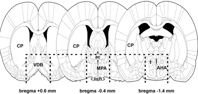
5.2.2. Microdissection of rat brain tissue samples
Thick coronal brain sections were prepared around the preoptic area with a razor blade cut immediately rostral to the optic chiasm and 2 mm caudal to this level (Fig. 3). A horizontal cut immediately above the anterior commissure, and sagittal cuts on both sides of the brain 2 mm lateral to the midline were used to dissect tissue blocks that contained the preoptic area of the hypothalamus as well as small parts of adjacent brain structures including parts of the diagonal band of Broca, the anterior commissure, the optic tract, the ventral pallidum (Fig. 3). The dissected tissue samples were quickly frozen on dry ice, and stored at - 80oC.
28
Fig. 3. Schematic figures demonstrate the brain region dissected for microarray and RT-PCR experiments. The rostral cut of coronally oriented tissue blocks was performed at bregma level +0.6 mm. The caudal surface of the microdissection was at bregma level -1.4 mm.
Dorsally, the dissection was made with a horizontal cut immediately above the anterior commissure (ac) as indicated by dashed line. Vertical dashed lines indicate the lateral border of the microdissection 2 mm lateral from the midline. The figure is from our previous publication (Dobolyi, 2009).
5.3. Microarray
RNA was purified from microdissected preoptic area 9 days after delivery from 4 primiparous lactating mothers and 4 mothers separated from pups immediately after parturition. Microarray measurements were performed in the Microarray Core Facility of the Semmelweis University using Agilent methods (Agilent Technologies, Palo Alto, CA).
Quality-checked total RNA of 500 ng was reverse transcribed by the Low-input RNA Linear Amplification Kit (Agilent Technologies) and then transcribed to either Cy5 or Cy3-labeled cRNA. The labeled cRNA was purified (RNeasy kit, Qiagen, Valencia, CA), the dye content (>8.0 pmol dye / g cRNA) and concentration of cRNA measured. For fluorescent microarray experiments, 4 pairs of mRNAs were arbitrarily formed. Each pair consisted of Cy5-labeled cRNA derived from a lactating mother and Cy3-labeled cRNA derived from a mother deprived of her pups. 1 g of labeled cRNA was hybridized to Agilent Rat Oligonucleotide 4x44K microarrays overnight at 60 oC and washed by the ozone-safe SSPE method. The
29
slides were treated with Stabilizing and Drying Solution (Agilent Technologies) and scanned by Agilent Microarray Scanner according to the instructions of the manufacturer. Data were normalized by the Feature Extraction software version 7.5 with default parameter settings for oligonucleotide microarrays and then transferred to GeneSpring 7.3 program (Agilent Technologies), with which normalization and data transformation steps were performed. The oligonucleotid sequence for amylin on the microarray chip corresponded to 354-413 bps of GenBank accession number NM_012586. This sequence shows about 47% sequence identity to the corresponding part of calcitonin, and 52-53% to other members of the CGRP peptide family.
5.4. RT-PCR
5.4.1. RT-PCR of the PTH2 receptor from human
Total mRNA was isolated using TrizolR Reagent (Invitrogen, Carlsbad, CA) from 50- 100 mg of brain tissue according to the manufacturer’s instructions. The degradation of RNA was assessed by running the purified RNAs on denaturing formaldehyde gels. Samples, in which the amount of 28S rRNA was at least equal to that of 18S rRNA, were processed further for RT-PCR. After diluting total RNA to 2 g / l, RNA was treated with Amplification Grade DNase I (Invitrogen) and cDNA was synthesized with a Superscript II reverse transcriptase kit (Invitrogen) according to the manufacturer’s instructions. After 10- fold dilution, 2.5 µl of the resulting cDNA was used as template in PCR reactions. The primer pair for PTH2 receptor was 5´-CAATTGCTTGGCTGTAGCTTT-3´ and 5´- ACAAAATCAATTTGCAGACACAA-3´ resulting in a PCR product of 440 bp that corresponds to bp 2162-2601 of the human PTH2 receptor (GenBank accession number NM_005048). The PCR product using this PTH2 receptor primer pair has been verified by sequencing to result in a product specific for the PTH2 receptor (Bagó et al., 2008). The primer pair for the housekeeping gene glyceraldehyde-3-phosphate dehydrogenase (GAPDH) was 5´-CCACCCAGAAGACTGTGGAT-3´ and 5´-CCCTGTTGCTGTAGCCAAAT-3´
resulting in a PCR product of 423 bp that corresponds to bp 650-1072 of human GAPDH (GenBank accession number NM_002046). The PCR reactions were performed with iTaq DNA polymerase (Bio-Rad Laboratories, Hercules, CA) in total volumes of 12.5 µl using primers at 300 nM final concentration under the following conditions: 95 oC for 3 min, followed by cycles of 95 oC for 0.5 min, 60 oC for 0.5 min and 72 oC for 1 min. The presence the PTH2 receptor was evaluated after 38 cycles while the presence of GAPDH after 33 cycles. Equal amounts (10 µl) of PCR products were run on gels and pictures taken with a
30
digital camera. Primers for PTH2 receptor are specific to sequences in different exons to allow recognition of potential genomic DNA contamination by the larger size of the product.
5.4.2. Real-time RT-PCR for TIP39 and amylin measurement in rat samples
Total RNA was isolated from the microdissected PVG, MPL, PIL, or preoptic area using TrizolR Reagent (Invitrogen, Carlsbad, CA) according to the manufacturer’s instructions. After diluting RNA to 2 g/l, it was treated with Amplification Grade DNase I (Invitrogen) and cDNA was synthesized with a Superscript II reverse transcriptase kit (Invitrogen) according to the manufacturer’s instructions. After 10-fold dilution, 2.5 µl of the resulting cDNA was used as template in PCR reactions. For TIP39, multiplex PCR was used
with dual-fluorescence labeled TAQMAN probes (6-FAM-
CGCTAGCTGACGACGCGGCCT-TAMRA for TIP39), and glyceraldehyde-3-phosphate- dehydrogenase (GAPDH) probe (JOE-ATGGCCTTCCGTGTTCCTACCCCC-TAMRA). The primers for TIP39 (CTGCCTCAGGTGTTGCCCT and TGTAAGAGTCCAGCCAGCGG) were used at 300 nM whereas the primers for GAPDH (CTGAACGGGAAGCTCACTGG and CGGCATGTCAGATCCACAAC) were used at 150 nM concentration. For amylin and corresponding GAPDH, PCR reactions were performed using SYBR Green dye (Sigma, St.
Louis, MO). The primers for amylin (ACATGTGCCACACAACGTCT and ACAAACACAGCAAGCACAGG corresponding to 222-241 and 493-512 bps of GenBank accession number NM_012586) and GAPDH (TGCCACTCAGAAGACTGTGG and GTCCTCAGTGTAGCCCAGGA corresponding to 540-559 and 812-831 bps of GenBank accession number M17701) were used at 300 nM final concentration. All PCR reactions were performed with iTaq DNA polymerase (Bio-Rad Laboratories, Hercules, CA) in total volumes of 12.5 µl under the following conditions: 95 oC for 3 min, followed by 35 cycles of 95 oC for 0.5 min, 60 oC for 0.5 min and 72 oC for 1 min. Cycle threshold values (CT values) were obtained from the linear region of baseline adjusted amplification curves. Standard curves obtained by measuring dilution series were used to calculate the amount of cDNA in the samples. Statistical analyses (Prism 4 for Windows, GraphPad Software, Inc.) were performed by one-way analysis of variance for the comparison of the 3 different groups followed by Bonferroni posttests for post hoc comparisons.
31 5.5. In situ hybridization histochemistry
5.5.1. Production of rat in situ hybridization probes for amylin, TIP39, and PTH2 receptor PCR products were produced from maternal preoptic cDNA using 2 different primer pairs for amylin corresponding to 222-241 and 493-512 bps, and to 54-73 and 243-262 bps of GenBank accession number NM_012586. For TIP39 and the PTH2 receptor, cDNA from diencephalon was used as a template for PCR reactions. Three regions of the rat TIP39 cDNA sequence were used to generate probes, corresponding to amino acids –55 to 17; –18 to 37, and –55 to 37, where amino acid 1 is the first residue of mature TIP39. Similarly, regions of the rat PTH2 receptor cDNA sequence corresponding to bases 482-864 and bases 1274-1828 was used to generate probes. The PCR products were purified from gel, inserted into TOPO TA cloning vectors (Invitrogen) and transformed chemically into competent bacteria. Selected plasmids were applied as templates in PCR reactions, using the specific primer pairs with the forward primers also containing a T3 for sense probes, while the reverse primers a T7 RNA polymerase recognition site for antisense probes. At the end, the identities of the cDNA probes were verified by sequencing. The different probes for the same genes produced equivalent hybridization patterns in all 3 cases. Comparisons between signals from antisense and sense (control) riboprobes were made on serial sections hybridized simultaneously and processed together. Sense probes did not result in specific labeling.
5.5.2. Macaque probe preparation for in situ hybridization
Template cDNA was prepared from macaque testis by an RT reaction, as described above. PCR reactions were also performed essentially as described above using primers with
human TIP39 (5´-GGGGACTGTGCGGGAAGC-3´ and 5´-
GCATGTACGAGTTCAGCCAGTGG-3´) and PTH2 receptor (5´-
TGTGGGGCTTCATCTTGATAGG-3´ and 5´-ATGGCGGTGTCCTTTTCCAGTC-3´)
sequences. The PCR products were cloned into plasmid vectors, and their identities were confirmed by DNA sequencing. A plasmid for each probe was used as template in PCR reactions, using primers that appended a T7 RNA polymerase recognition sequence (5´- GCGCGTAATACGACTCACTATAGGG-3´) into the 5’ end of the antisense primer. The TIP39 probe was a 372 base amplicon, which differed from the predicted human sequence (GenBank accession number NM_178449) at 16 scattered base positions. It also lacked codons for two amino acid residues in the predicted leader sequence. The PTH2-R probe was a 500 base amplicon. It differed from the predicted human sequence (GenBank accession number NM_005048) at 14 bases. It also contained a 49 base insertion that corresponds to
32
predicted intronic sequence. The small number of differences between the macaque probes and predicted human sequences may reflect PCR error or artifacts that do not affect their use as probes, or genuine species differences.
5.5.3. In situ hybridization protocol
The freshly dissected tissue was quickly frozen on dry ice. Serial coronal sections (12
m) were cut using a cryostat, mounted on positively charged slides (SuperfrostPlus®, Fisher Scientific, Pittsburgh, PA), dried, and stored at -80°C until use. Antisense [35S]UTP-labeled riboprobes were generated using T7 RNA polymerase of the MAXIscript transcription kit (Ambion, Austin, TX) from polymerase chain reaction-amplified fragments of the amylin or TIP39 cDNA subcloned into TOPO TA vectors. For hybridization, we used 80 µl hybridization buffer and 1 million DPM of labeled probe per slide. Washing procedures included a 30 min incubation in RNase A, followed by decreasing concentrations of sodium- citrate buffer (pH = 7.4) at room temperature, and then at 65°C. After drying, the slides were dipped in NTB2 nuclear track emulsion (Eastman Kodak) and stored at 4°C for 3 weeks for autoradiography. Then, the slides were developed and fixed with Kodak Dektol developer and Kodak fixer, respectively, counterstained with Giemsa and coverslipped with Cytoseal 60 (Stephens Scientific, Riverdale, NJ).
5.5.4. Quantitation of in situ hybridization data by counting autoradiography grains
Amylin mRNA-expressing neurons, above which more than 9 autoradiography grains (3 times the average background) were detected, were typically present in 3 coronal sections cut at 216 m distances. The total number of these cells was counted on 21st day of pregnancy, 1, 9, and 23 days after parturition. In addition, the number of autoradiography grains were counted in 20-20 evenly distributed but otherwise randomly selected amylin mRNA-expressing neurons in each of the 3-3 brains on 21st day of pregnancy, 1, 9, and 23 days after parturition. Both the total number of amylin mRNA-expressing neurons in the 3 consecutive sections and the average number of autoradiography grains per cell were calculated on one side of the 3-3 brain sections. Statistical analyses were performed using Prism 5 for Windows (GraphPad Software Inc.). Both cell numbers and the number of autoradiography grains in the 4 groups (21st day of pregnancy, 1, 9, and 23 days after parturition) were compared using one-way ANOVA followed by Tukey´s multiple comparison post-hoc test.
33
5.5.5. Densitometric analysis of in situ hybridization histochemistry
Dark-field photomicrographs were taken of the sections where the TIP39 signal was the highest in the PIL using a 10x objective. Each image was divided into 2 halves with identical size, such that one half contained all the observed TIP39 autoradiography signals, while the other half served as background control. The pixel number of white area (lighter than an arbitrary grayness used for all the images) was calculated for both halves of the images using ImageJ 1.47v (National Institutes of Health, USA) software. The difference between the 2 values (the half picture containing TIP39-expressing cells – the half picture containing only background autoradiography signal) was used to quantify the TIP39 mRNA level. TIP39 mRNA levels at the 5 different time points were compared using one-way ANOVA followed by Bonferroni´s multiple comparison test.
5.6. Histology
5.6.1. Tissue collection
Rats were deeply anesthetized and perfused transcardially with 150 ml saline followed by 300 ml of ice-cold 4% paraformaldehyde prepared in phosphate buffer (PB, 100 mM, pH=7.4). Brains were removed and postfixed in 4% paraformaldehyde for 24h and then transferred to PB containing 20% sucrose for 2 days. Serial coronal sections were cut at 50
m on a sliding microtome (SM 2000R, Leica Microsystems, Nussloch, Germany). Sections were collected in PB containing 0.05% sodium-azide and stored at 4C.
5.6.2. Cresyl-violet staining
Sections were mounted consecutively on gelatin-coated slides and dried. Sections were stained in 0.1% cresyl-violet dissolved in PB, then differentiated in 96% ethanol containing acetic acid. Sections were then dehydrated and coverslipped with Cytoseal 60 (Stephens Scientific, Riverdale, NJ).
5.6.3. Luxol fast blue staining
Sections were collected and mounted consecutively on gelatin-coated slides and dried.
Myelinated fibers were visualized with the sulphonated copper ohthalocyanine Luxol Fast Blue with a modification of the Kluver-Barrera method as described before (McIlmoyl, 1965).
Briefly, 25 µm thick sections were stained in 0.1% Luxol fast blue dissolved in 96% ethanol containing 0.05% acetic acid, and differentiated in 0.05% lithium-chloride followed by 70%