BBA - Molecular Basis of Disease 1867 (2021) 166243
Available online 8 August 2021
0925-4439/© 2021 The Author(s). Published by Elsevier B.V. This is an open access article under the CC BY-NC-ND license
(http://creativecommons.org/licenses/by-nc-nd/4.0/).
Fetal oxygen supply can be improved by an effective cross-talk between fetal erythrocytes and vascular endothelium
Payal Chakraborty, Ali Khamit, Edit Hermesz
*Department of Biochemistry and Molecular Biology, Faculty of Science and Informatics, University of Szeged, Szeged, Hungary
A R T I C L E I N F O Keywords:
Birth weight Endothelial dysfunction Nitric oxide synthase Twin pregnancy Umbilical cord
A B S T R A C T
In twin/multiple pregnancy, siblings experience an adverse intrauterine environment which forms the major etiological factor leading to pathological conditions. The status of the developing fetus is highly determined by the nitric oxide (NO) level, that facilitates vasodilation which in turn modulates the oxygen and nutrition supply.
As the umbilical cord (UC) lacks innervation, activation of the endothelial nitric oxide synthase (NOS3) is fundamental to maintain adequate NO production. Recent ground breaking fact showed that under stress con- ditions, circulating red blood cells (RBCs) can actively produces NO as a “rescue mechanism”. Therefore, this study majorly focused on the molecular mechanisms that affected the redox environment by altering NOS3 activation - both in the UC arteries and vein endothelium and RBCs - that have impacts on developmental pa- rameters, like birth weight. In connection to that, we pursued the communication efficiency between the vessels' endothelium and the circulating RBCs in demand of bioavailable NO. Our results indicated that twinning itself at stage 33–35 weeks, does not reduce the NOS3 level and its phosphorylation status in the cord vessels. However, RBC-NOS3 activation is highly upregulated during this period - providing additional evidence for the active regulatory role of fetal RBCs in the rate of blood flow - and this functional activity highly correlates with the birth weight of the fetuses. Detailed analysis on NOS3 signalling at different time points of gestation could establish a benchmark in understanding of the pathophysiological mechanisms involved in the process of developing neonatal vascular diseases.
1. Introduction
The physiological state of pregnancy is always in demand for excess oxygen supply which can easily disrupt the feto-maternal redox ho- meostasis balance. In connection to that, there are increasing evidences of enhanced oxidative insult conditions, like preeclampsia, ectopic pregnancy, placental abruption and so on. Moreover, in cases of multi- ple/twin pregnancy, additional trauma exists with greater risks of spontaneous miscarriage, preterm delivery, gestational hypertensive disorders, intrauterine growth restriction, perinatal death and related postnatal and neonatal illnesses. Modern usage of in-vitro fertilization (IVF) therapies has led to higher incidence of multiple pregnancies.
There have been relevant epidemiological data on the increasing fetal, neonatal and perinatal mortality rate of 3–6 times in twin and 5–15 times in other multiple pregnancies with comparison to singleton de- liveries [1]. The mechanism in twinning depends on zygosity; twins can either develop from one zygote (monozygotic) or from two separate zygotes (dizygotic). Monozygotic twins make up approximately 30% of twin pregnancies [2,3]. In general, ~16% of twin gestations have discordance i.e. difference in the weights of siblings higher than 15–25%
[4]. As per the American College of Obstetricians and Gynaecologists (ACOG) discordant growth gets associated with anomalies like, intra- uterine growth restriction, preterm birth, infection of one of the fetus, stillbirth, umbilical arterial pH <7.1, respiratory distress, and death
Abbreviations: ACOG, American College of Obstetricians and Gynaecologists; ANOVA, One-way Analysis of Variance; ED, Endothelial Dysfunction; 4-HNE, 4- Hydroxynonenal; H2O2, Hydrogen Peroxide; NO, Nitric Oxide; NOS2, Inducible Nitric Oxide Synthase; NOS3, Endothelial Nitric Oxide Synthase; O2•−, Superoxide Anion; ONOO−, Peroxynitrite; PB, Phosphate Buffer; PrD-Hwt, Premature Discordant High birth weight neonates; PrD-Lwt, Premature, Discordant, Low birth weight neonates; Pr-Hws, Premature High birth weight singletons; Pr-Hwt, Premature, High birth weight twins; Pr-Lwt, Premature, Low birth weight twins; RBCs, Red Blood Cells; ROS, Reactive Oxygen Species; UC, Umbilical Cord.
* Corresponding author at: Department of Biochemistry and Molecular Biology, Faculty of Science and Informatics, University of Szeged, P.O. Box 533, H-6701 Szeged, Hungary.
E-mail address: hermesz@bio.u-szeged.hu (E. Hermesz).
Contents lists available at ScienceDirect
BBA - Molecular Basis of Disease
journal homepage: www.elsevier.com/locate/bbadis
https://doi.org/10.1016/j.bbadis.2021.166243
Received 28 April 2021; Received in revised form 30 July 2021; Accepted 3 August 2021
within 1 week of birth [5].
Fetal homeostatic condition and the intrauterine environment criti- cally depend on the efficiency of the feto-placental circulation. The UC vascular system is the sole pathway of oxygen and nutrient transport to the fetus through the placenta. The UC circulation system is usually made up of one vein and two arteries. From the placenta, the oxygenated blood and nutrition are transported by the vein to the fetus and arteries carry the deoxygenated blood back to the placenta. In this scenario, it is clear that the UC vein is primarily and directly exposed to the toxic materials/free radicals unfiltered by or generated in the placenta. Any alteration in it may serve as fingerprints of the damages affecting the in–utero development. Major complications in fetal development can be directly or indirectly connected to the umbilical cord disorders causing intrauterine hypoxia and/or impaired blood flow to the developing fetus.
The major part of the umbilical cord lacks innervation and hence the vascular tone mainly depends on the level of NO, i.e. synthesized by the NOS3 [6]. The NO, being a potent vasoactive agent, causes vasodilation and increases the rate of oxygen perfusion and nutrient supply [7]. Any kind of impaired response and loss in the bioactivity of endothelium- derived NO within the umbilical cord vascular system causes intra- uterine hypoxia, increased feto-placental vascular resistance, and retardation in the fetal growth [8,9]. NOS3 activation gets regulated by its substrate L-arginine concentration, availability of cofactors, rate of electron transfers, subcellular localization, post-translational modifica- tions and diverse interacting proteins [10,11]. The process of NOS3 coupling/dimerization is the most crucial step towards its activation;
low concentration of L-arginine substrate causes NOS3 uncoupling and dysfunctionality [12,13]. NOS3 is in direct competition with Arginases for their common substrate, therefore an increased expression of Argi- nases indirectly influences the NOS3 activation process [14,15]. Among the post-translational modification steps, phosphorylation of NOS3 specific at Ser1177 residue is the prime site for the enzyme activation whereas the Thr495 residue be the negative regulatory site. Under shear stress conditions the phosphorylation step gets induced in the cells due to key factors like vascular endothelial growth factor and increased influx of electrons through the reductase domain from NADPH to the haem group in the oxygenase domain [16,17].
Under severe oxidative stress conditions, influenced by any intrinsic or extrinsic pathophysiological factors, there are obvious production of reactive oxygen species (ROS)/strong oxidants; like superoxide anion (O2•−), hydrogen peroxide (H2O2) and hydroxyl radical. The O2•−, in elevated level, undergoes spontaneous reactions and readily scavenges the bioavailable NO in the vascular system by forming deleterious pro- oxidant entity peroxynitrite (ONOO−) [18–20]. One of the pivotal fac- tor in the development of endothelial dysfunction (ED) is the reduced production/bioavailability of NO, resulting in an impaired endothelial growth and vasodilation.
RBCs were mainly ascribed as carriers for transmitting oxygen and NO into the vascular bed, recently emerged with a ground breaking publication showing the evidence for a NO synthase protein within the red blood cells (RBC-NOS3) [21,22]. Supposedly, the occurrence of ED can be sensed by the circulating RBCs, and thereby under acute shear stress conditions NO bioavailability might be increased by the RBC- NOS3 activation pathway. As the UC endothelium directly comes in contact with all the blood components and most predominantly the circulating red blood cells (RBCs), it intends to establish an effective crosstalk mechanism between them.
In our earlier work, we demonstrated that RBCs originated from twin neonates exhibit quite a high oxidative stress condition with the accu- mulation of NO3−/NO2− and strong oxidant molecules (H2O2 and ONOO−), compared to samples originated from the single pregnancy [23]. The developmental status of neonates is determined by a number of multifactorial and complex processes, particularly involving the sig- nalling pathway of NOS3 activation, which significantly determines the bioavailable NO level, and facilitates vasodilation which ultimately
modulate oxygen and nutrition supply for the developing fetus. In our recent work, we exhibited that during twin pregnancy, both the UC vessels and the RBCs of mature fetuses are impaired in their NOS3-NO producing capacity [24]. Here, we further pursued and examined the molecular consequences due to oxidative stress in neonates born from preterm twin pregnancies and attempted to establish a possible link between a certain alteration in the NOS3-NO pathway and the neonatal birth weight. We followed the status of premature twins with 3 different birth weight groups. In the appropriate weight for age category, samples of siblings born with low and high weight were compared to their age and weight matched neonates, from single pregnancy. Additionally, we also compared the samples that originated from the preterm discordant siblings.
2. Materials and methods 2.1. Human samples
According to the Declaration of Helsinki, informed consent was requested from pregnant women to collect a small portion of the um- bilical cord along with fetal cord blood samples. The study protocol was approved by the Ethics Committee from the Department of Obstetrics and Gynaecology at the University of Szeged, Hungary (16/2014). The study protocol strictly followed several exclusion criteria to collect the clinical samples such as (i) maternal age below 18 years, (ii) gestational diabetes, obesity, infection and inflammatory conditions or disorders such as cardiovascular diseases, (iii) complications or difficulty during delivery, (iv) dizygotic twin birth (v) fetal distress, (vi) malformations or evidence of genetic disorders and (vii) neonates born from mothers addicted to alcohol and /or smoking habit. The nutritional status of the mothers during pregnancy was satisfactory; no case of malnutrition occurred.
Clinical samples were grouped under different inclusion criteria. In the first group, 16 pairs of clinical samples from pre-mature, non- discordant, monozygotic, dichorionic twins were collected. Neonates were categorized based on their maturity and birth weight (percentile value); 2430–2570 g/33 +3–34 +6 weeks/95th percentile (Premature, High birth weight twins - Pr-Hwt) and 1940–1990 g/33–33 +6 weeks/
50th percentile (Premature, Low birth weight twins - Pr-Lwt). As control for the Pr-Hwt group, 15 samples of age and weight matched 2200–2500 g/33–35 weeks/95th percentile singletons were collected (Pr-Hws). The clinical parameters of the study group and the maternal age are presented in the Supplementary as Table S1.
In the second group, 20 pairs of clinical samples from premature, monozygotic, dichorionic, discordant twin category were collected. The criteria for discordance was followed as per the guidelines laid down by ACOG USA, with a minimum of 25% discordance between the siblings' birth weight. Siblings were grouped according to their birth weight;
2600–2890 g/35–35 +4 weeks/90–95th percentile in the high weight group (Premature, Discordant, High birth weight neonates - PrD-Hwt) and 1860–2040 g/35–35 + 4 weeks/10–25th percentile (Premature, Discordant, Low birth weight neonates - PrD-Lwt). The clinical param- eters of the study group and the maternal age are presented in the Supplementary as Table S2.
2.2. Blood smear image processing and data analysis
Using the validated calculation software MatLab we processed and analysed eosin stained blood smear images, originated from each of the collected sample groups. Identification of different phenotypic varia- tions in the RBC populations was conducted with an automated bio- image analysis tool CellProfiler™ (available on http://cellprofiler.org) [25].
2.3. Preparing umbilical cord and arterial cord blood samples for immunostaining
Small dissected portions of the umbilical cord were fixed with 4%
(w/v) paraformaldehyde in 0.05 M phosphate buffer pH 7.4 (PB) and cryopreserved with 30% (w/v) sucrose in PB with addition of 0.1% (w/
v) Na-azide. Specimen samples were embedded in Tissue-Tek® O.C.T.™ (Sakura Europe 4583, Alphen aan den Rijn, Netherlands) and cryosec- tioned with slice thickness of 16 μm. Further it gets mounted on Superfrost™ ultra plus® (Thermo scientific J3800AMNZ, Massachu- setts, United States) microscope slides and kept on − 80 ◦C till further processing [26].
Whole blood, derived from the cord arteries, was subjected to centrifuge at 200 ×g for 10 min at 4 ◦C. The lower two-thirds of the RBC phase was collected, washed with physiological salt solution (0.9%
NaCl, w/v) and fixed with 4% (w/v) paraformaldehyde in 0.05 M PB at 4 ◦C for 1 h and further processed for immunostaining [27]. To test the purity of the isolated RBCs, the cells were stained with RBC-specific marker anti Glycophorin A, where the purity of the samples was
>95% for RBCs [28].
2.4. Immunolabelling on umbilical cord sections and isolated red blood cells
Umbilical cord sections and fixed RBCs were stained by standard protocols [24]. Briefly, samples were permeabilized by 0.1% Triton X- 100 at room temperature for 20 min. Non-specific antibody binding sites were blocked with 4% (w/v) bovine serum albumin and 5% (v/v) normal goat serum in PB, and double labelled with primary antibodies overnight at 4 ◦C. After the incubation step, samples were washed and further incubated with goat anti-mouse Alexa®488/anti-rabbit Alexa®647- or goat anti-rabbit Alexa®488/anti-mouse Alexa® 647-con- jugated secondary antibodies in 1:2000 dilution, for 2 h in dark at room temperature (see Supplementary Table S3 for the detailed antibody list).
Finally, the slides were counterstained with 4′, 6-diamidino-2-phenylin- dole (D9542 from Sigma-Aldrich, St. Louis, MO, USA) in a final con- centration of 1 μg/ml, for 5 min in dark, washed, dried and mounted, using Antifading, BrightMount/Plus aqueous mounting medium (Abcam ab103748, Cambridge, United Kingdom), and examined under epi- fluorescence microscope (Nikon Eclipse 80i, 100×immersion objective;
Nikon Zeiss Microscopy GmbH, Jena, Germany) with a QImaging RETIGA 4000R camera, using Capture Pro 6.0 software (QImaging, Surrey, BC, Canada). Images were processed and analysed using ImageJ® software (http://imagej.nih.gov/ij/).
The stained RBCs, following the secondary antibody incubation step, were washed and processed for quantitative analysis (FACS, BD FACS- Calibur™, BD Biosciences, New Jersey, United States) [28,29]. The FACS data were analysed using FlowJo™ (FlowJo™ Software for Win- dows Version 10, Oregon, United States).
2.5. Assay and detection for viability in the RBCs
Fresh arterial cord blood was collected and centrifuged at 200 ×g for 5 min. From the lower two-thirds of the RBC phase 10 μl was collected and diluted by 10×in 0.9% (w/v) physiological salt solution having pH 7.4. Briefly, 1 μl of the diluted RBCs were mixed with 250 μl of Annexin binding buffer and 2.5 μl Annexin V using the Annexin V-FITC Apoptosis/Staining Detection kit (Abcam ab14085, Cambridge, United Kingdom). The quantitative measurement was done by FACS (FACS, BD FACSCalibur™, BD Biosciences, New Jersey, United States) [30] and all data were analysed using FlowJo™ (FlowJo™ Software for Windows Version 10, Oregon, United States).
2.6. Statistical analysis
All statistical analysis was considered with one-way analysis of
variance (ANOVA) and Newman-Keuls multiple comparison test using GraphPad Statistical Software version 6.0. (GraphPad Software, San Diego, CA, USA) The level of statistical significance was accepted at *p
≤0.05, **p ≤0.01, ***p ≤0.001 and ****p ≤0.0001.
3. Result
3.1. Expression of endothelial and inducible NOS, and Arginase1 in the non-discordant study populations
In this study, molecular parameters of UC vessels and cord blood, originated from twin pairs with high birth weight (95th percentile) were compared to age and weight matched singletons and age-matched twin pairs born with low birth weight (50th percentile).
Sections of the UCs originated from non-discordant twin pairs and singletons were immunolabelled and subjected to Image J© evaluation.
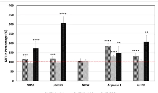
In the Hwt versus Hws comparison, a significant increase was measured in the NOS3 expression and also in its phosphorylation in- tensity level, within the Hwt arteries (~13 and ~17% respectively). In the vein, both values stayed unaltered (Fig. 1) (Supplementary Figs. S1 A–F and S2 A–F).
Considering the Hw versus Lw twin populations, NOS3 expression in the Lw arteries to some extent remained behind the Hw values (~28%) with about the same phosphorylation intensity level. In the vein, the NOS3 expression was unaltered, with a significant increase in the phosphorylation intensity (~26%) (Fig. 2) (Supplementary Figs. S1 D–I and S2 D–I). As a consequence of it, the phosphorylation status of NOS3 (NOS3/pNOS3 ratio) in both the vessels of Lwt samples was increased compared to the Hwt samples.
The NOS3 activation is influenced by Arginase1, which competes for the common substrate, L-arginine. Arginase1 expression in both the ar- teries and veins of the Hw twins exhibited significant elevation compared to their age and weight-matched singletons, ~85%, and
~30%, respectively (Fig. 1). In the Lw and Hw twin correlation, there was a ~22% increase in Lw arteries while in the veins no significant difference was detected (Fig. 2 and Supplementary Figs. S3 A, D, G and S4 A, D, G).
Upregulation of the inducible NOS (NOS2) expression could serve as a compensatory mechanism to increase the bioavailable NO level in the vascular endothelial layer under shear stress conditions. Comparison of the singleton versus Hwt values, there were no significant difference in the NOS2 expression for any of the vessels (Fig. 1). In case of Hwt versus Lwt comparison, the NOS2 value was significantly higher in the Lwt arteries (~47%), while in the vein it stayed unchanged (Fig. 2 and Supplementary Figs. S3 B, E, H and S4 B, E, H).
Following the NOS3 level and its phosphorylation status at position Ser1177 by FACS analysis in the isolated arterial RBC population, (where in each case according to the blank sample, an arbitrary borderline was considered along the x-axis, that divided the total RBC population into basal and high intensity levels) the Hwt group showed a 73.64% higher NOS3 expression and 206.35% higher phosphorylation intensity level when compared to their age and weight matched sin- gletons (Fig. 1). While, there was no significant variation in these values, between the Hw and Lw twin neonates (Fig. 2 and Supplementary Fig. S5).
Following the expression of Arginase1 by FACS analysis, clearly indicated a 48% elevation in the Hwt values when compared to their matched singletons (Fig. 1). Between the twin groups, there was no significant difference in Arginase1 expression level (Fig. 2 and Supple- mentary Fig. S6 A–D).
3.2. NOS3 expression and activation, NOS2 and Arginase1 level in the discordant study populations
In this part of the study, molecular parameters of twin siblings, born with significantly high weight differences, were compared. The high
Fig. 1. Estimation of the expression of NOS3 and phosphorylated NOS3, NOS2, Arginase1 and 4-HNE level in the UC vascular endothelium and fetal RBCs of the high weight preterm twin neonates. Measure- ment of the mean fluorescence intensity (MFI) were expressed in percentage of NOS3 and its activated level i.e. phosphorylated NOS3 (pNOS3), NOS2, Arginase1 and 4-HNE level of the preterm non- discordant high birth weight twin neonates in com- parison to their age and weight matched singletons taken as control. The red dotted line represents the control intensity level at 100%. The significant dif- ferences were accepted at **p <0.01, ***p <0.001 and ****p <0.0001 based on one-way ANOVA using the Newman-Keuls multiple comparison test.
Fig. 2.Quantification of the expression of NOS3 and phosphorylated NOS3, NOS2, Arginase1 and 4-HNE level in the UC vascular endothelium and fetal RBCs of the low weight preterm twin neonates. Measure- ment of the MFI were expressed in percentage of NOS3, phosphorylated NOS3, NOS2, Arginase1 and 4-HNE level of the preterm non-discordant low birth weight twin neonates in comparison to their high weight twin group taken as control. The red dotted line represents the control intensity level at 100%.
The significant differences were accepted at ***p <
0.001 and ****p <0.0001 based on one-way ANOVA using the Newman-Keuls multiple comparison test.
Fig. 3. Estimation of the expression of NOS3 and activated pNOS3, NOS2, Arginase1 and 4-HNE level in the UC vascular endothelium and fetal RBCs be- tween the premature discordant twin pair. Measure- ment of the MFI were expressed in percentage of NOS3, phosphorylated NOS3, NOS2, Arginase1 and 4-HNE level of the preterm discordant low birth weight twin neonates in comparison to their high weight twin pair, considered as control. The red dotted line represents the control intensity level at 100%. Statistical significances were accepted at *p <
0.05, **p <0.01, ***p <0.001 and ****p <0.0001 based on one-way ANOVA using the Newman-Keuls multiple comparison test.
weight neonates belonged to the 90–95, and their siblings into the 10–25 percentile categories.
In a similar manner, umbilical cord sections were double-labelled, first for NOS3/pNOS3, and subjected to Image J© evaluation. The NOS3 and pNOS3 intensities were significantly lower in both UC vessels of the low weight neonates, compared to their high weight siblings; by
~37% (NOS3) and ~45% (pNOS3) in the artery and by ~19% (NOS3) and ~26% (pNOS3) in the vein. The phosphorylation status of NOS3 (ratio NOS3/pNOS3) were reduced in both the vessel of the Lw-siblings (Fig. 3 and Supplementary Figs. S7 and S8).
Arginase1, being a key player in the NOS3 regulation, was expressed at a significantly lowered level in both the vessels (by ~17–18%) of the low weight siblings (Fig. 3 and Supplementary Figs. S9 A, D and S10 A, D). NOS2, serving as an alternative source for bioavailable NO, was present at a significantly higher level in the cord arterial endothelium of the low weight siblings (by ~24%) while in the vein, a significantly lower level (by ~37%) was detected (Fig. 3 and Supplementary Figs. S9 B, E and S10 B, E).
In a parallel study, isolated cord arterial RBCs were immunostained and analysed by FACS. No significant difference was found in the NOS3 expression, and also in the frequency of the high NOS3 expressing cells between the siblings. However, the pNOS3 intensity level was signifi- cantly higher in the low weight neonates (by ~29%), consequently, the phosphorylated status of NOS3 was significantly higher in these cases (Fig. 3 and Supplementary Fig. S11). The Arginase1 expression was significantly higher (by ~33%) in the low weight group, due to a significantly higher frequency (by ~8–10%) of the high Arginase1 expressing cells (Fig. 3 and Supplementary Fig. S12 A–C).
3.3. Quantitative measurement of lipid peroxidation, marked with 4- hydroxynonenal and Annexin V positive population
The most common products of lipid peroxidation, due to excessive ROS production, are the oxygenated α, β-unsaturated aldehydes such as 4-hydroxynonenal (4-HNE). The formation of this aldehyde was fol- lowed by immunolabelling the aldehyde-protein adduct with anti-4- HNE antibody. Furthermore, due to excess rate of lipid peroxidation, the level of phosphatidylserine gets altered in the outer leaflet of RBC plasma membranes which gets easily detected by Annexin V and quantified using FACS analysis.
3.3.1. Premature non-discordant study population
The intensity levels of 4-HNE evaluated by Image J©, showed a significant increase (by 31%) in the arteries, and stayed unchanged, in the vein of the Hw twin neonates, compared to their matched singletons (Fig. 1). In the Hwt versus Lwt comparison, there was a significant 17%
increase in the arteries, while there was no alteration between these values in the veins (Fig. 2 and Supplementary Figs. S3 C, F, I and S4 C, F, I). The 4-HNE intensity level was followed by FACS analysis in the isolated RBCs. The Hwt samples, compared to their matched singletons, showed a ~108% increase in the 4-HNE intensity level (Fig. 1). RBCs originated from the Lwt samples, exhibited 71% increase in comparison to the Hwt populations (Fig. 2 and Supplementary Fig. S6 E–H).
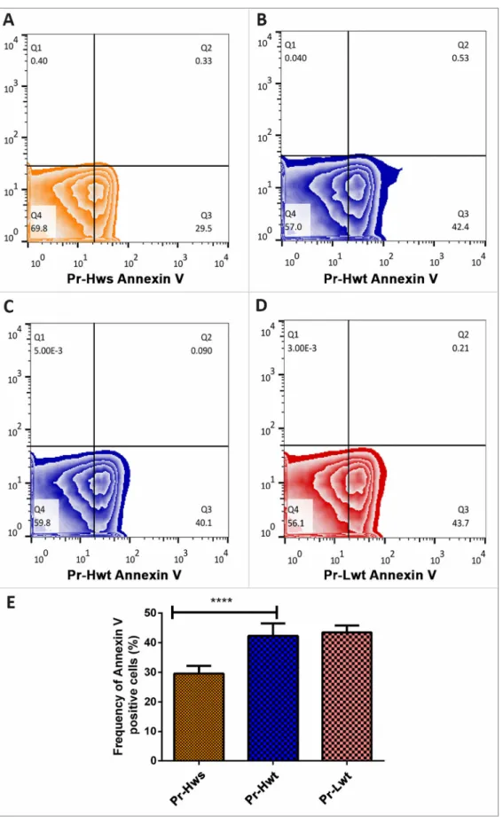
Simultaneously, RBC populations were tested for Annexin V posi- tivity by FACS analysis. These measurements revealed an increased frequency (~12.5%) in the Annexin-V positive RBCs originated from Hwt compared to Hws populations. However there was no significant difference between the Hwt and Lwt populations (Fig. 4 A–E).
3.3.2. Premature discordant study population
Measuring the 4-HNE intensity level, the Lw twins showed a signif- icant increase in both the artery and vein by 42% and 25% respectively (Fig. 3 and Supplementary Figs. S9 C, F and S10 C, F).
FACS analysis of HNE intensity in the isolated RBCs revealed a 69%
increase in the Lwt population in comparison to the Hwt group (Fig. 3 and Supplementary Fig. S12 D–F).
Furthermore, RBC marked by Annexin V showed a significant in- crease in the frequency of Annexin V positive cells by ~10% in the Lwt cell population compared to those of Hwt origin (Fig. 5 A–C).
3.4. Analysis of RBC morphological variants
Increased frequency of morphological variants in the RBC pop- ulations can be kind of an indication for certain pathological conditions.
Altogether, we screened over 3000 RBCs from the singletons, ~2000 from the mature and 4000 from the premature twin populations, and looked for Burr cells, Elliptocytes, and Rouleaux formation, among the regular, biconcave shaped RBCs. Between the mature and premature singleton groups, there was no major significant difference in the fre- quency of the phenotypical variants. However, a significant difference was found between singleton and twin neonates; such as in the mature twin RBCs, there was a 3-fold, while in the premature group a 2-fold increase occurred in the Burr cell populations. The Elliptocyte and Rouleaux variants were virtually absent in the singletons. In relation to the mature versus premature twins there was a 2–2.5 fold increase in the Elliptocytes and a 5–fold increase in the Rouleaux phenotypes in the premature populations, irrespective of their birth weight (Fig. 6).
4. Discussion
Twin siblings, in comparison to the age and weight-matched healthy singletons, undoubtedly experience an enhanced intrauterine hypoxic state with advanced oxidative stress [31–33], that gets directly or indirectly associated with umbilical cord disorders and contributes to the occurrence of pathological conditions in post-natal or adult life [34,35]. Such condition necessitates an efficient cross-talk between the vascular endothelium and the circulating RBCs that can enhance vaso- dilation and improve the rate of blood flow to the fetuses. Therefore, in this study we majorly focused on molecular mechanisms that affect the redox environment by altering NOS3 activation - both in the UC endo- thelial layer and RBCs - that have impact on the developmental pa- rameters. Our result highlighted, that a) twinning itself, does not disturb the NOS3 level and its phosphorylation at Ser1177 position in the cord vessels. Moreover, in case of twin neonates born with 95th percentile value, NOS3 expression and its activation was comparable to the age and weight-matched singletons. b) Further, in twins with a lower percentile value (50th and 25th), the NOS3 expression lagged behind in the arterial cord endothelium, but its phosphorylation at position Ser1177 increased in both the cord arteries and in the circulating arterial RBCs. c) Ana- lysing the discordant twin pairs, it seems, that in the lower weight sib- lings, the vascular NOS3 system is markedly impaired. d) Finally, we also pointed out in this study, that although a significant part of the RBC population seems to be functionally active, an increased appearance of phenotypic variants indicates the extent of damage in the plasma membrane structure and most likely also in its functional activity.
During twin pregnancy, most likely even the highest NOS3-NO production capacity of the UC vessels could not suffice to maintain the desired NO levels. The concept for an increased demand for NO under highly hypoxic conditions might be supported with the facts, that the so called “built in” alternative NO producing pathways were activated in all the investigated categories. The extent of the elevated NO demand can be deduced from the gradual activation of these pathways. In the high weight twin pairs with no obvious sign for ED, only the RBC-NOS3- NO pathway was activated, both at the translational and phosphoryla- tion levels. In case of the lower weight twin pairs it is even more important to ensure the availability of rescue mechanisms. In addition to activation of the RBC-NOS3-NO pathway, an increased phosphorylation at the Ser1177 position was observed in both the arteries and the veins.
Moreover, an inducible NOS isoform, the NOS2 was triggered in the arterial endothelium. NOS2 is induced under pathological conditions
Fig. 4. Viability assay on the isolated arterial cord RBCs derived from preterm non-discordant twin siblings. Representative zebra plots on isolated RBCs treated with FITC (Fluorescein IsoThioCyanate) - conjugated Annexin V of (A) premature high weight singleton (Hws), (B and C) premature high weight twin (Hwt) and (D) premature low weight twin (Lwt). Based on the blank value of the appropriate control and twin sample, an arbitrary borderline was considered along the x-axis, where the Q3 quadrant represents the Annexin V positive cells. The graphical summary (E) shows the percentage distribution of Annexin V positive RBCs localized in Q3 quadrant. **** marks the significant differences based on the Newman-Keuls multiple comparison test at p <0.0001.
like inflammation, sepsis or oxidative stress [36], and even a moderate increase in its expression could sufficiently increase the NO level, regarding to the higher rate of NOS2 catalytic activity i.e. (100–1000 fold that of the NOS3) [37,38]. As for the discordant population, in the lower weight siblings, both the activated RBC-NOS3-NO pathway and the upregulated arterial NOS2 system might be available as the compensatory mechanisms.
At the first glance, extended NO production seems to be beneficial when there is an insufficient level of bioavailable NO in the vascular system, however, increased synthesis of NO may indicate to a higher degree of oxidative stress. Under severe oxidative stress conditions the increased level of O2•− through a spontaneous reaction with the bioavailable NO, instantaneously initiates the formation of ONOO−, which signifies the “Janus faced” feature of NO [39]. This assumption was clearly supported by our data on the viability assays that were performed with RBC populations, showing increased frequency of the Annexin V positive cells which indicated cell death by apoptosis and also further by the higher extent of the macromolecular damages as rein- forced by the elevated formation of 4-HNE adducts.
Studying the morphological variants in the twin RBC population, we
exhibited a significant rise in the Burr cell population irrespective of birth weight and maturity. Losing their characteristic biconcave shape might be the consequences of both altered cytoskeletal network along with disproportional distribution of fatty acids, lysophospholipids in the RBC plasma membrane [40]. Furthermore, specifically in the preterm twin category we cited a significant distribution of Elliptocytes and Rouleaux phenotypes. Based on wide range of clinical studies morpho- logical differentiation of Elliptocytes, indicated iron deficiency and megaloblastic anaemia whereas the formation of RBC aggregates, due to high fibrinogen level, results in lower blood flow rates that might lead to pathogenesis of circulatory and thromboembolic complications in clin- ical cases like pregnancy, diabetes, etc. [41,42]. The increased level of phenotypic variations in the RBC populations could be a warning sign for their impaired functional and/or physiological properties, however, it is most likely that there is no apparent link between the varying morphological alteration and the RBC-NOS3 activation capacity. In our earlier study, we already provided evidence on elevated level of morphological variants in the fetal RBC populations exposed to toxic materials, due to maternal smoking [43]. In those cell populations the RBCs were functionally impaired far before their detectable Fig. 5. Viability Assay on the isolated arterial cord RBCs derived from the preterm discordant twin pairs. Panel (A–B) represents the zebra plots of the isolated arterial cord RBCs derived from preterm discordant twin pairs that are treated with FITC (Fluorescein IsoThioCyanate) - conjugated Annexin V. Based on the blank value of the appropriate control and twin sample, an arbitrary borderline was considered along the x-axis, where the Q3 quadrant represents the Annexin V positive cells. The graphical summary (C) shows the percentage distribution of Annexin V positive RBCs localized in Q3 quadrant. Statistical significance was accepted at
****p <0.0001 using the Newman-Keuls multiple comparison test.
Fig. 6.Distribution of morphological variants of RBCs derived from mature and premature twin sib- lings. Distribution of morphological variants in iso- lated RBC from singletons in comparison to mature and premature twin populations. Screening of RBCs (N =600 cells/sample groups) were counted from both singleton and twin origins. Statistical signifi- cance in the frequencies of the regular Biconcave at (***p <0.001), Burr at (***p <0.001 and ****p <
0.0001), Elliptocytes at (****p <0.0001 and **p <
0.01) and Rouleaux at (****p < 0.0001) were accepted by one-way ANOVA using the Newman- Keuls multiple comparison test. Sample number =5 from each groups.
morphological alterations. So we hypothesized that the NOS3 activation pathway was impaired independently from their phenotypical appear- ance [43].
Comparing the pre-mature data set with the values experienced in mature twin samples [24], the most striking and distinct difference lies in the veins. In exception, to the low weight discordant neonates most likely there were no alterations in the venous NOS3 expression/activa- tion of the pre-mature twin groups whereas, in both, the high and low weight mature twin populations a drastic decrease was observed in the vein endothelium. Possibly, it might be explained by the enhanced level of an adverse intrauterine environment during the progressive preg- nancy period; where the uterus becomes highly expanded, the oxygen pressure greatly reduced and the materno-fetal oxygen transfer gets significantly lowered [44–46]. All these parameters together point out towards an elevated pre-placental hypoxia and as a result of hypoxia- induced ROS generation, a cascade of harmful processes appears [47].
One of the harmful consequences of the highly elevated ROS might induce the Arginase1 expression in the circulating RBCs, and as its sequel probably influences on the RBC-NOS3 regulatory pathway.
Indeed, in the RBC populations of mature twins the Arginase1 level was double of the age and weight matched singletons. Moreover, higher level of Arginase1 not only affects the NOS3-NO pathway in the circulating RBCs, but also in the vessels' endothelium. In the mature twin popula- tion, together with neonates born to smoking mothers the high Argi- nase1 level was paralleled with increased ED, as a result of impairment of the NOS3-NO pathway in the UC vessels [24,43,48]. In line with this fact, Pernow and his co-workers previously presented the evidence of efficient cross-talk between the circulating RBCs and vessel endothe- lium, where RBCs became the protagonist player in augmenting the formation of ED, through the Arginase-dependent regulation process [49,50].
5. Conclusion
In this study we provided additional evidence for the active regula- tory role of fetal RBCs in the rate of blood flow, by their increased expression/activation of NOS3 under hypoxic conditions. This data set, similarly to recent publication [51], also points out that independently, both the endothelial cells and RBCs are found to be important regulators and separately contribute to the NO metabolism, vascular function, systemic hemodynamics and blood pressure homeostasis via NOS3.
Additionally, comparing our data set on premature twins (33–35 weeks) to our previous works [24,43], indicated that the NOS3-activation ca- pacity of the cord vessels and fetal RBCs may be more efficient than in case of the mature twin population (37–39 weeks) or in neonates, born to smoking mothers. There had been a clear observation, that irre- spectively of a common, highly hypoxic environment which character- izes premature or mature twin pregnancies and maternal smoking induced conditions in the neonates, the mediated oxidative stress response varies based on the distinguishable level of Arginase1. The direct correlation between Arginase1 and NOS3 for their competition to the common substrate L-arginine becomes complicated with the emerging paradox that states L-arginine concentrations in endothelial cells remain sufficiently high to support NO synthesis on the approach of compartmentalization of intracellular L-arginine into distinct, poorly interchangeable pools [52]. However, apart from other cell types, RBCs hold a unique cytoskeletal structure and are devoid of major intracel- lular organelles where the chances of intracellular L-arginine compart- mentalization into distinct interchangeable pools become negligible.
Hence we supposed that Arginase1 dependent alterations in the RBC- NOS pathway seem to be a determining factor for the further develop- ment of ED in the vascular system.
Furthermore, the understanding of NOS3 signalling at different time points of gestation also seemed to be essential from the clinical purview, due to rapid changes in the mechanisms, during the progressive preg- nancy period.
Funding
This work was supported by the European Union and the Hungarian Government in the framework of the GINOP-2.3.2-15-2016-00035 project.
CRediT authorship contribution statement
P.C. and A.K. performed the experiments; E.H. planned and super- vised the project and wrote the manuscript with the support from P.C.
All authors have read and agreed to the published version of the manuscript.
Data availability
The data will be available upon request.
Declaration of competing interest
The authors declare that they have no known competing financial interests or personal relationships that could have appeared to influence the work reported in this paper.
Acknowledgement
The authors are grateful to Prof. Hajnalka Orvos for the arrangement of clinical samples collection.
Appendix A. Supplementary data
Supplementary data to this article can be found online at https://doi.
org/10.1016/j.bbadis.2021.166243.
References
[1] A. Umranikar, D. Parmar, S. Davies, S. Fountain, Multiple births following in vitro fertilization treatment: redefining success, Eur. J. Obstet. Gynecol. Reprod. Biol.
170 (2013) 299–304, https://doi.org/10.1016/j.ejogrb.2013.06.031.
[2] M.A. Weber, N.J. Sebire, Genetics and developmental pathology of twinning, Semin. Fetal Neonatal Med. 15 (2010) 313–318, https://doi.org/10.1016/j.
siny.2010.06.002.
[3] G. Machin, Non-identical monozygotic twins, intermediate twin types, zygosity testing, and the non-random nature of monozygotic twinning: a review, Am. J.
Med. Genet. Part C Semin. Med. Genet. 151 (2009) 110–127, https://doi.org/
10.1002/ajmg.c.30212.
[4] J. Miller, S.P. Chauhan, A.Z. Abuhamad, Discordant twins: diagnosis, evaluation and management, Am. J. Obstet. Gynecol. 206 (2012) 10–20, https://doi.org/
10.1016/j.ajog.2011.06.075.
[5] ACOG, Multifetal gestations: twin, triplet, and higher-order multifetal pregnancies, Am. Coll. Obstet. Gynecol. Bull. 128 (4) (2016) e131–e146, https://doi.org/
10.1097/AOG.0000000000001709.
[6] P.M. Vanhoutte, H. Shimokawa, M. Feletou, E.H.C. Tang, Endothelial dysfunction and vascular disease – a 30th anniversary update, Acta Physiol. 219 (2017) 22–96, https://doi.org/10.1111/apha.12646.
[7] W. Hu, R. Jin, J. Zhang, T. You, Z. Peng, X. Ge, R.T. Bronson, J.A. Halperin, J. Loscalzo, X. Qin, The critical roles of platelet activation and reduced NO bioavailability in fatal pulmonary arterial hypertension in a murine hemolysis model, Blood. 116 (2010) 1613–1622, https://doi.org/10.1182/blood-2010-01- 267112.
[8] E.A. Herrera, B. Krause, G. Ebensperger, R.V. Reyes, P. Casanello, M. Parra- Cordero, A.J. Llanos, The placental pursuit for an adequate oxidant balance between the mother and the fetus, Front. Pharmacol. 5 JUN (2014) 149, https://
doi.org/10.3389/fphar.2014.00149.
[9] D. Tomasian, J.F. Keaney, J.A. Vita, Antioxidants and the bioactivity of endothelium-derived nitric oxide, Cardiovasc. Res. 47 (2000) 426–435, https://
doi.org/10.1016/S0008-6363(00)00103-6.
[10] J.B. Michel, O. Feron, K. Sase, P. Prabhakar, T. Michel, Caveolin versus calmodulin.
Counterbalancing allosteric modulators of endothelial nitric oxide synthase, J. Biol. Chem. 272 (1997) 25907–25912, https://doi.org/10.1074/
jbc.272.41.25907.
[11] X. Shu, T.C.S. Keller, D. Begandt, J.T. Butcher, L. Biwer, A.S. Keller, L. Columbus, B.E. Isakson, Endothelial nitric oxide synthase in the microcirculation, Cell. Mol.
Life Sci. 72 (2015) 4561–4575, https://doi.org/10.1007/s00018-015-2021-0.
[12] U. F¨orstermann, T. Münzel, Endothelial nitric oxide synthase in vascular disease:
from marvel to menace, Circulation. 113 (2006) 1708–1714, https://doi.org/
10.1161/CIRCULATIONAHA.105.602532.
[13] H. Li, S. Horke, U. Forstermann, Oxidative stress in vascular disease and its ¨ pharmacological prevention, Trends Pharmacol. Sci. (2013), https://doi.org/
10.1016/j.tips.2013.03.007.
[14] J. Pernow, C. Jung, Arginase as a potential target in the treatment of cardiovascular disease: reversal of arginine steal? Cardiovasc. Res. 98 (2013) 334–343, https://doi.org/10.1093/cvr/cvt036.
[15] W. Durante, F.K. Johnson, R.A. Johnson, Arginase: a critical regulator of nitric oxide synthesis and vascular function, Clin. Exp. Pharmacol. Physiol. 34 (2007) 906–911, https://doi.org/10.1111/j.1440-1681.2007.04638.x.
[16] B. Ozüyaman, M. Grau, M. Kelm, M.W. Merx, P. Kleinbongard, RBC NOS: ¨ regulatory mechanisms and therapeutic aspects, Trends Mol. Med. (2008), https://
doi.org/10.1016/j.molmed.2008.05.002.
[17] U. F¨orstermann, W.C. Sessa, Nitric oxide synthases: regulation and function, Eur.
Heart J. 33 (2012) 829–837, https://doi.org/10.1093/eurheartj/ehr304.
[18] S.B.A. Cau, F.S. Carneiro, R.C. Tostes, Differential modulation of nitric oxide synthases in aging: therapeutic opportunities, Front. Physiol. (2012), https://doi.
org/10.3389/fphys.2012.00218.
[19] Z. Yang, X.F. Ming, Arginase: the emerging therapeutic target for vascular oxidative stress and inflammation, Front. Immunol. (2013), https://doi.org/
10.3389/fimmu.2013.00149.
[20] R. Lucas, D. Fulton, R.W. Caldwell, M.J. Romero, Arginase in the vascular endothelium: friend or foe? Front. Immunol. 5 (2014) 589, https://doi.org/
10.3389/fimmu.2014.00589.
[21] M.M. Cortese-Krott, M. Kelm, Endothelial nitric oxide synthase in red blood cells:
key to a new erythrocrine function? Redox Biol. 2 (2014) 251–258, https://doi.
org/10.1016/j.redox.2013.12.027.
[22] P. Kleinbongard, R. Schulz, T. Rassaf, T. Lauer, A. Dejam, T. Jax, I. Kumara, P. Gharini, S. Kabanova, B. Ozüyaman, H.G. Schnürch, A. G¨ ¨odecke, A.A. Weber, M. Robenek, H. Robenek, W. Bloch, P. Rosen, M. Kelm, Red blood cells express a ¨ functional endothelial nitric oxide synthase, Blood (2006), https://doi.org/
10.1182/blood-2005-10-3992.
[23] K.N. Dugmonits, ´A. Ferencz, S. Zahor´an, R. L´azar, P. Talapka, H. Orvos, ´ E. Hermesz, Elevated levels of macromolecular damage are correlated with increased nitric oxide synthase expression in erythrocytes isolated from twin neonates, Br. J. Haematol. 174 (2016) 932–941, https://doi.org/10.1111/
bjh.14156.
[24] P. Chakraborty, K.N. Dugmonits, H. Orvos, E. Hermesz, Mature twin neonates exhibit oxidative stress via nitric oxide synthase dysfunctionality: a prognostic stress marker in the red blood cells and umbilical cord vessels, Antioxidants. 9 (2020) 1–12, https://doi.org/10.3390/antiox9090845.
[25] A.E. Carpenter, T.R. Jones, M.R. Lamprecht, C. Clarke, I.H. Kang, O. Friman, D.
A. Guertin, J.H. Chang, R.A. Lindquist, J. Moffat, P. Golland, D.M. Sabatini, CellProfiler: image analysis software for identifying and quantifying cell phenotypes, Genome Biol. 7 (2006), https://doi.org/10.1186/gb-2006-7-10-r100.
[26] E. Nishikawa, T. Matsumoto, M. Isige, T. Tsuji, H. Mugisima, S. Takahashi, Comparison of capacities to maintain hematopoietic stem cells among different types of stem cells derived from the placenta and umbilical cord, Regen. Ther. 4 (2016) 48–61, https://doi.org/10.1016/j.reth.2015.12.002.
[27] M.M. Cortese-Krott, A. Rodriguez-Mateos, R. Sansone, G.G.C. Kuhnle, S. Thasian- Sivarajah, T. Krenz, P. Horn, C. Krisp, D. Wolters, C. Heiß, K.D. Kr¨oncke, N. Hogg, M. Feelisch, M. Kelm, Human red blood cells at work: identification and visualization of erythrocytic eNOS activity in health and disease, Blood. 120 (2012) 4229–4237, https://doi.org/10.1182/blood-2012-07-442277.
[28] P. Chakraborty, K.N. Dugmonits, A.G. V´egh, R. Hollandi, P. Horv´ath, J. Mal´eth, P. Hegyi, G. N´emeth, E. Hermesz, Failure in the compensatory mechanism in red blood cells due to sustained smoking during pregnancy, Chem. Biol. Interact. 313 (2019), 108821, https://doi.org/10.1016/j.cbi.2019.108821.
[29] O. Mizrahi, E. Ish Shalom, M. Baniyash, Y. Klieger, Quantitative flow cytometry:
concerns and recommendations in clinic and research, Cytom. Part B Clin. Cytom.
(2018), https://doi.org/10.1002/cyto.b.21515.
[30] F. Fan, L. Sun, D. Zhang, L. Zhu, S. Wang, D. Wang, Original article effects of red blood cell supernatants on hypoxia/reoxygenation injury in H9C2 cells, Int. J. Clin.
Exp. Med. 11 (2018) 3612–3619. www.ijcem.com/.
[31] M. Argyraki, P. Damdimopoulou, K. Chatzimeletiou, G.F. Grimbizis, B.C. Tarlatzis, M. Syrrou, A. Lambropoulos, In-utero stress and mode of conception: impact on regulation of imprinted genes, fetal development and future health, Hum. Reprod.
Update (2019), https://doi.org/10.1093/humupd/dmz025.
[32] G. Acharya, S.E. Sonesson, K. Flo, J. R¨as¨anen, A. Odibo, Hemodynamic aspects of normal human feto-placental (umbilical) circulation, Acta Obstet. Gynecol. Scand.
95 (2016) 672–682, https://doi.org/10.1111/aogs.12919.
[33] K.H. Al-Gubory, P.A. Fowler, C. Garrel, The roles of cellular reactive oxygen species, oxidative stress and antioxidants in pregnancy outcomes, Int. J. Biochem.
Cell Biol. 42 (2010) 1634–1650, https://doi.org/10.1016/j.biocel.2010.06.001.
[34] A. Bak, K. Roszkowski, Oxidative stress in pregnant women, Arch. Perinat. Med. 19 (2013) 150–155.
[35] B.C. Feltes, J. de F. Poloni, D.L. Notari, D. Bonatto, Toxicological effects of the different substances in tobacco smoke on human embryonic development by a systems chemo-biology approach, PLoS One 8 (2013), e61743, https://doi.org/
10.1371/journal.pone.0061743.
[36] H. Kleinert, J. Art, A. Pautz, Regulation of the expression of inducible nitric oxide synthase, in: Nitric Oxide, 2010, pp. 211–267, https://doi.org/10.1016/B978-0- 12-373866-0.00007-1.
[37] Y.J. Geng, Q. Wu, M. Muszynski, G.K. Hansson, P. Libby, Apoptosis of vascular smooth muscle cells induced by in vitro stimulation with interferon-γ, tumor necrosis factor-α, and interleukin-1β, Arterioscler. Thromb. Vasc. Biol. (1996) https://doi.org/10.1161/01.ATV.16.1.19.
[38] X.M. Liu, G.B. Chapman, K.J. Peyton, A.I. Schafer, W. Durante, Carbon monoxide inhibits apoptosis in vascular smooth muscle cells, Cardiovasc Res. (2002), https://
doi.org/10.1016/S0008-6363(02)00410-8.
[39] B.L. Tan, M.E. Norhaizan, W.P.P. Liew, H.S. Rahman, Antioxidant and oxidative stress: a mutual interplay in age-related diseases, Front. Pharmacol. 9 (2018) 1162, https://doi.org/10.3389/fphar.2018.01162.
[40] N. Mohandas, P.G. Gallagher, Red cell membrane: past, present, and future, Blood.
112 (2008) 3939–3948, https://doi.org/10.1182/blood-2008-07-161166.
[41] J. Ford, Red blood cell morphology, Int. J. Lab. Hematol. 35 (2013) 351–357, https://doi.org/10.1111/ijlh.12082.
[42] G.J.C.G.M. Bosman, Disturbed red blood cell structure and function: an exploration of the role of red blood cells in neurodegeneration, Front Med. 5 (2018) 198, https://doi.org/10.3389/fmed.2018.00198.
[43] K.N. Dugmonits, P. Chakraborty, R. Hollandi, S. Zahor´an, G. Pankotai-Bod´o, P. Horv´ath, H. Orvos, E. Hermesz, Maternal smoking highly affects the function, membrane integrity, and rheological properties in fetal red blood cells, Oxid. Med.
Cell Longev. 2019 (2019), https://doi.org/10.1155/2019/1509798.
[44] D. Hutter, J. Kingdom, E. Jaeggi, Causes and mechanisms of intrauterine hypoxia and its impact on the fetal cardiovascular system: a review, Int. J. Pediatr. 2010 (2010) 1–9, https://doi.org/10.1155/2010/401323.
[45] J.C.P. Kingdom, P. Kaufmann, Oxygen and placental villous development: origins of fetal hypoxia, Placenta. 18 (1997) 613–621, https://doi.org/10.1016/S0143- 4004(97)90000-X.
[46] J. Stanek, Hypoxic patterns of placental injury: a review, Arch. Pathol. Lab. Med.
137 (2013) 706–720, https://doi.org/10.5858/arpa.2011-0645-RA.
[47] N.S. Chandel, E. Maltepe, E. Goldwasser, C.E. Mathieu, M.C. Simon, P.
T. Schumacker, Mitochondrial reactive oxygen species trigger hypoxia-induced transcription, Proc. Natl. Acad. Sci. U. S. A. 95 (1998) 11715–11720, https://doi.
org/10.1073/pnas.95.20.11715.
[48] S. Zahor´an, P.R. Sz´ant´o, N. Bodi, M. Bagy´ ´anszki, J. Mal´eth, P. Hegyi, T. S´ari, E. Hermesz, Sustained maternal smoking triggers endothelial-mediated oxidative stress in the umbilical cord vessels, resulting in vascular dysfunction, Antioxidants (2021), https://doi.org/10.3390/antiox10040583.
[49] Z. Zhou, A. Mahdi, Y. Tratsiakovich, S. Zahor´an, O. K¨ovamees, F. Nordin, A.
E. Uribe Gonzalez, M. Alvarsson, C.G. Ostenson, D.C. Andersson, U. Hedin, ¨ E. Hermesz, J.O. Lundberg, J. Yang, J. Pernow, Erythrocytes from patients with type 2 diabetes induce endothelial dysfunction via arginase I, J. Am. Coll. Cardiol.
72 (2018) 769–780, https://doi.org/10.1016/j.jacc.2018.05.052.
[50] J. Pernow, A. Mahdi, J. Yang, Z. Zhou, Red blood cell dysfunction: a new player in cardiovascular disease, Cardiovasc. Res. 115 (2019) 1596–1605, https://doi.org/
10.1093/cvr/cvz156.
[51] F. Leo, T. Suvorava, S.K. Heuser, J. Li, A. LoBue, F. Barbarino, E. Piragine, R. Schneckmann, B. Hutzler, M.E. Good, B.O. Fernandez, L. Vornholz, S. Rogers, A. Doctor, M. Grandoch, J. Stegbauer, E. Weitzberg, M. Feelisch, J.O. Lundberg, B.
E. Isakson, M. Kelm, M.M. Cortese-Krott, Red blood cell and endothelial eNOS independently regulate circulating nitric oxide metabolites and blood pressure, Circulation 49 (2021), https://doi.org/10.1161/CIRCULATIONAHA.120.049606.
[52] S. Elms, F. Chen, Y. Wang, J. Qian, B. Askari, Y. Yu, D. Pandey, J. Iddings, R.
B. Caldwell, D.J.R. Fulton, Insights into the arginine paradox: evidence against the importance of subcellular location of arginase and eNOS, Am. J. Physiol. Heart Circ. Physiol. 305 (2013), https://doi.org/10.1152/ajpheart.00755.2012.