15 plant
diseases caused
by nematodes
introduction
Nematodes are one of the plant parasites belonging to the animal kingdom that are studied in plant pathology. Nematodes, sometimes called eel- worms, are wormlike in appearance but quite distinct taxonomically from the true worms. Most of the several thousand species of nematodes live in great numbers freely in fresh or salt waters or in the soil feeding on microscopic plants and animals. Numerous species of nematodes attack and parasitize man and animals, on which they cause various diseases.
Several hundred species, however, are known to feed on living plants as parasites and to cause a variety of plant diseases.
characteristics of
plant-pathogenic nematodes
MORPHOLOGY Plant-parasitic nematodes are small, 3 0 0 to 1 0 0 0 μτη with some up to 4 mm long by 1 5 to 3 5 μτη wide (Fig. 234). Their small diameter makes them invisible to the naked eye, but they can be easily observed under the microscope. Nematodes are, in general, eel-shaped, and round in cross section, with smooth, unsegmented bodies, without legs or other appendages. The females of some species, however, become swollen at maturity and have pear-shaped or spheroid bodies (Fig. 235).
ANATOMY The nematode body is more or less transparent (Fig. 2 3 4 ) .
It is covered by a colorless cuticle which is usually marked by striations 6 1 2 or other markings. The cuticle molts when nematodes go through their
T
Stylet Muscles
Head-Face Vie w
Stylet ti p
Lips Mout h
•Esophagus
•Median bulb s of esophagu s
-Salivary gland s
Mouth Lips
Head Lateral
View
Lip region
Stylet Cuticle
Nerve rin g
"Intestine
Ovary
Stylet knob s
Testis Salivary Nerv e
gland duct s
Esophagus Body cavity
Cuticle Hypodermal cord
Body muscl e
Cuticular annulations
Egg
Spermatheca Uterus
•Vulva
Sperm
Cross sectio n o f nematod e Spicule-
Male an d femal e adult s o f a typical plant-parasiti c nematod e
•Anus -Phasmid Bursa
FIGURE 234.
Morphology and main characteristics of typical male and female plant parasitic nematodes.
I
2 3
4 5 6 7 8 9
10 II 12 13 14 15 16
0 250// 50Όμ 750μ \000μ. \250μ 1500/χ 1750/χ 2000/ζ 2250μ. 2500/χ 2750μ ΖΟΟΟμ
FIGURE 235.
Morphology and relative size of the most important plant-parasitic nematodes.
successive larval stages. The cuticle is produced by the hypodermis, which consists of living cells and extends into the body cavity as four chords separating four bands of longitudinal muscles. These muscles enable the nematode to move. Additional specialized muscles exist at the mouth and along the digestive tract and the reproductive structures.
The body cavity contains a fluid through which circulation and respi
ration take place. The digestive system is a hollow tube extending from the mouth through the esophagus, intestine, rectum, and anus. Lips, usually six in number, surround the mouth. All plant-parasitic nematodes have a hollow stylet or spear which is used to puncture plant cells.
The reproductive systems are well developed. Female nematodes have one or two ovaries followed by an oviduct and uterus terminating in a vulva. The male reproductive structure is similar to the female but there is a testis, seminal vesicle, and a terminus in a common opening with the intestine. A pair of protrusible, copulatory spicules are also present in the male. Reproduction in nematodes is through eggs and may be sexual, hermaphroditic, or parthenogenetic. Many species lack males.
LIFE CYCLES The life histories of most plant parasitic nematodes are, in general, quite similar. Eggs hatch into larvae, whose appearance and structure are usually similar to those of the adult nematodes. Larvae grow in size and each larval stage is terminated by a molt. All nematodes have
CHARACTERISTICS OF PLANT-PATHOGENIC NEMATODES 615 four larval stages, with the first molt usually occurring in the egg. After
the final molt the nematodes differentiate into adult males and females.
The female can then produce fertile eggs either after mating with a male or, in the absence of males, parthenogenetically, or can produce sperm herself.
A life cycle from egg to egg may be completed within 3 or 4 weeks under optimum environmental, especially temperature, conditions, but will take longer in cooler temperatures. In some species of nematodes the first or second larval stages cannot infect plants and depend for their metabolic functions on the energy stored in the egg. When the infective stages are produced, however, they must feed on a susceptible host or starve to death. Absence of suitable hosts may result in the death of all individuals of certain nematode species within a few months, but in other species the larval stages may dry up and remain quiescent, or the eggs may remain dormant in the soil for years.
ECOLOGY AND SPREAD Almost all plant-pathogenic nematodes live part of their lives in the soil. Many of these live freely in the soil, feeding superficially on roots and underground stems, but even in the specialized sedentary parasites, the eggs, the preparasitic larval stages, and the males are found in the soil for all or part of their lives. Soil temperature, moisture, and aeration affect survival and movement of nematodes in the soil. Nematodes occur in greatest abundance in a layer of soil from 0 to 1 5 cm deep, although distribution of nematodes in cultivated soil is irregular and is greatest in or around roots of susceptible plants, which they follow sometimes to considerable depths ( 3 0 to 1 5 0 cm or more). The greater concentration of nematodes in the region of host plant roots is due primarily to their more rapid reproduction on the food supply available and also to attraction of nematodes by substances released into the rhizophere. To these must be added the so-called hatching factor effect of substances originating from the root which diffuse into the surrounding soil and markedly stimulate the hatching of eggs of certain species. Most nematode eggs, however, hatch freely in water in the absence of any special stimulus.
Nematodes spread through the soil very slowly under their own power.
The overall distance traveled by a nematode probably does not exceed a meter per season. Nematodes move faster in the soil when the pores are lined with a thin (a few micrometers) film of water than when the soil is waterlogged. In addition to their own movement, however, nematodes can be easily spread by anything that moves and can carry particles of soil. Farm equipment, irrigation, flood or drainage water, animal feet, and dust storms spread nematodes in local areas, while over long distances nematodes are spread primarily with farm produce and nursery plants. A few nematodes that attack aboveground parts of plants not only spread through the soil as described above, but they are also splashed to the plants by falling rain or overhead watering, or they ascend wet plant stem or leaf surfaces on their own power. Further spread takes place upon contact of infected plant parts with adjacent healthy plants.
CLASSIFICATION All plant-parasitic nematodes (Fig. 2 3 5 ) belong to
the phylum Nemathelminthes, class Nematoda. Most of the important parasitic genera belong to the subclass Secernentea, order Tylenchida:
Superfamily: Tylenchoidea Family:
Tylenchidae
Genus: Anguina, wheat or seed-gall nematode
Ditylenchus, stem or bulb nematode of alfalfa, onion, narcis- sus, etc.
Tylenchorhynchus, stunt nematode of tobacco, corn, cotton, etc.
Heteroderidae
Genus: Heterodera, cyst nematode of potato, tobacco, soybean, sugar beets, cereals, etc.
Meloidogyne, root-knot nematode of almost all crop plants Hoplolaimidae
Genus: Helicotylenchus and Rotylenchus, spiral nematodes of vari- ous plants
Hoplolaimus, lance nematode of corn, sugarcane, cotton, alfalfa, etc.
Pratylenchus, lesion nematode of almost all crop plants and trees
Radopholus, burrowing nematode of banana, citrus, coffee, sugarcane, etc.
Rotylenchulus, reniform nematode of cotton, papaya, tea, tomato, etc.
Belonolaimus, sting nematode of cereals, legumes, cucurbits, etc.
Dolichodorus, awl nematode of celery, corn, bean, etc.
Tylenchulidae
Genus: Tylenchulus, citrus nematode of citrus, grapes, olive, lilac, etc.
Criconematidae
Genus: Criconema and Criconemoides, ring nematodes of woody perennials, turf, peanuts, etc.
Paratylenchus, pin nematode of various plants Hemicycliophora, sheath nematode of various plants Superfamily: Aphelenchoidea
Family:
Aphelenchoididae
Genus: Aphelenchoides, foliar nematode of chrysanthemum, straw- berry, begonia, rice, coconut, etc.
Three important genera of nematodes belong to the subclass Adenophorea, order Dorylaimida:
Family:
Tylencholaimidae
Genus: Longidorus, needle nematode of some plants
Xiphinema, dagger nematode of trees, woody vines, and of many annuals
Trichodoridae
Genus: Trichodorus, stubby root nematode of vegetables and field crops
ISOLATION OF NEMATODES 61 7
In terms of habitat, pathogenic nematodes are either ectoparasites, i.e., species that do not normally enter root tissue but feed only on the cells near the root surfaces, or endoparasites, i.e., species that enter the host and feed from within. Both of these can be either migratory, i.e., they live freely in the soil and feed on plants without becoming attached, or move around inside the plant, or sedentary, i.e., species that, once within a root, do not move about. The ectoparasitic nematodes include the ring nematodes (sedentary) and the dagger, stubby root, and sting nematodes (all migratory). The endoparasitic nematodes include the root knot, cyst, and citrus nematodes (all sedentary), and the lesion, stem and bulb, burrowing, leaf, stunt, lance, and spiral nematodes (all somewhat mi- gratory). Of these, the cyst, lance, and spiral nematodes may be somewhat ectoparasitic, at least during part of their lives.
isolation of nematodes
Plant-parasitic nematodes are generally isolated from the roots of plants they infect or from the soil surrounding the roots on which they feed (Fig.
236). A few kinds of nematodes, however, attack aboveground plant parts, e.g., chrysanthemum foliar nematode, grass and gall nematode, and the stem, leaf, and bulb nematode, and these can be isolated primarily from the plant parts they infect.
FROM SOIL
Using a freshly collected soil sample of about 100 to 300 cc, the nematodes in it can be isolated by either the Baermann funnel method or by sieving.
A Baermann funnel consists of a fairly large (12- to 15-cm diameter) glass funnel to which a piece of rubber tubing is attached, with a pinch- cock placed on the tubing. The funnel is placed on a stand and filled with water. The soil sample is placed in the funnel on porous, wet- strength paper, sometimes supported by a 5- to 6-cm circular piece of screen, or in a beaker over which a piece of cloth is fastened with a rubber band. The beaker is then inverted in the funnel with the cloth and all the soil below the surface of the water and allowed to stand overnight or for several hours. The live nematodes move actively and migrate through the cloth or porous paper into the water and sink to the bottom of the rubber tubing just above the pinchcock. Over 90 percent of the live nematodes are recovered in the first 5 to 8 ml of water drawn from the rubber tubing and this sample is placed in a shallow dish for examination and, if desired, single nematode isolation.
The sieving method is based on the fact that when a small soil sample, e.g., 300 cc, is mixed with considerably more water, e.g., 2 liters, the nematodes float in the water and can be collected on sieves with pores of certain sizes. Thus, the soil-water mixture is stirred and then allowed to
T DISEASES CAUSED BY NEMATODES 1. Boerman n funne l metho d r _ ^ \ _ /
Beaker content s ν o r I Beake r place d in ^ I 1 ~ j ^ψψ V^!y.f ?4 4R
placed i n funne l 1 ^^<*e r leve l j f u n n e l ^ ^ JL^ ... ,eve| | V Nematode s V hour s mos t
^ JS^;|rsP?ePeenr I _<Clot h cove r f Η sinkin g t o Π nematode s llp^Nematodes j r g-Rubbe r bon d V : : / wir e scree n botto m o f ar e recovere d
****** V movin g int o wate r ; Y ; V ; ^ ( r u b b e r tubin g i n 5- 8 m l wate r i Rubbe r tubin g j ^Nematode s ί °t P i n shallo w dis h
ο , ·Α. _ L__^J P movin g int o ί s
Beaker wit h «H * Clam p ι \ wate r !
soil o r plan t Jl ! 1* ! y=== \
tissue piece s /i j / L . 1/ _ j <GEiD
2. Sievin g metho d I 1
\ ^ Μ Lara e debri s !^ejrat0(?es cau9ht ^Wate r bottl e
U —l?
Stirrer^ ) «η^π β siev e n \fed/ BeQker p|Qce(j Qn
V? ^^A^ , discarde d VJ270'325 mesh)/ ^ / V / Baerman n funne,
f~zH\ ft^I^ M"""^ Vy ] / /Nematode s an d Γ an d nematodes
L^/J-Water leve l \ A J Wate r fin e soi l if J -fe 7 residu e washe d f- collecte d a s abov e
)a:U/\ an d nematode s /^~T_Wate r an d \ fro m siev e int o \
—SoiI fc J beaker 7 7 *
3. Centrifug e o r Suga r Flotatio n metho d
/ ^ nRII tub e 1/ 2 fif Stopper tub e ™ ^
place beake r / / D i s c a r d / /ful l wit h s u g a r / v / an d shak e f^lT^
contents int o
fJ ^^rAX
/ / supernatan t L J solutio n unti l pelle t //^^gO U ί/ο^ρ^η ° centrifuge (C^^^O / J I / ( 1 lb/ I water ) / < < w i s suspendedl^UV j 1/ 2 t 0 2 min-tubes ^\\^rUv w fcy V ^I^V
Spin a t 3000RPM\J-tX Nematode s for 4 min . an d debri s
f
/\\ yT\ Nematode s A ^<^Λ /TlZ^fV/ /caugh t o n siev e _y -f\ /f\ / J I /
^ / /"V / / / \ / J /Clea n nematode s flushe d Nematodes V / / / ///Suga r solutio n ^ $χ / int o dis h o r tub e fo r
suTp'ension Supernatan t decante d V / ^ ^ haetdodfs £ C0Untin g ° nd observations
into fin e siev e quick ly but ' ·
gently f/^^ S
FIGURE 236.
Methods of isolation of nematodes from soil or plant tissues.
HOW NEMATODES AFFECT PLANTS 619
stand for 30 seconds. The supernatant is poured through a 20-mesh sieve (20 holes per sq. inch) which holds large debris but allows the nematodes to pass into a bucket. The liquid containing the nematodes is then poured through a 60-mesh sieve which holds the larger nematodes and some debris but lets the smaller ones pass through into another bucket. The latter is then passed through a 200-mesh sieve which holds the small nematodes and some debris. Both the 60- and the 200-mesh sieves are washed 2 or 3 times to remove as much of the debris as possible and the nematodes are then washed into shallow dishes for direct examination and further isolation.
FROM PLANT MATERIAL
Regardless of the type of plant material containing the nematodes, it is cut into very small pieces by hand or by use of a blender for a few seconds, and is then placed in the Baermann funnel as described above. The nematodes leave the tissue and move into the water in the tubing from where they are collected in a shallow dish.
symptoms caused by nematodes
Nematode infections of plants result in the appearance of symptoms on roots as well as on aboveground parts of plants (Fig. 237). Root symptoms may appear as root knots or root galls, root lesions, excessive root branch- ing, injured root tips, and root rots when nematode infections are accom- panied by plant-pathogenic or saprophytic bacteria and fungi.
These root symptoms are usually accompanied by noncharacteristic symptoms in the aboveground parts of plants appearing primarily as reduced growth, symptoms of nutrient deficiencies such as yellowing of foliage, excessive wilting in hot or dry weather, reduced yields, and poor quality of products.
Certain species of nematodes invade the aboveground portions of plants rather than the roots, and on these they cause galls, necrotic lesions and rots, twisting or distortion of leaves and stems, and abnor- mal development of the floral parts. Certain nematodes attack grains or grasses forming galls full of nematodes in place of seed.
how nematodes affect plants
Nematodes damage plants only slightly by direct mechanical injury inflicted upon the plants during feeding. Most of the damage seems to be caused by a secretion of saliva injected into the plants while the
PLANT DISEASES CAUSED BY NEMATODES
FIGURE 237.
Types of symptoms caused by the most important plant parasitic nematodes.
INTERRELATIONSHIPS BETWEEN NEMATODES AND OTHER PATHOGENS 621 nematodes are feeding. Some nematode species are rapid feeders. They
puncture a cell wall, inject saliva into the cell, suck part of the cell contents, and move on within a few seconds. Others feed much more slowly and may remain at the same puncture for several hours or days.
These, as well as the females of species which become permanently established in or on roots, inject saliva intermittently as long as they are feeding.
The feeding process causes the affected plant cells to react resulting in dead or devitalized root tips and buds, lesion formation and tissue break- down, swellings and galls of various kinds, and crinkled and distorted stems and foliage. Some of these manifestations are caused by dissolution of infected tissues by nematode enzymes, which, with or without the help of toxic metabolites, cause tissue disintegration and death of cells.
Others are caused by abnormal cell enlargement (hypertrophy), by sup- pression of cell divisions, or by stimulation of cell division proceeding in a controlled manner and resulting in the formation of galls or of large numbers of lateral roots at or near the points of infection.
Plant disease syndromes caused by nematodes are complex. Root- feeding species probably decrease the ability of plants to take up water and nutrients from soil and thus cause symptoms of water and nutrient deficiencies in the aboveground parts of plants. However, it is the plant- nematode biochemical interactions, which impair the overall physiology of plants, and the role nematodes play in providing courts for entry of other pathogens that are primarily responsible for plant injury; the mechanical damage or withdrawal of food from plants by nematodes is generally less significant.
in terrela tionships between nematodes and other plant pathogens
Although nematodes can cause diseases to plants by themselves, most of them live and operate in the soil where they are constantly surrounded by fungi and bacteria, many of which can also cause plant diseases. In many cases an association develops between nematodes and certain of the other pathogens. Nematodes then become a part of an etiological complex resulting in a combined pathogenic potential far greater than the sum of the damage either of the pathogens can produce individually.
Several nematode-fungus disease complexes are known. Fusarium wilt of several plants increases in incidence and severity when the plants are also infected by the root knot, lesion, sting, reniform, burrowing, or stunt nematodes. Similar effects have also been noted in disease com- plexes involving nematodes and Verticillium wilt, Pythium damping off, Rhizoctonia and Phytophthora root rots, and in some other instances. In none of these cases is the fungus transmitted by the nematode. However, plant varieties susceptible to the respective fungi are damaged even more
when the plants are infected with the nematodes, the combined damage being considerably greater than the sum of the damage caused by each pathogen acting alone. Also, varieties ordinarily resistant to the fungi apparently become infected by them after previous infection by nematodes. The importance of nematodes in these complexes is indicated by the fact that soil fumigation aimed at eliminating the nematode, but not the fungus, greatly reduces the incidence and the damage caused by the fungus-induced disease.
Although it seems quite probable that the mechanical wounding caused to plants by nematodes is an important factor in providing av- enues of entry for the fungus, the continuation of the effect that nematodes have on host susceptibility in later stages of plant develop-
ment suggests that the nematodes may also cause some host response that lowers natural resistance to the fungus. It should also be noted that, in at least some such complexes, there is a greater mass of mycelium present in nematode-infected than in nematode-free tissues of the same plant and also that higher populations of nematodes are present in fungus-infected than in fungus-free tissues of a diseased plant.
Relatively few cases of nematode-bacterial disease complexes are known. Thus, the root-knot nematode increases the frequency and sever- ity of the bacterial wilt of tobacco caused by Pseudomonas sol- anacearum, of the bacterial wilt of alfalfa caused by Corynebacterium insidiosum, and of the bacterial scab of gladiolus caused by Pseudomonas marginata. In most of these the nematode role seems to be that of providing the bacteria with an infection court and to assist bacterial infection by wounding the host. On the other hand, root infection of plum trees with the ring nematode Criconemoides xenoplax changed the physiology of the trees and resulted in the development of more extensive cankers by the bacterium Pseudomonas syringae on branches of nematode-infected trees than on nematode-free trees.
Much better known are the interrelationships between nematodes and viruses. Several plant viruses such as grapevine fanleaf, arabis mosaic, tobacco ringspot, tomato ringspot, tomato black ring, raspberry ringspot, tobacco rattle, and pea early browning virus are transmitted through the soil by means of nematode vectors. All these viruses, however, are transmitted by only one or more of the three genera of dagger, needle, and stubby root nematodes: Xiphinema, Longidorus, and Trichodorus. Xiphinema and Longidorus transmit only round, i.e., polyhedral viruses, which include most of the nematode-transmitted viruses, while Trichodorus transmits two rod- or tubular-shaped viruses, tobacco rattle and pea early browning viruses. These nematodes can transmit some of the viruses after feeding on infected plants for as short a time as one hour, but the percentage of transmission increases with longer feedings up to four days. Once they have acquired virus from an infected plant the nematodes remain infective for periods of 2 to 4 months and sometimes even longer. All stages, larval and adult nematodes, can transmit viruses, but the virus is not carried from one larval stage to another and to adults through molts nor does the virus pass from adults, through eggs, to larvae. Although nematodes can ingest and
CONTROL OF NEMATODES 623
carry within them several plant viruses, they can only transmit certain of them to healthy plants, which suggests that there is a close biological association between the nematode vectors and the viruses they can transmit.
control of nematodes
Several methods of effectively controlling nematodes are available al- though certain factors, such as expense and types of crops, limit their applicability in some cases. Four general types of control methods are employed. Control through cultural practices, biological control through resistant varieties and certain other means, control by means of physical agents, e.g., heat, and control through chemicals. Usually a combination of several of these methods is employed for controlling nematode diseases of plants in practice.
CULTURAL PRACTICES
They result in partial or complete control of nematodes and include:
CROP ROTATION TO NONHOST PLANTS Several nematode species can infect only a few crops. Since plant-pathogenic nematodes are obligate parasites, the absence of susceptible hosts from the soil for 2 to 3 years results in elimination of the nematodes from that area through starvation and inability to reproduce. This method, of course, requires knowledge of the kinds of nematodes present in the soil and of what plants are resistant or susceptible to them. It is limited by the fact that many other nematodes can attack a wide variety of plants, thus limiting the choice of nonhost plants,- it is impractical for permanent plants, such as orchards, and when the nonhost crops are drastically different from the one previ- ously grown and require specialized production know-how and equip- ment.
SANITARY PRACTICES They include cleaning of all machinery thoroughly before moving into an uncontaminated area,- taking care not to bring nematodes into a field by means of contaminated nursery stock, seed, containers, etc.,- maintaining soil free from host plants which de- prive nematodes of roots on which to feed.
FLOODING Some species of nematodes have become adapted to living in the normal soil under ordinary moisture content and depend on a certain amount of aeration present in it. Flooding of the land for a period of several months results in the death of these nematodes and thus frees the land from these pathogens. However, in only very few fields is it possible or practical to flood the land for long periods and, therefore, the applicability of this control method is quite limited.
RESISTANT VARIETIES
Several crop varieties resistant to nematodes are available and others are under development. When resistant varieties with the desired horticul- tural qualities are available, their cultivation, instead of the susceptible ones, provides the most convenient and least expensive way of combat- ting nematodes. Experimental control of nematodes has also been ob- tained by interplanting with plants such as marigolds that are toxic to nematodes or with nematode-trapping plants such as Hesperis for the sugarbeet nematode and Crotalaria for the root-knot nematode. Trap crops attract the nematodes away from the susceptible crop plants and although the nematodes enter the trap plants they fail to develop in them.
Addition of organic matter or mulching has also controlled nematodes experimentally, presumably by increasing the populations of certain fungi and predatory nematodes which feed on plant parasitic nematodes.
HEAT TREATMENT
Two types of heat treatment are effective in controlling nematodes.
Raising the temperature of the soil to about 50°C for 30 minutes by means of steam or hot water is sufficient to kill most nematodes and nematode eggs. The commonly practiced soil "sterilization" at 82°C for 30 minutes eradicates all nematodes along with practically all other soil organisms. Heat treatment is the most effective and most commonly used method for treating soil to be used for a container or greenhouse bench crop. It is sometimes used for treatment of ground beds and small outdoor areas but is too expensive and impractical for use in large acre- ages. Its use, even in the greenhouse, is limited to preplan ting applications.
Hot-water dip treatments are used to a limited extent to eradicate nematodes from within roots, bulbs, etc., and also those clinging to the surfaces of roots or other propagative materials of vegetatively propagated nursery stock before they are planted in nematode-free soil. Tempera- tures varying from 43 to 53°C for periods of time varying from a few minutes to 30 minutes and up to 4 hours for the lower temperatures are usually employed for this purpose. The ability to withstand such temper- atures varies from plant to plant and with the stage of growth of the plant, dormant plants or plant organs being more tolerant to high temperatures than actively growing ones. Frequently the margin of safety between the temperature lethal to nematodes and injurious to plants is very narrow, and extreme precautions must be taken to avoid damaging the plants treated.
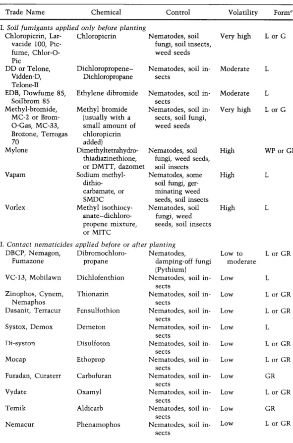
CHEMICAL CONTROL
The most promising method of controlling nematodes in the field has been through the use of chemicals called nematicides. Some of these, including chloropicrin, methyl bromide, Mylone, Vapam, and Vorlex, give off
CONTROL OF NEMATODES 625 gases after application to the soil and are general-purpose preplant fumi-
gants; they are effective against a wide range of soil microorganisms including, in addition to all nematodes, many fungi, insects, and weeds.
Other nematicides, e.g., Nemagon, Zinophos, Dasanit, Furadan, Mocap, and Temik, are of low volatility, are effective against nematodes and insects, and can be applied before and after planting of many, particularly non-food, crops which are tolerant to these chemicals.
Nematicides used as soil fumigants are available as liquids, emulsi- fiable concentrates, or granules. Application of nematicides in the soil is made either by applying the chemical evenly over the entire field (broad- cast) or by applying it only to the rows to be planted with the crop (row treatment). In both cases the fumigant is applied through delivery tubes attached at the back of tractor-mounted chisel-tooth injection shanks or disks spaced at variable widths and usually reaching 6 inches below the soil surface. The nematicide is covered instantly by a smoothing and firming drag or can be mixed into the soil with disk harrows or rototillers.
Highly volatile nematicides should be immediately covered with polyethylene sheeting (Fig. 238), and this should be left in place for at least 48 hours. When small areas are to be fumigated, the most conve- nient method is through injection of the chemical with a hand applicator or by placement of small amounts of granules in holes 6 inches deep, 6 to 12 inches apart, and immediately covering the holes with soil. In all cases
FIGURE 238.
Soil fumigation for the control of nematodes. Plastic sheet covers soil to keep volatile nematicides from escaping too soon. (Photo courtesy U.S.D.A.)
of preplant soil fumigation with phytotoxic nematicides, at least 2 weeks must elapse from the time of treatment before seeding or planting in the field to avoid plant injury.
In the above types of nematicide application, only a small portion of the soil and its microorganisms come in contact with the chemical immediately. The effectiveness of the fumigants, however, is based on the diffusion of the nematicides in a gaseous state through the pores of the soil throughout the area in which nematode control is desired. The distance of movement of the vapors is influenced by the size and con
tinuity of soil pores, soil temperature (best range between 10 and 20°C), soil moisture (best at about 80 percent of field capacity), and by the type of soil (more material is required for soils rich in colloidal or organic matter). Nematicides with low volatility, such as Nemagon, Furadan, and Zinophos, do not diffuse through the soil to any great extent and must be mixed with the soil mechanically or by irrigation water or rainfall. Most nematicides, with the exception of the highly volatile ones, can be applied in irrigation water as soaks or drenches, but only low-volatility nematicides can be applied through overhead sprinkler systems.
The most common nematicides and some of their properties are listed in Table IV. In practice, nematode control in the field is generally ob
tained by preplant soil fumigation with one of the nematicides listed in Table IV as "applied only before planting." These chemicals are nonspecific, i.e., they control all types of nematodes, although some nematodes are harder to control than others no matter what the nematicide. Chloropicrin, methyl bromide, Mylone, and Vapam are ex
pensive, broad spectrum nematicides that must be covered upon applica
tion and are therefore used for treatment of seedbeds and small areas. On the other hand, DD and EDB are cheaper, need not be covered upon application and are used for treatment of large fields. DD and EDB, however, control only nematodes and, therefore, mixtures of these with Vapam, Chloropicrin, or methyl bromide are often used to increase their fungicidal action. The chemicals listed as "contact nematicides applied before or after planting" can be used as preplant soil treatment of all types or crops, but their use after planting is limited to nonfood crops such as turf, ornamentals, nurseries, and young nonbearing orchard trees, and to a few, specific food crops for which each chemical has received clearance by the Food and Drug Administration. It should be noted that most of the contact nematicides were developed as insecticides, and nematicidal dos
ages are much higher than insecticidal ones. All nematicides are ex
tremely toxic to humans and animals and should be handled with great caution.
SELECTED REFERENCES
Anonymous. 1972 and annually afterwards. "Commonw. Inst, of Helminthology Descriptions of Plant-Parasitic Nematodes." Commonw. Agric. Bureaux, En
gland.
Christie, J. R. 1959. "Plant Nematodes, Their Bionomics and Control." Fla. Univ.
Agr. Expt. Sta. (Gainesville). 256 p.
Endo, Β. Y. 1975. Pathogenesis of nematode-inlected plants. Ann. Rev.
Phytopathol. 12:213-238.
" L = liquid; G = gas; WP = wettable powder; GR = granules.
N O T E: Some of the above chemicals are presently being reviewed by FDA and EPA for possible side effects and may be discontinued.
TABLE IV.
NAMES A N D PROPERTIES OF THE MOST C O M M O N NEMATICIDES
Trade Name Chemical Control Volatility Form"
I. Soil fumigants applied only before planting
Chloropicrin, Lar- Chloropicrin Nematodes, soil Very high L or G
vacide 100, Pic- fungi, soil insects,
fume, Chlor-O- weed seeds
Pic
DD or Telone, Dichloropropene- Nematodes, soil in Moderate L Vidden-D, Dichloropropane sects
Telone-Π
EDB, Dowfume 85, Ethylene dibromide Nematodes, soil in Moderate L
Soilbrom 85 sects
Methyl-bromide, Methyl bromide Nematodes, soil in Very high L or G MC-2 or Brom- (usually with a sects, soil fungi,
O-Gas, MC-33, small amount of weed seeds Brozone, Terrogas chloropicrin
70 added)
Mylone Dimethyltetrahydro- Nematodes, soil High WP or GR
thiadiazinethione, fungi, weed seeds, or DMTT, dazomet soil insects
Vapam Sodium methyl- Nematodes, some High L
dithio- soil fungi, ger
carbamate, or minating weed
SMDC seeds, soil insects
Vorlex Methyl isothiocy- Nematodes, soil High L
anate-dichloro- fungi, weed propene mixture, seeds, soil insects or MITC
II. Contact nematicides applied before or after planting
DBCP, Nemagon, Dibromochloro- Nematodes, Low to L or GR
Fumazone propane damping-off fungi moderate
(Pythium) VC-13, Mobilawn Dichlofenthion Nematodes,
sects
soil in- Low L
Zinophos, Cynem, Thionazin Nematodes, soil in- Low L or GR
Nemaphos sects
Dasanit, Terracur Fensulfothion Nematodes, sects
soil in- Low L or GR
Systox, Demox Demeton Nematodes,
sects
soil in- Low L
Di-syston Disulfoton Nematodes,
sects
soil in- Low L or GR
Mocap Ethoprop Nematodes,
sects
soil in- Low L or GR
Furadan, Curaterr Carbofuran Nematodes, sects
soil in- Low GR
Vydate Oxamyl Nematodes,
sects
soil in- Low L or GR
Temik Aldicarb Nematodes,
sects
soil in- Low GR
Nemacur Phenamophos Nematodes,
sects
soil in- Low L or GR
Jenkins, W. R., and D. P. Taylor. 1967. "Plant Hematology." Reinhold, New York, 270 p.
Krusberg, L. R. 1963. Host response to nematode infection. Ann. Rev.
Phytopathol. 1 : 2 1 9 - 2 4 0 .
Nusbaum, C. J., and H. Ferris. 1973. The role of cropping systems in nematode population management. Ann. Rev. Phytopathol. 1 1 : 4 2 3 - 4 4 0 .
Peachy, J. E. (Ed.). 1969. "Nematodes of Tropical Crops." Tech. Commun. Com- monw. Bur. Helminth. No. 40. 355 p.
Powell, Ν. T. 1971. Interactions between nematodes and fungi in disease com
plexes. Ann. Rev. Phytopathol. 9 : 2 5 3 - 2 9 4 .
Rohde, R. A. 1972. Expression of resistance in plants to nematodes. Ann. Rev.
Phytopathol. 1 0 : 2 3 3 - 2 5 2 .
Southey, J. F. (Ed.). 1959. "Plant Nematology." Ministry of Agr., Fisheries Food.
Tech. Bull. 7.
Smart, G. C , Jr., and V. G. Perry (Eds.). 1968. "Tropical Nematology." Univ. Fla.
Press, Gainesville. 153 p.
Thorne, G. 1961. "Principles of Nematology." McGraw-Hill, New York, 553 p.
Webster, J. M. 1969. The host-parasite relationships of plant-parasitic nematodes.
Advan. Parasitol. 7 : 1 - 4 0 .
Webster, J. M. (Ed.). 1972. "Economic Nematology." Academic Press, New York, 563 p.
Zuckerman, Β. M., W. F. Mai, and R. A. Rohde (Eds.). 1971. "Plant Parasitic Nematodes." Academic Press, New York, 2 volumes.
• Root-Knot Nematodes: Meloidogyne
R o o t - k n o t n e m a t o d e s o c c u r t h r o u g h o u t t h e world, but are found m o r e frequently and in greater n u m b e r s in areas w i t h w a r m or hot c l i m a t e s and short or mild winters. Root-knot nematodes are also found in g r e e n h o u s e s e v e r y w h e r e w h e n nonsterilized soil is used. T h e y a t t a c k m o r e t h a n 2 0 0 0 species of plants including a l m o s t all c u l t i v a t e d plants.
R o o t - k n o t n e m a t o d e s d a m a g e plants by devitalizing root tips and either stopping their g r o w t h or causing e x c e s s i v e root production, but primarily by causing f o r m a t i o n of swellings of t h e roots w h i c h n o t only deprive plants of n u t r i e n t s but also disfigure and r e d u c e t h e m a r k e t value of m a n y root crops. W h e n susceptible plants are infected at t h e seedling stage, losses are h e a v y and m a y result in c o m p l e t e d e s t r u c t i o n of t h e crop.
Infections of older plants m a y h a v e only slight effects o n yield or t h e y m a y r e d u c e yields considerably.
Symptoms. T h e aboveground s y m p t o m s are s i m i l a r t o t h o s e caused by m a n y o t h e r root diseases or e n v i r o n m e n t a l factors that result in reduced a m o u n t s of w a t e r available to t h e plant. Infected plants s h o w reduced g r o w t h and fewer, small, pale green, or y e l l o w i s h leaves t h a t tend to wilt in w a r m w e a t h e r . B l o s s o m s and fruits are either lacking or are dwarfed and of poor quality. Affected plants usually linger t h r o u g h t h e growing season and are s e l d o m killed p r e m a t u r e l y .
T h e m o s t c h a r a c t e r i s t i c s y m p t o m s of t h e disease are t h o s e appearing on t h e underground parts of t h e plants. Infected roots swell at t h e point of invasion and develop into t h e typical r o o t - k n o t galls w h i c h are t w o or t h r e e t i m e s as large in d i a m e t e r as t h e h e a l t h y root (Fig. 2 3 9 A ) . Several i nf e c t i on s t a k e place along t h e s a m e root and t h e developing galls give t h e
ROOT-KNOT NEMATODES 629
FIGURE 239.
(A) Galls on the roots of tomato plant caused by the root-knot nematode Meloidogyne sp. (B) Healthy and root-knot nematode-infected carrots. (C) Cross section of young tomato root showing part of root-knot nematode (arrow) and giant cells in the stele. (D) Section of tomato root showing a root-knot nematode feeding on the giant cells surrounding its head. (E) Female root-knot nematode feeding on young root and laying its egg mass in a matrix outside the root. (Photos A and Β courtesy U.S.D.A. Photos C, D, and Ε courtesy R. A. Rohde.)
root a rough, clubbed appearance. Roots infected by certain species of this nematode develop, in addition to galls, several short root branches which rise from the upper part of the gall and result in a dense, bushy root system (Fig. 239B). Usually, however, infected roots remain smaller and show various stages of necrosis. Rotting of the roots frequently develops, particularly late in the season. When tubers or other fleshy underground organs are attacked, they produce small swellings over their surface which become quite prominent at times and may cause distortion of the organs or cracking of their skin.
The pathogen: Meloidogyne sp. The adult male and female root-knot nematodes are easily distinguishable morphologically (Figs. 239 and 240).
The males are wormlike and about 1.2 to 1.5 mm long by 30 to 36 /zm in diameter. The females are pear shaped and about 0.40 to 1.30 mm long by 0.27 to 0.75 mm wide. Each female lays approximately 500 eggs in a gelatinous substance produced by the nematode. The first-stage larva
L a t e JL s t a g e l a r v a e
FIGURE 240.
Disease cycle of root knot caused by nematodes of the genus Meloidogyne.
develops inside e a c h egg and after undergoing t h e first m o l t w i t h i n t h e egg it b e c o m e s second-stage larva. T h e latter e m e r g e s from t h e egg into t h e soil, w h e r e it m o v e s until it finds a susceptible root. T h e second-stage larva is w o r m l i k e and is t h e only infective stage of this n e m a t o d e . If a susceptible h o s t is present in its vicinity, t h e larva enters t h e root, b e c o m e s sedentary, and grows in t h i c k n e s s , a s s u m i n g a sausage-shaped form. T h e n e m a t o d e feeds on t h e cells around its head by inserting its stylet and secreting saliva into t h e s e cells. T h e saliva s t i m u l a t e s cell e n l a r g e m e n t and also liquefies part of t h e c o n t e n t s of t h e cells, w h i c h are t h e n s u c k e d by t h e n e m a t o d e through its stylet. T h e n e m a t o d e undergoes a second m o l t and gives rise to t h e third-stage larva, w h i c h is similar to, but l a c k s a stylet and is s t o u t e r than, t h e second-stage larva. T h e third- stage larva goes t h r o u g h t h e third m o l t and gives rise to t h e fourth-stage larva, w h i c h c a n be distinguished as either m a l e or f e m a l e . A m a l e fourth-stage larva b e c o m e s w o r m l i k e and is coiled w i t h i n t h e third cuti- cle. It undergoes t h e fourth and final m o l t and e m e r g e s f r o m t h e root as t h e w o r m l i k e adult m a l e w h i c h b e c o m e s free living in t h e soil. T h e fourth-stage f e m a l e larva c o n t i n u e s to grow in t h i c k n e s s and s o m e w h a t in length, undergoes t h e fourth and final m o l t , and b e c o m e s an adult f e m a l e w h i c h appears pear shaped. T h e adult f e m a l e c o n t i n u e s to swell and, w i t h or w i t h o u t fertilization by a m a l e , produces eggs w h i c h are laid
ROOT-KNOT NEMATODES
in a gelatinous protective coat. The eggs may be laid inside or outside the root tissues depending on the position of the female. Eggs may hatch immediately or they may overwinter and hatch in the spring. A life cycle is completed in 25 days at 27°C, but it takes longer at lower or higher temperatures. When the eggs hatch, the infective second-stage larvae may migrate from within galls to adjacent parts of the root and cause new infections in the same root, or they may emerge from the root and infect other roots of the same plants or roots of other plants. The greatest numbers of root-knot nematodes are usually in the root zone from 5 to 25 cm below the surface, but galls have been found on peach and other roots 2 to 2.5 m deep. The ability of root-knot nematodes to move on their own power is limited, but they can be spread by water or by soil clinging to farm equipment or otherwise transported into uninfested areas.
Development of disease. Infective second-stage larvae usually enter roots behind the root tip, and push their way between or through cells until they reach positions behind the growing point. There they become permanently established with their head in the plerome (Fig. 240). In older roots the head is usually in the pericycle. Some cell damage occurs along the path of the larva and, if several larvae have entered, the cells near the root tip cease to divide and growth of the root stops. On the other hand, cortical cells near the point of entry begin to enlarge as sometimes do cells of the pericycle and endodermis near the path of the larvae. Two or three days after the larva has become established, some of the cells around its head begin to enlarge. Their nuclei divide but no cell walls are laid down. The existing walls between some of the cells break down and disappear and the protoplasmic contents of several coalesce, giving rise to giant cells (Figs. 239, C, D, and 240). Enlargement and coalescing of cells continues for 2 to 3 weeks, and the giant cells invade the surrounding tissues irregularly. Each gall usually contains 3 to 6 giant cells, which may form in the cortex as well as in the stele. The enlargement of the cells seems to be brought about by the substances contained in the saliva secreted by the nematode in the giant cells during feeding. The giant cells degenerate when nematodes cease to feed or die. When giant cells form in the stele, irregular xylem elements develop or their development may be interrupted. Xylem elements already present may be crushed by the mechanical pressure exerted by the enlarging cells. In the early stages of gall development the cortical cells enlarge in size but, during the later stages, they also divide rapidly. Swelling of the root results also from hypertrophy and hyperplasia of the vascular parenchyma, pericycle, and endodermis cells surrounding the giant cells and from enlargement of the nematode. As the females enlarge and egg sacs are formed, they push outward, split the cortex, and may become exposed on the surface of the root or may remain completely covered, depending on the position of the nematode in relation to the root surface.
In addition to the disturbance caused to plants by the nematode galls themselves, frequently damage to infected plants is increased by certain parasitic fungi, which can easily attack the weakened root tissues and the hypertrophied, undifferentiated cells of the galls. Moreover some fungi,
631
e.g., Pythium, Fusarium, and Rhizoctonia, grow and reproduce much faster in the galls than in other areas of the root, thus inducing an earlier breakdown of the root tissues.
Control. Root knot can be effectively controlled in the greenhouse with steam sterilization of the soil or soil fumigation with nematicides.
In the field the best control of root knot is obtained by fumigating the soil with chemicals such as DD, DBCP, or EDB. Several newer nematicides such as aldicarb, oxamyl, and phenamiphos are being used effectively.
Each treatment usually gives satisfactory control of root knot for one season. Varieties resistant to root-knot nematodes are also available in several crops.
SELECTED REFERENCES
Bird, A. F. 1974. Plant response to root-knot nematode. Ann. Rev. Phytopathol.
1 2 : 6 9 - 8 5 .
Carter, W. W., and S. Nieto, Jr. 1975. Population development of Meloidogyne incognita as influenced by crop rotation and fallow. Plant Dis. Reptr. 5 9 : 4 0 2 - 403.
Christie, J. R. 1936. The development of root-knot nematode galls. Phytopathol- ogy 2 6 : 1 - 2 2 .
Dropkin, V. H., and P. E. Nelson. 1960. The histopathology of root-knot nematode infections in soybeans. Phytopathology 5 0 : 4 4 2 - 4 4 7 .
Sasser, J. N. 1954. Identification and host-parasite relationships of certain root- knot nematodes [Meloidogyne sp.). Maryland Agr. Expt. Sta. Bull. A-77:30 p.
• Cyst Nematodes: Heterodera
Cyst nematodes cause a variety of plant diseases mostly in temperate regions of the world. Some species of cyst nematodes attack only a few plant species and are present over limited geographic areas while others attack a large number of plant species and are widely distributed. The most common cyst nematodes and their most important hosts are Heterodera avenae on cereals, H. glycines on soybeans, H. rostochiensis on potato (Fig. 241 A), tomato, and eggplant, H. schachtii on sugar beets (Fig. 24IB), crucifers, and spinach, H. tabacum on tobacco, andH. trifolii on clover. The diagnostic feature of cyst nematode infections is the presence of cysts on the roots and usually the proliferation of roots and production of shallow, bushy root systems.
• Soybean Cyst Nematode: Heterodera glycines
The soybean cyst nematode has been found in northeastern Asia, Japan, and in the U.S. in an area from Virginia to Florida to Arkansas to Missouri and Illinois. It continues to spread slowly to new areas in spite of the strict quarantine measures imposed on the presently infested areas. The most severely affected host is soybean, but several other legumes, such as common bean, vetch, lespedeza, lupine, and a few nonleguminous plants are also attacked by this nematode. Depending on the degree of infesta- tion, it can cause losses varying from slight to complete destruction of the crop. Usually, however, in heavily infested fields yield is reduced from 30 to 75 percent.
CYST NEMATODES 633
FIGURE 241.
(A) Larva and egg of the golden nematode of potato [Heterodera rostochiensis). (B) Bare spots in sugar beet field caused by injury by the sugar beet nematode Heterodera schachtii. (Photos courtesy U.S.D.A.)
Symptoms. Infected soybean plants appear stunted and have an un- thrifty appearance. The foliage turns yellow prematurely and falls off early. The plants bear only a few flowers and a few small seeds. Infected plants growing on sandy soil usually die. Infected plants growing on fertile soils with plenty of moisture may show only slight chlorosis of the older leaves, little or no stunting, and may produce a nearly normal yield for a year or two. In subsequent years, however, due to the tremendous buildup of nematodes in the soil, plants in these areas also become severely chlorotic and dwarfed.
The root system of infected plants appears smaller than that of healthy plants, but no macroscopic lesions, galls, or other type of abnormalities are evident on infected roots. Roots of infected plants usually have con- siderably fewer bacterial nodules than those of healthy plants. The most characteristic symptom of this disease is the presence of female nematodes in varying stages of development and of cysts attached on the soybean roots (Fig. 242). Young females are small, white, and partly buried in the root with only part of them protruding on the surface. Older females are larger, almost completely on the surface of the root, and appear yellowish or brown depending on maturity. Dead, brown cysts are also present on the roots.
The pathogen: Heterodera glycines. The soybean cyst nematode overwinters as a brown cyst in the upper 90 to 100 cm of soil. The cysts are the leathery skins of the females and are filled with eggs. The eggs contain fully developed second-stage larvae (Fig. 243). When temperature and moisture become favorable in the spring, the larvae emerge from the cysts and infect roots of host plants.
At 4 to 6 days after penetrating the roots, the larvae molt and produce the third-stage larvae. The third-stage larvae are much stouter than the second-stage larvae and 5 to 6 days later fourth-stage larvae begin to appear. The female fourth-stage larva loses its somewhat slender appear-
FIGURE 242.
Lemon-shaped encysted female nematodes attached to soybean roots. (Photo courtesy U.S.D.A.)
ance and develops the typical flask shape, measuring approximately 0.40 mm in length by 0.12 to 0.17 mm in width. By day 12 to 15, adult males and females appear.
The adult male is wormlike, about 1.3 mm long by 30 to 40 μτη in diameter. The males remain in the root for a few days, during which they may or may not fertilize the females, then move into the soil and soon die.
The adult females when fully developed are lemon shaped, measuring 0.6 to 0.8 mm in length and 0.3 to 0.5 mm in diameter. They are white to pale yellow at first, becoming yellowish-brown as they mature. The body cavity of the female is almost completely filled by the ovaries, and as the ova gradually develop into fully formed eggs, the body cavity of the female becomes completely filled with eggs. As the female body distends during egg production, it crushes cortical cells, splits the root surface, and pro
trudes until it is almost entirely exposed through the root surface. A gelatinous mass, usually mixed with dirt and debris, surrounds the poste-
FIGURE 243.
Disease cycle of the soybean cyst nematode Heterodera glycines.
635
rior end of the females and the nematodes deposit some of their eggs in it.
Each female produces 300 to 600 eggs, most of which remain inside her body when the female dies. Eggs in the gelatinous matrix may hatch immediately and the emerging second-stage larvae may cause new infec- tions. Finally, the old body wall, darkening to brown, becomes the cyst.
Approximately 21 to 24 days are required for the completion of a life cycle of this nematode. The cyst consists of the female cuticle transformed through the secretions of the nematode into a tough, brown sac that persists in the soil for many years and protects the eggs which have been formed within the body.
Development of disease. The infective second-stage larvae penetrate young primary roots or apical meristerns of secondary roots directly (Fig.
243). The advance into the cortex is mostly intracellular and results in distortion and death of invaded cells. The larvae often pass through the cortex and pierce their stylets into cells of the endodermis or the pericy- cle. Within 2 days from penetration larvae come to rest and feed on cells of the cortex and stele tissues causing the enlargement of these cells.
Such groups of enlarged cells, called syncytia, are surrounded by a single layer of small, hyperplastic cells the walls of which undergo further dissolution and allow enlargement of the syncytia. During the develop- ment of the third larval stage cortical cells surrounding the nematode are crushed by the expanding nematode body, especially by developing females. Syncytial development is either restricted largely to pericyclic tissue or occurs in tissues of the phloem and secondary cambium. Syn- cytia in contact with developing third- or fourth-stage males begin to show signs of degeneration indicating cessation of feeding. Syncytia in contact with females remain active up to and beyond the stage of egg deposition.
Degeneration of syncytia is accompanied by reduction of syncytial vol- ume and results in the receding and collapse of the syncytial wall. The resulting space is only partly occupied by surrounding parenchymatous tissue.
When soybean varieties resistant to the soybean cyst nematode are attacked, there is no apparent inhibition of penetration of the organism into the host tissues. Syncytia are formed within 2 to 3 days from inocu- lation, but by day 5 many of them degenerate and most second-stage larvae associated with them are dead. A few nematodes advance to the third stage, but no adult males or females are produced. Development of syncytia and subsequent degeneration and necrosis is restricted to the periphery of the stele and to regions in the cortex that are invaded and stimulated hv infective larvae. The root regions vacated by degenerate syncytia are qi ickly filled by adjacent rejuvenated parenchyma cells.
Syncytial development into the secondary cambial region of suscepti- ble varieties results in inhibition of secondary growth of both phloem and xylem. Since a short portion of a root may be attacked by many larvae, the large number of syncytia that develop may cause widespread reduction of the conductive elements, resulting in the restricted growth and yield of soybean plants, especially under stresses of moisture.
Control. Soil fumigation of soybean cyst nematode-infested fields
SUGAR BEET NEMATODE 637 with a variety of nematocides temporarily increases plant growth and
soybean yield. Nematode cysts and larvae, however, are almost never eradicated from a field completely by fumigation and a small nematode population left over after fumigation can build up rapidly on the vigorous soybean grown in newly fumigated soil. In addition, the cost of fumiga
tion per acre makes its use impractical.
The most practical method of control of the soybean cyst nematode is through a 2- to 3-year crop rotation, since some legumes are the only other cultivated crops that are hosts of this nematode. The effectiveness of crop rotation is increased by planting the more resistant soybean varieties which do not allow a quick and excessive buildup of nematode popula
tions.
Quarantine regulations are presently enforced to prevent the parasite from spreading into nematode-free areas by means of contaminated soil, products, machinery, or other articles.
• Sugar Beet Nematode: Heterodera schachtii
It occurs wherever sugarbeets are grown in North America, Europe, Mid
dle East, and Australia and is the most important nematode pest of sugar beet production. It also affects spinach and crucifers. The sugar beet nematode causes yield losses of 25 to 50 percent or more, especially in warmer climates or late planted crops. The losses on sugar beet are mostly the result of reduced root weight but in warm climates the sugar content is also reduced and, generally, the nematode agravates losses caused by other pathogens such as Cercospora, Rhizoctonia, and beet viruses. In fields infested with the sugar beet nematode, small to large patches of wilting or dead young plants or stunted older sugar beets appear (Fig. 241B). The latter have an excessive number of hairlike roots.
Small white or brownish cysts of female nematodes can be seen clinging to the roots. The morphology, biology, and spread of the sugar beet nematode is similar to that of the soybean cyst nematode. Control of the sugar beet nematode is based on early sowing so that plants can grow as much as possible at temperatures at which the nematodes are more or less inactive, on crop rotations with alfalfa, cereals, or potatoes which are not hosts of this nematode, and soil fumigation with DCP, EDB, or DBCP. No sugar beet varieties resistant to this nematode are commer
cially available yet.
SELECTED REFERENCES
Anonymous. 1961. Soybean cyst nematode. U.S. Dept. Agr., Agr. Res. Serv. Spec.
Reptr. 2 2 - 7 2 : 2 0 p.
Endo, Β. V. 1964. Penetration and development of Heterodera glycines in soybean roots and related anatomical changes. Phytopathology 5 4 : 7 9 - 8 8 .
Endo, Β. V. 1965. Histological responses of resistant and susceptible soybean varieties and backcross progeny to entry and development of Heterodera glycines. Phytopathology 5 5 : 3 7 5 - 3 8 1 .
Franklin, Mary T. 1972. Heterodera schachtii. C.I.H. Descriptions of Plant- Parasitic Nematodes. Set 1, No. 1. 4 p.