DOI: 10.1556/066.2018.47.3.4
DOSE DEPENDENT HEPATOTOXIC EFFECTS OF DRY SEED PHASEOLUS VULGARIS LINN. (RED KIDNEY BEANS) ON RABBITS
A. SIDDIQa, A.M. HASANb and S. ALAMa*
aDepartment of Pharmacology, Faculty of Pharmacy and Pharmaceutical Sciences, University of Karachi, Karachi-75270. Pakistan
bDepartment of Pharmacognosy, Faculty of Pharmacy and Pharmaceutical Sciences, University of Karachi, Karachi-75270. Pakistan
(Received: 31 July 2017; accepted: 29 December 2017)
Growing research on beans suggest that they are good for health, as they reduce the risk and/or help in the management of chronic diseases. Beans are valuable parts of several dietary recommendations. Saponins, α-galactosides, phytates, and lectins are currently drawing attention because of their diverse properties, both adverse and benefi cial. The aim was to determine the hepatotoxic effect of dry red kidney beans, i.e. Phaseolus vulgaris, on rabbits at 4 g kg–1, 6 g kg–1, and 8 g kg–1 dosing for the period of 30 days. The histopathological examination revealed infl ammation of hepatocyte at all three doses along with congestion. The biochemical testing of liver enzymes revealed elevated alanine transaminase (ALT), aspartate transaminase (AST) and gamma-glutamyl transferase (γ-GT). The dose-dependent increase in liver enzymes indicates the hepatotoxic effect of dry red kidney beans, and various studies have demonstrated that lectins are the toxic components responsible for infl ammation of body organs.
Keywords: Phaseolus vulgaris, hepatotoxic, liver enzymes, infl ammation
The Gene Banks of the World maintain the record of 40 000 bean cultivars, of which only a small part is massively cultivated for regular consumption (MCGINNIS & SUSZKIW, 2006).
There are 600 different cultivars of beans grown all around the world, of which 62 are commercially marketed and only 15 are recognized internationally. Beans are considered as
“perfect” food because they are nutritionally strong. Moreover, beans are promising sources of complex carbohydrates, dietary fi bre, folic acid, and proteins as well as some minerals (JONES, 2011). Phaseolus vulgaris (beans) are used less because of their antinutrient constituents, like contents inhibiting the activity of chymotrypsin, trypsin and α-amylase, fl atulence stimulating factors, phytic acid (PA), saponins, and lectins, and they also need prolong cooking time (SOETAN, 2008). Saponins, α-galactosides, phytates, and lectins are currently drawing attention because of their diverse properties, which are sometimes benefi cial as well as adverse for human body (GUPTA et al., 2016).
Growing research on beans suggests that they are good for health as they reduce the risk and/or help in the management of chronic diseases. Beans are incorporated in many dietary recommendations, Produce for Better Health Foundation, American Cancer Society, Dietary Approaches to Stop Hypertension (DASH), Dietary Guidelines for Americans (DGA) 2005, American Heart Association (AHA), and MyPyramid (WINHAM et al., 2008). AHA has included beans in their recommended diet designed to prevent heart diseases (LICHTENSTEIN et al, 2006). Phytochemicals bearing antioxidant properties include total phenolic compounds,
tannins, flavonoids, and phenolic acids. They work by interfering chain reactions of oxidation by eradicating free radicals and blocking further oxidative processes (PAREDES et al., 2009;
JEON et al., 2012).
It is a common perception of individuals that the use of beans as meal can lead to gas and bloating. On the other hand, the very same components that are responsible for gas and bloating possess health-improving properties. Beside this, they may be involved in enhancing the growth of gut fl ora. According to some studies, the fl atulence resulting from consumption of Phaseolus vulgaris is reduced after some period (LIANG, 2014).
Lectins are the toxic components of beans. They are responsible for infl ammation and food poisoning. Lectins from various foodstuffs aggravate various infl ammatory and digestive diseases. They act as carrier for transportation of various antigenic foreign proteins, which leads to destruction of different organs like brain, joints, skin, and other glands (HAMID &
MASOOD, 2009). Metabolic and tissue changes have also been associated with lectins. They are reported to modify gut and systemic metabolism (DE MOYA et al., 2003) and induce hyperplasia as well as hypertrophy of small intestine along with alteration of organs’ weight and function (MARZO et al., 2002). High concentration of lectin triggers abnormalities in brush border cells of small intestine (MIYAKE et al., 2007). Neutral fi bres and acid fi bres are able to decrease the availability of iron, copper, and zinc for the process of absorption (GARCIA-LOPEZ, 1984). PA and few proteins also hinder the absorption of minerals like magnesium, zinc, iron, and calcium (REYES-MORENO et al., 1993).
Because of diversifi ed effects including both benefi cial and adverse, and beans being a routine food for consumption in various parts of world, we aimed to determine the hepatotoxic effect of dry seeds of red kidney beans on rabbits.
1. Materials and methods
1.1. Animal selection
The study was performed on forty white, healthy male rabbits, weighing 1000–1500 grams.
Rabbits were provided with diet (fresh hay grass) and water daily. The reason for inducting rabbits as experimental animal is that several parameters, for instance biochemical and histopathological changes observed in rabbits, are similar to humans (SMITH & TAYLOR, 1995). The study was approved by animal ethical committee of Department of Pharmacology, Faculty of Pharmacy, University of Karachi and Board of Advance Studies and Research University of Karachi, Resol. No. 10(P) 05 dated: 13-10-2014.
1.2. Dosing
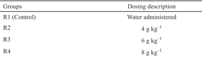
Rabbits were divided into four groups, R1, R2, R3, and R4, each group of 10 rabbits. Group R1 was assigned as control group to which water was administered (SHAH et al., 1992). The maximum dose of dry powder seeds of Phaseolus vulgaris used is 2.5 g kg–1 for a period of 28 days (CHOKSHI, 2007). Therefore, the doses were designed as 4 g kg–1, 6 g kg–1, and 8 g kg–1 of dry powder seeds of Phaseolus vulgaris for group R2, R3, and R4, respectively, as shown in Table 1. These doses were administered once a day every morning through oral route using gastric gavage for 30 days, diluted with a small amount of water to ease the administration.
All rabbits were acclimatized for a period of 10 days before the administration of doses. The study has been conducted from 1st to 30th September 2016.
Table 1. Dosing groups
Groups Dosing description
R1 (Control) Water administered
R2 4 g kg–1
R3 6 g kg–1
R4 8 g kg–1
1.3. Liver function test (LFT)
The ear of rabbits was phlebotomized for the collection of blood using sterile disposable syringe. The sample of blood was obtained in siliconized glass tube and was instantly separated by centrifugation at 3000 r.p.m. for 15 min by Humax 14 K (Germany). Auto- chemistry analyser was used to determine the liver enzymes from the collected plasma.
Enzymes estimated include Serum Glutamic-Pyruvic Transaminase (ALT), Serum Glutamic Oxaloacetic Transaminase (AST), Alkaline phosphatase (ALP), Gamma-Glutamyl Transferase (γ-GT) as well as Direct Bilirubin (DBR), and total Bilirubin (TBR). Liver functions test was used to diagnose the diseases of liver (GEORGE-GAY & PARKER, 2003).
1.4. Histopathological examination
Diagnostic Lab and Clinical Research Facility of Dr. Panjwani Center for Molecular Medicine and Drug Research Faculty was used in order to carry out histopathological examination of rabbit’s liver.
1.5. Statistical analysis
Statistical analysis was accomplished by using Statistical Package for the Social Sciences (SPSS) 20. All values of control and test groups were compared by using mean and standard deviation to the mean by applying one-way analysis of variance (ANOVA) with post hoc Tukey’s Honest Signifi cant Difference (HSD) test. Data in the form of mean ± standard deviation to the mean through 95% confi dence of interval and P-values were noticed. Values of P≤0.05 were considered as signifi cant, P≤0.01 were considered as highly signifi cant. Total no. of rabbits (n) in each group was 10.
2. Results and discussion
Gross examination did not show any macroscopic and microscopic alteration in liver in any group. However, there was evidence of mild to moderate infl ammation in portal tract of liver in all animal groups except control group along with congestion in sinusoids as shown in Figure 1. No fatty acid changes were observed in any of the groups. Table 2 illustrates the
Table 2. Scoring of histopathological examination of liver
Parameters No. of rabbits in the group
R1 (Control) R2 R3 Group R4
Hepatic portal tract infl ammation
No changes 9 1 0 1
Mild 1 8 7 4
Moderate 0 1 3 5
Congestion in sinusoids
No changes 9 1 0 1
Mild 1 8 7 4
Moderate 0 1 3 5
Fig. 1. Infl amed hepatic portal tract
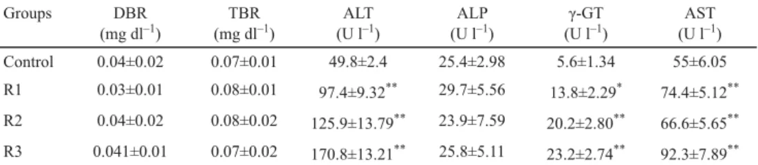
Table 3 reveals the comparison of DBR, TBR, ALT, ALP, and γ-GT levels of rabbits in control group and rabbits of test groups.
Table 3. Effects of different dosing groups of Phaseolus vulgaris on liver enzymes
Groups DBR
(mg dl–1)
TBR (mg dl–1)
ALT (U l–1)
ALP (U l–1)
γ-GT (U l–1)
AST (U l–1)
Control 0.04±0.02 0.07±0.01 49.8±2.4 25.4±2.98 5.6±1.34 55±6.05
R1 0.03±0.01 0.08±0.01 97.4±9.32** 29.7±5.56 13.8±2.29* 74.4±5.12**
R2 0.04±0.02 0.08±0.02 125.9±13.79** 23.9±7.59 20.2±2.80** 66.6±5.65**
R3 0.041±0.01 0.07±0.02 170.8±13.21** 25.8±5.11 23.2±2.74** 92.3±7.89**
Values expressed as mean ± SEM; number of animals (N)=10; *: P≤0.05 signifi cant as compared to control; **:
P≤0.01 highly signifi cant as compared to control
The levels of ALT increased in highly signifi cant (P≤0.01) manner in groups A, B, and C, i.e. 97.4±9.32 units per litre (U l–1), 125.9±13.79 U l–1 and 170.8±13.21 U l–1, respectively, as compared to control group, i.e. 49.8±2.4 U l–1.
The animals of group A showed signifi cant (P≤0.05) increase in the levels of γ-GT (13.8±2.29 U l–1) when compared to control group, i.e. 5.6±1.34 U l–1. And when comparing control to groups B and C, the levels of γ-GT increased in highly signifi cant (P≤0.01) manner, i.e. 20.2±2.80 U l–1 and 23.2±2.74 U l–1, respectively.
The levels of AST increased in highly signifi cant (P≤0.01) manner when levels of AST from control group, i.e. 55±6.05 U l–1, were compared to groups A, B, and C, i.e. 74.4±5.12 U l–1, 66.6±5.65 U l–1, and 92.3±7.89 U l–1, respectively.
However, the levels of TBR, DBR, and ALP do not show any signifi cant differences when the animals in control group are compared with groups A, B, and C.
The liver of rabbit consists of caudate lobe along with a narrow stalk. They have a gall bladder, which is primarily responsible for the storage of bile. It has been observed that no specifi c enzyme exists for the liver. However, in the majority of mammals, ALT is only present in liver, but in rabbits, this enzyme is present in both the liver and cardiac muscles.
AST is also found in various tissues, including muscles, liver, kidneys, and pancreas.
Therefore, the elevation of these enzymes can be ruled out through histopathological examination (MITCHELL & TULLY, 2008).
In the human body, liver is the largest and most complex organ, which plays a key role in the metabolism of various molecules like carbohydrate, protein, and fat. It is also referred to as detoxifi cation centre of drug and chemicals. Synthesis and secretion of bile also takes place in the liver. It also brings about the production of lipoproteins and plasma proteins and regulates blood glucose level (GIANNINI et al., 2005). A set of tests exists to check suspected hepatic disorder, measure the outcome of disease, or simply to evaluate the status of liver function. These tests involve the screening of blood, which gives information about a wide range of disease conditions (HALL & CASH, 2012). It is well understood that any destruction or damage to liver causes the release of enzymes from hepatocytes into the serum and later on the condition advances to necrosis or leads to alteration in cell membrane permeability (KYZNIETSOVA et al., 2015). ALT is predominantly present in hepatocellular cytoplasm, on the other hand, 80% of AST exists in the mitochondria of cells. Because of this fact, detection and prognosis of liver diseases are easily carried out. In slight hepatocellular injury, the principal form of enzyme in serum is cytoplasmic, whereas in serious injury, discharge of the mitochondrial enzyme occurs in the bloodstream. The γ-GT enzyme is associated with hepato-biliary disorders with greater specifi city as compared to other hepatic enzymes such as alkaline phosphatase and transaminases (PEREIRA et al., 2012).
The tests included in this research for accessing liver functions were for ALT, AST, γ-GT, ALP, along with direct bilirubin and total bilirubin. In this study, increase in levels of ALT, AST, and γ-GT was observed, which is attributed to the liver injury. The seed cotyledons of Phaseolus vulgaris are rich in lectins (NATARAJAN et al., 2013), and they are proved to be toxic and are responsible for infl ammation and food poisoning. Numerous diseases related to infl ammation and some digestive diseases arise from lectin containing food, grains, cereals, dairy, and legumes (HAMID & MASOOD, 2009). A study on mice, whose diet contained lectins, reported that lectins are accountable for bringing about structural and enzymatic alterations in liver (IKEGWUONU & BASSIR, 1976; CIRNATU et al., 2011). Diet containing lectin as protein, when incompatible with the type of antigen present in the blood may lead to destructions of
Therefore, it is suggested that lectins from Phaseolus vulgaris may increase the amount of liver enzymes, possibly by damaging the liver. Mild to moderate infl ammation in hepatic portal tract and sinusoidal congestions, as observed in histopathological study of liver, support the likelihood of hepatic adverse effect. The release of cytoplasmic enzyme, i.e. ALT, as well as mitochondrial enzyme, i.e. AST, indicates that dry seed powder of Phaseolus vulgaris causes severe hepatic injury. It has also been observed that with the subsequent increase in the dose of dry seed powder of Phaseolus vulgaris, the levels of ALT and γ-GT tend to increase signifi cantly. Increase in the level of γ-GT suggests the occurrence of hepato- biliary disorders as reported by PEREIRA and co-workers (2012). Hyperactivity of liver enzymes might also arise, because of increased catabolism of amino acids in the liver caused by lima beans. Release of AST and ALT from hepatocytes was observed in a study, in which dietary intake of lima beans was increased compared to normal intake (VASCONCELOS &
OLIVEIRA, 2004). It has been reported in a study that toxic manifestation of Phaseolus vulgaris is due to two main constituents, i.e. lectins and phytohaemagglutinins. The interaction of lectins with intracellular and extracellular glycoproteins disturbs the homeostasis of the body.
Besides, lectins inhibit the repair of plasma membrane, and they also inhibit the exocytosis, which results in cellular damage (KUMAR et al., 2013).
3. Conclusions
It is concluded that at doses of 4 g kg–1, 6 g kg–1, and 8 g kg–1, dry seeds of Phaseolus vulgaris have adverse effects on liver as indicated by dose-dependent elevation of hepatic enzymes.
Histopathological examination further supports the evidence of hepatotoxic effect, revealing infl ammation. The suggested mechanism for this adverse effect could be hyper-protein catabolic activity, which reportedly increases the amount of liver enzymes, disturbes homeostasis, inhibits cellular exocytosis and repair of plasma membrane. This work can be extended to understand further cellular mechanisms of action of constituents responsible for hepatotoxic effects as well as the culprit constituent underlying this adverse effect.
References
CHOKSHI, D. (2007): Subchronic oral toxicity of a standardized white kidney bean (Phaseolus vulgaris) extract in rats. Food Chem. Toxicol., 45(1), 32–40.
CIRNATU, D., JOMPAN, A., SIN, A.I. & ZUGRAVU, C.A. (2011): Multiple organ histopathological changes in broiler chickens fed on genetically modifi ed organism. Rom. J. Morphol. Embryo., 52(1), 475–480.
DE MOYA, C.C., GRANT, G., FRÜHBECK, G., URDANETA, E., GARCÍA, M., MARZO, F. & SANTIDRIÁN, S. (2003): Local (gut) and systemic metabolism of rats is altered by consumption of raw bean (Phaseolus vulgaris L. var.
athropurpurea). Brit. J. Nutr., 89(03), 311–318.
GARCIA-LOPEZ, J.S. (1984): Binding of minerals by cooked pinto beans (Phaseolus vulgaris) fi ber, infl uence of fi ber on iron absorption by normal and anemic rat intestinal segments. Doctoral dissertation, University of Wisconsin, Madison.
GEORGE-GAY, B. & PARKER, K. (2003): Understanding the complete blood count with differential. J. Perianesth.
Nurs., 18(2), 96–117.
GIANNINI, E.G., TESTA, R. & SAVARINO, V. (2005): Liver enzyme alteration: A guide for clinicians. Can. Med. Assoc.
J., 172(3), 367–379.
GUPTA, R.K., GUPTA, K., SHARMA, A., DAS, M., ANSARI, I.A. & DWIVEDI, P.D. (2016): Health risks and benefi ts of chickpea (Cicer arietinum) consumption. J. Agr. Food Chem., 65(1), 6–22.
HALL, P. & CASH, J. (2012): What is the real function of the liver ‘function’ tests. Ulster. Med. J., 81(1), 30–36.
HAMID, R. & MASOOD, A. (2009): Dietary lectins as disease causing toxicants. Pakistan J. Nutr., 8(3), 293–303.
IKEGWUONU, F.I. & BASSIR, O. (1976): Alterations in function, enzyme activities, and histopathology of the liver of the rat after administration of phytohemagglutinins (lectins). Toxicol. Appl. Pharm., 37(2), 211–216.
JEON, S., HAN, S., LEE, J., HONG, T. & YIM, D.S. (2012): The safety and pharmacokinetics of cyanidin-3-glucoside after 2-week administration of black bean seed coat extract in healthy subjects. Korean J. Physiol. Pha., 16(4), 249–253.
JONES, A.L. (2011): Phaseolus bean. Post-harvest operation. Food and Agriculture Organization of the United Nations. 24 pages.
KUMAR, S., VERMA, A.K., DAS, M., JAIN, S.K. & DWIVEDI, P.D. (2013): Clinical complications of kidney bean (Phaseolus vulgaris L.) consumption. Nutrition, 29(6), 821–827.
KYZNIETSOVA, M.Y., MAKIEIEVA, O.M., LAVROVSKA, D.O., TYMOSHENKO, M.O., SHEVEROVA, D.P., HALENOVA, T.I., MYCHOLAYOVYCH, O. & SAVCHUK, L.I.O. (2015): Effect of aqueous extract from Phaseolus vulgaris pods on lipid peroxidation and antioxidant enzymes activity in the liver and kidney of diabetic rats. J. Appl. Pharm.
Sci. (JAPS), 5(5), 1–6.
LIANG, Y. (2014): Effect of regular dietary consumption of beans or peas on body weight, body composition, and blood pressure in men and women with mild hypercholesterolemia. Doctoral dissertation, University of Alberta
LICHTENSTEIN, A.H., APPEL, L.J., BRANDS, M., CARNETHON, M., DANIELS, S., FRANCH, H.A., FRANKLIN, B., KRIS- ETHERTON, P., HARRIS, W.S., HOWARD, B. & KARANJA, N. (2006): Diet and lifestyle recommendations revision, 2006. A scientifi c statement from the American Heart Association nutrition committee. Circulation, 114(1), 82–96.
MARZO, F., ALONSO, R., URDANETA, E., ARRICIBITA, F.J. & IBANEZ, F. (2002): Nutritional quality of extruded kidney bean (Phaseolus vulgaris L. var. Pinto) and its effects on growth and skeletal muscle nitrogen fractions in rats.
J. Anim. Sci., 80(4), 875–879.
MCGINNIS, L. & SUSZKIW, J. (2006): Breeding better beans. Agric. Res., 54(6), 12.
MITCHELL, M. & TULLY JR, T.N. (2008): Manual of exotic pet practice. Elsevier Health Sciences. pp. 127–143.
MIYAKE, K., TANAKA, T. & MCNEIL, P.L. (2007): Lectin-based food poisoning: A new mechanism of protein toxicity.
PLoS ONE, 2(8), e687.
NATARAJAN, S.S., PASTOR-CORRALES, M.A., KHAN, F.H. & GARRETT, W.M. (2013): Proteomic analysis of common bean (Phaseolus vulgaris L.) by two-dimensional gel electrophoresis and mass spectrometry. J. Basic Appl.
Sci., 9, 424.
PAREDES, C., BECERRA, V. & TAY, U. (2009): Inorganic nutritional composition of common bean (Phaseolus vulgaris L.) genotypes race Chile. Chil. J. Agr. Res., 69(4), 486–495.
PEREIRA, L.L.S., PEREIRA, C.A., VICENTEDESOUSA, R., DOS SANTOS, C.D., FERREIRADEMORAES, C. & SATIRO, L.C.
(2012): White bean fl our (Phaseolus vulgaris): Therapeutic and toxicological research in Wistar rats. J. Appl.
Pharm. Sci., 2(3), 1.
REYES-MORENO, C., PAREDES-LÓPEZ, O. & GONZALEZ, E. (1993): Hard-to-cook phenomenon in common beans – A review. Crit. Rev. Food Sci. Nutr., 33(3), 227–286.
SHAH, A.K., BRUNDAGE, R.C., GRATWOHL, A. & SAWCHUK, R.J. (1992): Pharmacokinetic model for subcutaneous absorption of cyclosporine in the rabbit during chronic treatment. J. Pharm. Sci., 81(6), 491–495.
SMITH, C.P. & TAYLOR, V. (1995): The behaviour of rabbits: Implications for their laboratory management. -in:
Standards in laboratory animal management. Proceedings (2) of UFAW Symposium, Potters Bar, Herts, UK, pp. 127–143.
SOETAN, K. O. (2008): Pharmacological and other benefi cial effects of antinutritional factors in plants – A review. Afr.
J. Biotechnol., 7(25), 4713-4721.
VASCONCELOS, I.M. & OLIVEIRA, J.T.A. (2004): Antinutritional properties of plant lectins. Toxicon., 44(4), 385–403.
WINHAM, D., WEBB, D. & BARR, A. (2008): Beans and good health. Nutr. Today, 43(5), 201–209.