Contents lists available atScienceDirect
European Journal of Pharmaceutical Sciences
journal homepage:www.elsevier.com/locate/ejps
Comparative biocompatibility and antimicrobial studies of sorbic acid derivates
Dániel Nemes
a, Renátó Kovács
b, Fruzsina Nagy
b, Zoltán Tóth
b, Pál Herczegh
c, Anikó Borbás
c, Viktor Kelemen
c, Walter P. P fl iegler
d, István Rebenku
e, Péter B. Hajdu
e, Pálma Fehér
a, Zoltán Ujhelyi
a, Ferenc Fenyvesi
a, Judit Váradi
a, Miklós Vecsernyés
a, Ildikó Bácskay
a,⁎aDepartment of Pharmaceutical Technology, Faculty of Pharmacy, University of Debrecen, Debrecen, 4032, Hungary
bDepartment of Medical Microbiology, Faculty of Medicine, University of Debrecen, Debrecen, 4032, Hungary
cDepartment of Pharmaceutical Chemistry, Faculty of Pharmacy, University of Debrecen, Debrecen, 4032, Hungary
dDepartment of Biotechnology and Microbiology, Faculty of Science and Technology, University of Debrecen, Debrecen, 4032, Hungary
eDepartment of Biophysics and Cell Biology, Faculty of Medicine, University of Debrecen, Debrecen, 4032, Hungary
A R T I C L E I N F O
Keywords:
Sorbates Preservatives Biocompatibility G. mellonella Caco-2 cells Antimicrobials
A B S T R A C T
Nowadays, the sorbates are the third largest group of antimicrobial preservatives in food and pharmaceutical industries, following the parabens and benzoates whose safety is questioned by recent publications. A dis- advantage of sorbates is their pH dependence, as their antimicrobial effect is greatly reduced in alkaline en- vironment. The main, widely used sorbate derivatives are sorbic acid and potassium sorbate, no sorbic acid esters are involved in current industrial application. We aimed to test whether the esters of sorbic acid are capable to extend the antimicrobial spectrum of the original molecule while maintaining its advantageous biocompatibility profile. A comparative biocompatibility study of different derivatives (sorbic acid, potassium sorbate, isopropyl sorbate and ethyl sorbate) was carried out. In vitro cell viability assays of MTT (2-(4,5- dimethyl-2-thiazolyl)-3,5-diphenyl-2H-tetrazolium bromide), Neutral Red (3-amino-7-dimethylamino-2-me- thylphenazine hydrochloride) andflow cytometry with propidium iodide and annexin were performed on Caco- 2 cells. In case of in vivo toxicity study,G. mellonellalarvae were injected with different concentrations of the test compounds. Time-kill tests were executed on reference strains ofC. albicans, E. coli, andS. aureus. According to the MTT-assay, the IC50values were the following: ethyl sorbate, sorbic acid <0.045%w/w, isopropyl sorbate 0.32%w/w, potassium sorbate >0.75%w/w, while Neutral Red values were >0.75%w/wfor the esters and potassium sorbate and 0.66%w/wfor sorbic acid. Flow cytometry results indicated the higher cell damage in case of isopropyl sorbate. However, the cytotoxic results of isopropyl sorbate, in vivo toxicity study onG.
mellonellalarvae did not show significant mortality. It was found, that the antimicrobial properties of isopropyl sorbate were outstanding compared to sorbic acid and potassium sorbate. These results indicate, that the use of sorbate esters can be advantageous, hence, further toxicity studies are needed to prove their safety.
1. Introduction
Many commonly used excipients are presented in pharmaceutical and food industries. One such jointly used group of compounds are the antimicrobial preservatives. As both liquid, oral pharmaceutical pre- parations and certain beverages and drinks can be opened and closed
multiple times until their expiration date, every interaction with the outer environment risks the contamination of the product. The alkyl esters of 4-hydroxybenzoic acid, the parabens are the most commonly used group of pharmaceutical preservatives. However, recent studies indicated that they could actively promote the proliferation of estrogen dependent cell lines (Roszak et al., 2017). Their interaction with human
https://doi.org/10.1016/j.ejps.2019.105162
Received 15 July 2019; Received in revised form 24 October 2019; Accepted 18 November 2019
⁎Corresponding author.
E-mail addresses:nemes.daniel@pharm.unideb.hu(D. Nemes),kovacs.renato@med.unideb.hu(R. Kovács),nagyfruzsina0429@gmail.com(F. Nagy), toth.zoltan@med.unideb.hu(Z. Tóth),herczegh.pal@pharm.unideb.hu(P. Herczegh),borbas.aniko@pharm.unideb.hu(A. Borbás),
kelemen.viktor@pharm.unideb.hu(V. Kelemen),pfliegler.valter@science.unideb.hu(W.P. Pfliegler),rebenku.istvan@med.unideb.hu(I. Rebenku), hajdup@med.unideb.hu(P.B. Hajdu),feher.palma@pharm.unideb.hu(P. Fehér),ujhelyi.zoltan@pharm.unideb.hu(Z. Ujhelyi),
fenyvesi.ferenc@pharm.unideb.hu(F. Fenyvesi),varadi.judit@pharm.unideb.hu(J. Váradi),vecsernyes.miklos@pharm.unideb.hu(M. Vecsernyés), bacskay.ildiko@pharm.unideb.hu(I. Bácskay).
Available online 20 November 2019
0928-0987/ © 2019 The Authors. Published by Elsevier B.V. This is an open access article under the CC BY license (http://creativecommons.org/licenses/BY/4.0/).
T
endocrine system was also described (Nishihama et al., 2016). These results question their safe use and numerous governments limit the utilization of these materials (European Commission Regulation (EU) No 1004/2014). Accordingly, preservatives performing favourable biocompatibility profiles combined with reliable antimicrobial activity may replace parabens in food, pharmaceutical and cosmetics industry.
2,4-Hexadienoic acid, better known as sorbic acid and its potassium salt are alternatives of parabens. They are already widespread throughout the food and pharmaceutical industries, their application is well-established. Originally extracted from rowanberry, sorbic acid can also be found in various other plants (Shabir et al., 2011; Esquivel- Ferriño et al., 2012), although nowadays it is synthetically produced for commercial purposes. Aqueous solutions, o/w emulsions, suspensions, gels or any other product with high water content in pharmaceutical or food industries can be preserved by these compounds considering the chemical or physical interactions. Sorbic acid can be applied in more lipophilic environment such as ointments as well. Nowadays, potassium sorbate and sorbic acid with the sole purpose of antimicrobial pre- servation in concentration range of 0.1–0.2% are widely used in phar- maceutical industry (Rowe et al., 2009). Generally, they are accepted to be safe for human use, although toxicity and biocompatibility data profiles are incomplete and fragmentary. Rats, fed by the ten times of acceptable daily intake (ADI) for 60 days developed medium levels of toxicity (Abo-EL-Sooud et al., 2018), while the liver tissue of mice, fed with lower concentrations of potassium sorbate, than ADI showed no elevation in inflammatory genes (Raposa et al., 2016). Other publica- tions revealed low ciliary toxicity in rabbits (Wang et al., 2012) and improved growth performance in swine through the increase of IGF-I (Lou et al., 2011). Also, the inhibition of gastrointestinal en- doproteinases was reported (Esimbekova et al., 2017). Cell line in- vestigations include low toxicity on HL7702 hepatocyte cells and high toxicity in acidic conditions onD. tertiocleta(Chen et al., 2017), and no toxicity on human primary nasal ciliary epithelial cells compared to benzalkonium-chloride (Jiao et al., 2014; Ho et al., 2008) and low genotoxicity on human lymphocytes (Mamur et al., 2010). These var- ious data show, that sorbates do not express serious toxicity and if the regulatory concentrations are not exceeded, the human health risk is minimal (Mpountoukas et al., 2008). Acceptable human daily intake of sorbates (sorbic acid and potassium sorbate) is a maximum of 25 mg/kg but the official regulations vary in different countries (Dehghan et al., 2018).
The antimicrobial action of sorbates is not well understood, yet it is considered to be basically based on the intracellular acidification of microbes (Bagar et al., 2009;Plumridge et al., 2004). After penetrating the cell membrane, at the pH level of the cytosol, as a weak carboxylic acid, it releases a proton, which acidifies the cytosol, thus leading to the disruption of catabolic pathways (Mira et al., 2010). One possible re- sistance mechanism is the preventive acidification of cytosol and the adaption to it, in order to decrease the uptake of sorbic acid (Stratford et al., 2014). Another method is the decarboxylation of sorbic
acid to 1,3-pentadiene (Plumridge et al., 2008, 2010). It was also proved, that with the increase of extracellular pH, the antimicrobial action of sorbates decreases, as only the nonionized form can enter the cells (Wang et al., 2018). Also, there are signaling pathways, that sense the intracellular presence of sorbate ion and upregulates certain specific defense mechanisms (Kim et al., 2019) and these mechanisms does not provide general resistance against all weak acids (Creamer et al., 2017).
Sorbates can also effectively reduce bacterial biofilm formation (Al- Ahmad et al., 2008;Arzweiler et al., 2008;). Cellular stress caused by sorbates can also result in increased toxin production (Fodil et al., 2018). In theory the antimicrobial effect is increased, if a more lipo- philic compound enters the cell more easily. In this case, the ester de- rivatives of sorbic acid act as a prodrug, as further enzymatic activation is needed, to release the carboxylic group (Larsen et Johnson, 2019).
Based on these previous studies, our aim was to test the alkyl esters of sorbic acid compared to potassium sorbate and sorbic acid.
Antimicrobial properties of the tested substances (Fig. 1.) were studied on frequent pathogens,C. albicans, S. aureusandE. coliwith time-kill method.
Meanwhile, cytocompatibility was assessed by MTT (2-(4,5-di- methyl-2-thiazolyl)-3,5-diphenyl-2H-tetrazolium bromide) and Neutral Red (3-amino-7-dimethylamino-2-methylphenazine hydrochloride) as- says on Caco-2 human colon adenocarcinoma cell line andG. mellonella larve survivability tests. Caco-2 cells can model the susceptibility of the gastrointestinal tract as they morphologically represent the intestinal epithelium (Mao et al., 2016;Medrano-Padial et al., 2019). MTT and Neutral Red assays are rapid cytotoxicity methods which complement each other, because their mechanisms of action are different (Fotakis et Timbrell 2006). G. mellonellalarvae is a recent, emerging method for in vivo toxicity testing. (Maguire et al., 2016)
2. Materials and methods
2.1. Materials
Ph. Eur. 9. quality sorbic acid was purchased from Hungaropharma (Budapest, Hungary). Potassium sorbate and Neutral Red (3-amino-7- dimethylamino-2-methylphenazine hydrochloride) was obtained from Alfa Aesar (Karlsruhe, Germany) and ethyl-sorbate from TCI (Zwijndrecht, Belgium). The MTT (2-(4,5-dimethyl-2-thiazolyl)-3,5-di- phenyl-2H-tetrazolium bromide)) dye, Dulbecco's Modified Eagle's Medium with high glucose andL-glutamin (DMEM), phosphate buffered saline (PBS), trypsin from porcine, ethylene-diamine-tetra-acetic acid (EDTA), heat-inactivated fetal bovine serum (FBS), Roswell Park Memorial Institute-1640 (RPMI-1640) and Mueller-Hinton broth, sorbic chloride and propidium iodide were purchased from Sigma-Aldrich (Budapest, Hungary). Non-essential amino acids solution and penicillin- streptomycin mix, GlutaMax™ supplement, cell culture flasks and Annexin V, Alexa Fluor™647 conjugate were obtained from Thermo- Fisher (Darmstadt, Germany). Propan-2-ol, pyridine, dichloromethane Fig. 1.Substances involved in our experiments.
were purchased from Molar Chemicals (Halásztelek, Hungary).
2.2. Cell culture
Caco-2 (COlon adenoCArcinoma) cell line was obtained from the European Collection of Cell Cultures (ECACC, No. 86010202). Cells were grown in Nunc™ EasyFlask™ (Thermo-Fisher, Darmstadt, Germany) surface-treated plastic cell culture flasks in Dulbecco's Modified Eagle's Medium, supplemented with 3,7 g/l NaHCO3, 10%
(v/v) heat-inactivated fetal bovine serum (FBS), 1% (v/v) non-essential amino acids solution, 0.584 g/L-glutamine, 4.5 g/LD-glucose, 100 IU/
mL penicillin, and 100 µg/mL streptomycin at 37 °C in an atmosphere of 5% CO2. The cells were routinely maintained by regular passaging and glutamine was supplemented by GlutaMax™. The cells used for cytotoxic experiments were between passage numbers 20 and 40.
2.3. Cell viability tests
The cytotoxic effects of the various solutions were evaluated using the MTT and Neutral Red methods. Caco-2 cells in complete medium were seeded on 96-well plates at afinal density of 10.000 cells/well.
After 7 days, the medium was removed, and the cells were incubated for 30 min with the test solutions. In case of MTT-assay, the samples were removed, and a 5 mg/mL MTT solution (MTT salt solved in PBS) was added to each well. The plates were incubated for 3 h, then the MTT solution was removed and 0.1 mL of a solution of isopropanol–1 M hydrochloride acid (25:1) was added to each well to dissolve the formed formazan crystals. In case of Netural Red assay, the test solu- tions were removed and a 33,3 mg/mL NR solution (NR solved in cell culture medium) was added to each well. The cells were incubated for 2 h then, the NR solution was removed and 0.1 mL of a solution of isopropanol–1 M hydrochloride acid (25:1) was added to each well to dissolve the cells. The absorbance compounds were measured at 565 nm for MTT-assay and 540 nm for NR-assay. We used empty wells of the plate as reference and all the measurements were carried out with a Thermo-Fisher Multiskan Go (Thermo-Fisher, USA) microplate reader. Cell viability was expressed as a percent of the cell viability of the untreated control cells, which were incubated with PBS for 30 min.
2.4. G. mellonella larvae survivability tests
Larvae of the sixth developmental stage ofG. mellonellawere ob- tained from Bugs World Inc. (Budapest, Hungary). Larvae were at 10 °C and in a dark environment prior to use. Larvae size was between 2 and 3 cm and they showed no sign of melanization. For each treatment, 20 healthy larvae were placed in sterile vented Petri dishes. The test compounds were dissolved in PBS 20μl of each sample was injected into theG. mellonella haemocoelthrough the last pro-leg using a 29 G needle. The injected larvae were incubated at 30 °C for 96 h in dark environment. For the assessment of larval viability, larvae were gently probed with a blunt-ended needle and if no response was observed, the larvae were considered to be dead. Viability was observed at 24 h, 48 h, 72 h, and 96 h.
2.5. In vitro time-kill antimicrobial tests
In killing studies, we tested E. coli (American Type Culture Collection® 25,922™), S. aureus (ATCC® 43,300™) and C. albicans (ATCC® 10,231™) reference strains. The activity of sorbates was de- termined againstC. albicansand bacterium strains in RPMI-1640 and Mueller-Hinton broth at 0.045%, 0.09%, 0.18%, 0.375%, 0.75%w/w
concentrations using a starting inoculum of 1 × 10⁵ cells/mL and 1 × 10⁶–107cells/mL, respectively, in afinal volume of 5 mL, pH set to 7 (Nagy et al., 2019). In case of C. albicans, aliquots of 100 µl were removed after 0, 4, 8, 12 and 24 h of incubation, tenfold serial dilutions were prepared, and samples of dilutions (4 × 30 µl) were plated onto a
single Sabouraud dextrose agar plate and incubated at 35 °C for 48 h. In case ofE. coliandS. aureus, aliquots of 100 µl were removed after 0, 2, 4, 6, 8, 10, 12 and 24 h of incubation, tenfold serial dilutions were prepared, and samples of dilutions (4 × 30 µl) were plated onto a single Mueller–Hinton plate and incubated at 35 °C for 48 h. Tests were car- ried out in duplicates and mean values were presented. In any give concentration, were results differed from each other more than 5%, a third experiment was carried out.
2.6. Synthesis of isopropyl sorbate
Isopropyl sorbate was synthetized in situ for our experiments. To a stirred solution of isopropyl alcohol (11.7 mL, 0.15 mmol) in dry di- chloromethane (100 mL) under inert argon atmosphere and cooled to 0 °C, 2.0 equivalent (24.2 mL, 0.3 mmol) of dry pyridine and 1.0 equivalent (20 mL, 0.15 mmol) of sorbic chloride was added. The re- action was stirred at room temperature overnight. After completion, 2 mL of water was added, and the reaction mixture was stirred for 1 h.
Then, the reaction mixture was diluted with dichloromethane (300 mL) and was washed with saturated solution of NaHSO4twice, and Na2CO3
twice as well. The organic layer was then separated, dried over MgSO4, filtered and distilled under vacuum to give isopropyl sorbate (9 g, 40%), yellow, fruity smell liquid.
2.7. Flow cytometry measurements
For the flow cytometry measurements, a BD FACSArray (BD Biosciences, Germany)flow cytometer were used. 5 × 3 million Caco-2 cells were harvested from cell cultureflasks with trypsin-EDTA solution and were treated with 0.75%w/wsolutions of the tested compounds, dissolved in cell culture media. After 30 min, the cells were centrifuged, the culture media was removed and the cells were gently washed with cold PBS and centrifuged again. Supernatant was removed and with annexin-binding puffer, 1 × 10⁶cells/mL cell suspension was created.
100 µl of this suspension was treated with 5 µl of Alexa Fluror™647 and 1 µl of 100 µg/mL propidium iodide solution. The cell suspension was stained for 15 min on ice then immediately analyzed with theflow cytometer. The propidium iodide were excited with the 532 nm laser line and detected between 564–606 (yellow parameter). The Alexa Fluor™647 were excited with the 635 nm laser line and detected be- tween 653–669 nm (red parameter). The evaluation was made with FCS Express 6 (De Novo Software, USA). On the FSC SSC scatterplot the non- cellular events were excluded. On FSC-A-FSC-W scatterplot the duplets were excluded. The remaining events (8000–10.000) were analysed on a propidium iodide-Alexa Fluor 647 scatterplot, the quadrant gates were determined on non-labeled samples. The double positive cells regarded as necrotic/late apoptotic cells. The annexin V positive po- pulation was regarded as early apoptotic, the double negative popula- tion regarded as viable cells.
2.8. Statistical analysis
All data were analysed using GraphPad Prism (version 6; GrapPad Software, San Diego, California, USA). In case of MTT-assay and NR- assay results, the data was presented as means ± SEM. Each cell viability value represents the mean of twelve independent, parallel wells, with the highest and lowest absorbance values were excluded when calculating the mean. After that, at each concentration, the means of different solutions where compared with Kruskal–Wallis test fol- lowed by Dunn's test when all solutions were compared to each other.
Previously, all data groups were analysed with Shapiro–Wilk test for Gaussian distribution and Bartlett's test for equal variances. In each case we used significance levelp< 0.05. In vivo survival curves ofG. mel- lonellalarvae were plotted according to the Kaplan-Meier analysis, the survival curves were compared with Mantel-Cox log-rank test, GraphPad's Logrank test for trend and Gehan–Breslow–Wilcoxon test.
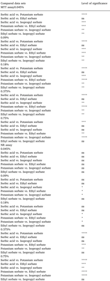
Table 1. shows the statistical analysis of the results of the MTT and NR assays. Flow cytometry tests were carried out as triplicates.
3. Results
3.1. Cell viability tests
Preservatives have high concentrations in the pharmaceutical pro- duct, to ensure the absolute inhibition of microbial growth. However, they are diluted in the stomach and later parts of the gastrointestinal tract. In order to compare both the antimicrobial and the biocompat- ibility tests, all compounds were tested in a wide range, setting 0.75%
w/was maximum value and halving the concentration of every further solution. According to the regulation of Hungarian pharmaceutical compounding formulation, the maximum applied dose of sorbic acid (only slightly soluble in water, but moderately in hot water) and po- tassium sorbate is 1%w/w. and tolerable according to Hungarian reg- ulations. Also, according to the European regulations, sorbic acid and potassium sorbate as food additives can be used from 0.02% (200 ppm) to 0.5% (5000 ppm) (Commission Regulation (EU) No 1129/2011). We aimed to investigate the biocompatibility and antimicrobial properties of the tested compounds above this approved range in order to get a more detailed view of such properties. Therefore, our concentrations were 0.045%, 0.09%, 0.18%, 0.375% and 0.75%w/wwhich cover the whole range of application. All of the sorbates for cytotoxicity tests were all diluted in PBS.
MTT assay (Fig. 2.) showed a dose-dependent toxicity of sorbates, where potassium sorbate was the least toxic compound, followed by isopropyl sorbate and ethyl sorbate, while sorbic acid had the lowest cell viability results. However, at the highest concentration, isopropyl sorbate, ethyl sorbate and the sorbic acid caused nearly total cell death.
Calculated IC50values are <0.045%w/wfor ethyl sorbate and sorbic acid, 0.32%w/wfor isopropyl sorbate and >0.75%w/wfor potassium sorbate.
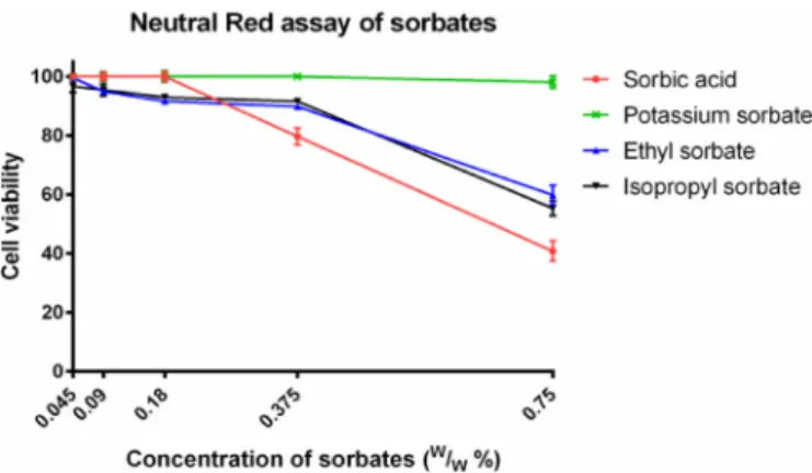
The lower concentrations of sorbates had only a minor impact on the viability of Caco-2 cells measured by Neutral Red assay (Fig. 3.).
Meanwhile, 0.375% and 0.75% drastically increased the toxicity of the test substances. Compared to the results of MTT assay, sorbic acid was Table 1
Results of Dunn's multiple comparison test generated by the data of MTT and NR assay. * =p< 0.05; ** =p< 0.01; *** =p< 0.001; **** =p< 0.0001.
Compared data sets Level of significance
MTT assay0.045%
Sorbic acid vs. Potassium sorbate ****
Sorbic acid vs. Ethyl sorbate ns
Sorbic acid vs. Isopropyl sorbate ***
Potassium sorbate vs. Ethyl sorbate ***
Potassium sorbate vs. Isopropyl sorbate **
Ethyl sorbate vs. Isopropyl sorbate **
0.09%
Sorbic acid vs. Potassium sorbate ****
Sorbic acid vs. Ethyl sorbate ns
Sorbic acid vs. Isopropyl sorbate ***
Potassium sorbate vs. Ethyl sorbate ***
Potassium sorbate vs. Isopropyl sorbate **
Ethyl sorbate vs. Isopropyl sorbate **
0.18%
Sorbic acid vs. Potassium sorbate ****
Sorbic acid vs. Ethyl sorbate ns
Sorbic acid vs. Isopropyl sorbate ***
Potassium sorbate vs. Ethyl sorbate ****
Potassium sorbate vs. Isopropyl sorbate **
Ethyl sorbate vs. Isopropyl sorbate **
0.375%
Sorbic acid vs. Potassium sorbate ****
Sorbic acid vs. Ethyl sorbate ns
Sorbic acid vs. Isopropyl sorbate **
Potassium sorbate vs. Ethyl sorbate ****
Potassium sorbate vs. Isopropyl sorbate **
Ethyl sorbate vs. Isopropyl sorbate **
0.75%
Sorbic acid vs. Potassium sorbate ***
Sorbic acid vs. Ethyl sorbate ns
Sorbic acid vs. Isopropyl sorbate ns
Potassium sorbate vs. Ethyl sorbate **
Potassium sorbate vs. Isopropyl sorbate ****
Ethyl sorbate vs. Isopropyl sorbate ns
NR assay 0.045%
Sorbic acid vs. Potassium sorbate ns
Sorbic acid vs. Ethyl sorbate ns
Sorbic acid vs. Isopropyl sorbate ns
Potassium sorbate vs. Ethyl sorbate ns
Potassium sorbate vs. Isopropyl sorbate ns
Ethyl sorbate vs. Isopropyl sorbate ns
0.09%
Sorbic acid vs. Potassium sorbate ns
Sorbic acid vs. Ethyl sorbate ns
Sorbic acid vs. Isopropyl sorbate *
Potassium sorbate vs. Ethyl sorbate *
Potassium sorbate vs. Isopropyl sorbate *
Ethyl sorbate vs. Isopropyl sorbate ns
0.18%
Sorbic acid vs. Potassium sorbate ns
Sorbic acid vs. Ethyl sorbate ns
Sorbic acid vs. Isopropyl sorbate *
Potassium sorbate vs. Ethyl sorbate *
Potassium sorbate vs. Isopropyl sorbate *
Ethyl sorbate vs. Isopropyl sorbate ns
0.375%
Sorbic acid vs. Potassium sorbate ****
Sorbic acid vs. Ethyl sorbate ns
Sorbic acid vs. Isopropyl sorbate ns
Potassium sorbate vs. Ethyl sorbate ***
Potassium sorbate vs. Isopropyl sorbate **
Ethyl sorbate vs. Isopropyl sorbate ns
0.75%
Sorbic acid vs. Potassium sorbate ****
Sorbic acid vs. Ethyl sorbate ***
Sorbic acid vs. Isopropyl sorbate **
Potassium sorbate vs. Ethyl sorbate ****
Potassium sorbate vs. Isopropyl sorbate ****
Ethyl sorbate vs. Isopropyl sorbate ns
Fig. 2.Cytotoxicity of sorbates measured by MTT assay. Cell viability expressed as the percentage of the absorbance of the untreated control cells. Data ex- pressed as mean ± SEM,n= 12.
Cell viability of the test samples at 0.045%, 0.09%, 0.18%, 0.375% and 0.75%
(w/w)%:
Sorbic acid: 3.6% ± 0.18%; 4.3% ± 0.1%; 3.8% ± 0.1%; 5.1% ± 0.3%;
4.0% ± 0.3%.
Potassium sorbate: 99.8% ± 1.5%; 96.6% ± 2.1%; 91.9% ± 2%;
89.3% ± 2%; 54.7% ± 2.2%.
Ethyl sorbate: 17.0% ± 2.2%; 22.6% ± 3.3%; 19.9% ± 4.3%;
10.7% ± 4.3%; 4.8% ± 1.2%.
Isopropyl sorbate: 59.1% ± 1.7%; 62.1% ± 3.7%; 57.4% ± 3.5%;
47.2% ± 2.6%; 3.8% ± 1.6%.
the most toxic compound in this experiment too. Calculated IC50values are above 0.75%w/wfor the esters and potassium sorbate and 0.66%
w/wfor sorbic acid.
3.2. In vivo toxicity tests
G. mellonellalarvae were injected with 20 µl of the four test sub- stances, dissolved in PBS. Throughout the 4 days of the experiment, their viability was observed every 24 h. Two concentrations of sorbates were used, 0.18% and 0.018%w/w. Each group consisted of 20 healthy larvae. Only a minor number of specimens died during the experiment and overall, the larvae showed no sign of melanisation or increased mortality (Fig. 4.). According to the statistical analysis, no curves were significantly different from PBS control and from each other.
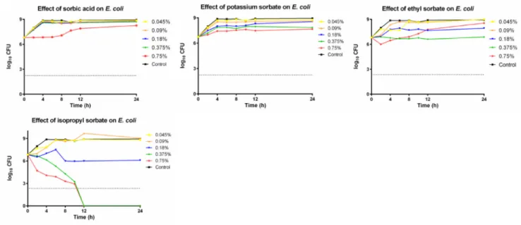
3.3. In vitro antimicrobial time-kill experiments
Time-kill tests were carried out, in order to study the antimicrobial
effect of sorbates.C. albicans, E. coliandS. aureuswere inoculated in RPMI-1640 or Mueller-Hinton broth at 0.045%, 0.09%, 0.18%, 0.375%, 0.75%w/wconcentrations of the different compounds. At given time- points, 100 µl of aliquots were plated on agar plates and counted.
Killing activity was determined by a threshold of 99.9% (log10
CFU = 2.24) extermination of initial CFU.
In case of the lowest concentration, C. albicans (Fig. 5A–D) was resistant to every tested compound. At 0.09%w/wconcentration iso- propyl sorbate (Fig. 5D) had a slight fungistatic effect, inhibiting the further growth of fungal cells. At 0.18%w/w, isopropyl sorbate termi- nated all pathogens after 12 h. No other tested substance had any effect onC. albicansat these concentrations. Potassium sorbate (Fig. 5B) had fungistatic effect at 0.375%w/wand above, while sorbic acid (Fig. 5A) and ethyl sorbate (Fig. 5C) could prevent the germination only at the highest tested concentration. Meanwhile, isopropyl sorbate had an in- creased killing effect above 0.18%w/w, as both higher concentrations identically eliminated all cells after 8 h.
S. aureuswas totally resistant to potassium sorbate, ethyl sorbate and sorbic acid (Fig. 6. A–C) as the inoculum size increased with time in case of every concentration. As such,S. aureuswas the least sensitive organism in our experiment. Isopropyl sorbate (Fig. 6D) had a bacter- iostatic effect at 0.375%w/wconcentration and above.
The growth of E. coli was heavily affected by isopropyl sorbate (Fig. 7D), as after twelve hours, no antimicrobial activity could be de- tected at 0.375%w/wconcentration and above. 0.18%w/wconcentra- tion of isopropyl sorbate was bacteriostatic. Sorbic acid and potassium sorbate (Fig. 7A and B) were totally ineffective against this species.
Meanwhile the results of ethyl sorbate (Fig. 7C) are contradictory, as 0.375%w/whad a stable static effect, while 0.75%w/wproved to be ineffective.
3.4. Flow cytometry measurements
Caco-2 cells were treated with 0.75%w/wsolutions of the tested substances for 30 min and stained with propidium iodide and annexin V.Figs. 8A–E shows the results the distribution of the gated cells. The double positive cells regarded as necrotic/late apoptotic cells, the an- nexin V positive population was regarded as early apoptotic, the double negative population regarded as viable cells. Propidium iodide negative and annexin positive cells were negligible. Isopropyl sorbate had in- creased cytotoxic effect, compared to the other compounds which had increased dead cell percentage than the untreated control.
4. Discussion
2,4-hexadienoic acid, as known as sorbic acid, is widely used as an antimicrobial preservative for food, cosmetic and pharmaceutical in- dustry. Its mechanism of action is stated to be based on the diffusion through the cell membrane and intracellular acidification of the tar- geted microbe (Stratford et al., 2013). As the sorbates can only enter the cell in unionized form, low pH greatly enhances their action, as they can be mostly found in that state at such conditions (Bayan, 2010). If the pH of a given product cannot be adjusted to acidic range, due to its stability, the effect of sorbates is reduced (Wang et al., 2018). The alkyl esters or sorbic acid might be the solution for the pH-dependency issue.
Thus, ethyl and isopropyl sorbates were involved in our study.
Tzatzarakis et al. (2000) and Charvalos et al. (2001) previously formulated different polyvinylpyrrolidone based polymers, to which sorbic acid was covalently bonded, and tested it against several fungi species. The inhibitory concentrations were promising, yet, no toxicity data is available, connected to the newly formed compounds. Moreover, they were not tested against bacterial strains either. Narasimhan et al.
synthesized 42 different sorbic acid esters and analysed their anti- microbial potential (Narasimhan et al., 2007). This publication sug- gested, that the increase of lipophilicity enhances the antibacterial and antifungal actions of the given compound. However, a disadvantage of Fig. 3.Cytotoxicity of sorbates measured by Neutral Red assay. Cell viability
expressed as the percentage of the absorbance of the untreated control cells.
Data expressed as mean ± SEM,n= 12.
Cell viability of the test samples at 0.045%, 0.09%, 0.18%, 0.375% and 0.75%
(w/w)%:
Sorbic acid: 100% ± 1%; 100% ± 1.4%; 100% ± 1.6%; 79.7% ± 2.9%;
40.8% ± 3.4%.
Potassium sorbate: 100% ± 1.1%; 100% ± 1.6%; 100% ± 1.9%;
100% ± 0.5%; 98.2% ± 2%.
Ethyl sorbate: 99.6% ± 0.4%; 94.9% ± 0.7%; 91.7% ± 0.9%;
89.9% ± 0.9%; 59.8% ± 3.3%.
Isopropyl sorbate: 96.7% ± 2.1%; 95.5% ± 2%; 93% ± 1%; 91.6% ± 1%;
55.4% ± 2.5%.
Fig. 4.Survival curve ofG. mellonellalarvae. Larvae were injected with 20 µl of test samples, each group had 20 larvae in it.
Death events of the experiment:
24 h: 0.
48 h: 0.
72 h: 1–0.18% isopropyl sorbate; 1–0.018% isopropyl sorbate.
96 h: 2–0.018% sorbic acid; 1–0.018% ethyl sorbate; 2–0.18% isopropyl sor- bate; 2–0.018% isopropyl sorbate.
these derivatives was the poor water solubility which limits their ap- plication in water-based systems. Our test substances were two esters with short alkyl chains, performing moderate water solubility.
The literature revealed, that all tested compounds are generally well tolerated.Qu et al. (2019) reported, that potassium sorbate had an IC50
value of 1.25 g/L after 24 h of incubation on HepG2 human liver cell line measured by MTT, while HUVEC cell line showed an 659.96 µM IC50 value of after 24 h of incubation measured by MTT (Mohammadzadeh-Agdash et al., 2018). These results match ourfind- ings (Fig. 2.), as after 30 min of incubation, 0.18% potassium sorbate concentration lowered the cell viability to 91.9%. The cell viability difference between potassium sorbate and sorbic acid might be ex- plained by the acidifying nature of the latter.
Smith et al. measured the cytotoxicity of potassium sorbate on Balb/
C 3T3 clone A31 embryonic mouse cells with Neutral Red and found that it was toxic only in extremely high concentrations, far over the generally applied concentrations (Smith et al., 2005). Our results (Fig. 3.) well correlates with this, as only the highest concentrations decreased cell viability. The high correlation of Neutral Red and MTT
cytotoxitcy tests was reported (Fotakis et Timbrell 2006). However, the differences between the assays in our experiments, were based on the acidification of the cytosol of Caco-2 cells. As the change of intracellular pH disrupted the metabolism of the cell, the enzymatic conversion of MTT is highly decreased (Berridge et al., 2005), but the lysosomal staining by Neutral Red was not inhibited (Elliott et Auersperg, 1993).
Another possible explanation of the cytotoxicity profile differences of sorbates is their binding to proteins, as it was proved that relatively similar molecules as carboxylic acids have various binding sites (Mohammadzadeh-Agdash, Akbari, Esazadeh and Dolatabadi, 2019).
Flow cytometry measurements revealed that compared to the con- trol, potassium sorbate, sorbic acid and ethyl sorbate could increase the amount of propidium iodide and annexin positive cells with 10%.
However, isopropyl sorbate was significantly more cytotoxic (68%
compared to the 28% of other tested substances), than any other deri- vatives. We suspected that this can be explained by the non-pH de- pendent mechanism of action and the higher membrane permeability of the isopropyl sorbate, which greatly exceeds the less lipophilic ethyl sorbate. MTT and NR assays did not certify that difference, as both Fig. 5.A-D Antimicrobial effect of sorbates onC. albicans.
Fig. 6.A-D Antimicrobial effect of sorbates onS. aureus.
substances showed similar cytotoxic effects. However, it was proved that the minimum change in the length of alkyl chain greatly modifies the biological activity and membrane passage in the case of salicylic acid derivatives (Li et al., 2019). Further investigation is needed to explain particularly the modification of sorbate esters membrane per- meability with different lengths of alkyl chains.
The use ofG. mellonellalarvae as a biocompatibility model organism
is relatively new. However, Maguire et al. found that the correlation between LD50values observed on this species and the results of pre- vious rat feeding toxicity and cytotoxicity results was linear (Maguire et al., 2016). Several recent publications concluded, that the use ofG. mellonellaelarvae not just complemented to cell culture studies in toxicity experiments, (Allegra et al., 2018;Bombarda et al., 2019) might be a good substitute of rodent model systems (Ignasiak et Fig. 7.A-D Antimicrobial effect of sorbates onE. coli.
Fig. 8.A-D. Flow cytometric measurement of Caco-2 cells, treated with 0.75%w/wsolutions of the test compounds, stained with propidium iodide (PI) and annexin (A). Data is represented as mean of triplicates. Mean percentage distribution of cells between the upper left (PI+, A-), upper right (PI+,A+), lower left (PI-, A-) and lower right (PI-,A+) quadrant, ± SEM:
control: 1.4% ± 0.1%, 18.1% ± 1.2%, 80.3% ± 1.3%, 0.3% ± 0.0%.
sorbic acid: 1.9% ± 0.1%, 29.3% ± 0.4%, 68.2% ± 0.4%, 0.5% ± 0.0%.
potassium sorbate: 4% ± 0.2%, 27.9% ± 2.1%, 67.5% ± 2.2%, 0.5% ± 0.1%.
ethyl sorbate: 6.5% ± 0.1%, 28.5% ± 0.3%, 64.3% ± 0.5%, 0.6% ± 0.0%.
isopropyl sorbate: 1.9% ± 0.1%, 67.8% ± 0.4%, 27.3% ± 0.4%, 3% ± 0.1%.
Maxwell, 2017), thus the prediction of human toxicity of tested com- pounds can be greatly enhanced. In our experiments, there was no significant difference between the mortality of different treated groups (Fig. 4), the larvae showed no sign of toxicity. As the injectable liquid volume is limited and 0.18% is a higher concentration, than sorbates are generally used at, we found that the further increase of the dose in not necessary.
Our results match thefindings of Narasimhan et al. who reported, that isopropyl sorbate was significantly more active againstS. aureus, E.
coliandC. albicans, than ethyl sorbate, which exceeded the original molecule only againstE. coliandC. albicans(Narasimhan et al., 2007).
In our experiment, MIC value was reached neither in the case of sorbic acid, nor with potassium sorbate (Figs. 5–7A and B) against the tested microbes. In many previous publications, it was found, that, the efficacy of sorbic acid and potassium sorbate decreases with the elevation of pH (Lues et Theron, 2012;Hwang et al., 2015). Wang et al. found, that potassium sorbate had a MIC value of 0.4w/w% againstE. coliandS.
aureus at pH 5, but 1.6 and 3.2w/w% if the pH was adjusted to 7 (Wang et al., 2018). Isopropyl sorbate could actively killE. coli(Fig. 7D) and C. albicans (Fig. 5D) cells and inhibit the growth of S. aureus (Fig. 6D) at pH 7, which is a remarkable feat compared to other sor- bates. Lipophilicity and long-term acidification of the cytosol are cri- tical in the antimicrobial mechanism of weak acids (Ullah et al., 2012) and we suspect, that isopropyl sorbate could more effectively pass through the cell membranes without the need of specific proteins (Piper, 2011), than the other tested compounds. Bacterial esterases are known to be part of antibiotic resistance in several species and thus (Egorov et al., 2018), they could possibly cleave the sorbate esters, as known in the case of parabens (Valkova et al., 2003).
Two generally accepted and applied preservatives, sorbic acid and potassium sorbate and two lipophilic sorbate derivates were tested on human colorectal cells,G. mellonellalarvae and various pathogens in order to test their biocompatibility and antimicrobial properties.
Further studies are needed, to specifically describe, the mechanism of action of sorbate esters, whether they have antimicrobial action on their own, or they act as prodrugs and can only be effective after en- zymatic conversion to sorbic acid. While ethyl sorbate had no sig- nificant inhibitory activity against the tested bacteria and fungi, iso- propyl sorbate demonstrated a significant bactericide and fungicide potential. As there is only one methyl group difference between the ethyl sorbate, which had limited effect against the tested microbes based on our experiments, an antimicrobial study with more sorbate esters would be able to clarify the lipophilicity-antimicrobial action correlations. Our results indicate, that the more lipophilic sorbate de- rivates could be promising antimicrobial preservatives, but their low water solubility can limit their application. In order to properly assess the safety biocompatibility profiles of these compounds, beside our study, different in vitro and in vivo genotoxicity and toxicity studies are also required including vertebrates and human cell lines.
Declaration of Competing Interest
The authors declare no conflict of interest.
Acknowledgments
The project was financed by the Gedeon Richter's Talentum Foundation (1103 Budapest, Gyömrői street 19-21.). The published work was also supported by EFOP-3.6.1-16-2016-00022 and EFOP- 3.6.3-VEKOP-16-2017-00009 projects of the European Union and ÚNKP-18-3 New National Excellence Program of the Ministry of Human Capacities of Hungary. The research was co-financed by the Higher Education Institutional Excellence Programmeof The Ministry of Human Capacities in Hungary, within the framework of the Research and Development on Therapeutic purposes thematic programme of the University of Debrecen (NKFIH-1150-6/2019).
Supplementary materials
Supplementary material associated with this article can be found, in the online version, atdoi:10.1016/j.ejps.2019.105162.
References
Abo-EL-Sooud, K., Hashem, M., M., Badr, Y., A., Eleiwa, M., M., E., Gab-Allaha, A., Q., Abd-Elhakim, Y., M., Bahy-EL-Diel, A., 2018. Assessment of hepato-renal damage and genotoxicity induced by long-term exposure tofive permitted food additives in rats.
Environ. Sci. Pollut. Res. Int. 25, 26341–26350.https://doi.org/10.1007/s11356- 018-2665-z.
Al-Ahmad, A., Wiedmann-Al-Ahmad, M., Auschill, T., M., Follo, M., Braun, G., Hellwig, E., Arweiler, N., B., 2008. Effects of commonly used food preservatives on biofilm formation of Streptococcus mutans in vitro. Arc. Oral Biol. 53, 765–772.https://doi.
org/10.1016/j.archoralbio.2008.02.014.
Allegra, E., Titball, R., W., Carter, J., Champion, O., L., 2018. Galleria mellonella larvae allow the discrimination of toxic and non-toxic chemicals. Chemosphere 198, 469–472.https://doi.org/10.1016/j.chemosphere.2018.01.175.
Arweiler, N., Lenz, B., Sculean, R., Al-Ahmad, A., Hellwig, A., Auschill, E, T., M., 2008.
Effect of food preservatives on in situ biofilm formation. Clin. Oral Investig. 12, 203–208.https://doi.org/10.1007/s00784-008-0188-6.
Bagar, T., Altenbach, K., Read, N., D., Benčina, M., 2009. Live-cell imaging and mea- surement of intracellular pH infilamentous fungi using a genetically encoded ratio- metric probe. Eukaryot. Cell 8, 703–712.https://doi.org/10.1128/EC.00333-08.
Bayan, AG., M, 2010. Inhibition of growth and caseinase production of Pseudomonas aeruginosa and Escherichia coli 28 by combination of low pH and NaCl, potassium sorbate or Thymus vulgaris extract. Acta Microbiol. Immunol. Hung. 57, 95–108.
https://doi.org/10.1556/AMicr.57.2010.2.3.
Berridge, M., V., Herst, P., M., Tan, A., S., 2005. Tetrazolium dyes as tools in cell biology:
new insights into their cellular reduction. Biotechnol. Annu. Rev. 11, 127–152.
https://doi.org/10.1016/S1387-2656(05)11004-7.
Bombarda, G., F., Rosalen, P., L., Paganini, E., R., Garcia, M., AR., Silva, D., R., Lazarini, J., G., Freires, I., A., Regasini, L., O., Sardi, J., CO., 2019. Bioactive molecule opti- mized for biofilm reduction related to childhood caries. Future Microbiol. 14https://
doi.org/10.2217/fmb-2019-0144.1207-1120.
Charvalos, E., Tzatzarakis, M., Tsatsakis, A., Petrikkos, G., 2001. Controlled release of water-soluble polymeric complexes of sorbic acid with antifungal activities. Appl.
Microbiol. Biotechnol. 57, 770–775.https://doi.org/10.1007/s00253-001-0853-z.
Chen, HH., XL., Xu, Shang, Y., Jiang, JG., 2017. Comparative toxic effects of butylparaben sodium, sodium diacetate and potassium sorbate to Dunaliella tertiolecta and HL7702 cells. Food Funct. 8, 4478–4486.https://doi.org/10.1039/C7FO01102D.
Creamer, K., E., Ditmars, F., S., Basting, P., J., Kunka, K., S., Hamdallah, I., N., Bush, S., P., Scott, Z., He, A., Penix, S., R., Gonzalez, A., S., Eder, E., K., Camperchioli, D., W., Berndt, A, Clark, M., W., Rouhier, K., A., Slonczewski, J., L., 2017. Benzoate- and salicylate-tolerant strains of Escherichia coli K-12 lose antibiotic resistance during laboratory evolution. Appl. Environ. Microbiol. 83https://doi.org/10.1128/AEM.
02736-16.e02736-16.
Dehghan, P., Mohammadi, A., Mohammadzadeh-Aghdash, H., Dolatabadi, J., E., N., 2018. Pharmacokinetic and toxicological aspects of potassium sorbate food additive and its constituents. Trends Food Sci. Tech. 80, 123–130.https://doi.org/10.1016/j.
tifs.2018.07.012.
Egorov, A., M., Ulyashova, M., M., Rubtsova, M., Y., 2018. Bacterial enzymes and anti- biotic resistance. Acta Naturae 10, 33–48 PMID: 30713760.
Elliott, W., M., Auersperg, N., 1993. Comparison of the neutral red and methylene blue assays to study cell growth in culture. Biotech. Histochem. 68, 29–35.https://doi.
org/10.3109/10520299309105573.
Esimbekova, E., N., Asanova, A., A., Deeva, A., A., Kratasyuk, V., A., 2017. Inhibition effect of food preservatives on endoproteinases. Food Chem 235, 294–297.https://
doi.org/10.1016/j.foodchem.2017.05.059.
Esquivel-Ferriño, P., C., Favela-Hernández, J., M., J., Garza-González, E., Waksman, N., Ríos, M., Y., Camacho-Corona, M., R., 2012. Antimycobacterial activity of con- stituents from Foeniculum vulgare var. dulce grown in Mexico. Molecules 17, 8471–8482.https://doi.org/10.3390/molecules17078471.
European Commission Regulation (EU)No 1004/2014 of 18September 2014amending Annex V to Regulation (EC) No 1223/2009 of the European Parliament and of the Council on cosmetic products.https://eur-lex.europa.eu/legal-content/EN/TXT/?
uri=CELEX%3A32014R1004.
European Commission Regulation (EU)No 1129/2011 of 11November 2011amending Annex II to Regulation (EC) No 1333/2008 of the European Parliament and of the Council by establishing a Union list of food additives.https://op.europa.eu/en/
publication-detail/-/publication/28cb4a37-b40e-11e3-86f9-01aa75ed71a1/
language-en.
Fodil, S., Delgado, J., Varvaro, L., Yaseen, T., Rodríguez, A., 2018. Effect of potassium sorbate (E‐202) and the antifungal PgAFP protein on Aspergillus carbonarius growth and ochratoxin A production in raisin simulating media. J. Sci. Food Agric. 98, 5785–5794.https://doi.org/10.1002/jsfa.9128.
Fotakis, G., Timbrell, J., A., 2006. In vitro cytotoxicity assays: Comparison of LDH, neutral red, MTT and protein assay in hepatoma cell lines following exposure to cadmium chloride. Toxicol. Lett. 160, 171–177.https://doi.org/10.1016/j.toxlet.
2005.07.001.
Ho, CY., Wu, MC., Lan, MY., Tan, CT., Yang, AH., 2008. In vitro effects of preservatives in nasal sprays on human nasal epithelial cells. Am. J. Rhinol. 22, 125–129.https://doi.
org/10.2500/ajr.2008.22.3154.
Hwang, CA., Huang, L., Juneja, V., 2015. Effect of acidified sorbate solutions on the lag- phase durations and growth rates of listeria monocytogenes on meat surfaces. J.
Food. Prot. 78, 1154–1160.https://doi.org/10.4315/0362-028X.JFP-14-408.
Ignasiak, K., Maxwell, A., 2017. Galleria mellonella (greater wax moth) larvae as a model for antibiotic susceptibility testing and acute toxicity trials. BMC Res. Notes. 10, 428.
https://doi.org/10.1186/s13104-017-2757-8.
Jiao, J., Meng, N., Zhang, L., 2014. The effect of topical corticosteroids, topical anti- histamines, and preservatives on human ciliary beat frequency. ORL 76, 127–136.
https://doi.org/10.1159/000363575.
Kim, M., S., Cho, K., H., Park, K., H., Jang, J., Hahn, JS., 2019. Activation of Haa1 and War1 transcription factors by differential binding of weak acid anions in Saccharomyces cerevisiae. Nucleic Acids Res 47, 1211–1224.https://doi.org/10.
1093/nar/gky1188.
Larsen, E., M., Johnson, R., J., 2019. Microbial esterases and ester prodrugs: An unlikely marriage for combating antibiotic resistance. Drug Dev. Res. 80, 33–47.https://doi.
org/10.1002/ddr.21468.
Li, L., Tavallaie, M., S., Xie, F., Xia, Y., Liang, Y., Jiang, F., Fu, L., 2019. Identification of lipid-like salicylic acid-based derivatives as potent and membrane-permeable PTP1B inhibitors. Bioorg. Chem. 93, 103296.https://doi.org/10.1016/j.bioorg.2019.
103296.
Lou, ZF., Fang, XL., Shu, G., Wang, SB., Zhu, XT., Gao, P., Chen, LL., Chen, CY., Xi, QY., Zhang, YL., Jiang, QY., 2011. Sorbic acid improves growth performance and reg- ulates insulin-like growth factor system gene expression in swine. J. Anim. Sci. 89, 2356–2364.https://doi.org/10.2527/jas.2010-3677.
Lues, J., F., R., Theron, M., M., 2012. Comparing organic acids and salt derivatives as antimicrobials against selected poultry-borne Listeria monocytogenes strains in vitro.
Foodborne Pathog. Dis. 9, 1126–1129.https://doi.org/10.1089/fpd.2012.1220.
Maguire, R., Duggan, O., Kavanagh, K., 2016. Evaluation of Galleria mellonella larvae as an in vivo model for assessing the relative toxicity of food preservative agents. Cell.
Biol. Toxicol. 32, 209–216.https://doi.org/10.1007/s10565-016-9329-x.
Mamur, S., Yüzbaşıoğlu, D., Ünal, F., Yilmaz, S., 2010. Does potassium sorbate induce genotoxic or mutagenic effects in lymphocytes? Toxicol. In Vitro 24, 790–794.
https://doi.org/10.1016/j.tiv.2009.12.021.
Mao, X., Nguyen, T., H., D., Lin, M., Mustapha, A., 2016. Engineered nanoparticles as potential food contaminants and their toxicity to Caco‐2 cells. J. Food Sci. 81, 2107–2113.https://doi.org/10.1111/1750-3841.13387.
Medrano-Padial, C., Puerto, M., Moreno, F., J., Richard, T., Cantos-Villar, E., Pichardo, S., 2019. In vitro toxicity assessment of stilbene extract for its potential use as anti- oxidant in the wine industry. Antioxidants 8, 467.https://doi.org/10.3390/
antiox8100467.
Mira, N., P., Teixeira, M., C., Sá-Correia, I., 2010. Adaptive response and tolerance to weak acids in Saccharomyces cerevisiae: a genome-wide view. OMICS 14, 525–540.
https://doi.org/10.1089/omi.2010.0072.
Mohammadzadeh-Agdash, H., Sohrabi, Y., Mohammadi, A., Shanehbandi, D., Dehghan, P., Dolatabadi, J.E.N., 2018. Safety assessment of sodium acetate, sodium diacetate and potassium sorbate food additives. Food Chem. 257, 211–215.https://doi.org/10.
1016/j.foodchem.2018.03.020.
Mohammadzadeh-Agdash, H., Akbari, N., Esazadeh, K., Dolatabadi, J., E., N., 2019.
Molecular and technical aspects on the interaction of serum albumin with multi- functional food preservatives Food Chem. 293, 491-498. 10.1016/j.foodchem.2019.
04.119.
Mpountoukas, P., Vantarakis, A., Sivridis, E., Lialiaris, T., 2008. Cytogenetic study in cultured human lymphocytes treated with three commonly used preservatives. Food Chem. Toxicol. 46, 2390–2393.https://doi.org/10.1016/j.fct.2008.03.021.
Nagy, F., Tóth, Z., Bozó, A., Czeglédi, A., Rebenku, I., Majoros, L., Kovács, R., 2019.
Fluconazole is not inferior than caspofungin, micafungin or amphotericin B in the presence of 50% human serum against Candida albicans and Candida parapsilosis biofilms. Med. Mycol. 57, 573–581.https://doi.org/10.1093/mmy/myy108.
Narasimhan, B., Judge, V., Narang, R., Ohlan, R., Ohlan, S., 2007. Quantitative struc- ture–activity relationship studies for prediction of antimicrobial activity of synthe- sized 2,4-hexadienoic acid derivatives. Bioorg. Med. Chem. Lett. 17, 5836–5845.
https://doi.org/10.1016/j.bmcl.2007.08.037.
Nishihama, Y., Yoshinaga, J., Iida, A., Konishi, S., Imai, H., Yoneyama, M., Nakajima, D., Shiraishi, H., 2016. Association between paraben exposure and menstrual cycle in female university students in Japan. Reprod. Toxicol. 63, 107–113.https://doi.org/
10.1016/j.reprotox.2016.05.010.
Piper, P., W., 2011. Resistance of yeasts to weak organic acid food preservatives. Adv.
Appl. Microbiol. 77, 97–113.https://doi.org/10.1016/B978-0-12-387044-5.
00004-2.
Plumridge, A., Hesse, S., J.A., Watson, A., J., Lowe, K., C., Stratford, M., Archer, D., B., 2004. The weak acid preservative sorbic acid inhibits conidial germination and mycelial growth of Aspergillus niger through intracellular acidification. Appl.
Environ. Microbiol. 70, 3506–3511.https://doi.org/10.1128/AEM.70.6.3506-3511.
2004.
Plumridge, A., Melin, P., Stratford, M., Novodvorska, M., Shunburne, L., Dyer, P.S., Roubus, J., A., Menke, H., Stark, J., Stam, H., Archer, D., B., 2010. The decarbox- ylation of the weak-acid preservative, sorbic acid, is encoded by linked genes in Aspergillus spp. Fungal. Genet. Biol. 47, 683–692.https://doi.org/10.1016/j.fgb.
2010.04.011.
Plumridge, A., Stratford, M., Lowe, K., C., Archer, D., B., 2008. The weak-acid pre- servative sorbic acid is decarboxylated and detoxified by a phenylacrylic acid dec- arboxylase, PadA1, in the spoilage mold Aspergillus niger. Appl. Environ. Microbiol.
74, 550–552.https://doi.org/10.1128/AEM.02105-07.
Qu, D., Jiang, M., Huang, D., Zhang, H., Feng, L., Chen, Y., Zhu, X., Wang, S., Han, J., 2019. Synergistic effects of the enhancements to mitochondrial ROS, p53 activation and apoptosis generated by aspartame and potassium sorbate in HepG2 cells.
Molecules 24, 457.https://doi.org/10.3390/molecules24030457.
Raposa, B., Pónusz, R., Gerencsér, G., Budán, F., Gyöngyi, Z., Tibold, A., Hegyi, D., Kiss, I., Koller, Á., Varjas, T., 2016. Food additives: Sodium benzoate, potassium sorbate, azorubine, and tartrazine modify the expression of NFκB, GADD45α, and MAPK8 genes. Physiol. Int. 103, 334–343.https://doi.org/10.1556/2060.103.2016.3.6.
Roszak, J., Smok-Pniążek, A., Domeradzka-Gajda, K., Grobelny, J., Tomaszewska, E., Ranoszek-Soliwoda, K., Celichowski, G., Stępnik, M., 2017. M. Inhibitory effect of silver nanoparticles on proliferation of estrogen-dependent MCF-7/BUS human breast cancer cells induced by butyl paraben or di-n-butyl phthalate. Toxicol. Appl.
Pharmacol. 337, 12–21.https://doi.org/10.1016/j.taap.2017.10.014.
Rowe, R., C., Sheskey, P., J., Quinn, M., E., 2009. Handbook of Pharmaceutical Excipients, sixth ed. Pharmaceutical Press and American Pharmacists Association, London, Chicago.
Shabir, G., Anwar, F., Sultana, B., Khalid, Z., M., Afzal, M., Khan, Q., M., Ashrafuzzaman, M., 2011. Antioxidant and antimicrobial attributes and phenolics of different solvent extracts from leaves,flowers and bark of gold hohar [Delonix regia (Bojer ex Hook.) Raf.]. Molecules 16, 7302–7319.https://doi.org/10.3390/molecules16097302.
Smith, C., N., Alexander, B., R., 2005. The relative cytotoxicity of personal care pre- servative systems in Balb/C 3T3 clone A31 embryonic mouse cells and the effect of selected preservative systems upon the toxicity of a standard rinse-offformulation.
Toxicol. In Vitro 19, 963–969.https://doi.org/10.1016/j.tiv.2005.06.014.
Stratford, M., Nebe-von-Caron, G., Steels, H., Novodvorska, M., Ueckert, J., Archer, D., B., 2013. Weak-acid preservatives: pH and proton movements in the yeast
Saccharomyces cerevisiae. Int. J. Food Microbiol. 161, 164–171.https://doi.org/10.
1016/j.ijfoodmicro.2012.12.013.
Stratford, M., Steels, H., Nebe-von-Caron, G., Avery, S., V., Novodvorska, M., Archer, D., B., 2014. Population heterogeneity and dynamics in starter culture and lag phase adaptation of the spoilage yeast Zygosaccharomyces bailii to weak acid preservatives.
Int. J. Food Microbiol. 181, 40–47.https://doi.org/10.1016/j.ijfoodmicro.2014.04.
017.
Tzatzarakis, M., N., Tzatsakis, A., M., Lotter, M., M., Shtilman, M., I., Vakalounakis, D., J., 2000. Effect of novel water-soluble polymeric forms of sorbic acid against Fusarium oxysporum f.sp. radicis-cucumerinum. Food Addit. Contam. 17, 965–971.https://
doi.org/10.1080/02652030010002289.
Ullah, A., Orij, R., Brul, S., Smits, G., J., 2012. Quantitative analysis of the modes of growth inhibition by weak organic acids in saccharomyces cerevisiae. Appl. Environ.
Microbiol. 78, 8377–8387.https://doi.org/10.1128/AEM.02126-12.
Valkova, N., Lépine, F., Labrie, L., Dupont, M., Beaudet, R., 2003. Purification and characterization of PrbA, a new esterase from enterobacter cloacae hydrolyzing the esters of 4-hydroxybenzoic acid (parabens). J. Biol. Chem. 278, 12779–12785.
https://doi.org/10.1074/jbc.M213281200.
Wang, C., Deng, Q., Han, D., Zhang, L., 2012. Effects of benzalkonium chloride and po- tassium sorbate on airway ciliary activity. ORL 74, 149–153.https://doi.org/10.
1159/000337830.
Wang, J., Ma, M., Yang, J., Chen, L., Yu, P., Wang, J., Gong, D., Deng, S., Wen, X., Zeng, Z., 2018. In vitro antibacterial activity and mechanism of monocaprylin against es- cherichia coli and staphylococcus aureus. J. Food Prot. 81, 1988–1996.https://doi.
org/10.4315/0362-028X.JFP-18-248.