TISSUE CHARACTERIZATION AND CUSTOM MANUFACTURED PHANTOMS
FOR MODELLING OF MEDICAL ULTRASOUND IMAGES
Kriszti´ an F¨ uzesi
Theses of the Ph.D. Dissertation
Supervisor: Dr. Mikl´ os Gy¨ ongy
P´ azm´ any P´ eter Catholic University Faculty of Information Technology and Bionics
Tam´ as Roska Doctoral School of Sciences and Technology
Budapest, 2019
1 Introduction
A sound wave having a frequency over the range of human perception (approxiamtely above 20 kHz) is called ultrasound (US). In nature many animals use it for navigation, for example bats or dolphins. Using this recognition the first technological application was developed by Paul Langevin in 1917, where the goal was the detection of submarines. [5]
Nowadays, US has a wide variety of applications such as non- destructive testing, range finding, security systems, low energy data-transfer, welding of plastic parts or biomedical applications.
[5–9]
In medicine US is one of the most widespread medical imaging techniques. In medical imaging the frequency range of 1-20 MHz is usually applied. Since penetration depth and resolution are inversely proportional to each other, lower frequencies are used where higher depths of penetration are required, while higher frequencies will achieve higher resolution at the cost of limited depth of penetration. The main advantage of using US is that it is a non-invasive technique in the classical sense. Namely, only heating and mechanical effects emerge as a safety issue during examinations. Nevertheless, thermal and mechanical indices are always kept under control and displayed on the screen during operation of the imager. [5]
The most recent advances in technology enables us to acquire high quality images (e.g. 4D fetal US). Furthermore, there are several attempts to follow some treatments providing temper- ature maps using US data [10–18]. However, without precise models and measurements, development of these areas would be impossible.
2 Challenges in Ultrasound Phantom Manufacture
From the very beginning of medical imaging there had al- ways been a need for test objects, which were usually placed in baths filled with water. In US imaging, regularly placed string or rod targets were imaged underwater to ensure standardized results and eliminate artefacts of the equipment. As the resolu- tion of these images improved along with hardware and software solutions, the need for smaller and more precise targets emerged.
For example nowadays, a typical US calibration phantom contains strings with a diameter smaller than 0.3 mm [19]. Also, main characteristics of the propagation medium such as speed of sound, attenuation and speckle density are well-adjusted. Never- theless, as commercial phantoms are improving over time, their price is also increasing. Moreover, commercial phantoms still of- fer tissue models only on a macroscopic level which are best to be used for the training of radiologists or calibration purposes but are less useful in research and development (R&D) and in the verification of the devices. [20]
These circumstances and the development attempts for spe- cialized high frequency applications such as acoustic microscopy as well as superficial US increase the demand for customizable in-house solutions.
The main requirements for creating optimal custom manufac- tured phantoms are high resolution (under 0.1 mm) at least in 2-dimensions; precise, reproducible placement of scatterers; and reasonable acoustic properties for successful tissue mimicking.
Using conventional manufacturing methods one can only create
phantoms that are only mimicking tissues on a macroscopic level, moreover in many cases only some of the acoustic properties can be attained adequately. [20] For this reason, during the develop- ment of an image enhancement algorithm, tests nowadays rely on computer simulations and qualitative results obtained from real US data.
Considering the temperature dependence of acoustic proper- ties of tissues our knowledge is still insufficient, which shows the necessity of further tissue charachterization as well.
In summary, the challenges in this field include the research of still unavailable or uncertain acoustic properties of tissues, searching for new, advanced materials which could be adequate as a propagation medium and developing new types of in-house phantom manufacturing methods. The current doctoral work aims to further scientific knowledge in these topics by performing tissue characterization of the temperature dependence of com- mon ultrasonic parameters as well as by developing cost-effective ultrasound phantom manufacturing technologies.
3 New Scientific Results
Thesis I: I characterized the propagation speed of sound and acoustic attenuation, as well as the temperature dependence of the aforesaid parameters in porcine myocardium. The results confirm the feasibility of ultrasound in the monitoring of thermal therapy and show what properties a realistic muscle-tissue model should have.
Corresponding publication: [1]
The temperature dependence of soft tissue acoustic prop- erties is relevant for monitoring tissue hyperthermia and also when manufacturing customized tissue-mimicking ultrasound phantoms.
Therefore I investigated the propagation speed and attenu- ation of healthy porcine left ventricular myocardia (N = 5) in a frequency range relevant for clinical diagnostic imaging, i.e.
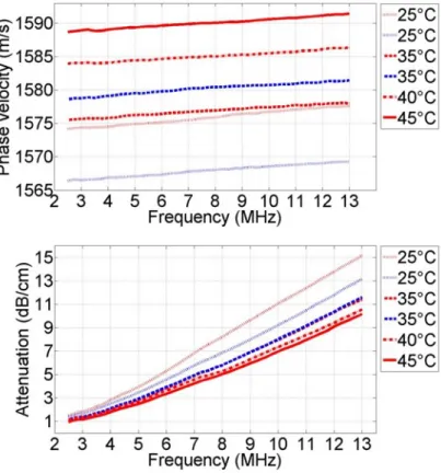
2.5−13.0 MHz. Each tissue sample was held in a water bath at a temperature T = 25◦C, heated to 45◦C, and allowed to cool back down to 25◦C. Due to initial tissue swelling, the data for decreasing temperatures was considered more reliable. In this case, the slope of the phase velocity versus temperature relation was measured to be 1.10±0.04 m/s/◦C, and the slope of the attenuation was 0.11 ± 0.04 dB/cm/◦C at 10 MHz, or −0.0041± 0.0015 dB/cm/MHz1.4336/◦C as a function of frequency.
Figure 1. Averaged phase velocity dispersion curves and attenuation coefficients calculated over al l samples in the frequency range of 2.5- 13.0 MHz. Red and blue colors indicate the data acquired during heating and cooling of the surrounding medium, respectively. Dispersion of the phase velocity is clearly visible and can be described by a linear function of frequency with a slope of 0.2705±0.042 m/s/MHz. The attenuation power law coefficientnis 1.4336±0.025.
Thesis II: I compared the feasibility of two rapid 3D- prototyping methods in creating ultrasound phantoms. I have shown that Fused Deposition Modelling (FDM) and Digital Light Processing (DLP) are able to print ultrasound wire phantoms for 2D imaging at the resolution of 0.3mm, which is suitable for ul- trasound imagers employing frequencies below 4 MHz.
Corresponding publication: [2]
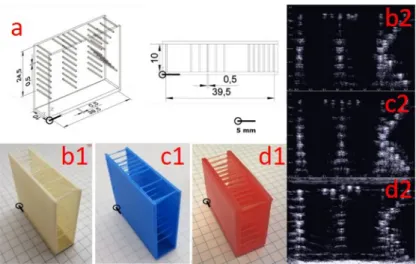
Figure 2. Filament target phantoms bearing the letters “ITK” and their US images. a) Technical drawing of the filament target phantom.
b1&b2) Acrylonitrile butadiene styrene (ABS) phantom and its US image.
c1&c2: Polylactic acid (PLA) phantom and its US image.
d1&d2: Photopolymer phantom and its US image.
US images were made after placing the phantoms in de-ionized water. The pattern of letters “ITK” is clearly identifiable. However, there is evidence of multiple reflections from each filament.
Recently, the use of 3D printing for manufacturing ultra- sound phantoms has only emerged using expensive and complex technology of photopolymer jetting. Keeping in mind the modest means of many research laboratories, two reliable and cost-effective 3D printing methods were developed for phantom
manufacturing, namely fused deposition modelling (FDM) and digital light processing (DLP) techniques.
After successful trials, wire target phantoms were printed and tested using both techniques. One photopolymer material was used in the custom-manufactured DLP printer and several other materials (ABS, PLA and thermoplastic urethane - TPU) in the FDM printer. Except TPU, the results of the prints were satisfactory and could be used as calibration phantoms, thus the achievements were published.
Thesis III: I developed a method with which ultrasound phantoms can be manufactured using Photopolymer Jetting tech- nology.
The use of photopolymer jetting in the manufacture of ultrasound phantoms has been shown previously by Jacquet et.
al. [21], however, a full description of the setup and parameters used was lacking. I developed a method with which FC705 support material could be printed to be a suitable propagation medium for ultrasound imaging. In my phantoms Vero White Plus serves as the material of scatterers and the enclosing wall.
Thesis III.a.: I have shown that such phantoms can be used to quantitatively test super-resolution algorithms.
Corresponding publication: [3]
Ultrasound images are usually covered with speckle noise.
This speckle pattern is originating from sub-wavelength scatter- ers, however, it makes the images hard to interpret to the user, moreover introduces additional uncertainty to measurements.
Super-resolution algorithms are aiming to improve image
quality and in parallel reduce measurement errors. Despite the importance to have quantitative feedback, testing of these algorithms usually includes only simulation results and presents in-vivo examples in a qualitative fashion. Using my custom manufactured phantoms I could measure quantitatively the effect of a super-resolution algorithm on FWHM and RMSE parameters of real ultrasound images.
B-mode Restored img. Restored img.
image (p= 0.5) (p= 2)
FWHMx (mm) 1.42 0.78 0.98
FWHMz (mm) 0.37 0.26 0.27
RMSE 2.34 1.87 1.93
Table 1. Axial and transverse ful l width at half maxima (FWHM) values of a single scatterer from the outer frame of the phantom. The results show an improvement in resolution using p = 0.5 and p = 2 norms.
Root mean square error (RMSE) values obtained using the inner random scatterer pattern.
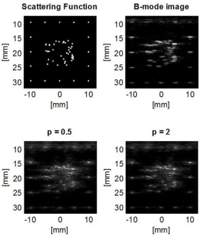
Figure 3. Scattering function of our phantom, the obtained B-mode image and two resulting images of the algorithms using p = 0.5 and p = 2 norm. It can be seen that the clusters of unresolvable scatterers in the center region became distinguishable using both norms.
Thesis III.b.: I developed an algorithm with which arbi- trary medical ultrasound images could be well (R2 ≥80 %) ap- proximated and physically realized.
Corresponding publication: [4]
In ultrasound imaging current phantoms can mimic tissues only on a macroscopic scale. One of the main reasons for this is the lack of usage of 3D printing methods – and looking into deeper, the lack of suitable 3D printing materials – in phantom manufacture. Relying on the previous results using photopolymer jetting technology I developed an iterative algorithm (Stippling Algorithm - SA), which is able to calculate the 2D (axial and lateral) position and intensity of scatterers based on a post-beamformed RF ultrasound image. A further advantage of the algorithm is that it is scalable to the resolution of any 3D-printer.
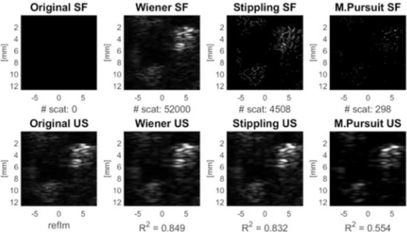
Figure 4. SA results validated on a 3D printed phantom. Images show the scattering function (SF), the original and resulting ultrasound images of the 3D printed phantom. SA performed better than matching pursuit (MP), even though for MP, local sparsity is not a requirement.
Figure 5. SA test results on a real US image. In this case an image of a carotid artery is used. For this reason the original scattering function is unknown. Despite the number of the scatterers on the Wiener-deconvolved image is one magnitude larger than in the case of SA, the resulting US image have nearly the same quality (R2 value) when comparing it to the initial US image.
References
Publications of the author
[1] K. F¨uzesi, N. Ilyina, E. Verboven, K. V. D. Abeele, M. Gy¨ongy, and J. D’hooge, “Temperature dependence of speed of sound and attenuation of porcine left ventricular myocardium,”Ultra- sonics, vol. 82, pp. 246–251, jan 2018.
[2] K. F¨uzesi and M. Gy¨ongy, “Comparison of two inexpensive rapid prototyping methods for manufacturing filament tar- get ultrasound phantoms,” Ultrasound in Medicine & Biology, vol. 43, no. 3, pp. 712–720, mar 2017.
[3] K. F¨uzesi, A. Basarab, G. Cserey, D. Kouam´e, and M. Gy¨ongy,
“Validation of image restoration methods on 3d-printed ultra- sound phantoms,” in2017 IEEE International Ultrasonics Sym- posium (IUS). IEEE, sep 2017.
[4] K. F¨uzesi, ´A. Makra, and M. Gy¨ongy, “A stippling algorithm to generate equivalent point scatterer distributions from ultra- sound images,” inProceedings of Meetings on Acoustics 6ICU. Acoustical Society of America, 2017.
Other references
[5] C. Hill, J. Bamber, and G. ter Haar, Physical Principles of Medical Ultrasonics, 2nd ed. Wiley, New York, 2004.
[6] J. Tsujino et. al., “Ultrasonic plastic welding using fundamental and higher resonance frequencies,”Ultrasonics, vol. 40, no. 1-8, pp. 375–378, may 2002.
[7] R. Adhami and P. Meenen, “Fingerprinting for security,”IEEE Potentials, vol. 20, no. 3, pp. 33–38, 2001.
[8] R. J. Przybyla, et. al., “In-air rangefinding with an AlN piezo- electric micromachined ultrasound transducer,” IEEE Sensors Journal, vol. 11, no. 11, pp. 2690–2697, nov 2011.
[9] S. C. Boon, et. al., “Method and apparatus for intra-body ul- trasound communication,” Patent, Oct. 16, 2012, uS Patent 8,290,598.
[10] G. Menikou and C. Damianou, “Acoustic and thermal char- acterization of agar based phantoms used for evaluating fo- cused ultrasound exposures,” Journal of Therapeutic Ultra- sound, vol. 5, no. 1, jun 2017.
[11] Y. Zhou, “Noninvasive thermometry in high-intensity focused ultrasound ablation,” Ultrasound Quarterly, vol. 33, no. 4, pp.
253–260, dec 2017.
[12] A. A. Anosov, et. al., “Passive estimation of internal temper- atures making use of broadband ultrasound radiated by the body,” The Journal of the Acoustical Society of America, vol.
137, no. 4, pp. 1667–1674, apr 2015.
[13] A. Anosov, P. Subochev, A. Mansfeld, and A. Sharakshane,
“Physical and computer-based modeling in internal tempera- ture reconstruction by the method of passive acoustic thermom- etry,” Ultrasonics, vol. 82, pp. 336–344, jan 2018.
[14] E. S. Ebbini, C. Simon, D. Liu, “Real-time ultrasound thermog- raphy and thermometry [life sciences],”IEEE Signal Processing Magazine, vol. 35, no. 2, pp. 166–174, mar 2018.
[15] W. Kwiecinski, F. Bessi`ere, E. C. Colas, W. A. N'Djin, M. Tan- ter, C. Lafon, and M. Pernot, “Cardiac shear-wave elastography using a transesophageal transducer: application to the mapping of thermal lesions in ultrasound transesophageal cardiac abla- tion,” Physics in Medicine & Biology, vol. 60, no. 20, p. 7829, 2015.
[16] C. H. Seo et. al., “The feasibility of using thermal strain imaging to regulate energy delivery during intracardiac radio-frequency ablation,” IEEE Transactions on Ultrasonics, Ferroelectrics and Frequency Control, vol. 58, no. 7, pp. 1406–1417, jul 2011.
[17] P. Baki, S. J. Sanabria, G. Kosa, G. Szekely, and O. Goksel,
“Thermal expansion imaging for monitoring lesion depth us- ing m-mode ultrasound during cardiac RF ablation: in vitro study,” International Journal of Computer Assisted Radiology and Surgery, vol. 10, no. 6, pp. 681–693, apr 2015.
[18] R. M. Arthur, W. L. Straube, J. W. Trobaugh, and E. G. Moros,
“Non-invasive estimation of hyperthermia temperatures with ultrasound,” International Journal of Hyperthermia, vol. 21, no. 6, pp. 589–600, sep 2005.
[19] M. M. Goodsitt, et. al., “Real-timeB-mode ultrasound quality control test procedures. report of AAPM ultrasound task group no. 1,”Medical Physics, vol. 25, no. 8, pp. 1385–1406, aug 1998.
[20] E. J. Boote, “Phantoms for ultrasound experimentation and quality control,” in The Phantoms of Medical and Health Physics. Springer New York, nov 2014, pp. 159–179.
[21] J.-R. Jacquet, F. Levassort, F. Ossant, and J.-M. Gr´egoire, “3d printed phantom for high frequency ultrasound imaging,” inUl- trasonics Symposium (IUS), 2015 IEEE International. IEEE, 2015, pp. 1–4.