Prothrombotic Effects of Manufactured Nanoparticles
Ph.D. Dissertation
Dr. Péter Bihari
Semmelweis University
Doctoral School of Basic Medicine
Supervisors: Dr. Béla Merkely, D.Sc.
Dr. Fritz Krombach, D.Sc.
Opponents: Dr. Zsuzsanna Bereczky, Ph.D.
Dr. János Szebeni, D.Sc.
Chairman of Committee: Dr. Péter Ferdinandy, D.Sc.
Members: Dr. Erzsébet Komorowicz, Ph.D.
Dr. József Kaszaki, Ph.D.
Budapest
2015
Table of contents
I. ABBREVIATIONS ... 5
II. INTRODUCTION ... 6
1. Overview ... 6
2. Nanoparticles ... 7
a) Definitions ... 7
b) Types of nanoparticles ... 8
c) Properties ... 9
d) Applications ... 11
e) Interactions with biological molecules ... 13
f) Interactions with cells ... 15
g) Exposure routes, biodistribution, and fate ... 17
3. Thrombus formation ... 20
4. Nanoparticles and thrombus formation ... 29
a) Thrombus formation on foreign surfaces ... 29
b) Epidemiology of particulate matter-associated diseases ... 30
c) Particulate matter and thrombus formation ... 32
d) Manufactured nanoparticles and thrombus formation ... 35
III. AIMS ... 37
IV. MATERIALS AND METHODS ... 39
1. Materials ... 39
a) Nanoparticles ... 39
b) Reagents ... 40
c) Mouse serum ... 40
d) Antibodies and fluorescent beads for flow cytometry ... 40
2. Characterisation of nanoparticles ... 41
a) Size distribution and zeta potential measurement ... 41
b) Transmission electron microscopy ... 42
c) Endotoxin measurement ... 42
3. Optimisation of the nanoparticle dispersion method ... 42
a) Effect of different ultrasound energies ... 43
b) Various sequences of preparation steps ... 43
c) Mediums and dispersion stabilisers ... 43
d) Different types of nanoparticles ... 43
e) Evaluation of the stability of nanoparticle dispersions ... 44
4. Dispersion of nanoparticles ... 44
5. Blood collection for in vitro studies ... 44
6. Optimisation of the platelet-granulocyte complex measurement ... 45
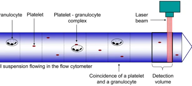
a) Experimental analysis of coincidence ... 45
b) Mathematical description of coincidence ... 47
7. Measurement of platelet activation in vitro ... 49
a) Incubation of whole blood with nanoparticles ... 49
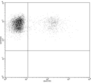
b) Flow cytometry ... 50
c) Aggregometry ... 51
8. Detection of in vivo thrombus formation ... 51
a) Animals ... 51
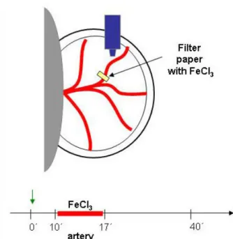
b) Ferric chloride-induced thrombosis in small mesenteric arteries ... 51
c) Light/dye-induced thrombosis in the cremasteric microcirculation ... 53
9. Statistics ... 55
V. RESULTS ... 57
1. Physical characterisation of nanoparticles and optimisation of the dispersion method ... 57
a) Measurement of polystyrene beads ... 57
b) Ultrasound energy ... 58
c) Sequence of preparation steps ... 59
d) Albumin and nanoparticle concentration ... 62
e) Stability ... 65
f) Different types of nanoparticles ... 66
g) Zeta potential ... 70
h) Transmission electron microscopy ... 70
2. Optimisation of platelet-granulocyte complex measurement ... 74
a) Flow cytometric analysis of platelet-granulocyte coincidence ... 74
b) Mathematical description of platelet-granulocyte complexes ... 79
3. Effect of nanoparticles on platelet activation in vitro ... 80
a) Platelet P-selectin expression ... 80
b) Platelet-granulocyte complexes ... 81
c) Platelet aggregometry ... 83
4. Effect of nanoparticles on thrombus formation in vivo ... 84
a) Mouse blood counts ... 84
b) Mesenteric thrombosis ... 84
c) Cremasteric thrombosis ... 86
VI. DISCUSSION ... 88
1. Optimisation of the nanoparticle dispersion method ... 88
2. Optimisation of platelet-granulocyte complex measurement ... 94
3. Prothrombotic effects of nanoparticles ... 97
VII. CONCLUSIONS ... 103
VIII. SUMMARY ... 105
IX. ÖSSZEFOGLALÁS ... 106
X. REFERENCES ... 107
XI. PUBLICATIONS ... 126
XII. ACKNOWLEDGEMENTS ... 129
I. Abbreviations
ADP adenosindiphosphate
BSA bovine serum albumin DEP diesel exhaust particles
EDTA ethylenediaminetetraacetic acid FITC fluoresceinisothiocyanate
HSA human serum albumin
LPS lipopolysaccharide
MSA mouse serum albumin
MWNT multi-walled carbon nanotubes PBS phosphate-buffered saline
PdI polydispersity index
PM particulate matter
PPP platelet-poor plasma
PRP platelet-rich plasma
PSGL-1 P-selectin glycoprotein ligand-1 ROS reactive oxygen species
SWNT single-walled carbon nanotubes
TF tissue factor
TFPI tissue factor pathway inhibitor vWF von Willebrand factor
II. Introduction
1. Overview
Nanoparticles are particles with length scales under 100 nanometres (Lewinski et al.
2008). Nanoparticles always existed in our ambient nature, but they have received a substantial boost of interest in the last 50 years with the emergence of nanotechnology.
Since then, nanotechnology has grown into a prominent industry and hundreds of different variants of nanomaterials and nanotech-based products are now commercially available (Maynard and Rejeski 2009).
The classification of nanoparticles as a new entity is justified by their physicochemical properties, which are different from the bulk material and the atoms and molecules from which they are built up. Although the unique physicochemical properties of manufactured nanoparticles enable the improvement of novel applications, they might also cause unusual types of interactions with biological materials and toxic effects not yet experienced.
Nanoparticles can come in contact with the human body through inhalation, ingestion, dermal deposition, but also through injection for medical applications.
Nanoparticles, having entered the body, can translocate into the systemic blood circulation, reach various remote organs, and may affect their function. The significance of this phenomenon is underlined by previous epidemiological studies identifying ambient nano-sized particles as a major contributor to adverse cardio-respiratory effects of air pollution, where impact on haemostasis has been found to play an important role.
(Oberdorster et al. 2005a, Oberdorster et al. 2005b)
Haemostasis is an important physiological function of the human body, maintaining integrity of blood vessels. Disruption of the vascular integrity, resulting in contact of human blood with any surfaces other then the inner wall of the vessels, induces thrombus formation to stop bleeding (Ruggeri 2002). A well-known consequence is unwanted thrombus formation on the surface of implanted foreign materials in the circulation (Gorbet and Sefton 2004). Although nanoparticles are small, they represent a very high cumulative surface that might also influence haemostasis.
The increasing utilisation of nanomaterials in technological and medical applications
The fact that nanoparticles represent a potentially thrombogenic large cumulative surface, and knowledge of the prothrombotic effects of ambient nanoparticles, raises the question of whether manufactured nanoparticles also influence thrombus formation.
Although circulating nanoparticles also reach the microcirculation, prothrombotic effects of manufactured nanoparticles in the microvasculature have not yet been examined. The aim of my dissertation was, therefore, to investigate the effects of manufactured nanoparticles on platelet activation and on thrombus formation in small arteries and in the microcirculation.
2. Nanoparticles
a) Definitions
Nanotechnology is a relatively new discipline where some of the definitions are still immature. The most often used definitions are listed below:
Nano is derived from the Greek word for dwarf and meaning extremely small.
In physics, it means 10-9.
Nanoparticles are just defined by size. There are, however different definitions in use. All of them share the criteria that nanoparticles should be smaller than 100 nm, but differ regarding how many dimensions should be considered (Oberdorster et al. 2005a, Lewinski et al. 2008).
In this dissertation, the following more generalised definition is used:
Nanoparticles are particles with lengths that range from 1 to 100 nanometres in two or three dimensions (Lewinski et al. 2008).
Most of the nanoparticles are spherical particles, but the above definition is suitable also for particles that are in one dimension bigger than 100 nm such as fibres, tubes, or rods. The best examples of such high aspect ratio nanoparticles are carbon nanotubes, which have a diameter of a few nanometres and a length of several micrometres.
Nanotechnology is the engineering and manufacturing of material at the atomic and molecular scale.
Manufactured nanoparticles are nanoparticles made by nanotechnology.
b) Types of nanoparticles
Nanoparticles can be categorized as ambient or manufactured nanoparticles (Table 1).
Ambient nanoparticles are nano-sized particles found in our environment that are generated by natural processes such as fires, volcanoes, sea spray, or erosion. In addition, they can originate from anthropogenic sources such as traffic or industry. In publications about air pollution, ambient nanoparticles are described as a fraction of particulate matter (PM). Particulate matter is categorized by size, where PM10 are particles under 10 µm, PM2.5 are particles under 2.5 µm, and PM0.1, also referred to as ultrafine particles, are nanoparticles present in the ambient air (Oberdorster et al. 2005a, Borm et al. 2006).
Although biological macromolecules are traditionally not considered as nanoparticles, many such molecules match the size criteria of nanoparticles. Thus, protein complexes (e.g. ribosome, transferrin), lipoproteins or viruses might be regarded as biological nanoparticles. Such biological particles are also recommended for the production of hybrid bio-nanoparticles (Douglas and Young 2006, Uchida et al. 2007).
In contrast to ambient nanoparticles, manufactured nanoparticles are produced intentionally by nanotechnology. They can be subcategorized according to their material (Table 1) (Oberdorster et al. 2005a, Borm et al. 2006).
Table 1. Type of nanoparticles
Source Subtype Examples
Ambient
natural processes (fires, volcanoes, sea spray, erosion)
ultrafine particles, or PM0.1 industrial processes (traffic,
industry)
biological nanoparticles ribosome, lipoproteins, viruses
manufactured
carbon nanoparticles
carbon black, single-walled carbon nanotubes (SWNT), multi-walled carbon nanotubes (MWNT), fullerenes
quantum dots
cadmium selenide, cadmium sulfide, indium arsenide, indium phosphide
metal, metal oxide nanoparticles
titanium dioxide, zinc oxide, gold, silver, iron oxide, silicium oxide organic nanoparticles liposome, gelatine
hybrid bio-nanoparticles nanoparticles in protein cage
c) Properties
Nanoscale material is an intermediate between atomic and bulk material. Some of the special features of nanomaterials are due to the high surface area-volume ratio (Fig. 1).
When particles are very small, most of the atoms are on the surface of the particle, resulting in a large active surface area with a high chemical reactivity. Another conse- quence of the high surface area-volume ratio is that the van der Waals forces between the relatively large surface areas are high enough to move the small mass nanoparticles together, resulting in the formation of agglomerates (Borm et al. 2006). The building of agglomerates is further exaggerated in biological fluids exerting high ionic strength and physiological pH (Nel et al. 2009).
Fig. 1. Inverse relationship between particle size and the percent of surface molecules (Nel et al. 2006).
Moreover, materials on the nanoscale can exert properties that are independent of the high surface area (Burda et al. 2005). An example is the quantum size effect of semiconductor nanocrystals, i.e. variations in electric and optical properties at a size that is comparable or smaller then the length scale of electron motion in bulk material (Bohr radius). Although the bulk material of quantum dots (cadmium selenide, cadmium sulphide, indium arsenide, and indium phosphide) do not exert fluorescent properties, nanocrystals of these semiconductors (quantum dots) have a very strong and narrow fluorescent signal band upon excitation and the wavelength of emission depends on the particle size (Biju et al. 2008). In metallic nanoparticles, surface plasmon resonance can be induced, whereby free electrons respond collectively by oscillating in resonance with the light wave, resulting in light absorption (Burda et al. 2005). Properties of ferromagnetic or ferrimagnetic materials also change at around the size of 10-20 nm, resulting in a single magnetic domain that shows superparamagnetic behaviour (Lu et al. 2007). Carbon in nanotube form gains outstanding mechanical properties with extremely high tensile strength (Kis and Zettl 2008). Carbon nanotubes also have peculiar electronic characteristics. Their electronic conductivity depends on the tube diameter and the wrapping angle (a measure of the helicity of the tube lattice), with only slight differences in these parameters causing a shift from metallic to semiconducting properties (Wilder et al. 1998). These examples, although not exhaustive, demonstrate why nanoparticles open up many possibilities for innovations in technology.
d) Applications
Based on these unique properties, nanomaterials are already widely used in a diverse array of applications including chemistry, environmental engineering, food, clothes, personal care products, energy production and storage, optics, information technology, and construction materials (Maynard et al. 2006, Maynard and Rejeski 2009). There are also many biomedical applications of nanoparticles (Salata 2004). Different types of nanoparticle constructs are under development, where drugs are delivered by a nanoparticle directly to a target cell, lowering unwanted side effects on other cells (Davis et al. 2008). Several such drug-containing nanoparticles are already in medical use mainly for cancer therapy (Zhang et al. 2008). Labelled magnetic nanoparticles are applied for the visualisation of atherosclerotic plaques or tumours by magnetic resonance imaging. These magnetic nanoparticles can also be heated by alternating magnetic fields, and thus they can be utilised to kill cancer cells (magnetic hyperthermia treatment) (Ito et al. 2005, Lu et al. 2007). Nanotechnology is also used in tissue engineering to produce biomaterials with nanostructured surfaces that can be better incorporated into living tissue, such as in bone implants (Salata 2004). Other biomedical applications of nanoparticles include fluorescent labelling, biodetection of pathogens, probing of DNA structures, purification of biological molecules, or phagokinetic studies (Salata 2004) (Table 2).
Table 2. Examples of nanoparticle applications already available on the market
(normal text), or under development (in italics).
Nanoparticle General applications Medical application
Carbon nanotubes
- construction materials e.g. in sport equipments or in wind turbine blades
- antifouling paints for ships - batteries for electronic devices - purification of drinking water
(De Volder et al. 2013)
- bone implants (Salata 2004) - drug delivery
- near infrared hyperthermia therapy
- gene therapy - tissue engineering (He et al. 2013)
Nanoparticle General applications Medical application
Metal, metal- oxide
nanoparticles
- transparent sunscreens (contains TiO2 or ZnO nanoparticles) - self-cleaning windows (Borm et
al. 2006)
- surface enhanced Raman spectroscopy (Burda et al.
2005)
- many food products contain TiO2 nanoparticles (Weir et al.
2012)
- nanoparticle-based therapeutics in preclinical development (Zhang et al. 2008)
Magnetic nanoparticles
- high density data storage - magnetically separable
catalysts
- magnetic separation of biomolecules (Lu et al. 2007)
- contrast agent for magnetic resonance imaging
(superparamagnetic iron oxide nanoparticles) (Ittrich et al.
2013)
- magnetic hyperthermia treatment (Ito et al. 2005) - magnetic drug delivery (Lu et
al. 2007)
Quantum dots
- different applications of bioimaging (Chen and Liang 2014)
- quantum dot-LED display - solar cells with quantum dots
(Hardman 2006)
- photodynamic therapy and - radiotherapy of cancer with
quantum dots (Juzenas et al.
2008)
Nanoparticle General applications Medical application
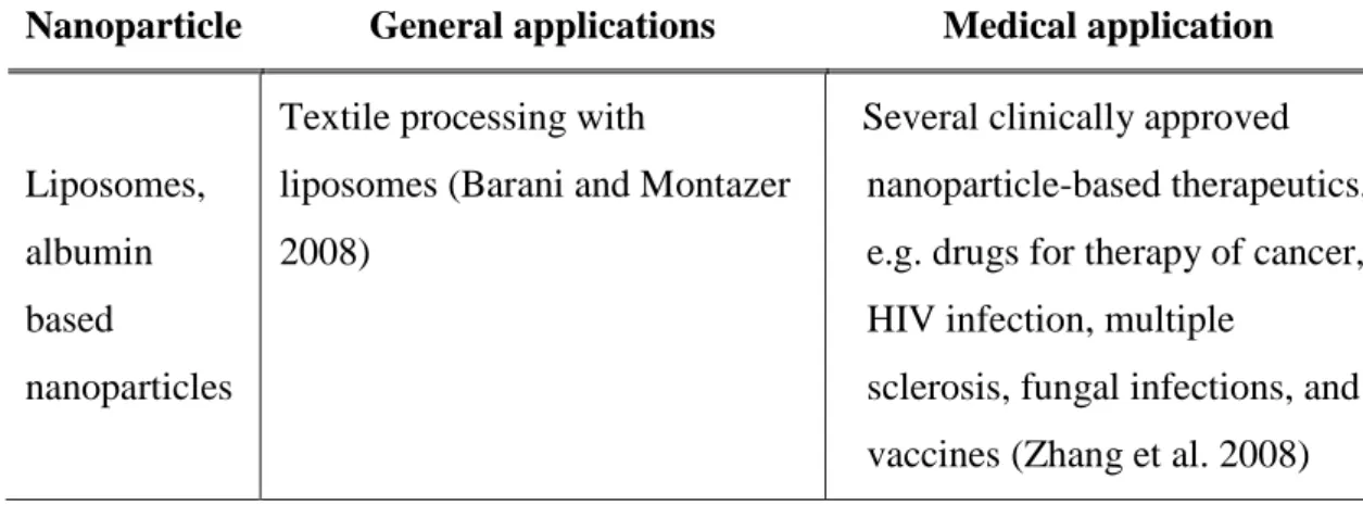
Liposomes, albumin based
nanoparticles
Textile processing with
liposomes (Barani and Montazer 2008)
Several clinically approved nanoparticle-based therapeutics, e.g. drugs for therapy of cancer, HIV infection, multiple
sclerosis, fungal infections, and vaccines (Zhang et al. 2008)
e) Interactions with biological molecules
Nanoparticles in organisms come into contact with biological molecules, and these interactions affect their fate and effects in the body (Lynch and Dawson 2008). Due to the relatively high surface area versus the small mass, the van der Waals forces cause biological molecules to adhere to the particles. Interestingly, the number and types of biomolecule that adhere to the curved nanoparticle surface differ in many cases from those which adhere to flat surfaces of the same material (Lynch et al. 2009).
Nanoparticles in a biological fluid (e.g., plasma) are covered by proteins. This organized protein structure that surrounds the nanoparticles is called the protein corona (Fig. 2).
Fig. 2. Nanoparticle protein corona. Adhesion of proteins to nanoparticles is determined by different equilibrium constants (Lynch and Dawson 2008).
The composition of the protein corona depends on the proteins surrounding the nanoparticles, and on the size and surface properties of the nanoparticles (Lundqvist et al. 2008). Additionally, the composition of the protein corona is in continuous exchange with the proteins of the environment. First, nanoparticles are covered with proteins that are abundant in the solution; later, proteins in lower concentration but with higher affinity occupy the nanoparticle surface. Accordingly, it has been shown that nanoparticles in plasma first bind albumin and fibrinogen molecules, and then bind proteins that are present in lower concentrations in plasma but have higher affinity toward nanoparticles, such as apolipoproteins (Cedervall et al. 2007b, Lynch and Dawson 2008). In the nanoparticle corona almost all of the plasma proteins can be found at different amounts according to the particle surface properties: i.e. albumin, fibrinogen, immunoglobulins, proteins of the complement system, apolipoproteins, as well as coagulation cascade and acute phase proteins (Lundqvist et al. 2008). As a consequence of binding, the conformation and function of adhered proteins can change.
This can lead to the exposure of new epitopes or alter protein function (Nel et al. 2009).
Nanoparticles can also induce protein fibrillation, i.e. certain proteins aggregate into long, thin fibrils called amyloid structures (Colvin and Kulinowski 2007, Linse et al.
2007). It is generally perceived that the properties of nanoparticles determine the composition of the protein corona. In turn, the composition of the protein corona determines the behaviour of nanoparticles in biological systems. The protein corona can influence the interaction with receptors, cellular uptake, organ distribution, cell and organ functions, and excretion. It has been demonstrated that inhibition of protein adhesion by coating nanoparticles with amphiphilic polyethylene glycol molecules leads to decreased cellular uptake, longer circulation, and altered biodistribution (Praetner et al. 2010, Jokerst et al. 2011).
Nanoparticles also interact with membrane phospholipids. Adhesion depends on the charge of nanoparticles (Verma and Stellacci 2010). It has been shown that nanoparticles of negative charge induce local gelation in otherwise fluid bilayers;
nanoparticles of positive charge induce otherwise gelled membranes to fluidize locally.
It has been hypothesised that charged nanoparticles alter the tilt angle of the phospho- choline head group, which is an electric dipole of phosphate and choline. The
reorganisation of the phosphocholine head group changes the fluidity of membranes.
(Wang et al. 2008)
Nucleic acids bind to nanoparticles as well. Cationic nanoparticles or nanoparticles bearing cationic ligands provide highly efficient DNA binding via electrostatic interaction. This kind of interaction can be utilised for electrostatic assembly of nanoparticles along DNA molecules (Saha et al. 2011). High affinity binding of single- stranded DNA to SWNT has been used for quantitative detection of SWNT in aqueous samples (Mota et al. 2013).
f) Interactions with cells
Nanoparticles are able to undergo diverse interactions with cells. Nanoparticles can adhere to cell surfaces, translocate into cells, and influence cellular functions through interactions with cell components (Zhao et al. 2011).
As described above (section II/2/e), nanoparticles can adhere to proteins and lipid macromolecules, which are the main components of cell membranes.
Cells can internalise nanoparticles by different mechanisms. Larger or aggregated nanoparticles are taken up by phagocytosis by specialized cells such as macrophages, monocytes, and neutrophils or by macropinocytosis by other cell types. Smaller particles can be endocytosed through the formation of endocytic vesicles of different sizes by clathrin-mediated (~120 nm), caveolae-mediated (~60 nm), or clathrin/caveolin independent (~90 nm) mechanisms (Conner and Schmid 2003, Dobrovolskaia and McNeil 2007, Verma and Stellacci 2010) (Fig 3). Moreover, some nanoparticles are able to penetrate cell membranes without any active process of the cell (Verma and Stellacci 2010).
Fig 3. Endocytosis mechanism depends on the size of nanoparticles (Conner and Schmid 2003).
Nanoparticles taken up by an active internalisation process of the cell are enclosed in endosomes and thus separated from the cytosol. There are, however, several mechanisms that lead to the escape of nanoparticles from the endosomes. Internalisation of nanoparticles with high aspect ratio such as MWNT, which are relatively long and stiff, can lead to disruption of the phagosome (frustrated phagocytosis). Macrophages, in an attempt to destroy the carbon nanotubes, release harmful oxygen radicals, and hydrolytic enzymes which are deposited in the surrounding medium, leading to chronic inflammation (Poland et al. 2008). Amine-modified nanoparticles were also described as being able to escape from the lysosomes to the cytoplasm. This phenomenon can be explained by the proton sponge hypothesis that states that unsaturated amines on the material surface are capable of sequestering protons. The lysosomal proton pump, which is responsible for acidification, tries to acidize the lysosome, keeping the pump going, and leading to the retention of one Cl– anion and one water molecule for each proton that enters the lysosome. Ultimately, this process causes lysosomal swelling and rupture, leading to particle deposition in the cytoplasm (Dobrovolskaia and McNeil 2007, Xia et al. 2008).
Oxidative stress is thought to play an important role in the mechanisms of nanoparticle-induced cytotoxicity. Nanoparticles can generate reactive oxygen species (ROS) through redox chemistry or interfere with the ROS production of the cell. The excited energy state in a semiconductor nanoparticle leads to the generation of superoxide radicals, e.g. titanium dioxide nanoparticles produce ROS upon ultraviolet light exposure. Transition metals on the nanomaterial surface (e.g. metal impurities in nanotubes) can generate superoxide anions through Fenton chemistry. Nanoparticles can
also alter phagocytosis leading to intracellular (proton sponge hypothesis) or extracellular release of ROS (frustrated phagocytosis). Furthermore, nanoparticles can lodge in mitochondria and interfere with the electron transport chain leading to the production of superoxide anions (Nel et al. 2006, Li et al. 2008).
Nanoparticles can also enter the nucleus either by penetrating the nuclear membrane, or being transported through the nuclear pore complexes, or becoming enclosed in the nucleus by chance during mitosis. Nanoparticles can cause genotoxic effect through ROS production, direct mechanical interference with the mitotic spindle and DNA or interaction with nuclear proteins and enzymes (Gonzalez et al. 2008, Singh et al. 2009).
Nanoparticles at different subcellular locations can interact with macromolecules and thus influence cellular functions.
g) Exposure routes, biodistribution, and fate
Nanoparticles can enter the human body through the skin, respiratory, or gastrointestinal tract, or by injection into the circulation (Oberdorster et al. 2005a). The actual exposure route depends on the exposure scenario (Fig. 4). Ambient nanoparticles from natural sources such as combustion processes enter the human body mainly via inhalation.
Manufactured nanoparticles can also be inhaled if they get into the air during production. Nanoparticles in food or water can get into the body through the gastrointestinal tract. Nanoparticles in cosmetics and sun creams come into contact with the skin. Nanoparticles can be cleared from these organs, e.g. from the respiratory tract, by the mucociliar system, or they might be deposited. Importantly, nanoparticles can translocate from these organs into the blood vessels and reach any tissue of the body through the systemic circulation.
Fig. 4. Confirmed (solid arrow) and potential (dashed arrow) exposure routes of nanoparticles in the human body. Nanoparticles can be taken up into the human body from the air through the respiratory tract, from food and water through the gastrointestinal tract, or they can be deposited from air, water, or clothes onto the skin.
Nanoparticles might translocate from these organs into the blood, or in medical applications they can also be directly injected into the systemic circulation. Circulating nanoparticles can reach any organs of the body and be excreted by the kidney, liver, or skin. GI tract: gastrointestinal tract, CNS: central nervous system, PNS: peripheral nervous system. (Oberdorster et al. 2005a)
Several experimental studies have shown that a small fraction of inhaled nanoparticles, depending on their size, can cross the air-blood barrier in the lungs and translocate into the systemic circulation. Kreyling and coworkers have shown that depending on particle size about 1-10 % of deposited 192Ir particles translocated from the lungs into the blood and deposited in secondary organs, such as liver, spleen, heart, and brain (Kreyling et al. 2002). Nemmar and coworkers confirmed this observation in a human study. They found that inhaled 99mTc-labelled ultrafine carbon particles pass rapidly into the systemic circulation (Nemmar et al. 2002a). The mechanism of translocation is supposed to be via pores (Conhaim et al. 1988, Hermans and Bernard
from the lungs was shown for different types of nanoparticles: 192Iridium particles (Kreyling et al. 2002), ultrafine particles (Nemmar et al. 2002a), Tc-labelled albumin molecules (Nemmar et al. 2001), carbon black (Shimada et al. 2006), gold nanoparticles (Lipka et al. 2010), nano-ceria (He et al. 2010), and fullerenes (Naota et al. 2009).
Another main exposure route is through the gastrointestinal tract. It is estimated that 1012-1014 fine and ultrafine particles, including mainly silicates and titanium dioxide, are ingested per person per day in the Western world (Lomer et al. 2002). Experiments in rats and also human studies have shown that TiO2 particles (150–500 nm) taken in via food can translocate into the blood and accumulate in the liver and spleen (Jani et al.
1990, Borm et al. 2006).
Regarding the contact of nanoparticles with the skin, numerous studies have demonstrated that TiO2 or ZnO nanoparticles do not penetrate into or translocate through normal skin (Nohynek et al. 2007, Nohynek et al. 2008, Nohynek et al. 2010).
However, small quantum dots can penetrate broken skin to lodge in other tissues and organs (Mortensen et al. 2008).
In diagnostic or therapeutic medical applications, nanoparticles are directly injected into the systemic circulation. Nanoparticles translocated from the respiratory or gastro- intestinal tract into the systemic circulation or nanoparticles directly injected into the bloodstream can reach all the organs of the body and finally pass the microcirculation of any tissue (Borm et al. 2006, Geiser and Kreyling 2010). It has been shown that circulating nanoparticles can also cross the blood brain barrier and accumulate in the brain (Kreuter 2001, Hu and Gao 2010). A special route to the central nervous system is the direct pathway of nanoparticles from the nasal airways into the brain through the olfactory nerves (Elder et al. 2006).
The biodistribution of nanoparticles depends on their properties. The main uptake organs are the lungs, liver, and spleen. Nanoparticles have different half-lives in the body depending on their type and surface modifications, and they are excreted through renal or hepatic pathways. The way of excretion depends on size, shape, charge, and composition of the nanoparticles. Nanoparticles which are smaller than 6 nm can be filtered in the glomerulus and excreted by the kidney in the same way as small biological molecules. Interestingly, high aspect ratio nanoparticles, such as carbon nanotubes, which have a mean diameter of few nm and lengths in the micrometer range,
have been reported to be cleared from the blood also through the renal pathway. The other main organ of nanoparticle excretion is the liver, which is able to excrete nanoparticles that do not undergo renal clearance. Hepatocytes can take up nanoparticles from the blood, and eliminate them via the biliary pathway. For example, polystyrene nanoparticles have been found to be eliminated by the hepatic pathway, which was mediated by ApoE receptors. Also hepatic Kupffer cells, together with other cells of the reticuloendothelial system, contribute to the elimination of nanoparticles, mainly by degradation processes. On the other hand, nanoparticles such as quantum dots, which can not be degraded, deposit in the phagocytes, leading to long-term retention of these nanoparticles in the body. (Choi et al. 2007, Hagens et al. 2007, Longmire et al. 2008, Landsiedel et al. 2012)
3. Thrombus formation
Haemostasis is an essential physiological function maintaining the integrity of vessels based on a well-tuned interplay between endothelial cells, platelets, and the coagulation system. Haemostatic processes are able to stop bleeding from injured vessels by platelet plug formation and blood coagulation. On the other hand, pathological thrombus formation may induce thromboembolic diseases.
Vascular injury
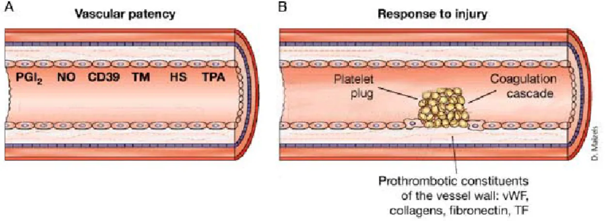
Intact endothelial cells provide a non-thrombogenic surface to which neither platelets nor fibrin mesh can adhere (Fig 5). Additionally, endothelial cells actively inhibit thrombus formation. They produce prostacyclin and nitric oxide, which both suppress platelet activation. Endothelial cells also express CD39, an ecto-ADPase that blocks adenosindiphosphate (ADP) dependent secondary platelet activation. Heparan sulphate and thrombomodulin-protein C complex on the surface of endothelial cells, as well as the endothelial production of tissue factor pathway inhibitor (TFPI), inhibit the coagulation system. Finally, the production of plasminogen activators (tissue plasminogen activator and urokinase-type plasminogen activator) leads to lysis of erroneously generated thrombi (Ruggeri 2002).
Fig 5. A. Vascular patency. Healthy endothelial cells inhibit thrombus formation.
Prostacyclin (PGI2), nitric monoxide (NO), and ecto-ADPase (CD39) suppress platelet activation. Thrombomodulin (TM), heparan sulphate (HS), and tissue plasminogen activator (TPA) counteract the coagulation process. B. Response to injury. Loss of antithrombogenic endothelial cells and interaction of platelets and plasma proteins with prothrombotic constituents of the subendothelial matrix induces thrombus formation:
generation of a platelet plug and a fibrin mesh. Modified after Ruggeri at al. (Ruggeri 2002).
At the site of vascular injury, the loss of antithrombogenic endothelial cells and the contact with the denuded subendothelial matrix evolve the formation of a platelet plug and initiate the coagulation cascade (Fig 5). The subendothelial matrix is, in contrast to endothelial cells, highly thrombogenic. Moreover, von Willebrand factor (vWF), the main ligand of platelet tethering, immediately deposits from plasma onto the denuded subendothelial layer. The interaction between subendothelial matrix proteins and platelets lead to the adhesion and activation of platelets. At the same time, tissue factor (TF) expressed on the surface of subendothelial cells initiates the coagulation process.
During thrombus formation, platelets form a plug and the coagulation cascade forms a fibrin mesh, and these two processes aid one another (Ruggeri 2002).
Platelet adhesion and initial platelet activation
Platelets are small (2-5 µm) anuclear cell fragments with armature for adhesion and aggregation (Broos et al. 2012). Adhesion is a multistep process between elements of the subendothelial matrix of the vessel wall and the receptors of platelets (Fig 6).
Platelets first tether through glycoprotein GPIb-V-IX to vWF immobilized on collagen fibres. This adhesive interaction is rapidly reversible and does not readily support stable platelet adhesion. However, the resulting rolling and deceleration of platelets allows the interaction of the two major collagen receptors GPVI and integrin α2β1 with the subendothelial matrix resulting in firm adhesion and platelet activation. (Ruggeri 2002, Varga-Szabo et al. 2008, Nuyttens et al. 2011)
Fig. 6. Formation of a platelet plug on the denuded subendothelial matrix. Platelets tether to vWF via GP-Ib-V-IX receptors. Interaction of platelet receptors with proteins of the subendothelial matrix induces platelet adhesion. Activation of αIIbβ3 receptors leads to aggregation of platelets bridged by fibrinogen molecules. (Jackson et al. 2000).
Collagen-GPVI and vWF-GPIb-V-IX interactions are the key initiators of platelet activation (Fig. 7). Signal transduction through phospholipase C γ2 results in the release of secondary activation molecules, integrin activation, and procoagulant changes in platelets (Li et al. 2010).
Fig. 7. Platelet signalling. Exposure to the subendothelial matrix activates platelets:
collagen-GPVI, collagen-α2β1, and vWF-GPIb-V-IX interactions activate phospholipase Cγ2 (PLCγ2) inducing the formation of inositol triphosphate (IP3) and diacylglycerol (DAG) second messengers. IP3 mobilise Ca2+ from intracellular stores through inositol triphosphate receptor (IP3R) which subsequently leads also to Ca2+ influx by store operated calcium entry (SOCE). Ca2+ and DAG activate calcium and DAG regulated guanine nucleotide exchange factor 1 (CalDEG-GEFI). This signal transduction results in degranulation, synthesis of thromboxane A2 (TXA2) by phospholipase A2 (PLA2), and inside-out activation of αIIbβ3 receptors. Platelet activation through second messengers and thrombin: ADP receptor P2Y1, TXA2 receptor TP, and thrombin receptors PAR1/4 activate PLCβ2, which further exaggerates the platelet activation. ADP can also bind to P2Y12 receptor, which blocks the synthesis of inhibitory cAMP molecules. Endothelial cells inhibit platelets: Prostacyclin (PGI2) binds to prostacyclin receptor (IP) and activates adenylate cyclase (AC) leading to increased cyclic adenosine monophosphate (cAMP) level. Nitric monoxide (NO) increases intracellular cyclic guanosine monophosphate (cGMP) through the soluble guanylate cyclase (sGC). Increased levels of cAMP and cGMP suppress platelet activation. Slightly modified after Broos et al.
(Broos et al. 2012).
Secondary platelet activation
After adhesion and activation of adhered platelets, secondary platelet activation molecules are released (Fig. 7). ADP and serotonin are released by degranulation of dense-granules upon activation of platelets. In contrast, thromboxane A2 is de novo synthesized from phospholipids. Thrombin, not a secondary platelet activator, is generated in the coagulation cascade and besides having a pivotal role in coagulation, contributes also to activation of platelets. All of these activator molecules act through G-protein coupled receptors. These autocrine and paracrine signals further excite already activated platelets and amplify the thrombus formation process by activating and recruiting other platelets to the growing thrombus. (Li et al. 2010, Broos et al. 2012) Platelet aggregation
Aggregation means the binding of platelets to each other, leading to a growing thrombus and finally to a haemostatic platelet plug. Platelets adhere to each other by binding to both sides of a fibrinogen molecule through αIIbβ3 receptors (Fig. 6). In resting platelets, the receptor only has a low affinity to fibrinogen, but the affinity dramatically increases upon platelet activation. This change in fibrinogen activity requires an inside-out activation of the αIIbβ3 integrin receptor (Fig 7). The activated αIIbβ3 receptor is able to bind fibrinogen, leading to the aggregation of platelets. Ligand binding of the αIIbβ3 receptor also induces signalling events, i.e. outside-in signalling leads to platelet spreading, granule secretion, and clot retraction. (Ruggeri 2002, Jackson 2007, Li et al. 2010).
Procoagulant changes in platelets
Platelet activation also induces procoagulant changes in platelets. Activation of scramblase leads to the exposure of negatively charged phosphatidylserine molecules on the plasma membrane, providing a platform for the coagulation cascade. Vitamin K- dependent clotting factors in the presence of Ca2+ can bind to the platelet plasma membrane. In these compartments, coagulation factors can build complexes that accelerate the speed of enzymatic reactions by 105-107 times. In addition, activated platelets produce procoagulant microparticles that further increase the active surface for the coagulation process (Machovich 2006, Tanaka et al. 2009, Broos et al. 2012,
Formation of platelet-leukocyte complexes
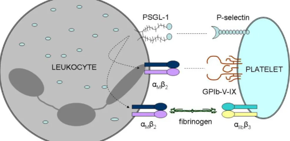
Activation of platelets does not only induce platelet-platelet adhesion but also the formation of heterotypic platelet-leukocyte aggregates (Fig. 8). Platelets can form complexes with granulocytes, monocytes, and with subsets of lymphocytes. These interactions play an important role in platelet-dependent leukocyte recruitment and also in the augmentation of thrombus formation.
Fig. 8. Platelet-leukocyte adhesion. Tethering of platelets to leukocytes through P-selectin and PSGL-1 induces activation of αMβ2receptors. Binding of activated αMβ2
to αIIbβ3 through fibrinogen and to GPIb-V-IX directly leads to firm adhesion. Modified after Ley at al. (Ley 2011)
The initial adhesion of platelets to leukocytes is mediated by P-selectin on platelets and P-selectin glycoprotein ligand-1 (PSGL-1) on leukocytes. Ligation of PSGL-1 leads to Src family kinase-dependent signalling that results in conformational change of leukocyte β2 integrins and also induces delayed responses in leukocytes by the activation of nuclear factor κB and the transcription of proinflammatory molecules (Totani and Evangelista 2010). On the other hand, the loose connection between P- selectin and PSGL-1 holds platelets and leukocytes close to each other, enabling the formation of firm connections, and also activates leukocyte integrins necessary for the strong adhesion. Firm platelet-leukocyte adhesion is mediated mainly due to the formation of a fibrinogen bridge between αIIbβ3 on platelets and macrophage 1-antigen (αMβ2 integrin) on leukocytes, and due to the direct interaction of leukocyte macrophage
1-antigen (αMβ2 integrin) and GPIb-V-IX receptor of platelets (May et al. 2007). These interactions recruit leukocytes to the site of the thrombus formation. In these heterotypic conjugates, the interaction of platelet P-selectin with leukocyte PSGL-1 and platelet CD40 ligand with leukocyte CD40 induces TF expression of leukocytes contributing to initiation of the coagulation cascade and consequently to fibrin production (Cerletti et al. 2012). Moreover, activated neutrophils recruited to the thrombus produce neutrophil extracellular traps. Neutrophil extracellular traps serve as a scaffold primarily composed of extracellular DNA fibres, which are able to trap pathogens circulating in blood.
Neutrophil extracellular trap fibres also contribute significantly to thrombus formation by adhering and activating platelets as well as by stimulating fibrin formation (Fuchs et al. 2012).
Initiation of the coagulation cascade
At the same time as platelet plug formation, blood coagulation factors become activated at the sites of vascular lesions. The initial step of the coagulation cascade (Fig. 9) is triggered by the interaction of activated factor VII plasma protein with subendothelial TF at the site of vascular injury (extrinsic pathway). The activated factor VII-TF complex cleaves factor X. Activated factor X together with activated cofactor V cleaves prothrombin to thrombin. In the initial phase of coagulation, activated factor X generates just a trace amount of thrombin. There are two main inhibitors of the initiation of the coagulation cascade: TFPI blocking factor X, and antithrombin III blocking factor X and thrombin. For the initialization of the coagulation cascade, the TF concentration has to be high enough to overcome the barrier given by TFPI and antithrombin III in order to propagate the coagulation reaction. This mechanism prevents the false activation of the coagulation cascade (Tanaka et al. 2009).
Fig. 9. Initiation and propagation of the coagulation cascade. The coagulation cascade is triggered by the interaction of TF with factor VII. Activated factor VII-TF complex (fVIIa/TF) leads to the activation of factor X (fXa) and factor II (fIIa). The propagation of the coagulation cascade is due to a positive feedback loop involving the activation of factor XII (fXIIa), XI (fXIa), X (fXa), II (fIIa), and cofactor VIII (fVIIIa) and V (fVa) on activated platelets. Activated factor II cleaves fibrin, resulting in fibrin polymerisation.
Activated factor XIII (fXIIIa) strengthens the fibrin mesh. Antithrombin III (AT) and TFPI are inhibitors of the coagulation cascade. Slightly modified after Tanaka et al.
(Tanaka et al. 2009)
Propagation of thrombin production
After initiation of the coagulation cascade, thrombin generation is further exaggerated due to positive feedback loops involving coagulation factors of the intrinsic pathway (Fig 9). Thrombin activates factor XI, VIII, and V. Activated factor XI mediates the activation of factor IX. Activated factor IX together with activated cofactor VIII mediates the cleavage of factor X leading to thrombin generation. Thus, thrombin exaggerates its own production by activating serine proteases of the intrinsic pathway.
Thrombin has a central role in haemostasis: it mediates the generation and stabilisation of the fibrin mesh and it is also a very potent platelet activator.
Contact activation
Coagulation might also be initiated in an alternative way due to contact activation. The biological relevance of this kind of activation has not been fully elucidated, but this
mechanism is relevant to thrombus formation upon the interaction of foreign materials with blood. This phenomenon is also widely utilised in the measurement of the activated partial thromboplastin time when silica or kaolin particles are added to blood to initiate the intrinsic coagulation. The contact system consists of factor XII, plasma kallikrein, as well as the cofactor high-molecular-weight-kininogen that assembles on negatively charged surfaces. When particles with a negatively charged solid surface are incubated with blood, factor XII is able to become activated non-enzymatically.
Alternatively, plasma kallikrein can cleave and activate the surface-bound factor XII.
Activated factor XII also cleaves prekallikrein to kallikrein, resulting in a positive amplification loop. Activated factor XII activates factor XI and thus initiates the intrinsic pathway of the coagulation process (Maas and Renne 2012).
Fibrin mesh
Once thrombin is generated, it cleaves fibrinogen molecules, leading to the release of fibrin monomers. These monomers aggregate spontaneously in a regular array, forming a fibrin clot. In addition to fibrin formation, thrombin activates factor XIII, a highly specific transglutaminase that introduces cross-links composed of covalent bonds between the glutamines and lysines in the fibrin monomers.
Fibrinolysis
Erroneously or excessively produced fibrin mesh can be degraded by fibrinolysis. At first plasminogen, the proenzyme of plasmin, binds to the lysine side chains of the fibrin mesh. This interaction leads to a conformational change of plasminogen molecules, enabling cleavage of plasminogen by plasminogen activators (tissue plasminogen activator and urokinase-type plasminogen activator). The generated plasmin cleaves fibrin into soluble fibrin degradation products, making more and more lysine side chains available where new plasminogen molecules can adhere and become activated. The fibrinolysis is controlled by plasminogen activator inhibitor-1, plasmin inhibitor and trombin activatable fibrinolysis inhibitor. The latter inhibits fibrinolysis by removing lysine side chains from the fibrin mesh. (Machovich 2006, Schaller and Gerber 2011, Foley et al. 2013)
Thrombus formation in the microcirculation
Disturbances in haemostasis can also occur in the microcirculation, inducing alteration of blood flow in tissues. Augmented microcirculatory thrombus formation has been implicated in the pathophysiology of sepsis, disseminated intravascular coagulation, and multiple organ dysfunctions (Gando 2010, Semeraro et al. 2012). Microvascular thrombosis also plays an important role in thrombotic microangiopathies, which comprise a spectrum of diseases such as typical and atypical haemolytic uremic syndrome, thrombotic thrombocytopenic purpura, antiphospholipid syndrome, paroxismal nocturnal haemoglobinuria, malignant hypertension, drugs or systemic autoimmune diseases or antibody-mediated rejection (Benz and Amann 2010).
4. Nanoparticles and thrombus formation
a) Thrombus formation on foreign surfaces
There is a great deal of information about the interaction between blood and foreign material devices such as oxygenators, plasmapheresis equipment, hemodialysers, catheters, stents, vascular grafts, or heart valves. Thrombosis and embolization are well- known complications of these cardiovascular devices. The interaction of blood with foreign surfaces induces thrombus formation by a combination of several mechanisms, involving the coagulation system, platelets, the complement system, and leukocytes (Gorbet and Sefton 2004)
Upon interaction with blood, biomaterials are immediately covered by plasma proteins. Among these proteins are fibrinogen, vWF, factor XII, high molecular weight kininogen, C3 complement, and IgG (Ekdahl et al. 2011).
Factor XII becomes non-enzymatically activated upon adhesion onto negatively charged surfaces and initiates the coagulation cascade (Gorbet and Sefton 2004, Vogler and Siedlecki 2009).
Platelets can adhere to foreign materials mediated by αIIbβ3 or GPIb-V-IX receptors binding to fibrinogen or vWF adhering to the surface of biomaterials. Adhesion leads to platelet activation. However, the interaction with biomaterials often induces platelet activation without adhesion, which is characterised by a decreased platelet count due to
the removal of activated platelets from the circulation. In the presence of cardiovascular devices, platelets also adhere to leukocytes and induce the formation of platelet- leukocyte aggregates (Mickelson et al. 1996, Bonomini et al. 1999, Gorbet and Sefton 2004) (see also section II/3). The amount of platelet-leukocyte aggregates can also be considered as a parameter for biocompatibility (Gorbet and Sefton 2004).
The complement cascade, which is part of the innate immune system, also contributes to the thrombus formation on biomaterials. The complement cascade results in the formation of the terminal C5b-9 complement complex that can be incorporated into platelet membranes inducing platelet activation. Platelets are also activated by the interaction of C1q and C3a ligands with their platelet receptors. (Gorbet and Sefton 2004, Markiewski et al. 2007, Ekdahl et al. 2011)
Leukocytes can also adhere to the surface of foreign materials. Upon adhesion and due to complement activation, leukocytes express TF. Interaction of TF with factor VII initiates the extrinsic pathway of coagulation. Adhered leukocytes also contribute to the recruitment of platelets by binding platelets to biomaterials through platelet-leukocyte interactions (Gorbet and Sefton 2004, Vogler and Siedlecki 2009).
b) Epidemiology of particulate matter-associated diseases
Relevant information about the toxicity of nanoparticles comes from the investigation of the detrimental effect of air pollution on the human body. Although air pollution is composed not just of nanosized particles, but also larger particulate matter, gaseous and liquid components, this knowledge gives some clues about the possible effects of nanoparticles.
There is a well-known epidemiological association between air pollution and cardiovascular or pulmonary diseases (Brook et al. 2010). The detrimental health effects of air pollution were first realised in the 20th century after major incidents involving acute air pollution (Meuse Valley, Belgium 1930; Donora, USA 1948; and Greater London, UK 1952,) caused sudden deaths, increased illness, and hospital admissions (Simkhovich et al. 2008).
Further investigations revealed that the particulate matter fraction of air pollution is the major contributor to these toxic effects. A number of epidemiological studies have demonstrated the close relationship between particulate matter levels and
cardiopulmonary mortality both in short and long-term studies. Overall evidence from meta-analysis of about 100 research papers since the early 1990s and recent comprehensive analysis of large number multicity studies assume that a short-term (1 to 5 days) 10 µg/m3 elevation of PM2.5 results in an ~1% increase in mortality (Pope and Dockery 2006, Samoli et al. 2008, Brook et al. 2010, Emmerechts and Hoylaerts 2012).
Long-term prospective cohort studies have provided more evidence about the health effects of particulate matter. The Harvard Six Cities Study investigated about 8000 persons over 14-16 years (Dockery et al. 1993), the American Cancer Society study analysed data from more than 500,000 adults over 16 years (Pope et al. 2002, Pope et al.
2004, Franchini and Mannucci 2012) and recent large cohort studies of 13.2 million US Medicare participants were analysed for the time period 2000 to 2005. The overall evidence from the cohort studies show on average 10% increase for all case mortality per 10 µg/m3 elevation in long-term PM2.5 exposure. The cardiovascular mortality risk ranges from 3% to 76% in the different cohort studies (Brook et al. 2010).
The role of the nano-sized fraction in particulate matter induced detrimental effects has been gathered from studies collecting data at the source of combustion-derived particles. Ultrafine particles generated by combustion-related processes have a very short life (minutes to hours) and rapidly grow to form a larger complex. Therefore, ultrafine particulate matter concentration is at its highest near the source of primary particles, such as in the vicinity of busy roads. Interestingly, Peters et al. found in a survey of 691 patients a correlation between exposure to traffic and the onset of a myocardial infarction within one hour afterwards (Peters et al. 2001, Peters et al. 2004).
An association was also found between short-term exposure to ultrafine particles and hospital admissions for stroke (Andersen et al. 2010). A linear relationship was described between the distance from major traffic roads and the risk of deep vein thrombosis (Baccarelli et al. 2009), indicating the role of ultrafine particles in this process.
There are also epidemiological data that substantiate the influence of air pollution on haemostasis (Emmerechts and Hoylaerts 2012). A positive association was found between particulate matter exposure and plasma fibrinogen concentration, plasminogen activator inhibitor-1 level, and changes of coagulation tests towards hypercoagulability (Pekkanen et al. 2000, Su et al. 2006, Baccarelli et al. 2007, Chuang et al. 2007, Bigert
et al. 2008). Particulate matter exposure is also associated with platelet activation.
Chronic inhalation of particulate matter emitted from biomass burning during household cooking in India was shown to induce platelet activation (increased CD62P expression) and to increase the amount of platelet-leukocyte aggregates (Ray et al. 2006). Positive associations were found between soluble P-selectin (shedding after platelet activation) and ambient ultrafine particles (Delfino et al. 2008). Also an immediate increase of a plasma soluble CD40 ligand and a decrease in platelet count was found to be associated with ultrafine particles (Ruckerl et al. 2007).
c) Particulate matter and thrombus formation
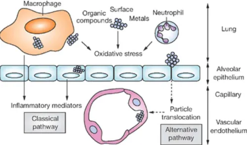
To find out how inhaled particulate matter induces detrimental cardiovascular effects several experimental studies have been carried out. Inhaled particles have been found to deposit in the lungs and to induce the production of inflammatory mediators. Based on these findings, the first (classical) hypothesis was that the release of inflammatory agents from the lungs into the blood circulation alters the cardiovascular system or haemostasis (Fig. 10) (Brook et al. 2004, Mills et al. 2009).
The role of inflammatory mediators in inducing prothrombotic effects is supported by experiments in which mice were intratracheally exposed to particulate matter. These studies observed shortened bleeding time and increased thrombus formation 24 hours after application (Mutlu et al. 2007). The prothrombotic effect was attributed to an IL-6- dependent increased activity of the coagulation cascade, and a tumour necrosis factor alpha-dependent decreased fibrinolysis due to increased plasminogen activator inhibitor-1 activity (Mutlu et al. 2007, Budinger et al. 2011). Also controlled human exposure to diesel exhaust particles (DEP) was shown to reduce the release of endothelial tissue plasminogen activator (Mills et al. 2005, Mills et al. 2007).
Furthermore, increased TF expression was demonstrated upon incubation of different cells with particulate matter in vitro and in the aorta after long-term exposure to particulate matter in vivo (Gilmour et al. 2005, Sun et al. 2008).
Fig. 10. Classical and alternative pathways through which airborne nanoparticles induce cardiovascular effects. Classical pathway: Nanoparticles deposited in the lung induce oxidative stress through chemically active organic or metal compounds given on the surface of nanoparticles or through activation of neutrophils and macrophages. In this way they initiate the production of inflammatory mediators from macrophages and alveolar epithelial cells, which are released into the systemic circulation and induce cardiovascular effects. Alternative pathway: Nanoparticles translocate from the lungs through the alveolar epithelium into the blood circulation, where they alter cardiovascular functions. (Mills et al. 2009)
In further epidemiological studies about the detrimental effects of particulate matter, the important role of its ultrafine fraction has been indicated. Knowing that nano-sized particles are able to translocate from the lungs into the blood (see section II/2/g), an alternative hypothesis was suggested emphasizing that translocated ultrafine particles can reach remote tissues via the circulation and influence organ functions directly (Fig.
10) (Brook et al. 2004, Mills et al. 2009).
The first experimental findings supporting the alternative hypothesis that intratracheally administered nanoparticles are able to augment thrombus formation were published by Nemmar et al. The authors demonstrated in an in vivo hamster model that intratracheally instilled or intravenously injected amine-modified polystyrene nanoparticles activate platelets and enhance thrombosis in femoral arteries and veins within 1 hour after application (Nemmar et al. 2002b). Interestingly, just nano-sized (60
nm) and not coarse (400 nm) polystyrene beads influenced thrombus formation, while both nano-sized and coarse polystyrene beads induced lung inflammation suggesting different mechanisms for these two processes (Nemmar et al. 2003b). Later, not just polystyrene beads but also DEP were shown to activate blood platelets and augment thrombus formation (Nemmar et al. 2003a). The prothrombotic effect of DEP was augmented even 6 and 24 hours after instillation and the instillation of DEP nanoparticles also induced an inflammatory reaction in the lung, i.e. increased granulocyte count and elevated histamine concentration in the bronchoalveolar lavage.
Interestingly, H1 histamine receptor antagonist inhibited the increase in granulocyte counts in the bronchoalveolar lavage and the late (6- and 24-hour) prothrombotic effects, but did not affect the acute (1 hour) DEP-induced thrombosis or platelet activation (Nemmar et al. 2003c). These findings suggest that both mechanisms contribute to the prothrombotic effect of DEP nanoparticles: translocated nanoparticles induce early prothrombotic effects by direct activation, while lung inflammatory mediators generate late prothrombotic effects.
The above investigations evoked further studies to find out how the interaction of ultrafine particles with blood can influence haemostasis. Several experiments demonstrated the role of platelet activation in the prothrombotic effects of ultrafine particles. In vitro experiments found increased P-selectin expression, augmented platelet aggregation, and the formation of platelet-leukocyte aggregates upon the incubation of blood with ultrafine particles (Nemmar et al. 2002b, Nemmar et al. 2003a, Nemmar et al. 2003b, Nemmar et al. 2003c, Radomski et al. 2005, Khandoga et al. 2010, Nemmar et al. 2010, Forestier et al. 2012, Kim et al. 2012). Using in vivo microscopy, platelet accumulation was found in the hepatic microcirculation in mice upon injection of carbon black ultrafine particles. Platelet adhesion to endothelial cells was strongly associated with fibrin deposition, and the blockade of the fibrinogen receptor αIIbβ3
inhibited platelet accumulation in the liver (Khandoga et al. 2004). In the recent work of our research group (Khandoga et al. 2010), a similar experiment was carried out, but instead of systemic injection, inhalative exposure was used for the application of carbon black nanoparticles. The experiment showed the same result: platelets accumulated in the microcirculation of the liver. Fibrin deposition was found on the endothelial surfaces of the microcirculation in the liver and heart. In contrast, according to data obtained
from the bronchoalveolar lavage, a significant pulmonary inflammatory response did not occur and neither the plasma levels of proinflammatory cytokines nor blood cell counts were affected. These data suggest that inhaled carbon black nanoparticles generate prothrombotic effects, also without inducing an inflammatory response in the lungs.
These data were further confirmed by controlled human exposure experiments.
Inhalation of elemental carbon ultrafine particles by patients with type 2 diabetes mellitus for 2 hours increased the CD40 ligand expression of platelets and the number of platelet-leukocyte aggregates (Stewart et al. 2010). In the experiment of Lucking et al. healthy volunteers were exposed to DEP, and thrombus formation was measured in a Badimon ex vivo perfusion chamber. The Badimon chamber consists of a pump to draw blood from the cubital vein through a perfusion chamber with a strip of porcine aorta.
Diesel exhaust particles increased thrombus formation and the formation of platelet- leukocyte aggregates (Lucking et al. 2008).
Ambient nanoparticles were also found to activate the coagulation system (Nemmar et al. 2010, Kilinc et al. 2011). The authors concluded that early procoagulant effects are dependent on the TF-driven extrinsic pathway, whereas long-lasting thrombogenic actions are due to translocated ultrafine particles inducing contact-dependent activation of the coagulation cascade (Kilinc et al. 2011).
Summarising the experimental data about prothrombotic mechanisms of inhaled particulate matter suggests that larger ambient particles deposit in the lung and induce procoagulative changes through inflammatory mediators, whereas nano-sized ambient particles immediately translocate in the circulation, directly activate platelets, and later on activate the coagulation system via contact activation.
d) Manufactured nanoparticles and thrombus formation
The research performed on biomaterials and particulate matter indicates that manufactured nanoparticles might also influence thrombus formation. In spite of the fact that manufactured nanoparticles are produced in high amounts, their effects on the thrombotic process have been investigated to a lesser extent.
Most information concerning the prothrombotic effects of nanoparticles have been obtained from in vitro experiments. Increased platelet activation and aggregation were
found to be induced by amine-modified polystyrene beads (Nemmar et al. 2002b, Mayer et al. 2009, McGuinnes et al. 2011), quantum dots (Geys et al. 2008), titanium dioxide nanorods (Nemmar et al. 2008), and gold nanoparticles (Deb et al. 2011).
However, in another study gold nanoparticles failed to affect platelet function or coagulation (Dobrovolskaia et al. 2009). Silver nanoparticles were found to prevent platelet activation, adhesion, and aggregation in a concentration-dependent manner, and to inhibit fibrin polymerisation (Shrivastava et al. 2009, Shrivastava et al. 2011).
Coagulation was induced by synthetic amorphous silica and organically modified silica nanoparticles (Tavano et al. 2010), polylactic acid nanoparticles (Sahli et al. 1997), and titanium dioxide nanotubes, whereas titanium dioxide nanoparticles inhibited the coagulation system (Roy et al. 2007). Nano-sized copper (II) oxide caused up-regulation of plasminogen activator inhibitor-1 in endothelial cells (Yu et al. 2010).
There is much less data about the effect of nanoparticles on thrombus formation in vivo. It has been shown that amine-modified polystyrene nanoparticles augmented and carboxyl-modified polystyrene nanoparticles inhibited thrombus formation in femoral vessels (Nemmar et al. 2002b). Carbon nanotubes were found to augment ferric chloride-induced thrombosis in vivo (Radomski et al. 2005). TiO2 rutile nanorods decreased the platelet count in vivo (Nemmar et al. 2008). Intravenous administration of quantum dots (Geys et al. 2008) or mesoporous silicate nanoparticles (Hudson et al.
2008) were shown to induce pulmonary thrombosis.
The above studies have investigated thrombus formation in large vessels. Although nanoparticles also reach the microcirculation, there are only a few recent publications that have analysed the effects of nanoparticles on thrombus formation in the microvasculature. Amine-modified polystyrene nanoparticles as benchmark particles were shown to induce a dose-dependent enhancement of thrombus formation in the microcirculation (Silva et al. 2005). Additionally, a previous study from our research group has demonstrated that systematically administered carbon black nanoparticles enhanced platelet accumulation on the endothelium of postsinusoidal venules and sinusoids in the hepatic microcirculation (Khandoga et al. 2004). However, the effects of manufactured nanoparticles on thrombus formation in the microcirculation have not been investigated yet.
III. Aims
The main objective of my research work was to investigate platelet activating and microcirculatory prothrombotic effects of manufactured nanoparticles. To achieve these aims, some methodological problems also had to be solved, i.e. the preparation of nanoparticle dispersions in physiological solutions and the optimisation of the platelet- granulocyte complex measurement.
1. Optimisation of the preparation method of nanoparticle dispersions
For investigations of in vitro and in vivo prothrombotic effects, nanoparticles have to be dispersed in physiological solutions. However, nanoparticles in solutions with physiological salt concentrations and pH values form coarse agglomerates. To avoid the formation of coarse agglomerates, a steric stabiliser has to be added to the nanoparticle dispersions. To optimise the dispersion method, we varied the following factors while preparing the nanoparticle dispersions:
i) ultrasound energy levels
ii) type of dispersion stabilizer: human, bovine, or mouse albumin, Tween 80, or mouse serum
iii) concentration of dispersion stabilizer iv) concentration of nanoparticles v) sequence of preparation steps vi) stability of the dispersion over time
vii) We also tested our method on a broad range of various types of nanoparticles.
2. Optimisation of platelet-granulocyte complex measurement
For the evaluation of platelet activation, also the amount of platelet-granulocyte complexes was measured. The measurement of platelet-granulocyte complexes with a flow cytometer is based on the simultaneous detection of fluorescent signals from both cell types. However, these double-positive signals can also originate from the coincidence of non-interacting platelets and granulocytes in the detection volume. Our aim was to develop a method that measures the real amount of platelet-granulocyte complexes without overestimating it due to coincidence.
3. Prothrombotic effects of nanoparticles
Ambient nanoparticles have been shown to exert prothrombotic effects, but manufactured nanoparticles are less well investigated in this regard. Although circulating nanoparticles also reach the microcirculation, the effects of manufactured nanoparticles on microcirculatory thrombus formation have not been investigated yet.
Thus the aim of this study was:
i) to characterize the effects of DEP, titanium dioxide rutile, and single-walled carbon nanotube nanoparticles on platelet activation and
ii) on the formation of platelet-granulocyte complexes in vitro
iii) to assess their impact on thrombus formation in small arteries and in the microcirculation in vivo, and
iv) to compare these effects with those induced by surface-modified polystyrene beads as benchmark particles.
IV. Materials and Methods
1. Materials
a) Nanoparticles
Table 3. Nanoparticles used in our study, name of company, and size of nanoparticles
as given by the manufacturer.
Nanoparticle Size Company
Titanium(IV) oxide nanopowder 99.5% rutile
~10 nm × 40 nm Sigma-Aldrich, Schnelldorf, Germany
Titanium(IV) oxide nanopowder 99.7%
anatase
<25 nm Sigma-Aldrich, Schnelldorf, Germany
Zinc oxide nanopowder <100 nm Sigma-Aldrich, Schnelldorf, Germany
Plain polystyrene beads 60 nm Bangs Laboratories, Fishers, USA
Carboxyl-modified polystyrene beads
60 nm Bangs Laboratories, Fishers, USA
Amine-modified polystyrene beads
65 nm Bangs Laboratories, Fishers, USA
S-purified SWNT outer diameter < 2 nm, length 1–5 μm
SES research, Houston, USA
S-purified MWNT outer diameter 10–30 nm, lengths 1–2 μm
SES research, Houston, USA