Vol.:(0123456789)
1 3
Archives of Virology (2020) 165:245–248 https://doi.org/10.1007/s00705-019-04441-4
ANNOTATED SEQUENCE RECORD
Coding‑complete genome sequencing suggests that Newcastle
disease virus challenge strain Herts’33 (IVMP) may represent a distinct genotype
Enikő Fehér1 · Ádám Bálint2 · Szilvia Marton1 · Krisztina Bali1 · Sándor Belák3 · Krisztián Bányai1
Received: 19 June 2019 / Accepted: 15 September 2019 / Published online: 8 November 2019
© The Author(s) 2019
Abstract
We determined the genomic sequence of a Newcastle disease virus (NDV) line obtained directly from the first NDV isolate, named Herts’33. This strain shared ≤ 90% nucleotide sequence identity with the NDV sequences available in the GenBank database, and formed a distinct branch in a phylogenetic tree. This branch may be considered to represent a separate NDV genotype. Our study indicates that investigation of the genomic sequences of old NDV strains that originated from the early outbreaks of Newcastle disease may alter the phylogenetic grouping of the NDV strains and provide data on the evolution of viral genomes over time.
Newcastle disease virus (NDV) (species Avian orthoavula- virus 1) belongs to the genus Orthoavulavirus, family Para- myxoviridae, order Mononegavirales. The enveloped virion has a single-stranded, negative-sense RNA genome with a length of ~ 15.2 kb [1, 2]. The viral genome is composed of six genes in the following order: 3’-NP-P-M-F-HN-L-5’
(letters designate the nucleoprotein (NP), the phosphopro- tein (P), the matrix protein (M), the fusion protein (F), the hemagglutinin-neuraminidase (HN) and the RNA poly- merase (L). Based on a number of criteria, the most recent genetic characterization classifies strains belonging to the species Avian orthoavulavirus 1 into genotypes I to XXI and numerous subgenotypes within a subset of genotypes [1].
NDV infection may affect a wide range of wild and domestic avian species, causing serious losses during the epizootics [2]. The disease is controlled by vaccination of poultry, typically using live attenuated strains [2, 3]. For example, the viscerotropic Herts’33 strain was originally isolated from a field sample after an outbreak in 1933 in Hertfordshire (UK) and subsequently became an important strain for control and prevention of Newcastle disease (ND).
The mesogenic H vaccine line was considered a derivate of the Herts’33 field strain and used as a potent live, attenu- ated vaccine on multiple continents, including Europe [4].
In addition, descendants of the original velogenic Herts’33 strain have been commonly used as challenge strains for test- ing ND vaccine efficacy.
With the introduction of molecular characterization methods, it has become possible to compare the genetic features of different descendants of Herts’33 and to analyze the homogeneities and differences in their genomes. In a 2003 paper, Czeglédi et al. [4] revealed that the descend- ant lines of Herts’33, including the vaccine strain H, dif- fered in their F protein gene coding sequences, and three genetic groups were identified by phylogenetic analysis. It was hypothesized that the phylogenetic lineage that includes the Weybridge-origin Herts’33 strain might be a descendant of the isolate from the first ND case, while the Herts’33 lines that clustered phylogenetically with genotype IV sequences were of unknown origin. Interestingly, the vaccine strain H clustered with genotype III strains, including other vaccine strains (e.g., Mukteswar) [4].
Handling Editor: Bert K. Rima.
* Krisztián Bányai bkrota@hotmail.com
1 Institute for Veterinary Medical Research, Centre
for Agricultural Research, Hungarian Academy of Sciences, Hungária krt. 21, Budapest 1143, Hungary
2 Veterinary Diagnostic Directorate, National Food Chain Safety Office, Budapest, Hungary
3 Department of Biomedical Sciences and Veterinary Public Health (BVF), The OIE Collaborating Centre for the Biotechnology-Based Diagnosis of Infectious Diseases in Veterinary Medicine, Swedish University of Agricultural Sciences (SLU), Uppsala, Sweden
246 E. Fehér et al.
1 3
In this study, we determined the complete coding sequence of a lineage Herts’33(W) [4] strain, which has been maintained in eggs and used as a challenge strain at the Insti- tute for Veterinary Medicinal Products, Budapest, Hungary [4]. This strain, referred to here as Herts’33 (IVMP), was purchased in 1999 from the Veterinary Laboratories Agency, Weybridge, and then aliquoted in the Budapest institute.
When starting the present study, an aliquot of the original seed was passaged once in eggs to obtain sufficient amount of material for genome sequencing. The viral RNA was extracted using a QIAamp Viral RNA Mini Kit (QIAGEN) and amplified by random RT-PCR as described elsewhere in detail [5]. The amplified cDNA was subjected to sequenc- ing using the Ion Torrent PGM system (Life Technologies).
Genome assembly was performed using CLC Genomic Workbench v7.0 (www.qiage nbioi nform atics .com/produ cts/
clc-genom ics-workb ench/). Trimmed sequence reads were mapped onto reference NDV sequences to produce the con- sensus genome sequence of Herts’33 (IVMP).
The consensus sequence of Herts’33 (IVMP) consisted of 15,166 bases (accession no. MK674396) and lacked sequence information for a ~ 20 nt-long fragment in the 3’
end untranslated region. The genome organization was typi- cal for NDV (Fig. 1), and the cleavage site in the F protein was typical for velogenic and mesogenic NDV strains (aa
112 RRQRR↓F117) [6, 7].
A BLAST search and phylogenetic analysis [8, 9] showed that the closest relative of Herts’33 (IVMP) was the geno- type I (class II) strain Ulster/67 (accession no. AY562991), with 90.2% genome-wide identity. Regarding the com- plete F gene, we used the newly recommended method to calculate the average sequence identity among strains;
as a result, the ≤ 88.3% nt sequence identity between the Herts’33 (IVMP) and the references fell below the cutoff value (i.e., 10% evolutionary distance) established for geno- type discrimination for NDV strains [1, 10]. The topology of a phylogenetic tree based on the F gene also suggested
that Herts’33 (IVMP) could be considered a separate geno- type (Fig. 2; [1, 10]). Despite the molecular evidence that Herts’33 (IVMP) represents a unique genetic lineage, unfor- tunately, independent isolates related to this historic labora- tory strain were not available for validation of our genetic classification [1, 10].
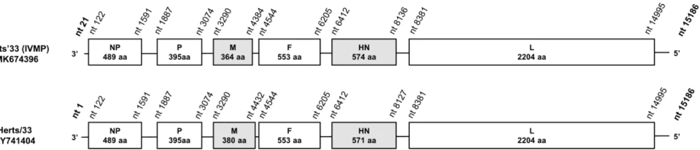
The strain Herts’33 (IVMP) and a genotype IV strain, Herts/33 (accession no. AY741404), shared 88.1% genome- wide and 85.7% F-gene-based nt sequence identity and clus- tered on different branches in the phylogenetic trees (Figs. 1, 2). Additional differences were seen when comparing the lengths of the deduced M and HN proteins (Fig. 1) [6]. The length of the M protein of Herts’33 (IVMP) was identical to that of most of the NDV isolates (364 aa) but shorter than the M protein of Herts/33 (380 aa). The HN protein of Herts’33 (IVMP) was 3 aa longer than the HN protein of other NDV strains (574 aa vs. 571 aa), including Herts/33 (Fig. 1), although additional size variation exists in other strains (e.g., Ulster/67, 616 aa). These data together imply a distinct evolutionary origin of the strains Herts’33 (IVMP) and Herts/33, a finding that corroborates earlier hypotheses [4].Apparently, some reference NDV strains were main- tained at various laboratories for decades without the precise knowledge of their genetic background. In this study, we provide genomic sequence data of an ‘old’ NDV challenge strain that likely represents an extinct genotype of NDV. This new genome sequence information will be useful to update schemes for classification of NDV isolates and to support more reliable evaluation of experimental data on the genetic diversity of various vaccine and challenge strains of NDV.
Fig. 1 Schematic representation and comparison of the Herts’33 (IVMP) and Herts/33 genomes. NP, nucleocapsid protein; P, phos- phoprotein; M, matrix protein; F, fusion glycoprotein; HN, hemagglu-
tinin-neuraminidase protein; L, large or RNA-dependent RNA poly- merase protein. The nt positions in Herts’33 (IVMP) were adjusted to those of the fully sequenced strain Herts/33
Fig. 2 Phylogenetic analysis based on full-length fusion gene sequences (A) and representative complete genome sequences (B) using the maximum-likelihood method and the general time-reversi- ble (G) model in MEGA6 software [9], with 100 bootstrap replicates.
The Herts’33 (IVMP) sequence is indicated by a black dot
◂
247 Newcastle disease virus strain Herts’33 (IVMP)
1 3
B A
248 E. Fehér et al.
1 3
Acknowledgements Open access funding provided by MTA Centre for Agricultural Research (MTA ATK).
Author contributions ÁB, SB, and KB conceived the study and pro- vided reagents and materials; EF, SM, and KB generated and analyzed data; and EF prepared the first manuscript draft. All authors have read and approved the final version of the manuscript and its submission for publication.
Funding Support was obtained from the Bolyai János Fellowship pro- gram awarded by the Hungarian Academy of Sciences (EF, SM), from the Swedish Research Council FORMAS Strong Research Environ- ments project, nr 2011–1692, “BioBridges” (SB), and from the Hun- garian Scientific Research Fund (NKFI-OTKA, K120201) (KB).
Compliance with ethical standards
Conflict of interest The authors declare no conflict of interest.
Open Access This article is distributed under the terms of the Crea- tive Commons Attribution 4.0 International License (http://creat iveco mmons .org/licen ses/by/4.0/), which permits unrestricted use, distribu- tion, and reproduction in any medium, provided you give appropriate credit to the original author(s) and the source, provide a link to the Creative Commons license, and indicate if changes were made.
References
1. Dimitrov KM, Abolnik C, Afonso CL, Albina E, Bahl J, Berg M, Briand FX, Brown IH, Choi KS, Chvala I, Diel DG, Durr PA, Ferreira HL, Fusaro A, Gil P, Goujgoulova GV, Grund C, Hicks JT, Joannis TM, Torchetti MK, Kolosov S, Lambrecht B, Lewis NS, Liu H, Liu H, McCullough S, Miller PJ, Monne I, Muller CP, Munir M, Reischak D, Sabra M, Samal SK, de Almeida SR, Shittu I, Snoeck CJ, Suarez DL, Van Borm S, Wang Z, Wong FYK (2019) Updated unified phylogenetic classification system and
revised nomenclature for Newcastle disease virus. Infect Genet Evol. 74:103917. https ://doi.org/10.1016/j.meegi d.2019.10391 7 2. Bello MB, Yusoff K, Ideris A, Hair-Bejo M, Peeters BPH, Omar
AR (2018) Diagnostic and vaccination approaches for Newcastle Disease Virus in poultry: the current and emerging perspectives.
Biomed Res Int 2018:7278459
3. Mayers J, Mansfield KL, Brown IH (2017) The role of vaccina- tion in risk mitigation and control of Newcastle disease in poultry.
Vaccine 35:5974–5980
4. Czeglédi A, Wehmann E, Lomniczi B (2003) On the origins and relationships of Newcastle disease virus vaccine strains Hertford- shire and Mukteswar, and virulent strain Herts’33. Avian Pathol 32:271–276
5. Bányai K, Kemenesi G, Budinski I, Földes F, Zana B, Marton S, Varga-Kugler R, Oldal M, Kurucz K, Jakab F (2017) Candidate new rotavirus species in Schreiber’s bats, Serbia. Infect Genet Evol 48:19–26
6. de Leeuw OS, Koch G, Hartog L, Ravenshorst N, Peeters BP (2005) Virulence of Newcastle disease virus is determined by the cleavage site of the fusion protein and by both the stem region and globular head of the haemagglutinin-neuraminidase protein. J Gen Virol 86:1759–1769
7. Gould AR, Hansson E, Selleck K, Kattenbelt JA, Mackenzie M, Della-Porta AJ (2003) Newcastle disease virus fusion and haemagglutinin-neuraminidase gene motifs as markers for viral lineage. Avian Pathol 32:361–373
8. Altschul SF, Gish W, Miller W, Myers EW, Lipman DJ (1990) Basic local alignment search tool. J Mol Biol 215:403–410 9. Tamura K, Stecher G, Peterson D, Filipski A, Kumar S (2013)
MEGA6: molecular evolutionary genetics analysis version 6.0.
Mol Biol Evol 30:2725–2729
10. Diel DG, da Silva LH, Liu H, Wang Z, Miller PJ, Afonso CL (2012) Genetic diversity of avian paramyxovirus type 1: proposal for a unified nomenclature and classification system of Newcastle disease virus genotypes. Infect Genet Evol 12:1770–1779 Publisher’s Note Springer Nature remains neutral with regard to jurisdictional claims in published maps and institutional affiliations.