FIRST IDENTIFICATION OF PORCINE PARVOVIRUS 3 IN A WILD BOAR IN ITALY BY VIRAL METAGENOMICS –
SHORT COMMUNICATION
Maria Grazia AMOROSO1*, Francesco CERUTTI2, Nicola D’ALESSIO1,
Maria Gabriella LUCIBELLI1, Anna CERRONE1, Pier Luigi ACUTIS2, Giorgio GALIERO1, Giovanna FUSCO1* and Simone PELETTO2
1Department of Animal Health, Istituto Zooprofilattico Sperimentale del Mezzogiorno, Via Salute 2, 80055 Portici, Naples, Italy; 2Istituto Zooprofilattico Sperimentale del
Piemonte, Torino, Italy
(Received 22 June 2018; accepted 20 November 2018)
Metagenomic analysis revealed the presence of porcine parvovirus 3 (PPV3) in the pool of the internal organs of a wild boar found dead in Southern Italy. Phylogenetic analysis based on the complete coding sequences showed that the newly detected virus is most closely related to those found also in wild boars in Romania during 2010–2011. Even though the death could not be associated with this virus, PPV3 could have contributed to lowering the host’s immunologi- cal defences.
Key words: Parvovirus, metagenomics, wild boar
Parvoviruses are small, non-enveloped viruses with a single-stranded DNA genome. Members of the family Parvoviridae are common pathogens causing a wide range of diseases in humans and animals, including swine as well. Among porcine parvoviruses, a novel emerging type, namely porcine parvovirus (PPV3), has been discovered in Hong Kong in 2007 (Lau et al., 2008). Phylogenetic anal- ysis showed that this virus (also referred to as porcine hokovirus, porcine partet- ravirus, porcine parvovirus type 4) was most similar to human parvoviruses (types 4 and 5), forming a distinct cluster among parvoviruses (Miranda et al., 2016). PPV3 has been identified in pigs on almost all continents (Sliz et al., 2015) and recently in cattle and also in wild boars in some countries of Europe like Germany (Adlhoch et al., 2010), Romania (Cadar et al., 2011), Slovakia (Sliz et al., 2015) and Portugal (Miranda et al., 2016). In October 2016, a wild boar found already dead by a hunter in the province of Avellino, Campania Re- gion, Southern Italy, was referred for necropsy to the Istituto Zooprofilattico Sperimentale del Mezzogiorno (IZSM). Organs were analysed by microbiologi- cal investigations following standard procedures to determine the possible cause
*Corresponding authors; E-mail: mariagrazia.amoroso@izsmportici.it;
giovanna.fusco@izsmportici.it; Phone: 0039 (0) 817-865116-113
of death. Aerobic culture of the brain, liver, lymph nodes, spleen and intestine was carried out together with anaerobic culture of the intestine. Furthermore, the presence of Chlamydophila spp., Coxiella burnetii and Neospora caninum was investigated by PCR using previously described protocols (Ossewaarde and Mei- jer, 1999; Perugini et al., 2009; Auriemma et al., 2014). Escherichia coli toxins were investigated on isolated E. coli according to Borriello et al. (2012). A pool of organs was subjected to metagenomic analysis. In brief, tissue samples from brain, spleen, lymph node and intestine were homogenised in ice-cold PBS and centrifuged. The clear supernatant was filtered through a 0.45-µm filter and treated with a DNase/RNase cocktail, then processed for total RNA extraction using Qiamp Viral RNA kit. The RNA was reverse transcribed and pre-amplified by the SISPA method (Allander et al., 2005) and the ds-cDNA was processed with the Nextera XT library preparation kit for sequencing on Illumina MiSeq platform with a 2 × 250-bp run and a MiSeq Reagent Kit v3-600. Data were fil- tered with Trim Galore! V0.4.3 (Trim_galore: https://www.bioinformatics.
babraham. ac.uk/projects/trim_galore/) and bbmap.sh v37.54 (bbmap: https://
sourceforge.net/projects/bbmap/) removing SISPA primers, low quality reads, and reads mapping to the host annotated transcriptome. The remaining reads were de novo assembled with MEGAHIT v1.1.1 (Li et al., 2016) and contigs were classi- fied with a blastn search. Reads were also mapped to reference African swine fe- ver virus (ASFV) genomes using Bowtie 2 (Langmead and Saltzberg, 2012).
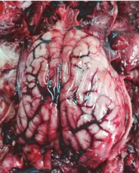
Fig. 1. External surface of the brain of the wild boar found dead in South Italy during the hunting season of 2016. A marked vascularisation can be clearly observed
Fig. 2. Phylogenetic tree of 42 genome sequences of porcine parvoviruses. The tree was inferred using algorithm MrBayes v3.2.6 based on the GTR substitution model. Country of origin and GenBank accession number are shown for each sequence. The tree is mid-point rooted and the in- ternal node support is expressed as posterior probability. The PPV3 genome sequence presented in
this work is marked in bold. The scale bar is expressed as nucleotide substitutions per site
The postmortem examination showed the presence of marked brain vascu- larisation (Fig. 1), pulmonary congestion and meningeal hyperaemia. The remain- ing organs appeared to be normal in terms of shape, size and consistency. Microbi-
ological investigation revealed no anaerobic bacteria. Aerobic cultures showed on- ly the presence of E. coli in the intestine. The isolated bacteria underwent toxin characterisation showing the presence of the VT2 toxins. All PCRs, targeting the different pathogens, were negative. As to the metagenomic analysis, the run gener- ated 27,275,964 raw reads that resulted in 27,168,344 reads after filtering. No reads mapped to the AFSV genome; this step was carried out to exclude AFSV that had just reached western Europe at the time of writing this manuscript, with two cases identified in wild boars in Belgium (unpublished). The PPV3 contig was 4,779 nucleotide (nt) long corresponding to 93.47% of the complete genome (5,113 bp); 2.75% reads mapped to the virus genome, with an average coverage of 21,900X. The sequence has been submitted to the GenBank database with Acces- sion Number MH884552. According to the blast output, the genome shared 98–
99% nucleotide sequence identity with other PPV3 genomes in the non-redundant database. The PPV3 genome was aligned for comparison with 41 complete ge- nomes of porcine parvoviruses available in GenBank with Muscle v3.8.31 (Edgar, 2004). The average identity of the Italian virus was 98.47%, with a minimum of 96.63% and a maximum of 99.35%, showing a limited diversity within this virus.
At the amino acid level, the average similarity was 98.80% (min.: 94.15%, max.:
99.87%). A phylogenetic tree was inferred using MrBayes v3.2.6 (Ronquist et al., 2012) based on the GTR substitution model. Our newly identified virus genome appeared within a clade of genomes originating from Romania and dating back to 2010–2011 (Cadar et al., 2011). Members of this clade share > 99% nt sequence identity and all of them originate from wild boar. In the Romanian study, 372 ani- mals were sampled during the 2010/2011 hunting season and 188 (50.54%) proved to be positive to PPV3 (originally called Porcine hokovirus). The phylogenetic analysis of the NP1 performed by Cadar et al. (2011) revealed a high similarity be- tween the Romanian virus sequences collected in 2006/2007 and others from Ger- many and UK, while the more recent ones were more similar to those from Hong Kong dating back to 2007 (samples reported as from China in Fig. 2). PPV3 was also detected in 12 out of 50 wild boars shot in the hunting season 2011/2012 in the districts of Vila Real and Braganc, Portugal, and the NP1 sequences were closely related to those of viruses from Europe, the United States, Brazil and Chi- na. According to the phylogenetic tree of the complete genome of PPV3, at least four clades can be identified (Fig. 2). The clades c and d include PPV3 sequences from Europe, while viruses from China are present in every clade. Surprisingly, most of the PPV3 detected in Europe (Germany, Portugal, Romania, and Slovakia) belonged to clade c, while only samples from Italy, Romania and Slovakia be- longed to clade d. Apparently, these two variants circulate in Europe, and both have been detected in Romanian and Slovakian wild boars. The high similarity of our Italian virus to Romanian samples may suggest that the virus has not a high evolutionary rate. This is the first identification of PPV3 in Italy, a country where there are large populations of wild boars often interacting with domestic animals
(Aprea et al., 2017). Further investigations on porcine parvoviruses in Italy may therefore be necessary to assess their prevalence in both wild and domestic popula- tions. Finally, further data are needed to better describe the epidemiology of this virus in the European setting.
References
Adlhoch, C., Kaiser, M., Ellerbrok, H. and Pauli, G. (2010): High prevalence of porcine Hokovirus in German wild boar populations. Virol. J. 7, 171-422X-7-171.
Allander, T., Tammi, M. T., Eriksson, M., Bjerkner, A., Tiveljung-Lindell, A. and Andersson, B.
(2005): Cloning of a human parvovirus by molecular screening of respiratory tract sam- ples. Proc. Natl Acad. Sci. U. S. A. 102, 12891–12896.
Aprea, G., Amoroso, M. G., Di Bartolo, I., D’Alessio, N., Di Sabatino, D., Boni, A., Cioffi, B., D’Angelantonio, D., Scattolini, S., De Sabato, L., Cotturone, G., Pomilio, F., Migliorati, G., Galiero, G. and Fusco, G. (2018): Molecular detection and phylogenetic analysis of hepatitis E virus strains circulating in wild boars in south-central Italy. Transbound. Emerg.
Dis. 65, e25–e31.
Auriemma, C., Lucibelli, M. G., Borriello, G., De Carlo, E., Martucciello, A., Schiavo, L., Gallo, A., Bove, F., Corrado, F., Girardi, S., Amoroso, M. G., Degli Uberti, B. and Galiero, G. (2014):
PCR detection of Neospora caninum in water buffalo foetal tissues. Acta Parasitol. 59, 1–4.
Borriello, G., Lucibelli, M. G., De Carlo, E., Auriemma, C., Cozza, D., Ascione, G., Scognamiglio, F., Iovane, G. and Galiero, G. (2012): Characterization of enterotoxigenic E. coli (ETEC), Shiga-toxin producing E. coli (STEC) and necrotoxigenic E. coli (NTEC) isolated from di- arrhoeic Mediterranean water buffalo calves (Bubalus bubalis). Res. Vet. Sci. 93, 18–22.
Cadar, D., Csagola, A., Lorincz, M., Tombacz, K., Spinu, M. and Tuboly, T. (2011): Distribution and genetic diversity of porcine hokovirus in wild boars. Arch. Virol. 156, 2233–2239.
Edgar, R. C. (2004): MUSCLE: multiple sequence alignment with high accuracy and high throughput. Nucleic Acids Res. 32, 1792–1797.
Langmead, B. and Salzberg, S. L. (2012): Fast gapped-read alignment with Bowtie 2. Nat. Meth- ods 9, 357–359. doi: 10.1038/nmeth.1923.
Lau, S. K., Woo, P. C., Tse, H., Fu, C. T., Au, W. K., Chen, X. C., Tsoi, H. W., Tsang, T. H., Chan, J. S., Tsang, D. N., Li, K. S., Tse, C. W., Ng, T. K., Tsang, O. T., Zheng, B. J., Tam, S., Chan, K. H., Zhou, B. and Yuen, K. Y. (2008): Identification of novel porcine and bo- vine parvoviruses closely related to human parvovirus 4. J. Gen. Virol. 89, 1840–1848.
Li, D., Luo, R., Liu, C. M., Leung, C. M., Ting, H. F., Sadakane, K., Yamashita, H. and Lam, T.
W. (2016): MEGAHIT v1.0: A fast and scalable metagenome assembler driven by ad- vanced methodologies and community practices. Methods 102, 3–11.
Miranda, C., Coelho, C., Vieira-Pinto, M. and Thompson, G. (2016): Porcine hokovirus in wild boar in Portugal. Arch. Virol. 161, 981–984.
Ossewaarde, J. M. and Meijer, A. (1999): Molecular evidence for the existence of additional mem- bers of the order Chlamydiales. Microbiology 145 (Pt 2), 411–417.
Perugini, A. G., Capuano, F., Esposito, A., Marianelli, C., Martucciello, A., Iovane, G. and Galiero, G. (2009): Detection of Coxiella burnetii in buffaloes aborted fetuses by IS111 DNA amplification: a preliminary report. Res. Vet. Sci. 87, 189–191.
Ronquist, F., Teslenko, M., van der Mark, P., Ayres, D. L., Darling, A., Hohna, S., Larget, B., Liu, L., Suchard, M. A. and Huelsenbeck, J. P. (2012): MrBayes 3.2: efficient Bayesian phylo- genetic inference and model choice across a large model space. Syst. Biol. 61, 539–542.
Sliz, I., Vlasakova, M., Jackova, A. and Vilcek, S. (2015): Characterization of porcine parvovirus type 3 and porcine circovirus type 2 in wild boars (Sus scrofa) in Slovakia. J. Wildl. Dis.
51, 703–711.