Which way does the citric acid cycle turn during hypoxia? The critical role of alpha-ketoglutarate dehydrogenase complex
Christos Chinopoulos
Department of Medical Biochemistry, Semmelweis University, Budapest, 1094, Hungary
Running title: Direction of Krebs cycle
Address correspondence to: Dr. Christos Chinopoulos, Department of Medical Biochemistry, Semmelweis University, Budapest, Hungary. Tel: +361 4591500 ext. 60024, Fax: +361 2670031. E-mail:
chinopoulos.christos@eok.sote.hu
Grant information: Work cited from the author's laboratory was supported by the Országos Tudományos Kutatási Alapprogram (OTKA) grants NNF 78905, NNF2 85658, K 100918 and the MTA-SE Lendület Neurobiochemistry Research Division grant 95003.
Abstract
The citric acid cycle forms a major metabolic hub and as such it is involved in many disease states implicating energetic imbalance. In spite of the fact that it is being branded as a 'cycle', during hypoxia, when the electron transport chain does not oxidize reducing equivalents, segments of this metabolic pathway remain operational but exhibit opposing directionalities. This serves the purpose of harnessing high energy phosphates through matrix substrate-level phosphorylation in the absence of oxidative phosphorylation. In this mini-review these segments are appraised, pointing to the critical importance of the alpha-ketoglutarate dehydrogenase complex dictating their directionalities.
Keywords: tricarboxylic acid cycle; Krebs cycle; energy metabolism; mitochondria, cancer
Background
The citric acid cycle (Krebs, 1940a) consists of sequential, reversible and irreversible biochemical reactions, shown in figure 1. There are many 'entry' points to this pathway but as the name 'cycle' implies, there is no end: every product of a reaction is a substrate for the next one eventually leading to the regeneration of each one of them. An exhaustive review on the cycle is beyond the scope of the present material; furthermore, the focus of this review is on energy harnessing, the role of the cycle in anabolism is hereby ignored. Two of the several benefits of this pathway are: i) the reduction of NAD+ and FAD+ by the respective dehydrogenases, and ii) the generation of high energy phosphates through matrix substrate- level phosphorylation. In the presence of oxygen, NADH and FADH2 are oxidized by the electron transport chain, leading to the development of an electrochemical gradient across the inner mitochondrial membrane. Ultimately, this electrochemical gradient is utilized by F0-F1 ATP synthase to make ATP.
During anoxia, NADH and FADH2 cannot be oxidized by the respiratory complexes, therefore, oxidative phosphorylation seizes. In the sections appearing below it is reviewed how reducing equivalents may still get oxidized by other means for the purpose of forming high energy phosphates through matrix substrate- level phosphorylation, causing segments of the citric acid cycle to run in different directions.
A naïve look on the citric acid cycle
It is unfortunate that the depiction of the citric acid cycle as a unidirectional pathway is widespread, whereas in fact the irreversible reactions are only those catalyzed by citrate synthase and the - ketoglutarate dehydrogenase complex, see figure 1. Ironically, Krebs himself was proving his theory on the cycle by fighting opposing views of other researchers who ignored the reversibility of several steps.
The below is an excerpt from a reply to the criticisms of F. L. Breusch and of J. Thomas (Breusch, 1937;
Breusch, 1939; Thomas, 1939):
"Several of the criticisms arise solely from a misinterpretation of the theory. Thomas as well as Breusch draw incorrect conclusions from the theory and argue against the theory when these conclusions are not confirmed by their experiments. This applies, for instance, to Thomas's statement that the formation of malate from oxaloacetate must be inhibited by malonate if the citric acid cycle is correct. In actual fact it was expressly stated that the reactions succinate ↔ fumarate ↔ malate ↔ oxaloacetate are reversible, and that oxaloacetate can be converted into the other C4 dicarboxylic acids in two ways, either anaerobically, or aerobically, the first being unaffected by malonate. Thomas omitted the reversibility symbol (↔) in his version of the theory and hence reaches incorrect conclusions", (Krebs, 1940b).
A key concept from the above excerpt is that oxaloacetate can be anaerobically converted into malate, fumarate, or succinate in the presence of malonate, a competitive inhibitor of succinate dehydrogenase.
The conversion of oxaloacetate to malate to fumarate, back to malate, back to oxaloacetate, which can condense with acetyl-CoA to form citrate has been called 'backflux' of the citric acid cycle (Sonnewald et al., 1993), (Brekke et al., 2012). This backflux has been supported by mathematical models (Merle et al., 1996a), (Merle et al., 1996b), (Oz et al., 2004), just as Krebs has purported, a concept that has been supported by the earliest experiments of Albert Szent-Györgyi (Szent-Gyorgyi, 1935).
Since Krebs' discovery of the citric acid cycle, some of the remaining steps of the cycle have been proven to operate reversibly and in segments, the directionalities of which are dictated by the presence or absence of oxygen. To address this in more detail these steps are examined individually in concert with relevant reactions, see figure 1.
Pyruvate is a main entry point to the citric acid cycle. In the cytosol, pyruvate may arise from lactate with which is in equilibrium through lactate dehydrogenase. Furthermore, pyruvate can be formed through the reversible cytosolic alanine aminotransferase reaction (isoform 1) (Sohocki et al., 1997), where - ketoglutarate and alanine give rise to glutamate and pyruvate (not shown). Most notably though, pyruvate is formed from phosphoenolpyruvate (PEP) by pyruvate kinase (PK), catalyzing a strongly exergonic and irreversible reaction. In turn, PEP may originate from glycolysis, or having exited from mitochondria
through a phosphate/phosphoenolpyruvate antiporter, a protein with isoforms in members C, D and E of the solute carrier family 35 (Venter et al., 2001), (Gerhard et al., 2004), (Ota et al., 2004), (Skarnes et al., 2011). Regeneration of pyruvate from PEP that has exited mitochondria is part of the so-called "pyruvate recycling pathway", elaborated below. PEP has also been shown to enter mitochondria (McCoy and Doeg, 1975) likely through the same antiporter, supporting citrate synthesis (Wiese et al., 1996). Pyruvate enters mitochondria through the recently identified pyruvate carrier incorporating MPC1 and MPC2 (Herzig et al., 2012), (Bricker et al., 2012). In the mitochondrial matrix pyruvate may be formed through a mitochondrial alanine aminotransferase (isoform 2) (Yang et al., 2002). There have also been reports of a mitochondrial pyruvate kinase in pig liver (Pizzuto et al., 2010), Jerusalem artichoke (de Bari L. et al., 2007), and the protozoon Toxoplasma gondii (Saito et al., 2008). It remains to be seen if a mitochondrial pyruvate kinase is confirmed by other, independent laboratories.
Inside the mitochondrial matrix, pyruvate will be processed by: i) pyruvate carboxylase, ii) malic enzyme, iii) the pyruvate dehydrogenase complex, or iv) undergo transamination with glutamate to yield - ketoglutarate and alanine. The enzyme methylmalonyl-CoA oxaloacetate transcarboxylase (EC 2.1.3.1) may also exchange pyruvate with oxaloacetate, but so far, this protein has been identified only in bacteria metabolizing propanoate (Swick and Wood, 1960).
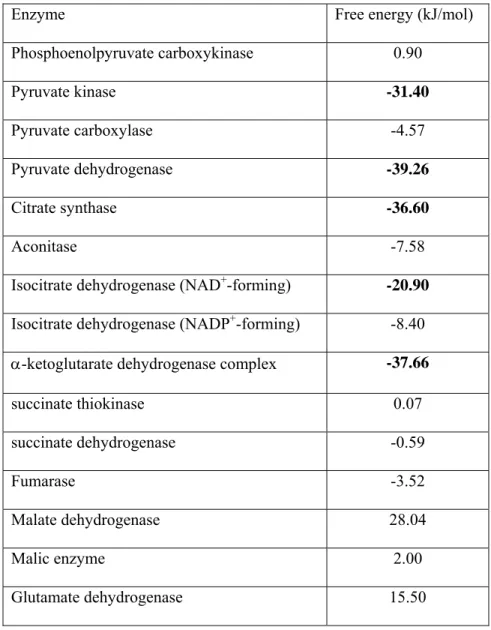
Carboxylation of pyruvate by pyruvate carboxylase yields oxaloacetate, and although this reaction is thermodynamically reversible (see table I), backflow towards pyruvate may occur only at a low rate (Freidmann et al., 1971), (McClure et al., 1971a), (McClure et al., 1971b), (Barden et al., 1972).
Pyruvate carboxylase activity shows a remarkable cell-specific distribution in the brain: neurons appear to exhibit very little if at all pyruvate carboxylase activity, while astrocytes adequately express the fully functional enzyme (Yu et al., 1983), (Shank et al., 1985), (Cesar and Hamprecht, 1995). It has been a long-standing question of this author, what purpose does it serve that, neurons lack pyruvate carboxylase?
This question is pressing, because neurons lose -ketoglutarate in the form of glutamate and GABA during neurotransmission, which are taken up by the astrocytes (Balazs et al., 1970), (Henn et al., 1974).
Evidence of neuronal pyruvate carboxylation emerged as early as 1966 (O'Neal and Koeppe, 1966), reviewed in (Hassel, 2001). In view of the absence of pyruvate carboxylase in neurons, alternative pathways were sought; it was found that neurons may carboxylate pyruvate through malic enzyme (Vogel et al., 1998b), (Hassel and Brathe, 2000), a reversible enzyme (Hsu and Lardy, 1969) exhibiting high pyruvate carboxylating activity (Salganicoff and Koeppe, 1968) that forms malate. In astrocytes, malic enzyme is mostly in the cytosol, while in neurons is mostly in the mitochondria (Kurz et al., 1993), (Bukato et al., 1994), (McKenna et al., 1995), (Vogel et al., 1998a), (Vogel et al., 1998b). However, most researchers consider the mitochondrial malic enzyme to operate in the decarboxylating direction in the brain (Vogel et al., 1998b), (Hertz et al., 1999), (McKenna et al., 2000). Furthermore, the group of Sonnewald concluded that neuronal pyruvate carboxylation is unlikely to be of quantitative significance (Waagepetersen et al., 2001); likewise, (Lieth et al., 2001) found that no pyruvate carboxylation occurs in neurons. It appears that the chapter on neuronal pyruvate carboxylation is still open. On the other hand, irreversible decarboxylation of pyruvate catalyzed by the pyruvate dehydrogenase complex yielding acetyl-CoA occurs in mitochondria of all cell types. Acetyl-CoA is also an indispensable activator of pyruvate carboxylase, see (Adina-Zada et al., 2012) and references therein. The oxaloacetate formed from pyruvate carboxylase will have four fates: i) it will get reversibly decarboxylated by a mitochondrial phosphoenolpyruvate carboxykinase, an enzyme that has been cloned and adequately characterized (Modaressi et al., 1996), (Modaressi et al., 1998); ii) it may get transaminated with glutamate, yielding - ketoglutarate and aspartate; iii) it may get converted to malate, through MDH; or iv) condense with acetyl-CoA to form citrate, a reaction catalyzed by citrate synthase. The latter reaction is irreversible, and represents the first committed step to citric acid cycle.
Citrate will then get converted to isocitrate by aconitase in a reversible reaction, which will then be dehydrogenated to -ketoglutarate by isocitrate dehydrogenase (IDH). There are two mitochondrial IDH isoforms: IDH2, which is NADP+-dependent and reversible (Des Rosiers C. et al., 1995; Lemons et al., 2010; Metallo et al., 2009; Metallo et al., 2012; Yoo et al., 2008), and IDH3 (Ehrlich and Colman, 1983),
(Des Rosiers C. et al., 1994), (Comte et al., 2002) which is NAD+-dependent, and considered irreversible (Gabriel et al., 1986). Splice variants have also been identified (Okamoto et al., 2003), (Kim et al., 1999).
IDH1, which is NADP+-dependent and reversible (Londesborough and Dalziel, 1968) is expressed predominantly in the cytosol (Nekrutenko et al., 1998). The reversibility of mitochondrial IDH has been inferred from experiments in isolated nerve from lobsters, bullfrogs and rabbits (Waelsch et al., 1965), and further on demonstrated in striatal slices (Cheng and Nakamura, 1972), cortical slices (Nakamura et al., 1970), (Patel, 1974b) brain homogenates (Salganicoff and Koeppe, 1968), rat liver (Des Rosiers C. et al., 1995) and rat heart (Randle et al., 1970), (Comte et al., 2002). Despite the fact that IDH3 is highly regulated by a variety of effectors (Gabriel et al., 1986), from the above an obvious futile cycle can be discerned, where isocitrate (plus NAD+) is oxidatively decarboxylated to -ketoglutarate (plus NADH) by IDH3, and then IDH2 may reverse this by converting -ketoglutarate plus NADPH to isocitrate plus NADP+; it has been proposed that this futility, in concert with the function of a proton-translocating transhydrogenase, fine-tune the citric acid cycle or produce heat, thereby contributing to thermoregulation in the animal at the expense of the transmembrane proton electrochemical potential gradient, or even contribute to the regulation of 'leak' currents across the inner mitochondrial membrane (Sazanov and Jackson, 1994). This is theoretically possible because the catalytic site of the transhydrogenase for oxidation and reduction of the nicotinamide nucleotides is facing the matrix, thus membrane potential generated by the respiratory chain is consumed during the reduction of NADP+ or NAD+ by NADH or NADPH, respectively (Jackson, 1991), which can be accounted as a 'leak' pathway, therefore contributing to metabolic thermogenesis. By the same token, membrane potential can be generated during the reverse operation, thereby decreasing this leak pathway, hence minimizing metabolic thermogenesis. Since the transhydrogenase links the redox state of NAD+ and NADP+ to membrane potential to the concerted actions of IDH2 and IDH3, it is conceivable that the transhydrogenase may impose on the rate of IDH reaction, effectively controlling the rate of the overall citric acid cycle. These theoretical considerations elaborated by Sazanov and Jackson could be easily tested since C57Bl/6J mice do not express the
transhydrogenase, while mice belonging to any of the following strains: C57BL/6JEi, C57BL/6N, C57BL/6NJ, C57BL/6ByJ, C57BL/10J, C57L/J, or C58/J express this protein (Toye et al., 2005). To the best of my knowledge, the results of such an experiment do not exist in the literature.
Apart from regenerating isocitrate by IDH2, -ketoglutarate could have three fates: i) transamination with aspartate, yielding oxaloacetate and glutamate, ii) dehydrogenation by glutamate dehydrogenase yielding glutamate, or iii) decarboxylation by KGDHC yielding succinyl-CoA. The reaction catalyzed by KGDHC requires CoASH and NAD+, is irreversible, and represents the second commitment step in the citric acid cycle. KGDHC deserves particular attention: this enzyme complex is subject to tight regulation: it is inhibited by increased [succinyl-CoA]/[CoA] and [NADH]/[NAD+] ratios (Garland and Randle, 1964), (Hamada et al., 1975), (McMinn and Ottaway, 1977), (LaNoue et al., 1983), (Markiewicz and Strumilo, 1995); it exhibits a biphasic response to Ca2+, low concentrations activating the complex (McCormack and Denton, 1979), (Lawlis and Roche, 1981) (McCormack and Denton, 1989), (Wan et al., 1989), (Panov and Scarpa, 1996) while high concentrations of the cation inhibit it (Lai and Cooper, 1986). The regulation of KGDHC reaction by the above and other effectors such as adenine nucleotides, pH and Mg2+ has been reviewed by (Strumilo, 2005) and (Starkov, 2012) and modeled by the group of Beard (Qi et al., 2011). The citric acid cycle is indeed regulated at several steps (LaNoue et al., 1970), (LaNoue et al., 1983), (Bukato et al., 1995), (Gibala et al., 2000), (Pagliarini and Dixon, 2006), (McKenna, 2007);
furthermore, different intermediates of the cycle exert different regulation depending on target isoform and tissue distribution (McKenna et al., 1995), (McKenna et al., 2000); equally importantly, the cycle is modulated by regulating the transport of substrates that enter the cycle (Safer and Williamson, 1973).
However, it is KGDHC that exhibits the highest flux-control coefficient of the citric acid cycle (Patel, 1974a), (Cooney et al., 1981), (Taegtmeyer, 1983), (Russell, III and Taegtmeyer, 1991), (Russell, III and Taegtmeyer, 1992) (Sheu and Blass, 1999). From the factors mentioned to regulate KGDHC reaction, two of them are emphasized: CoASH and NAD+. Availability of CoASH is known to limit KGDHC reaction (HUELSMANN et al., 1964), (Taegtmeyer, 1983), (Russell, III and Taegtmeyer, 1991), (Russell, III and Taegtmeyer, 1992). The availability of CoASH and the competition for it between pyruvate
dehydrogenase (PDH) and KGDHC (HUELSMANN et al., 1964), (Randle et al., 1970), led to the suggestion that the citric acid cycle operates in two sequential segments; the first being from acetyl-CoA until -ketoglutarate, (CoASH required for the PDH reaction), and the second one being from - ketoglutarate to oxaloacetate (CoASH required for the KGDHC reaction), (Randle et al., 1970). This was probably the first notion implying that the citric acid cycle may run in segments. Regarding NAD+ availability, it is already a textbook definition that NADH oxidized by the respiratory complexes yields NAD+, which will be re-reduced by the dehydrogenases of the cycle. However, as mentioned above, during hypoxia, when the electron transport chain is not operational, NADH may get oxidized by other means such as by the reverse operation of malate dehydrogenase, forming malate from oxaloacetate. This concept forms the basis for the continuous operation of KGDHC during hypoxia and will be discussed in greater detail below, (see under "What happens to NADH during hypoxia?").
The product of KGDHC, succinyl-CoA is the substrate for 'substrate-level phosphorylation" performed by succinate thiokinase, a reversible enzyme (also known as succinyl-CoA ligase). Succinate thiokinase is a heterodimer enzyme being composed of an invariant α subunit encoded by SUCLG1 and a substrate- specific β subunit, encoded by either SUCLA2 or SUCLG2. With the input of inorganic phosphate this dimer combination results in either an ATP-forming SUCL (EC 6.2.1.5) or a GTP-forming SUCL (EC 6.2.1.4). GTP may transphosphorylate to ATP by nucleoside-diphosphate kinase. In either case, succinate thiokinase also generates CoASH and succinate. Generation of high-energy phosphates by this reaction proceeds even in the absence of oxygen and perhaps under these conditions with a higher rate, because hypoxia activates the enzyme (Phillips et al., 2009). This concept will be reviewed in more detail below, (see under " Hypoxia: all roads lead to succinate?").
Succinate will be processed by the reversible succinate dehydrogenase to yield fumarate, and since this protein is also respiratory complex II, FAD+ will be reduced to FADH2. Fumarate is in equilibrium with malate through the reaction catalyzed by fumarase, an enzyme that is also found in the cytosol (Raimundo et al., 2011).
Malate that emerges from fumarase will have two fates, both catalyzed by reversible reactions: i) dehydrogenation by malate dehydrogenase to oxaloacetate, thereby completing the citric acid cycle or ii) decarboxylation by malic enzyme to pyruvate; this reaction is part of the 'pyruvate-recycling pathway', see below under "The 'pyruvate-recycling pathway'".
Apart from the mitochondrial malate dehydrogenase, there is a cytosolic malate dehydrogenase; both enzymes contribute to the operation of the malate-aspartate shuttle (Borst, 1962). In addition to these two malate dehydrogenases, the shuttle recruits aspartate aminotransferases in the mitochondrial matrix and in the cytosol, the malate--ketoglutarate antiporter and glutamate-aspartate antiporter in the inner mitochondrial membrane. The shuttle mediates the transfer of NADH from the cytosol formed in i) the glyceraldehyde-3-phosphate dehydrogenase reaction in the glycolysis and ii) that coming from oxidation of lactate by the lactate dehydrogenase reaction to pyruvate, into the mitochondrial matrix. Through augmented transamination of aspartate and -ketoglutarate, the malate-aspartate shuttle has been implicated in a cascade of metabolic reactions leading to anaerobic production of ATP (Pisarenko et al., 1995), see below under "Hypoxia: all roads lead to succinate?". Blockade of the shuttle by the transaminase inhibitor aminooxyacetate (Kauppinen et al., 1987) during ischemia and at the beginning of reperfusion confered cardioprotection in ischemia–reperfusion injury similar to that observed during ischemic preconditioning (Stottrup et al., 2010). However, sustained inhibition of the shuttle during reperfusion carried a negative outcome (Lofgren et al., 2010). As a word of caution, inhibiting transamination with aminooxyacetate confers very different effects on the oxidation of energy substrates in different preparations, suggesting different regulation of metabolism (McKenna et al., 1993).
The 'pyruvate-recycling pathway'
The 'pyruvate-recycling pathway' was described in liver and kidney 40 years ago (Freidmann et al., 1971), (Cohen, 1987), (Rognstad and Katz, 1972), discovered as part of the dicarboxylic acid cycle (Freidmann
et al., 1971). Later on, it was found to operate in brain tissue (Cerdan et al., 1990), (Kunnecke et al., 1993), where it has been extensively characterized, (Bakken et al., 1997a), (Haberg et al., 1998), and especially in astrocytes (Bakken et al., 1997b),. Basically, it refers to the fact that downstream metabolism of pyruvate will eventually regenerate pyruvate. This may occur either through PC forming oxaloacetate, that in turn will form PEP through mitochondrial PEPCK, and then PEP will exit mitochondria, where it can regenerate pyruvate through PK. GTP for the mitochondrial PEPCK may originate from the GTP- forming succinate thiokinase, see below and (Stark et al., 2009). Alternatively, oxaloacetate will be reduced to malate through malate dehydrogenase (MDH) that will be then catalyzed by malic enzyme yielding pyruvate. The pathway followed for recycling of pyruvate will depend on the tissue because of the tissue-dependent distribution, regulation and equilibria of the participating enzymes.
What happens to NADH during hypoxia?
The reversibility of MDH not only assists in the completion of the 'pyruvate-recycling pathway', it also contributes to re-oxidation of NADH when the electron transport chain is inhibited due to lack of oxygen.
Having said that, the question arises, why is there a need for continuing oxidation of NADH? As mentioned above, the citric acid cycle not only provides reducing equivalents for the respiratory complexes, it also generates high energy phosphates through matrix substrate-level phosphorylation. This occurs at the level of succinate thiokinase. However, since this is a highly reversible reaction, formation of ATP or GTP will happen only if the equilibrium favors succinate production and succinyl-CoA is readily available. Succinyl-CoA may arise from either propionyl-CoA metabolism, or KGDHC; provision of succinyl-CoA through KGDHC is much higher than that originating from propionyl-CoA metabolism (Stryer L, 1995). For KGDHC to operate, the inputs of CoASH, -ketoglutarate and NAD+ are necessary.
-ketoglutarate may arise from glutamate, an amino acid which is abundant in at least two pools in the CNS (McKenna, 2007). CoASH will re-emerge from succinate thiokinase, if the equilibrium favors ATP
or GTP production (Allen and Ottaway, 1986). Therefore, the key ingredient is NAD+. Formation of NAD+ through MDH, converting oxaloacetate to malate is a viable possibility. Indeed, MDH is known to be upregulated in KGDHC deficiencies (Bubber et al., 2011), (Shi et al., 2008), (Bubber et al., 2005), (Shi and Gibson, 2011). Another viable possibility that, to the best of my knowledge, has not been adequately characterized is NADH oxidation by mitochondrial diaphorases. This concept has been cultivated by the group of Iaguzhinskii, where the stimulatory effect of diaphorase substrates was examined during cyanide-resistant respiration of isolated mitochondria (Kolesova et al., 1987), (Kolesova et al., 1989), (Kolesova et al., 1991), (Kolesova et al., 1993). Whichever the source of NAD+, the continuous operation of KGDHC will supply succinate thiokinase with succinyl-CoA that will generate high-energy phosphates, and succinate. The high-energy phosphates will support the hydrolytic function of the Fo-F1 ATPase which operates in reverse during anoxic conditions, thus maintaining a modest membrane potential (Rouslin et al., 1986), (Rouslin et al., 1990). Generation of high-energy phosphates in the mitochondrial matrix will also lead to an increase of their local concentration, thereby maintaining the reversal potential of the adenine nucleotide translocase in a range (termed the 'B space' of mitochondrial phosphorylation) where it will prevent the translocase from reversing, hence sparing the extramitochondrial ATP pools from mitochondrial consumption (Chinopoulos et al., 2010), (Chinopoulos, 2011a), (Chinopoulos, 2011b).
Hypoxia: all roads lead to succinate?
From the above one may deduce that during hypoxia, the segment [-ketoglutarate → succinyl-CoA → succinate] may 'collide' with the product of malate dehydrogenase towards NAD+ formation -malate- which is in equilibrium with fumarate and succinate, since both fumarase and succinate dehydrogenase catalyze reversible reactions, a consideration that has been well established to occur in vivo (Brekke et al., 2012). This would practically mean that succinate is a 'dead-end' metabolite in hypoxia. Activity of the citric acid cycle in anoxia has been unequivocally demonstrated in 1966 (Randall, Jr. and Cohen, 1966).
Anaerobic metabolism was found to change the level of citric acid cycle intermediates during the first few seconds (Goldberg et al., 1966) and even more so within the first few minutes (Norberg and Siesjo, 1975).
Accumulation of succinate as an end product of anaerobic metabolism of glutamate was hypothesized even before the discovery of the citric acid cycle (Needham, 1930), a hypothesis that received solid foundation as early as 1937 (Weil-Malherbe, 1937), still prior to the establishment of the cycle. Since then, many publications emerged in the literature in support of this hypothesis: succinate was reported to accumulate in several hypoxic tissues (Hoberman and Prosky, 1967), (Folbergrova et al., 1974), (Freminet et al., 1980), (Freminet, 1981), (Benzi et al., 1982), (Pisarenko et al., 1986). This accumulation of succinate was associated with fumarate reduction and -ketoglutarate oxidation (HUNTER, Jr., 1949), (Chance and HOLLUNGER, 1960), (Randall, Jr. and Cohen, 1966), (SANADI and FLUHARTY, 1963) (Penney and Cascarano, 1970), (Wilson and Cascarano, 1970), (Taegtmeyer et al., 1977b) (Taegtmeyer, 1978). Succinate accumulates during anaerobic exercise, and also in diving animals (Hochachka and Dressendorfer, 1976), (Hochachka and Storey, 1975) and invertebrates performing facultative anaerobiosis (Hochachka and Mustafa, 1972). As early as 1949, it was recognized that anaerobic mitochondrial metabolism can generate ATP and maintain mitochondrial energization via substrate-level phosphorylation during the conversion of α-ketoglutarate to succinate (HUNTER, Jr., 1949), (Gronow and Cohen, 1984), (Penney and Cascarano, 1970), (Peuhkurinen, 1982), (Snaith et al., 1992). The benefits of mitochondrial substrate-level phosphorylation substantiated by the shift of equilibrium of the succinate thiokinase reaction towards succinate accumulation during anaerobiosis have been also demonstrated in hemorrhagic shock (Chick et al., 1968), and in hypoxic injury of the proximal kidney tubules (Weinberg et al., 2000b). During oxygen deprivation, an increase in glutamate utilization has been observed in several mammalian tissues, sometimes to the point of depletion (Williamson et al., 1967), (Edington et al., 1973), (Gailis and Benmouyal, 1973), (Taegtmeyer et al., 1977a) (Taegtmeyer et al., 1977b), (Pisarenko et al., 1987). In cardiac ischemia, malate increases 4-fold (Pisarenko et al., 1988), together with pronounced conversion of -ketoglutarate to succinate (Peuhkurinen et al., 1983), (Sanborn et al.,
1979), (Pisarenko et al., 1985), (Pisarenko et al., 1987); as a result, succinate exits the ischemic organ (Pisarenko et al., 1988). Succinate can be transported out of mitochondria in exchange of malate or inorganic phosphate (LaNoue et al., 1970), though its transport is relatively slow (Jans and Willem, 1991). Increased myocardial uptake of glutamate was demonstrated in humans after myocardial hypoxic episodes (Mudge, Jr. et al., 1976), (Thomassen et al., 1983). This was later confirmed in a rat (Pisarenko et al., 1985), and dog model (Lazar et al., 1980). However, it must also be pointed out that in cell cultures of astrocytes exposed to hypoxia, NMR spectroscopic studies reported no accumulation of succinate (Sonnewald et al., 1994), nor increased consumption of glutamate (Bakken et al., 1998).
In view of the fact that supporting the conversion of -ketoglutarate and its precursors to succinate benefits anaerobic metabolism in many tissues, several regimens boosting substrate-level phosphorylation have been examined, with impressive outcomes: exogenous glutamate, aspartate, and -ketoglutarate were reported to improve the performance of anoxic or ischemic myocardium via mitochondrial synthesis of ATP coupled to succinate and alanine formation (Pisarenko et al., 1983) (Pisarenko et al., 1985), (Bittl and Shine, 1983), (Matsuoka et al., 1986). Provision of -ketoglutarate during blood cardioplegia improved myocardial protection in patients undergoing coronary operations (Kjellman et al., 1995), (Kjellman et al., 1997). Infusion with -ketoglutarate after coronary operation in patients that underwent cardiopulmonary bypass enhanced renal blood flow (Jeppsson et al., 1998). Fibroblasts from patients with no F0-F1 ATPase activity could be rescued by administration of -ketoglutarate plus aspartate (Sgarbi et al., 2009). Beneficial effects of -ketoglutarate plus aspartate were also shown in hypoxic kidney proximal tubules, with an associated increase in succinate formation (Weinberg et al., 2000a). However, perfusion of ischemic kidney with -ketoglutarate and malate, worsened the situation (Bienholz et al., 2012), possibly due to cardiovascular depressive effects.
The question arises, what will happen with all this succinate? Succinate exits mitochondria and will inhibit HIF- prolyl hydroxylase, thereby stabilizing and activating HIF-1, initiating a hypoxic response (Selak et al., 2005), (Semenza, 2007); this phenomenon is extremely important in tumorigenesis
(Raimundo et al., 2011). Furthermore, succinate (as well as -ketoglutarate) have been found to act as ligands for G-coupled receptors, outside the cell (He et al., 2004), (Deen and Robben, 2011). From the metabolic point of view, succinate could only be metabolized when the electron transport chain regains functionality when adequate oxygen concentrations are restored, and follow the route towards fumarate
→ malate → oxaloacetate generating FADH2 and NADH in the process, or malate → pyruvate, generating NADPH through malic enzyme. Perhaps succinate accumulation contributes to hyperventilation after anaerobic exercise?
One very important aspect of metabolism of succinate upon restoration of anaerobiosis, is the potential for forming reactive oxygen species in excess (Starkov, 2008). Catabolism of succinate by mitochondria exhibiting a sufficiently high membrane potential leads to generation of hydrogen peroxide and superoxide radical (Korshunov et al., 1997); there are a number of reviews on this subject, the reader is referred elsewhere (Brand et al., 2004), (Andreyev et al., 2005), (Adam-Vizi and Chinopoulos, 2006), (Drose and Brandt, 2012).
Can succinate formation through the 'NAD+-fumarate reductase system' generate ATP through the forward operation of the F0-F1 ATP synthase during hypoxia?
The conversion of fumarate to succinate as part of an own entity, termed the 'NAD+-fumarate reductase system' has been reported to exist in bacteria, lower eucaryotes, shellfish and cancer cells (Kroger et al., 1992), (Kita et al., 2007), (Tomitsuka et al., 2009) (Tomitsuka et al., 2010), (Tomitsuka et al., 2012), composed of complex I and the reverse reaction of complex II. This system does not need oxygen, and only complex I functions as a proton pump; it has been suggested that during anaerobiosis, the ensuing formation of a transmembrane electrochemical proton gradient through this system supports ATP synthesis by F0-F1 ATP synthase. However, in my opinion, in mammalian mitochondria this system cannot support formation of ATP through the F0-F1 ATP synthase, for the following reasons: i) the
of ubiquinol (QH2) to ubiquinone (Q) by FAD+ is possible, however, downstream interactions of Q with complex I does not necessarily translate to proton pumping through this complex by this mechanism; ii) nonetheless, assuming that proton pumping may occur at the level of complex I by this mechanism, when complexes III and IV are not pumping any protons, this is not sufficient for generating a membrane potential that would exceed the reversal potential of the F0-F1 ATP synthase (Chinopoulos et al., 2010), (Chinopoulos, 2011a); therefore F0-F1 ATP synthase would still pump protons out of the matrix, hydrolyzing ATP in the process; iii) in the experiments outlined in (Chinopoulos et al., 2010) using respiration-impaired mammalian mitochondria where the concept of the 'NAD+-fumarate reductase system' could be considered, ATP was formed in the mitochondrial matrix by succinate thiokinase in the presence of rotenone; in this case, complex I was completely blocked and therefore an 'NAD+-fumarate reductase system' is redundant. Likewise, in a study on ischemic myocardium (Peuhkurinen et al., 1983) the potential energy-yielding reduction of fumarate to succinate was found to be insignificant. Perhaps in bacteria, lower eucaryotes and cancer cells other kind of ubiquinone analogues are operational, that thermodynamically may support the 'NAD+-fumarate reductase system', and there are evidence in support of this notion (Tomitsuka et al., 2012), (Tomitsuka et al., 2009), (Tomitsuka et al., 2010).
Citric acid cycle: the segments
From the above considerations one can deduce three segments of the citric acid cycle:
(1) Oxaloacetate + acetyl-CoA→ citrate ↔ isocitrate ↔ -ketoglutarate (2) -ketoglutarate → succinyl CoA ↔ succinate
(3) Oxaloacetate ↔ malate ↔ fumarate ↔ succinate
Segment (1) may occur towards the direction of -ketoglutarate synthesis only during aerobic metabolism; acetyl-coA will originate from pyruvate through PDH or from fatty acids, and it will reach KGDHC, the enzyme expressing the highest flux-control coefficient of the citric acid cycle. The two key components dictating if KGDHC will be operational are CoASH and NAD+. During anaerobiosis, - ketoglutarate (that may originate from glutamine and/or glutamate) may also backflux to isocitrate, which is in equilibrium with citrate (Comte et al., 2002), (Des Rosiers C. et al., 1995), (Des Rosiers C. et al., 1994).
During aerobiosis, segment (2) will commence, where -ketoglutarate will be metabolized by KGDHC, until the emergence of succinate. In anaerobiosis, segment (2) will commence only if sufficient CoASH and NAD+ are available. It is highly likely that during anaerobiosis NAD+ will originate from MDH, operating towards malate formation.
The magnitude of succinate concentration emerging from segment (2) will be 'weighted' against that coming from segment (3), where all participating substrates appear to exist in equilibrium. In aerobic metabolism, the formation of oxaloacetate is favored. In anaerobic conditions, succinate formation is favored. The direction favored during anaerobiosis generates NAD+, which is critical for the operation of KGDHC, which will supply succinyl CoA to succinate thiokinase that will yield high-energy phosphates also regenerating CoASH.
From the above, it is obvious that KGDHC plays a critical role in determining whether a segment will be operational, and if yes, towards which direction, see figure 2. Bearing that in mind, the question arises as to the usefulness of this information. For once, increased flux of KGDHC by substrates such - ketoglutarate or glutamate has shown a beneficial outcome in diverse pathological situations involving hypoxia, see above, " Hypoxia: all roads lead to succinate?". Secondly, certain pathological conditions may emerge by inhibition of KGDHC, either by reactive oxygen species made elsewhere, or by the enzyme complex itself (Starkov et al., 2004), (Tretter and Adam-Vizi, 2004) upon reoxygenation, or by
inherent mutations of a gene encoding at least one of the subunits of the complex (Ambrus et al., 2011), reviewed in (Starkov, 2012).
Conclusions
On 1953, when Hans Krebs was cycling down the stairs on the right to climb them up again on the left in order to receive the Nobel Prize from King Gustaf VI for "his discovery of the citric acid cycle", little did he know that nearly 60 years later there would be a flare of interest to investigate the cycle and its directionality in vivo (Schroeder et al., 2009), (Chen et al., 2012), (Zacharias et al., 2012), (http://www.nobelprize.org/mediaplayer/index.php?id=633). The benefits of knowing the directionality of the cycle during hypoxia are elaborated above, however, a field that gains momentum rapidly involves the adulteration of this biochemical pathway for the purpose of cancer cell survival (Wise et al., 2011), (Mullen et al., 2012). It is perhaps in this pathway where cancer finds metabolic support when growing in hypoxic environments, while also exhibiting a number of defects in the electron transport chain (Tomlinson et al., 2002), (Hao et al., 2009), (Linehan et al., 2010), (Weinberg et al., 2010) rendering its harboring mitochondria as defective. The latest findings pave exciting new ways for researching on one of the most fundamental discovery of biochemistry, the citric acid cycle.
Acknowledgements: I thank Prof. Mary C McKenna and Dr. Anatoly A Starkov for helpful discussions, and Ilana Zholobovsky for translations of the articles in Russian.
Reference List
Adam-Vizi V, Chinopoulos C. 2006. Bioenergetics and the formation of mitochondrial reactive oxygen species. Trends Pharmacol. Sci. 27:639-645.
Adina-Zada A, Zeczycki TN, Attwood PV. 2012. Regulation of the structure and activity of pyruvate carboxylase by acetyl CoA. Arch. Biochem. Biophys. 519:118-130.
Allen DA, Ottaway JH. 1986. Succinate thiokinase in pigeon breast muscle mitochondria. FEBS Lett.
194:171-175.
Ambrus A, Torocsik B, Tretter L, Ozohanics O, Adam-Vizi V. 2011. Stimulation of reactive oxygen species generation by disease-causing mutations of lipoamide dehydrogenase. Hum. Mol. Genet.
20:2984-2995.
Andreyev AY, Kushnareva YE, Starkov AA. 2005. Mitochondrial Metabolism of Reactive Oxygen Species. Biochemistry (Moscow) 70:200-214.
Bakken IJ, Sonnewald U, Clark JB, Bates TE. 1997a. [U-13C]glutamate metabolism in rat brain mitochondria reveals malic enzyme activity. Neuroreport 8:1567-1570.
Bakken IJ, White LR, Aasly J, Unsgard G, Sonnewald U. 1997b. Lactate formation from [U- 13C]aspartate in cultured astrocytes: compartmentation of pyruvate metabolism. Neurosci. Lett.
237:117-120.
Bakken IJ, White LR, Unsgard G, Aasly J, Sonnewald U. 1998. [U-13C]glutamate metabolism in astrocytes during hypoglycemia and hypoxia. J. Neurosci. Res. 51:636-645.
Balazs R, Machiyama Y, Hammond BJ, Julian T, Richter D. 1970. The operation of the gamma- aminobutyrate bypath of the tricarboxylic acid cycle in brain tissue in vitro. Biochem. J. 116:445- 461.
Barden RE, Fung CH, Utter MF, Scrutton MC. 1972. Pyruvate carboxylase from chicken liver. Steady state kinetic studies indicate a "two-site" ping-pong mechanism. J. Biol. Chem. 247:1323-1333.
Benzi G, Pastoris O, Dossena M. 1982. Relationships between gamma-aminobutyrate and succinate cycles during and after cerebral ischemia. J. Neurosci. Res. 7:193-201.
Bienholz A, Petrat F, Wenzel P, Ickerott P, Weinberg JM, Witzke O, Kribben A, de GH, Feldkamp T.
2012. Adverse effects of alpha-ketoglutarate/malate in a rat model of acute kidney injury. Am. J.
Physiol Renal Physiol 303:F56-F63.
Bittl JA, Shine KI. 1983. Protection of ischemic rabbit myocardium by glutamic acid. Am. J. Physiol 245:H406-H412.
Borst P. 1962. The aerobic oxidation of reduced diphosphopyridine nucleotide formed by glycolysis in Ehrlich ascites-tumour cells. Biochim. Biophys. Acta 57:270-282.
Brand MD, Buckingham JA, Esteves TC, Green K, Lambert AJ, Miwa S, Murphy MP, Pakay JL, Talbot DA, Echtay KS. 2004. Mitochondrial superoxide and aging: uncoupling-protein activity and superoxide production. Biochem. Soc. Symp.:203-213.
Brekke E, Walls AB, Norfeldt L, Schousboe A, Waagepetersen HS, Sonnewald U. 2012. Direct measurement of backflux between oxaloacetate and fumarate following pyruvate carboxylation.
Glia 60:147-158.
Breusch FL. 1937. Citronensäure im Gewebsstoffwechsel. Hoppe-Seyler's Zeitschrift für physiologische Chemie 250:262-282.
Breusch FL. 1939. The fate of oxaloacetic acid in different organs. Biochem. J. 33:1757-1770.
Bricker DK, Taylor EB, Schell JC, Orsak T, Boutron A, Chen YC, Cox JE, Cardon CM, Van Vranken JG, Dephoure N, Redin C, Boudina S, Gygi SP, Brivet M, Thummel CS, Rutter J. 2012. A mitochondrial pyruvate carrier required for pyruvate uptake in yeast, Drosophila, and humans.
Science 337:96-100.
Bubber P, Haroutunian V, Fisch G, Blass JP, Gibson GE. 2005. Mitochondrial abnormalities in Alzheimer brain: mechanistic implications. Ann. Neurol. 57:695-703.
Bubber P, Hartounian V, Gibson GE, Blass JP. 2011. Abnormalities in the tricarboxylic acid (TCA) cycle in the brains of schizophrenia patients. Eur. Neuropsychopharmacol. 21:254-260.
Bukato G, Kochan Z, Swierczynski J. 1994. Subregional and intracellular distribution of NADP-linked malic enzyme in human brain. Biochem. Med. Metab Biol. 51:43-50.
Bukato G, Kochan Z, Swierczynski J. 1995. Different regulatory properties of the cytosolic and mitochondrial forms of malic enzyme isolated from human brain. Int. J. Biochem. Cell Biol.
27:1003-1008.
Cerdan S, Kunnecke B, Seelig J. 1990. Cerebral metabolism of [1,2-13C2]acetate as detected by in vivo and in vitro 13C NMR. J. Biol. Chem. 265:12916-12926.
Cesar M, Hamprecht B. 1995. Immunocytochemical examination of neural rat and mouse primary cultures using monoclonal antibodies raised against pyruvate carboxylase. J. Neurochem.
64:2312-2318.
Chance B, Hollunger G. 1960. Energy-linked reduction of mitochondrial pyridine nucleotide. Nature 185:666-672.
Chen AP, Hurd RE, Schroeder MA, Lau AZ, Gu YP, Lam WW, Barry J, Tropp J, Cunningham CH.
2012. Simultaneous investigation of cardiac pyruvate dehydrogenase flux, Krebs cycle metabolism and pH, using hyperpolarized [1,2-(13)C2]pyruvate in vivo. NMR Biomed. 25:305- 311.
Cheng SC, Nakamura R. 1972. Metabolism related to the tricarboxylic acid cycle in rat brain slices.
Observations on CO 2 fixation and metabolic compartmentation. Brain Res. 38:355-370.
Chick WL, Weiner R, Cascareno J, Zweifach BW. 1968. Influence of Krebs-cycle intermediates on survival in hemorrhagic shock. Am. J. Physiol 215:1107-1110.
Chinopoulos C. 2011a. Mitochondrial consumption of cytosolic ATP: not so fast. FEBS Lett. 585:1255- 1259.
Chinopoulos C. 2011b. The "B Space" of mitochondrial phosphorylation. J. Neurosci. Res. 89:1897-1904.
Chinopoulos C, Gerencser AA, Mandi M, Mathe K, Torocsik B, Doczi J, Turiak L, Kiss G, Konrad C, Vajda S, Vereczki V, Oh RJ, Adam-Vizi V. 2010. Forward operation of adenine nucleotide translocase during F0F1-ATPase reversal: critical role of matrix substrate-level phosphorylation.
FASEB J. 24:2405-2416.
Cohen SM. 1987. Effects of insulin on perfused liver from streptozotocin-diabetic and untreated rats: 13C NMR assay of pyruvate kinase flux. Biochemistry 26:573-580.
Comte B, Vincent G, Bouchard B, Benderdour M, Des Rosiers C. 2002. Reverse flux through cardiac NADP(+)-isocitrate dehydrogenase under normoxia and ischemia. Am. J. Physiol Heart Circ.
Physiol 283:H1505-H1514.
Cooney GJ, Taegtmeyer H, Newsholme EA. 1981. Tricarboxylic acid cycle flux and enzyme activities in the isolated working rat heart. Biochem. J. 200:701-703.
de Bari L., Valenti D, Pizzuto R, Atlante A, Passarella S. 2007. Phosphoenolpyruvate metabolism in Jerusalem artichoke mitochondria. Biochim. Biophys. Acta 1767:281-294.
Deen PM, Robben JH. 2011. Succinate receptors in the kidney. J. Am. Soc. Nephrol. 22:1416-1422.
Des Rosiers C., Di DL, Comte B, Laplante A, Marcoux C, David F, Fernandez CA, Brunengraber H.
1995. Isotopomer analysis of citric acid cycle and gluconeogenesis in rat liver. Reversibility of isocitrate dehydrogenase and involvement of ATP-citrate lyase in gluconeogenesis. J. Biol.
Chem. 270:10027-10036.
Des Rosiers C., Fernandez CA, David F, Brunengraber H. 1994. Reversibility of the mitochondrial isocitrate dehydrogenase reaction in the perfused rat liver. Evidence from isotopomer analysis of citric acid cycle intermediates. J. Biol. Chem. 269:27179-27182.
Drose S, Brandt U. 2012. Molecular mechanisms of superoxide production by the mitochondrial respiratory chain. Adv. Exp. Med. Biol. 748:145-169.
Edington DW, Ward GR, Saville WA. 1973. Energy metabolism of working muscle: concentration profiles of selected metabolites. Am. J. Physiol 224:1375-1380.
Ehrlich RS, Colman RF. 1983. Separation, recombination, and characterization of dissimilar subunits of the DPN-dependent isocitrate dehydrogenase from pig heart. J. Biol. Chem. 258:7079-7086.
Folbergrova J, Ljunggren B, Norberg K, Siesjo BK. 1974. Influence of complete ischemia on glycolytic metabolites, citric acid cycle intermediates, and associated amino acids in the rat cerebral cortex.
Brain Res. 80:265-279.
Freidmann B, Goodman EH, Jr., Saunders HL, Kostos V, Weinhouse S. 1971. An estimation of pyruvate recycling during gluconeogenesis in the perfused rat liver. Arch. Biochem. Biophys. 143:566- 578.
Freminet A. 1981. Carbohydrate and Amino-Acid-Metabolism During Acute-Hypoxia in Rats - Blood and Heart Metabolites. Comparative Biochemistry and Physiology B-Biochemistry & Molecular Biology 70:427-433.
Freminet A, Leclerc L, Poyart C, Huel C, Gentil M. 1980. Alanine and succinate accumulation in the perfused rat heart during hypoxia. J. Physiol (Paris) 76:113-117.
Gabriel JL, Zervos PR, Plaut GW. 1986. Activity of purified NAD-specific isocitrate dehydrogenase at modulator and substrate concentrations approximating conditions in mitochondria. Metabolism 35:661-667.
Gailis L, Benmouyal E. 1973. Endogenous alanine, glutamate, aspartate, and glutamine in the perfused guinea-pig heart: effect of substrates and cardioactive agents. Can. J. Biochem. 51:11-20.
Garland PB, Randle PJ. 1964. Regulation of glucose uptake by muscles. 10. Effects of alloxan-diabetes, starvation, hypophysectomy and adrenalectomy, and of fatty acids, ketone bodies and pyruvate, on the glycerol output and concentrations of free fatty acids, long-chain fatty acyl-coenzyme A, glycerol phosphate and citrate-cycle intermediates in rat heart and diaphragm muscles. Biochem.
J. 93:678-687.
Gerhard DS, Wagner L, Feingold EA, Shenmen CM, Grouse LH, Schuler G, Klein SL, Old S, Rasooly R, Good P, Guyer M, Peck AM, Derge JG, Lipman D, Collins FS, Jang W, Sherry S, Feolo M, Misquitta L, Lee E, Rotmistrovsky K, Greenhut SF, Schaefer CF, Buetow K, Bonner TI, Haussler D, Kent J, Kiekhaus M, Furey T, Brent M, Prange C, Schreiber K, Shapiro N, Bhat NK, Hopkins RF, Hsie F, Driscoll T, Soares MB, Casavant TL, Scheetz TE, Brown-stein MJ, Usdin TB, Toshiyuki S, Carninci P, Piao Y, Dudekula DB, Ko MS, Kawakami K, Suzuki Y, Sugano S, Gruber CE, Smith MR, Simmons B, Moore T, Waterman R, Johnson SL, Ruan Y, Wei CL, Mathavan S, Gunaratne PH, Wu J, Garcia AM, Hulyk SW, Fuh E, Yuan Y, Sneed A, Kowis C, Hodgson A, Muzny DM, McPherson J, Gibbs RA, Fahey J, Helton E, Ketteman M, Madan A, Rodrigues S, Sanchez A, Whiting M, Madari A, Young AC, Wetherby KD, Granite SJ, Kwong PN, Brinkley CP, Pearson RL, Bouffard GG, Blakesly RW, Green ED, Dickson MC, Rodriguez AC, Grimwood J, Schmutz J, Myers RM, Butterfield YS, Griffith M, Griffith OL, Krzywinski MI, Liao N, Morin R, Palmquist D, Petrescu AS, Skalska U, Smailus DE, Stott JM, Schnerch A, Schein JE, Jones SJ, Holt RA, Baross A, Marra MA, Clifton S, Makowski KA, Bosak S, Malek J.
2004. The status, quality, and expansion of the NIH full-length cDNA project: the Mammalian Gene Collection (MGC). Genome Res. 14:2121-2127.
Gibala MJ, Young ME, Taegtmeyer H. 2000. Anaplerosis of the citric acid cycle: role in energy metabolism of heart and skeletal muscle. Acta Physiol Scand. 168:657-665.
Goldberg ND, Passonneau JV, Lowry OH. 1966. Effects of changes in brain metabolism on the levels of citric acid cycle intermediates. J. Biol. Chem. 241:3997-4003.
Gronow GH, Cohen JJ. 1984. Substrate support for renal functions during hypoxia in the perfused rat kidney. Am. J. Physiol 247:F618-F631.
Haberg A, Qu H, Bakken IJ, Sande LM, White LR, Haraldseth O, Unsgard G, Aasly J, Sonnewald U.
1998. In vitro and ex vivo 13C-NMR spectroscopy studies of pyruvate recycling in brain. Dev.
Neurosci. 20:389-398.
Hamada M, Koike K, Nakaula Y, Hiraoka T, Koike M. 1975. A kinetic study of the alpha-keto acid dehydrogenase complexes from pig heart mitochondria. J. Biochem. 77:1047-1056.
Hao HX, Khalimonchuk O, Schraders M, Dephoure N, Bayley JP, Kunst H, Devilee P, Cremers CW, Schiffman JD, Bentz BG, Gygi SP, Winge DR, Kremer H, Rutter J. 2009. SDH5, a gene required for flavination of succinate dehydrogenase, is mutated in paraganglioma. Science 325:1139-1142.
Hassel B. 2001. Pyruvate carboxylation in neurons. J. Neurosci. Res. 66:755-762.
Hassel B, Brathe A. 2000. Cerebral metabolism of lactate in vivo: evidence for neuronal pyruvate carboxylation. J. Cereb. Blood Flow Metab 20:327-336.
He W, Miao FJ, Lin DC, Schwandner RT, Wang Z, Gao J, Chen JL, Tian H, Ling L. 2004. Citric acid cycle intermediates as ligands for orphan G-protein-coupled receptors. Nature 429:188-193.
Henn FA, Goldstein MN, Hamberger A. 1974. Uptake of the neurotransmitter candidate glutamate by glia. Nature 249:663-664.
Hertz L, Dringen R, Schousboe A, Robinson SR. 1999. Astrocytes: glutamate producers for neurons. J.
Neurosci. Res. 57:417-428.
Herzig S, Raemy E, Montessuit S, Veuthey JL, Zamboni N, Westermann B, Kunji ER, Martinou JC.
2012. Identification and functional expression of the mitochondrial pyruvate carrier. Science 337:93-96.
Hoberman HD, Prosky L. 1967. Evidence of reduction of fumarate to succinate in perfused rat liver under conditions of reduced O2 tension. Biochim. Biophys. Acta 148:392-399.
Hochachka PW, Dressendorfer RH. 1976. Succinate accumulation in man during exercise. Eur. J. Appl.
Physiol Occup. Physiol 35:235-242.
Hochachka PW, Mustafa T. 1972. Invertebrate facultative anaerobiosis. Science 178:1056-1060.
Hochachka PW, Storey KB. 1975. Metabolic consequences of diving in animals and man. Science 187:613-621.
Hsu RY, Lardy HA. 1969. Malic enzyme. Methods Enzymol. 13:230-235.
Huelsmann WC, Siliprandi D, Ciman M, Siliprandi N. 1964. Effect of carnitine on the oxidation of alpha- oxoglutarate to succinate in the presence of acetoacetate or pyruvate. Biochim. Biophys. Acta 93:166-168.
Hunter FE, Jr. 1949. Anaerobic phosphorylation due to a coupled oxidation-reduction between alpha- ketoglutaric acid and oxalacetic acid. J. Biol. Chem. 177:361-372.
Jackson JB. 1991. The proton-translocating nicotinamide adenine dinucleotide transhydrogenase. J.
Bioenerg. Biomembr. 23:715-741.
Jans AW, Willem R. 1991. Metabolism of [2-13C]succinate in renal cells determined by 13C NMR. Eur.
J. Biochem. 195:97-101.
Jeppsson A, Ekroth R, Friberg P, Kirno K, Milocco I, Nilsson FN, Svensson S, Wernerman J. 1998.
Renal effects of alpha-ketoglutarate early after coronary operations. Ann. Thorac. Surg. 65:684- 690.
Kauppinen RA, Sihra TS, Nicholls DG. 1987. Aminooxyacetic acid inhibits the malate-aspartate shuttle in isolated nerve terminals and prevents the mitochondria from utilizing glycolytic substrates.
Biochim. Biophys. Acta 930:173-178.
Kim YO, Koh HJ, Kim SH, Jo SH, Huh JW, Jeong KS, Lee IJ, Song BJ, Huh TL. 1999. Identification and functional characterization of a novel, tissue-specific NAD(+)-dependent isocitrate dehydrogenase beta subunit isoform. J. Biol. Chem. 274:36866-36875.
Kita K, Shiomi K, Omura S. 2007. Advances in drug discovery and biochemical studies. Trends Parasitol.
23:223-229.
Kjellman U, Bjork K, Ekroth R, Karlsson H, Jagenburg R, Nilsson F, Svensson G, Wernerman J. 1995.
Alpha-ketoglutarate for myocardial protection in heart surgery. Lancet 345:552-553.
Kjellman UW, Bjork K, Ekroth R, Karlsson H, Jagenburg R, Nilsson FN, Svensson G, Wernerman J.
1997. Addition of alpha-ketoglutarate to blood cardioplegia improves cardioprotection. Ann.
Thorac. Surg. 63:1625-1633.
Kolesova GM, Kapitanova NG, Iaguzhinskii LS. 1987. [Stimulation by quinones of cyanide-resistant respiration in rat liver and heart mitochondria]. Biokhimiia. 52:715-719.
Kolesova GM, Karnaukhova LV, Iaguzhinskii LS. 1991. [Interaction of menadione and duroquinone with Q-cycle during DT-diaphorase function]. Biokhimiia. 56:1779-1786.
Kolesova GM, Karnaukhova LV, Segal' NK, Iaguzhinskii LS. 1993. [The effect of inhibitors of the Q- cycle on cyano-resistant oxidation of malate by rat liver mitochondria in the presence of menadione]. Biokhimiia. 58:1630-1640.
Kolesova GM, Vishnivetskii SA, Iaguzhinskii LS. 1989. [A study of the mechanism of cyanide resistant oxidation of succinate from rat liver mitochondria in the presence of menadione]. Biokhimiia.
54:103-111.
Korshunov SS, Skulachev VP, Starkov AA. 1997. High protonic potential actuates a mechanism of production of reactive oxygen species in mitochondria. FEBS Lett. 416:15-18.
Krebs HA. 1940a. The citric acid cycle and the Szent-Gyorgyi cycle in pigeon breast muscle. Biochem. J.
34:775-779.
Krebs HA. 1940b. The citric acid cycle: A reply to the criticisms of F. L. Breusch and of J. Thomas.
Biochem. J. 34:460-463.
Kroger A, Geisler V, Lemma E, Theis F, Lenger R. 1992. Bacterial Fumarate Respiration. Archives of Microbiology 158:311-314.
Kunnecke B, Cerdan S, Seelig J. 1993. Cerebral metabolism of [1,2-13C2]glucose and [U-13C4]3- hydroxybutyrate in rat brain as detected by 13C NMR spectroscopy. NMR Biomed. 6:264-277.
Kurz GM, Wiesinger H, Hamprecht B. 1993. Purification of cytosolic malic enzyme from bovine brain, generation of monoclonal antibodies, and immunocytochemical localization of the enzyme in glial cells of neural primary cultures. J. Neurochem. 60:1467-1474.
Lai JC, Cooper AJ. 1986. Brain alpha-ketoglutarate dehydrogenase complex: kinetic properties, regional distribution, and effects of inhibitors. J. Neurochem. 47:1376-1386.
LaNoue K, Nicklas WJ, Williamson JR. 1970. Control of citric acid cycle activity in rat heart mitochondria. J. Biol. Chem. 245:102-111.
LaNoue KF, Schoolwerth AC, Pease AJ. 1983. Ammonia formation in isolated rat liver mitochondria. J.
Biol. Chem. 258:1726-1734.
Lawlis VB, Roche TE. 1981. Inhibition of bovine kidney alpha-ketoglutarate dehydrogenase complex by reduced nicotinamide adenine dinucleotide in the presence or absence of calcium ion and effect of adenosine 5'-diphosphate on reduced nicotinamide adenine dinucleotide inhibition. Biochemistry 20:2519-2524.
Lazar HL, Buckberg GD, Manganaro AJ, Becker H, Maloney JV, Jr. 1980. Reversal of ischemic damage with amino acid substrate enhancement during reperfusion. Surgery 88:702-709.
Lemons JM, Feng XJ, Bennett BD, Legesse-Miller A, Johnson EL, Raitman I, Pollina EA, Rabitz HA, Rabinowitz JD, Coller HA. 2010. Quiescent fibroblasts exhibit high metabolic activity. PLoS.
Biol. 8:e1000514.
Li X, Wu F, Qi F, Beard DA. 2011. A database of thermodynamic properties of the reactions of glycolysis, the tricarboxylic acid cycle, and the pentose phosphate pathway. Database. (Oxford) 2011:bar005.
Lieth E, LaNoue KF, Berkich DA, Xu B, Ratz M, Taylor C, Hutson SM. 2001. Nitrogen shuttling between neurons and glial cells during glutamate synthesis. J. Neurochem. 76:1712-1723.
Linehan WM, Srinivasan R, Schmidt LS. 2010. The genetic basis of kidney cancer: a metabolic disease.
Nat. Rev. Urol. 7:277-285.
Lofgren B, Povlsen JA, Rasmussen LE, Stottrup NB, Solskov L, Krarup PM, Kristiansen SB, Botker HE, Nielsen TT. 2010. Amino acid transamination is crucial for ischaemic cardioprotection in normal and preconditioned isolated rat hearts--focus on L-glutamate. Exp. Physiol 95:140-152.
Londesborough JC, Dalziel K. 1968. The equilibrium constant of the isocitrate dehydrogenase reaction.
Biochem. J. 110:217-222.
Markiewicz J, Strumilo SA. 1995. Some regulatory properties of the 2-oxoglutarate dehydrogenase complex from European bison heart. Acta Biochim. Pol. 42:339-346.
Matsuoka S, Jarmakani JM, Young HH, Uemura S, Nakanishi T. 1986. The effect of glutamate on hypoxic newborn rabbit heart. J. Mol. Cell Cardiol. 18:897-906.
McClure WR, Lardy HA, Kneifel HP. 1971a. Rat liver pyruvate carboxylase. I. Preparation, properties, and cation specificity. J. Biol. Chem. 246:3569-3578.
McClure WR, Lardy HA, Wagner M, Cleland WW. 1971b. Rat liver pyruvate carboxylase. II. Kinetic studies of the forward reaction. J. Biol. Chem. 246:3579-3583.
McCormack JG, Denton RM. 1979. The effects of calcium ions and adenine nucleotides on the activity of pig heart 2-oxoglutarate dehydrogenase complex. Biochem. J. 180:533-544.
McCormack JG, Denton RM. 1989. The role of Ca2+ ions in the regulation of intramitochondrial metabolism and energy production in rat heart. Mol. Cell Biochem. 89:121-125.
McCoy GD, Doeg KA. 1975. Characterization of the phosphoenolpyruvate inhibition of mitochondrial protein synthesis. J. Biol. Chem. 250:3510-3514.
McKenna MC. 2007. The glutamate-glutamine cycle is not stoichiometric: fates of glutamate in brain. J.
Neurosci. Res. 85:3347-3358.
McKenna MC, Stevenson JH, Huang X, Tildon JT, Zielke CL, Hopkins IB. 2000. Mitochondrial malic enzyme activity is much higher in mitochondria from cortical synaptic terminals compared with mitochondria from primary cultures of cortical neurons or cerebellar granule cells. Neurochem.
Int. 36:451-459.
McKenna MC, Tildon JT, Stevenson JH, Boatright R, Huang S. 1993. Regulation of energy metabolism in synaptic terminals and cultured rat brain astrocytes: differences revealed using aminooxyacetate. Dev. Neurosci. 15:320-329.
McKenna MC, Tildon JT, Stevenson JH, Huang X, Kingwell KG. 1995. Regulation of mitochondrial and cytosolic malic enzymes from cultured rat brain astrocytes. Neurochem. Res. 20:1491-1501.
McMinn CL, Ottaway JH. 1977. Studies on the mechanism and kinetics of the 2-oxoglutarate dehydrogenase system from pig heart. Biochem. J. 161:569-581.
Merle M, Martin M, Villegier A, Canioni P. 1996a. Mathematical modelling of the citric acid cycle for the analysis of glutamine isotopomers from cerebellar astrocytes incubated with [1(- 13)C]glucose. Eur. J. Biochem. 239:742-751.
Merle M, Martin M, Villegier A, Canioni P. 1996b. [1-13C]glucose metabolism in brain cells: isotopomer analysis of glutamine from cerebellar astrocytes and glutamate from granule cells. Dev. Neurosci.
18:460-468.
Metallo CM, Gameiro PA, Bell EL, Mattaini KR, Yang J, Hiller K, Jewell CM, Johnson ZR, Irvine DJ, Guarente L, Kelleher JK, Vander Heiden MG, Iliopoulos O, Stephanopoulos G. 2012. Reductive glutamine metabolism by IDH1 mediates lipogenesis under hypoxia. Nature 481:380-384.
Metallo CM, Walther JL, Stephanopoulos G. 2009. Evaluation of 13C isotopic tracers for metabolic flux analysis in mammalian cells. J. Biotechnol. 144:167-174.
Modaressi S, Brechtel K, Christ B, Jungermann K. 1998. Human mitochondrial phosphoenolpyruvate carboxykinase 2 gene. Structure, chromosomal localization and tissue-specific expression.
Biochem. J. 333 ( Pt 2):359-366.
Modaressi S, Christ B, Bratke J, Zahn S, Heise T, Jungermann K. 1996. Molecular cloning, sequencing and expression of the cDNA of the mitochondrial form of phosphoenolpyruvate carboxykinase from human liver. Biochem. J. 315 ( Pt 3):807-814.
Mudge GH, Jr., Mills RM, Jr., Taegtmeyer H, Gorlin R, Lesch M. 1976. Alterations of myocardial amino acid metabolism in chronic ischemic heart disease. J. Clin. Invest 58:1185-1192.
Mullen AR, Wheaton WW, Jin ES, Chen PH, Sullivan LB, Cheng T, Yang Y, Linehan WM, Chandel NS, DeBerardinis RJ. 2012. Reductive carboxylation supports growth in tumour cells with defective mitochondria. Nature 481:385-388.
Nakamura R, Cheng SC, Naruse H. 1970. A study on the precursors of the acetyl moiety of acetylcholine in brain slices. Observations on the compartmentalization of the acetyl-coenzyme A pool.
Biochem. J. 118:443-450.
Needham DM. 1930. A quantitative study of succinic acid in muscle: Glutamic and aspartic acids as precursors. Biochem. J. 24:208-227.
Nekrutenko A, Hillis DM, Patton JC, Bradley RD, Baker RJ. 1998. Cytosolic isocitrate dehydrogenase in humans, mice, and voles and phylogenetic analysis of the enzyme family. Mol. Biol. Evol.
15:1674-1684.
Norberg K, Siesjo BK. 1975. Cerebral Metabolism in Hypoxic Hypoxia .2. Citric-Acid Cycle Intermediates and Associated Amino-Acids. Brain Research 86:45-54.
O'Neal RM, Koeppe RE. 1966. Precursors in vivo of glutamate, aspartate and their derivatives of rat brain. J. Neurochem. 13:835-847.
Okamoto K, Matsuzaka Y, Yoshikawa Y, Takaki A, Kulski JK, Tamiya G, Inoko H. 2003. Identification of NAD+-dependent isocitrate dehydrogenase 3 gamma-like (IDH3GL) gene and its genetic polymorphisms. Gene 323:141-148.
Ota T, Suzuki Y, Nishikawa T, Otsuki T, Sugiyama T, Irie R, Wakamatsu A, Hayashi K, Sato H, Nagai K, Kimura K, Makita H, Sekine M, Obayashi M, Nishi T, Shibahara T, Tanaka T, Ishii S, Yamamoto J, Saito K, Kawai Y, Isono Y, Nakamura Y, Nagahari K, Murakami K, Yasuda T, Iwayanagi T, Wagatsuma M, Shiratori A, Sudo H, Hosoiri T, Kaku Y, Kodaira H, Kondo H, Sugawara M, Takahashi M, Kanda K, Yokoi T, Furuya T, Kikkawa E, Omura Y, Abe K, Kamihara K, Katsuta N, Sato K, Tanikawa M, Yamazaki M, Ninomiya K, Ishibashi T, Yamashita H, Murakawa K, Fujimori K, Tanai H, Kimata M, Watanabe M, Hiraoka S, Chiba Y, Ishida S, Ono Y, Takiguchi S, Watanabe S, Yosida M, Hotuta T, Kusano J, Kanehori K, Takahashi-Fujii A, Hara H, Tanase TO, Nomura Y, Togiya S, Komai F, Hara R, Takeuchi K, Arita M, Imose N, Musashino K, Yuuki H, Oshima A, Sasaki N, Aotsuka S, Yoshikawa Y, Matsunawa H, Ichihara T, Shiohata N, Sano S, Moriya S, Momiyama H, Satoh N, Takami S, Terashima Y, Suzuki O, Nakagawa S, Senoh A, Mizoguchi H, Goto Y, Shimizu F, Wakebe H, Hishigaki H, Watanabe T, Sugiyama A, Takemoto M, Kawakami B, Yamazaki M, Watanabe K, Kumagai A, Itakura S, Fukuzumi Y, Fujimori Y, Komiyama M, Tashiro H, Tanigami A, Fujiwara T, Ono T, Yamada K, Fujii Y, Ozaki K, Hirao M, Ohmori Y, Kawabata A, Hikiji T, Kobatake N, Inagaki H, Ikema Y, Okamoto S, Okitani R, Kawakami T, Noguchi S, Itoh T, Shigeta K, Senba T, Matsumura K, Nakajima Y, Mizuno T, Morinaga M, Sasaki M, Togashi T, Oyama M, Hata H, Watanabe M, Komatsu T, Mizushima-Sugano J, Satoh T, Shirai Y, Takahashi Y, Nakagawa K, Okumura K, Nagase T, Nomura N, Kikuchi H, Masuho Y, Yamashita R, Nakai K, Yada T, Nakamura Y, Ohara O, Isogai T, Sugano S. 2004. Complete sequencing and characterization of 21,243 full- length human cDNAs. Nat. Genet. 36:40-45.
Oz G, Berkich DA, Henry PG, Xu Y, LaNoue K, Hutson SM, Gruetter R. 2004. Neuroglial metabolism in the awake rat brain: CO2 fixation increases with brain activity. J. Neurosci. 24:11273-11279.
Pagliarini DJ, Dixon JE. 2006. Mitochondrial modulation: reversible phosphorylation takes center stage?
Trends Biochem. Sci. 31:26-34.
Panov A, Scarpa A. 1996. Independent modulation of the activity of alpha-ketoglutarate dehydrogenase complex by Ca2+ and Mg2+. Biochemistry 35:427-432.
Patel MS. 1974a. Inhibition by the branched-chain 2-oxo acids of the 2-oxoglutarate dehydrogenase complex in developing rat and human brain. Biochem. J. 144:91-97.
Patel MS. 1974b. The relative significance of CO2-fixing enzymes in the metabolism of rat brain. J.
Neurochem. 22:717-724.
Penney DG, Cascarano J. 1970. Anaerobic rat heart. Effects of glucose and tricarboxylic acid-cycle metabolites on metabolism and physiological performance. Biochem. J. 118:221-227.
Peuhkurinen KJ. 1982. Accumulation and disposal of tricarboxylic acid cycle intermediates during propionate oxidation in the isolated perfused rat heart. Biochim. Biophys. Acta 721:124-134.
Peuhkurinen KJ, Takala TE, Nuutinen EM, Hassinen IE. 1983. Tricarboxylic acid cycle metabolites during ischemia in isolated perfused rat heart. Am. J. Physiol 244:H281-H288.
Phillips D, Aponte AM, French SA, Chess DJ, Balaban RS. 2009. Succinyl-CoA synthetase is a phosphate target for the activation of mitochondrial metabolism. Biochemistry 48:7140-7149.
Pisarenko O, Studneva I, Khlopkov V. 1987. Metabolism of the tricarboxylic acid cycle intermediates and related amino acids in ischemic guinea pig heart. Biomed. Biochim. Acta 46:S568-S571.
Pisarenko O, Studneva I, Khlopkov V, Solomatina E, Ruuge E. 1988. An assessment of anaerobic metabolism during ischemia and reperfusion in isolated guinea pig heart. Biochim. Biophys. Acta 934:55-63.
Pisarenko OI, Khlopkov VN, Ruuge EK. 1986. A 1H NMR study of succinate synthesis from exogenous precursors in oxygen-deprived rat heart mitochondria. Biochem. Int. 12:145-153.
Pisarenko OI, Solomatina ES, Ivanov VE, Studneva IM, Kapelko VI, Smirnov VN. 1985. On the mechanism of enhanced ATP formation in hypoxic myocardium caused by glutamic acid. Basic Res. Cardiol. 80:126-134.
Pisarenko OI, Solomatina ES, Studneva IM, Ivanov VE, Kapelko VI, Smirnov VN. 1983. Effect of exogenous amino acids on the contractility and nitrogenous metabolism of anoxic heart. Adv.
Myocardiol. 4:309-318.
Pisarenko OI, Studneva IM, Shulzhenko VS, Korchazhkina OV, Kapelko VI. 1995. Substrate accessibility to cytosolic aspartate aminotransferase improves posthypoxic recovery of isolated rat heart. Biochem. Mol. Med. 55:138-148.
Pizzuto R, Paventi G, Atlante A, Passarella S. 2010. Pyruvate kinase in pig liver mitochondria. Arch.
Biochem. Biophys. 495:42-48.
Qi F, Pradhan RK, Dash RK, Beard DA. 2011. Detailed kinetics and regulation of mammalian 2- oxoglutarate dehydrogenase. BMC. Biochem. 12:53.
Raimundo N, Baysal BE, Shadel GS. 2011. Revisiting the TCA cycle: signaling to tumor formation.
Trends Mol. Med. 17:641-649.
Randall HM, Jr., Cohen JJ. 1966. Anaerobic CO2 production by dog kidney in vitro. Am. J. Physiol 211:493-505.
Randle PJ, England PJ, Denton RM. 1970. Control of the tricarboxylate cycle and its interactions with glycolysis during acetate utilization in rat heart. Biochem. J. 117:677-695.
Rognstad R, Katz J. 1972. Gluconeogenesis in the kidney cortex. Quantitative estimation of carbon flow.
J. Biol. Chem. 247:6047-6054.
Rouslin W, Broge CW, Grupp IL. 1990. ATP depletion and mitochondrial functional loss during ischemia in slow and fast heart-rate hearts. Am. J. Physiol 259:H1759-H1766.
Rouslin W, Erickson JL, Solaro RJ. 1986. Effects of oligomycin and acidosis on rates of ATP depletion in ischemic heart muscle. Am. J. Physiol 250:H503-H508.
Russell RR, III, Taegtmeyer H. 1991. Changes in citric acid cycle flux and anaplerosis antedate the functional decline in isolated rat hearts utilizing acetoacetate. J. Clin. Invest 87:384-390.
Russell RR, III, Taegtmeyer H. 1992. Coenzyme A sequestration in rat hearts oxidizing ketone bodies. J.
Clin. Invest 89:968-973.
Safer B, Williamson JR. 1973. Mitochondrial-cytosolic interactions in perfused rat heart. Role of coupled transamination in repletion of citric acid cycle intermediates. J. Biol. Chem. 248:2570-2579.
Saito T, Nishi M, Lim MI, Wu B, Maeda T, Hashimoto H, Takeuchi T, Roos DS, Asai T. 2008. A novel GDP-dependent pyruvate kinase isozyme from Toxoplasma gondii localizes to both the apicoplast and the mitochondrion. J. Biol. Chem. 283:14041-14052.
Salganicoff L, Koeppe RE. 1968. Subcellular distribution ot pyruvate carboxylase, diphosphopyridine nucleotide and triphosphopyridine nucleotide isocitrate dehydrogenases, and malate enzyme in rat brain. J. Biol. Chem. 243:3416-3420.
Sanadi DR, Fluharty AL. 1963. On the mechanism of oxidative phosphorylation. vii. the energy-requiring reduction of pyridine nucleotide by succinate and the energy-yielding oxidation of reduced pyridine nucleotide by fumarate. Biochemistry 2:523-528.
Sanborn T, Gavin W, Berkowitz S, Perille T, Lesch M. 1979. Augmented conversion of aspartate and glutamate to succinate during anoxia in rabbit heart. Am. J. Physiol 237:H535-H541.
Sazanov LA, Jackson JB. 1994. Proton-translocating transhydrogenase and NAD- and NADP-linked isocitrate dehydrogenases operate in a substrate cycle which contributes to fine regulation of the tricarboxylic acid cycle activity in mitochondria. FEBS Lett. 344:109-116.
Schroeder MA, Atherton HJ, Ball DR, Cole MA, Heather LC, Griffin JL, Clarke K, Radda GK, Tyler DJ.
2009. Real-time assessment of Krebs cycle metabolism using hyperpolarized 13C magnetic resonance spectroscopy. FASEB J. 23:2529-2538.
Selak MA, Armour SM, MacKenzie ED, Boulahbel H, Watson DG, Mansfield KD, Pan Y, Simon MC, Thompson CB, Gottlieb E. 2005. Succinate links TCA cycle dysfunction to oncogenesis by inhibiting HIF-alpha prolyl hydroxylase. Cancer Cell 7:77-85.
Semenza GL. 2007. Hypoxia-inducible factor 1 (HIF-1) pathway. Sci. STKE. 2007:cm8.
Sgarbi G, Casalena GA, Baracca A, Lenaz G, DiMauro S, Solaini G. 2009. Human NARP mitochondrial mutation metabolism corrected with alpha-ketoglutarate/aspartate: a potential new therapy. Arch.
Neurol. 66:951-957.