Full Terms & Conditions of access and use can be found at
https://www.tandfonline.com/action/journalInformation?journalCode=ionc20
Acta Oncologica
ISSN: 0284-186X (Print) 1651-226X (Online) Journal homepage: https://www.tandfonline.com/loi/ionc20
Loss of histidine decarboxylase as a marker of malignant transformation and dedifferentiation of B-cells infiltrating the skin. A case report of a therapy-resistant multiple myeloma complicated by skin infiltration
Judit Várkonyi, István Karádi, Katalin Szőcs, István Sugár, Zoltán Sápi, Márta Marschalko, Éva Pállinger, Zsuzsanna Darvas & András Falus
To cite this article: Judit Várkonyi, István Karádi, Katalin Szőcs, István Sugár, Zoltán Sápi, Márta Marschalko, Éva Pállinger, Zsuzsanna Darvas & András Falus (2008) Loss of histidine decarboxylase as a marker of malignant transformation and dedifferentiation of B-cells infiltrating the skin. A case report of a therapy-resistant multiple myeloma complicated by skin infiltration, Acta Oncologica, 47:3, 458-461, DOI: 10.1080/02841860701491066
To link to this article: https://doi.org/10.1080/02841860701491066
Published online: 08 Jul 2009.
Submit your article to this journal
Article views: 226
View related articles
LETTERS TO THE EDITOR
Loss of histidine decarboxylase as a marker of malignant
transformation and dedifferentiation of B-cells infiltrating the skin.
A case report of a therapy-resistant multiple myeloma complicated by skin infiltration
JUDIT VA ´ RKONYI
1, ISTVA ´ N KARA´DI
1, KATALIN SZO ˝ CS
1, ISTVA ´ N SUGA´R
2,
ZOLTA ´ N SA´PI
3, MA ´ RTA MARSCHALKO
4, E ´ VA PA´LLINGER
5, ZSUZSANNA DARVAS
5&
ANDRA ´ S FALUS
513rd Department of Internal Medicine, Semmelweis University, Budapest, Hungary,22nd Department of Surgery, Semmelweis University, Budapest, Hungary,3Department of Pathology, St. John’s Hospital, Budapest, Hungary,
4Department of Dermatology, Semmelweis University, Budapest, Hungary and5Department of Genetics and Cell Immunbiology, Semmelweis University, Budapest, Hungary
To the Editor
Multiple myeloma (MM) is a B-cell lymphoma of matured plasmacytic origin. There is a well de- scribed spectrum of cutaneous diseases in MM including cell infiltration and amyloid deposition [1]. Little is known about the basic processes involved that allow malignant plasma cells or lym- phomas to grow outside the bone marrow environ- ment. Several experimental models have been used to study this issue [2]. Hedvat et al. found altered gene expression profile pattern in plasma cells growing in extramedullary sites mostly involved in angiogenesis and adhesion [3]. Identification of other tumour-specific alterations required for extra- medullary growth would confer better understand- ing of tumour metastasis. In an earlier study authors found that histidine decarboxylase (HDC)- that is the only enzyme capable of histamine synthesis- is absent from B-lymphocytes infiltrating the skin in a B-cell chronic lymphocytic leukemia patient who developed cutaneous infiltrations in the course of the disease. In contrast to this, those cells that remained in the bone marrow had their HDC activity been preserved [4]. There is evidence that besides hista- mine being a well known mediator of allergic reactions, may also be involved in certain types of cell proliferations like wound healing, embryonic
development and tumor growth [5,6]. There are data confirming that a functioning HDC gene is important in maintaining immune homeostasis [7].
The relation of histamine metabolism and metasta- tising human plasma cell malignancies has not been examined so far. The case reported here served an appropriate occasion for that.
Case report
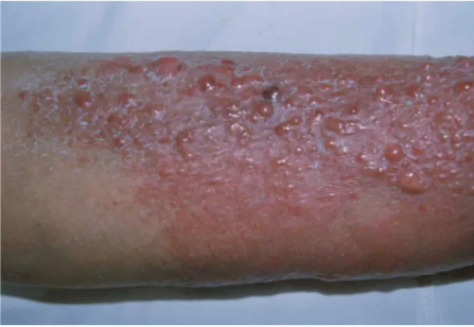
A 70 year-old female multiple myeloma patient type IgG lambda, stage IA, developed purple coloured papular skin infiltrates involving both legs after 2 months ineffective thalidomide and 5 months mel- phalan plus prednisolone therapy (Figure 1).
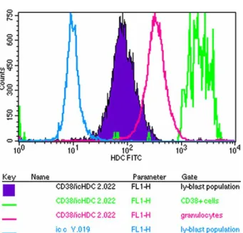
The skin infiltrating cells were shown to be plasma cells (Figure 2). There was a significant difference in HDC content comparing the skin infiltrating plasma cells to bone marrow plasma cells: plasma cells infiltrating the skin became HDC negative during tissue invasion (Figures 3, 4). A very strong HDC positivity was found among bone marrow plasma cells identified by CD38 and CD138 in contrast to other mononuclear cells (Figures 5, 6).
Skin infiltrates came along with other signs of progression, like extension of bony lesions, appearance of subcutaneous nodules, fibronodular
Correspondence: Judit Varkonyi, Kutvolgyi u´ t 4.,1125. Budapest, Hungary. Tel/Fax:361 275 24 20. E-mail: varkjud@kut.sote.hu
Acta Oncologica, 2008; 47: 458480
(Received 20 April 2007; accepted 1 June 2007)
ISSN 0284-186X print/ISSN 1651-226X online#2008 Taylor & Francis DOI: 10.1080/02841860701491066
lung manifestations and the development of heart and renal amyloidosis causing intractable oedema leading eventually to the patient’s death.
The disease was refractory to various chemother- apeutic protocols including continuous low dose Melphalan, cyclophosphamide, CVP, M2, VAD, high dose dexamethasone and local bone irradiation.
The administration of the cytokine interferon alpha (IFN-a) had to be stopped because of intolerance.
Twenty two months from diagnosis the patient died of cardiac and renal insufficiency.
Laboratory results
Initially WBC: 4.3l09/l, PCV: 39%, Hgb 133 mg/
dl, Platelet count 236l09/l, bone marrow infiltra- tion rate: 30%, IgG: 1925, IgA: 15, IgM: 28 mg/dl.
Preterminally the degree of proteinuria was over 1.0 g/24 h, pancytopenia developed and the IgG concentration had also risen up to 3 090 mg/dl.
(Between the two end points the M component was around 11 mg/dl for 8 months.) ECG showed low voltage. There were focal sparkling, pericardial fluid, dilated atria and concentric left ventricular hypertrophy seen on echocardiography as evidences of amyloidosis.
Discussion
The case presented here raises three questions:
1) the pathologic mechanism of epidermoinvasivity;
2) the role of histamine metabolism in the malignant process and 3) the therapy for secondary B-cell cutaneous lesions.
Secondary cutaneous infiltration developing in the course of a patient with B-cell neoplasm is a relatively rare event. In contrast to primary cutaneous infiltrations the secondary form in a
Figure 1. Myelomatous skin infiltrates. On the extensor surfaces of the legs ten serous bullae are seen, of size 1020 mm overlying erythemato-edematous plaques; this appearance developed in the 11th month of the disease course.
Figure 2. Biopsy specimen of the involved skin with a plasma cell infiltrate. HE staining.
Figure 3. Histidine decarboxylase activity in bone marrow smear with 30% plasma cell infiltration rate. Plasma cells are shown to be HDC positive. Immunohistochemical staining.
Figure 4. The lack of Histidine decarboxylase immunoreactivity in the skin specimen infiltrated with plasma cells. Immunohisto- chemical staining.
Letters to the editor 459
lymphoma patient usually means transformation of the disease, heralding a poor prognosis [4,8]. In all those cases had already been published, the absence of HDC in the cutaneous lesions was confirmed in contrast to lymphoma cells residing in the bone marrow, similarly to the case presented here. This phenomenon may be one of the alterations that allow the cells to become epidermotropic.
The presence of HDC in both benign and malignant cell proliferations has been well documen- ted. Intracellular histamine may bind to binding sites
other than histamine receptors. Ligands for the intracellular histamine receptor/binding sites appear to represent a new class of tumor promoting agents or perhaps might act as inhibitors [9]. The possible role of antihistamines and HDC inhibitors as anti- proliferative agents is controversial and still under evaluation. Histidinol, an intracellular histamine receptor HIC antagonist, has an antiproliferative effect itself and also enhances cytotoxicity of anti- neoplastic drugs in combination. This effect is not uniform; it depends on cell type and dose [10].
Histamine potentiates alpha IFNmediated antitu- mor effects, which may be due to histamine en- hancement of NK and T-cell cytotoxicity [11]. This effect had been exploited in a trial where histamine was give to NHL patients previously treated with chemotherapy, to maintain remission [12].
IFN-a does not eliminate malignant cells but inhibits their overgrowth in culture [13]. This could be one possible explanation for the clinical finding in the B-CLL case with cutaneous infiltrations re- sponding to INF-a as it has been reported earlier [4]. In that case the skin infiltrations disappeared within a month without healing of the leukemia process itself.
Conclusion
What we could learn from the case presented here is that epidermoinvasive plasma cells lose their HDC content. This is an acquired characteristic similar to the upregulation of adhesion molecules like CD44 which renders them able to disseminate and capable of skin invasion [14]. The acquired loss of HDC expression in the infiltrative cells points out that changes may occur not only on cell surface level- involving adhesion and angiogenesis- but even in the metabolism of such conservative molecules as HDC.
Thus, extramedullary myeloma can be biologically altered from myeloma remaining in situ suggesting that the therapeutic approach should also be differ- ent. Hopefully further studies on histamine metabo- lism might lead to the development of more targeted therapy to lymphoma patients including those with cutanous infiltrations.
Abbreviations
HDC: histidine decarboxylase, IgY: polyclonal chicken antibody
IFN-a: interferon alpha, MM: multiple myeloma NHL: non-Hodgkin lymphoma
CVP: Cyclophosphamide, Vincristine, Prednison, M2 protocol: Melphalan, BCNU, Vincristine and Prednisolone, VAD-protocol: Vincristine, Adriamy- cin, Prednisolone
Figure 5. Flow cytometric analysis of bone marrow cells of the myeloma patient in respect of their HDC content. Plasma cells are identified by CD38. In contrast to the FITC bound anti-chicken antiserum showing background activity, all the cells had HDC content but the CD38 malignant plasma cells had the highest one.
Figure 6. Flow cytometric analysis of bone marrow cells of the myeloma patient in respect of their HDC content. Plasma cells are identified by CD138 whose HDC content is the highest of all.
References
[1] Bayer-Garner IB, Smoller BR. The spectrum of cutaneous disease in multiple myeloma. J Am Acad Dermatol 2003;/48:/ 497507.
[2] Gado K, Silva S, Pa´lo´czi K, et al. Mouse plasmocytoma: An experimental model of human multiple myeloma. Haemato- logica 2001;/86:/22736.
[3] Hedvat CV, Comenzo RL, Teruya-Feldstein J, Olshen AB, Ely SA, Osman K, et al. Insight into extramedullary tumour cell growth revealed by expression profiling of human plasmocytomas and multiple myeloma. Brit J Haematol 2003;/122:/72844.
[4] Va´rkonyi J, Zalatnai A, Tı´ma´r J, et al. Secondary cutaneous infiltration in B cell chronic lymphocytic leukemia. Acta Haematol 2000;/103:/11621.
[5] Kahlson G. A place for histamine in normal physiology.
Lancet 1960;/I:/6771.
[6] Bartholeyns J, Bouclier M. Involvement of histamine in growth of mouse and rat tumors: Antitumoral properties of monofluoromethylhistidine, an enzyme-activated irreversible inhibitor of histidine decarboxylase. Carcer Res 1984;/44:/ 63945.
[7] Quintana FJ, Buzas E, Proha´szka Z, Bı´ro´ A, Kocsis J, Fu¨ st G, et al. Knock-out of the histidine decarboxylase gene modofies the repertoire of natural autoantibodies. J Auto- immun 2004;/22:/297305.
[8] Tarkova´cs G, Ujvary B, Panczel P, Berczi L, Palinger E, Matolcsy A, et al. Losing histidine decarboxylase immun- reactivity appears to be a regularity in the development of secondary cutaneous B-cell lymphoma. EORTC Cutaneous Lymphoma Task Force Clinical Meeting Budapest, 2224 September 2006.
[9] Bencsa´th M, Pa´lo´czi K, Szalai CS, Szenthe A, Szebere´nyi J, Falus A. Histidine decarboxylase in peripheral lymphocytes of healthy individuals and chronic lymphoid leukemia patients. Pathol Oncol Res 1998;/4:/1214.
[10] Brandes LJ, LaBella FS, Warrington RC. Increased ther- apeutic index of antineoplastic drugs in combination with intracellular histamine antagonists. J Natl Cancer Inst 1991;/ 83:/132936.
[11] Naredi P. Histamine as adjunct to immunotherapy. Semin Oncol 2002;/29:/314.
[12] Ahlberg R, MacNamara B, Andersson M, et al. Stimulation of T-cell cytokine production and NK-cell function by IL-2, IFN-aand histamine treatment. Hematol J 2003;/4:/295302.
[13] Cornelissen JJ, Ploemacher RE, Wognum BW, Borsboom A, Kluin-Nelemans HC, Hagemeijer A, et al. An in vitro model for cytogenetic conversion in CML. IFNa preferentially inhibits the outgrowth of malignant stem cells preserved in long term culture. J Clin Invest 1998;/102:/97683.
[14] Drillenburg P, Pals ST. Cell adhesion receptors in lymphoma dissemination. Blood 2000;/95:/190010.
EBV positivity in primary cutaneous large B-cell lymphoma with immunophenotypic features of leg type: An isolated incidence or something more significant?
SUJATA GAITONDE
1, SRAVANKUMAR KAVURI
1, VICTORIA ALAGIOZIAN- ANGELOVA
1, DAVID PEACE
2& SOPHIE WOROBEC
31Department of Pathology, University of Illinois, Chicago, USA,2Department of Medicine, University of Illinois, Chicago, USA, and3Department of Dermatology, University of Illinois, Chicago, USA
To the Editor,
The WHO-EORTC classification divides cutaneous B-cell lymphomas into four categories of which primary cutaneous diffuse large B-cell lymphoma, leg type (PCLBCL, leg type) characteristically involves lower legs of elderly women and shows a predominance of confluent sheets of medium sized to large cells with round nuclei and prominent nucleoli resembling centroblasts and/ or immuno- blasts. The neoplastic B-cells of PCLBCL, leg type have characteristic immunophenotype which readily
differentiates them from other subtypes of primary cutaneous diffuse large B-cell lymphomas. The overall prognosis in this group is poor with a tendency to extracutaneous dissemination. An asso- ciation of PCLBCL, leg type with Epstein Barr virus (EBV) has not been previously described. We report an unusual case of PCLBCL with immunopheno- typic features of leg type with EBV positivity in the neoplastic B-cells by in situ hybridization. A fifty-five year old Caucasian male presented with a subcuta- neous nodule on his right thigh which was comple-
Correspondence: University of Illinois at Chicago, Department of Pathology (M/C 847), Medicine and Dermatology, 840 South Wood Street, Room 110 CSN, Chicago, IL 60612 USA. Tel:1 312 996 4206. E-mail: sgaitond@uic.edu
Letters to the editor 461
(Received 13 June 2007; accepted 6 August 2007)
ISSN 0284-186X print/ISSN 1651-226X online#2008 Taylor & Francis DOI: 10.1080/02841860701621241