PHYSIOLOGICAL AND STRUCTURAL MODIFICATIONS IN SNAIL MEDIC (MEDICAGO SCUTELLATA L.) PLANTS
EXPOSED TO SALINITY
Houneida attia,1,2 * K. H. AlAmer,2 Imen SelmI,1 W. DjebAlI,1 WIDeD CHAïbI1 and mouHIbA ben nASrI1
1Physiology and Biochemistry of Plant Response to Abiotic Stress, Faculty of Science of Tunis, Tunis El Manar University, 2092 Tunis, Tunisia
2Biology Department, Faculty of Science, Taif University, Kingdom of Saudi Arabia (Received: December 4, 2017; accepted: April 19, 2018)
Seeds of snail medic (Medicago scutellata L.) were assessed for their response to salt at the germination and seedling stages. NaCl at concentrations 86 and 170 mM decreased the final germination percentage.
Embryonic axis length, water content and dry weight of embryonic axis and cotyledons were also reduced by salt treatment. Furthermore, 28-d-old plants were grown hydroponically with different NaCl concen- trations (0, 86 and 170 mM). After 7 days of treatment, growth, water content and development of the different organs of M. scutellata plant were affected especially at the highest NaCl concentration (170 mM). However, NaCl did not affect root length and the number of stem shoots but reduced stem length and total leaf area. Salt treatment increased markedly the concentration of Na+ in leaf and root tissues while reduced that of K+ only in root and stem tissues. Lipid peroxidation revealed the damage of the membranes of roots and leaves. Moreover, showed a more intense suberization and lignification at the cambial zone of roots of M. scutellata, were observed under the effect of NaCl.
Keywords: Salinity – anatomical changes – growth – mineral nutrition – Medicago scutellata
INTRODUCTION
Salinity is one of the major factors that affect plant growth in Tunisia, where there is a wide variety of saline-sodic soils in depressions and in the main sebkhas and chotts [20, 23, 25]. Moreover, in irrigated areas, the low quality of irrigation water charged with dissolved salts has resulted unfortunately in soil secondary salinization respon- sible for decline in productivity [25]. Legumes like most crop plants are susceptible to salinity [7, 10]. These plants are widely grown for grain and forage purposes, their worldwide economic importance is only secondary for grasses [14]. In addition, leg- umes can establish root symbioses with nitrogen-fixing soil bacteria, enabling the plants to grow in nitrogen-poor soils. This ability to colonize soils where other plants cannot thrive makes the study of legumes and their symbioses important for agricul- ture.
Snail medic (Medicago scutellata) is a diploid annual forage legume cultivated in Mediterranean areas and Australia in order to improve pasturelands, and used in crop
* Corresponding author; e-mail address: houneida_attia@yahoo.fr
Acta Biologica Hungarica 69, 2018
rotation [28]. Annual medic species because of nitrogen fixation and proper quality of fodder have high crop values [12]. Together with M. truncatula and M. polymor- pha, M. scutellata represents an important pasture legume for increasing forage pro- duction in marginal areas where the environmental conditions limit the growth of perennial species as M. sativa.
In this paper, we studied the response of M. scutellata to different NaCl concentra- tions during vegetative growth. Dry matter production of leaves, stems and roots, and ion accumulation were measured, along with chlorophyll contents. Electrolyte leak- age and malondialdehyde (MDA) content were determined as proxies of oxidative stress, and we have especially studied the effect of NaCl on structural modifications, including suberization and lignification in M. scutellata.
MATERIALS AND METHODS Plant material and germination conditions
The seeds of Medicago scutellata were provided by Seed Laboratory of the Tunisian Ministry of Agriculture. For germination, seeds were disinfected in oxygenated water for 20 min and rinsed in distilled water. The seeds were then placed in Petri dishes supplied with double layer filter paper initially moistened with a solution of the respective salt concentration: 0, 86 and 170 mM [20]. The seeds were then incubated for 9 d in darkness at room temperature (25 ± 1 °C). Each treatment was performed on 25 seeds per Petri dish and was replicated three times. Seeds with emerged radicle were counted daily. Final germination percentage (FG%) was calculated as 100×
number of germinated seeds divided by the number of sown seeds. After 9 d, seed- lings were divided into embryonic axis and cotyledons for determination of growth parameters. Fresh weights (FW) of all samples were recorded. Plant material was dried at 60 °C for 2 days and the dry weight (DW) was measured. Tissue water con- tent was obtained from the (FW-DW/FW) ratio.
Culture conditions
Seeds of Medicago scutellata were disinfected in oxygenated water for 20 min and rinsed in distilled water and germinated at 25 °C in Petri dishes on filter paper mois- tened with distilled water. Nine-d-old seedlings were transferred in pots containing 2 L of aerated, 2-fold diluted nutritive solution [16], with one plant per pot. The solu- tion contained 0.50 mM MgSO4, 0.25 mM KH2PO4, 1.25 mM Ca(NO3)2, 1.25 mM KNO3, 50 µM KCl, 1 µM MnSO4, 0.5 µM CuSO4, 0.5 µM ZnSO4, 10 µM H3BO3, 0.05 µM (NH4)Mo7O24, and 1.5 µM Fe-EDTA. It was renewed weekly. The photo- period was 16 h with a photosynthetic photon flux density of 150 µmol m–2 s–1 at the plant level. The day/night temperature and relative humidity regimes were 25 °C/18 °C and 60/80%, respectively.
Plant morphology and growth
Twenty-eight-day-old plants grown on control medium were exposed to different (0, 86 and 170 mM) NaCl treatments during 7 days. Five plants per treatment were sam- pled (final harvest) and roots, stems, and leaves were separated. Roots were gently surface dried. Shoot and root fresh weights were immediately determined.
Morphological parameters (stem length, root length and leaf area) were measured using a scanner and Optimas® software and number of stem shoots was also deter- mined by a simple counting. The samples were then oven-dried for 48 h at 70 °C and weighted.
Ion accumulation
At the final harvest, roots (but not other organs) were rinsed in 100 mL cold distilled water (three successive baths, 30 s each) then gently blotted between paper towels.
Ions (K+ and Na+) were extracted from 25 mg samples of dried tissues with 0.5%
(v/v) HNO3 for 48 h, and assayed by flame photometer (Jenway PFP7, ELE Instrument Co. Ltd) using butane-air flame.
Chlorophyll content
For each treatment, four plants were used and treated individually. Whole set of fresh leaves of each plant was separately incubated in the dark for 72 h at 4 °C in acetone 80% (v/v). Absorbance of acetone extracts was measured at 460, 645, and 663 nm with a DU 640 Beckman spectrophotometer. Concentrations ofchlorophyll a, b, and total chlorophylls were calculated using the equations proposed by McKinney [22].
Membrane permeability (electrolyte leakage, EL)
Electrolyte leakage from leaf cells was determined as described by Dionisio-Sese and Tobita [9]. Leaf samples were taken and cut into 1 cm2 sections (approximately 200 mg FW). The samples were then placed in individual stoppered vials containing 10 mL of distilled water. The tubes were incubated in a water bath at 32 °C for 2 h and the initial electrical conductivity of the medium (EC1) was measured using digi- tal electrical conductivity meter (DDS-11A). The samples were autoclaved at 121 °C for 20 min to release the remaining electrolytes, then cooled to 25 °C, and the final electrical conductivity (EC2) was measured. The electrolyte leakage (EL) was calcu- lated as (EC1/EC2) × 100. Six replicates from six different plants were used.
EL from roots was determined after the immersion of root in 0.5 mM CaCl2 solu- tion for 5 min and incubation in darkness with 25 mL of water for 1 h (EC1 determi-
Acta Biologica Hungarica 69, 2018
nation). Roots were then transferred into 25 mL of boiling water for 5 min, cooled to 25 °C and the final electrical conductivity (EC2) was measured. EL was calculated according to Shalata and Neumann [32]: EL = (EC1/(EC1 + EC2)) × 100.
Lipid peroxidation
Lipid peroxidation was determined using the thiobarbituric acid (TBA) reaction to measure the tissue content of MDA [15]. An amount of 250 mg of leaves, stems or roots were ground in a cold mortar using 50 mM Tris–HCl (pH 7.5) as buffer contain- ing 0.2 mM EDTA and 0.2% (v/v) Triton X-100. The homogenate was centrifuged at 10,000 × g for 15 min. 2.5 mL of 0.25% (w/v) thiobarbituric acid containing 10%
(w/v) trichloroacetic acid and 0.01% butylated hydroxytoluene was added to aliquots of supernatant. The mixture was heated at 95 °C for 15 min and then quickly cooled in an ice bath. After centrifugation at 5500 g for 5 min, the absorbance of the super- natant was read at 535 and 600 nm. The MDA content was calculated by using a molar extinction coefficient of 155 mM–1 cm–1. Six different plants were used as replicates.
Histology
Histological observations were made on root, stem and leaf transverse sections in two ways. In the first case, sections were cut on a freezing microtome (Leitz Kryomat 1700), and placed in sodium hypochlorite (12°) for 10 to 20 min to remove the cell contents. After treatment with acetic acid 30% (v/v) for 5 min, the sections were stained with a mixture of alum carmine and methyl-green. In the second case, the leaf sections were fixed in a mixture of alcohol and acetic acid for 24 h. Tissues were dehydrated stepwise in alcohol solution series, then in toluene. After their embedding in paraffin the samples were stained with hematoxylin, safranin and aniline blue. The observations, which covered at least five samples taken from control or treated plants, were performed using a light microscope (Leica RM2165) equipped with a micromet- ric rule. The micrographs were digitized with a scanner (HP Scanjet 3800, Hewlett Packard, USA) and analysed with a software (ImageJ, National Institutes of Health Bethesda, MD), to measure mesophyll thickness and vascular bundle diameter of leaves. A minimum of three samples were examined per each treatment.
Statistical analysis
Statistical analysis was performed with StatisticaTM software, using ANOVA and mean comparison with Duncan’s test. Values were calculated at the p ≤ 0.05 probabil- ity level.
RESULTS
Effect of salt treatment on germination
All germination curves reached a plateau (FG%max : maximum FG%), but they dif- fered on the value of FG%max and on the time necessary to reach this value (Fig. 1).
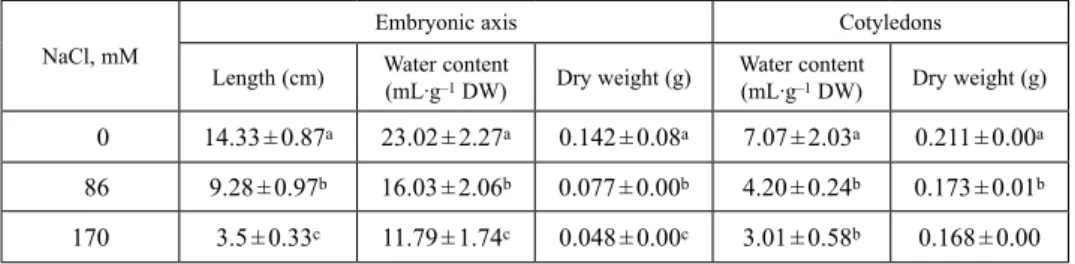
For M. scutellata seeds, FG%max exceeded 80% at 0 and 86 mM NaCl, but it was reduced to 60% at 170 mM NaCl. Furthermore, the latency for radicle emergence augmented with salt concentration. Embryonic axis biomass was more affected by salt than cotyledons (Table 1). At 170 mM NaCl, embryonic axis biomass was restricted by 66% as compared with control (no salt), whereas cotyledon biomass was restricted only by 20%. Embryonic axis length was still more sensitive to salt inhibi- tion: Embryonic axis length was diminished by 35% at 86 mM NaCl and by 76% at 170 mM NaCl. In embryonic axis and cotyledon, salt limited water content (Table 1).
Fig. 1. Effect of different NaCl concentrations (0, 86 and 170 mM) on germination percentage of M. scutellata after 9 days of treatment
Table 1
Effect of different NaCl concentrations (0, 86 and 170 mM) on length, water content and dry weight in embryonic axis and cotyledons of M. scutellata. Seeds were germinated on moist filter paper for 9 d in the absence (control) or in the presence of NaCl. Data are the means of three replicates ± confidence interval. Means sharing a same letter are not significantly different at p = 0.05 (ANOVA and mean com-
parison with Duncan’s test)
NaCl, mM
Embryonic axis Cotyledons
Length (cm) Water content
(mL∙g–1 DW) Dry weight (g) Water content
(mL∙g–1 DW) Dry weight (g) 0 14.33 ± 0.87a 23.02 ± 2.27a 0.142 ± 0.08a 7.07 ± 2.03a 0.211 ± 0.00a 86 9.28 ± 0.97b 16.03 ± 2.06b 0.077 ± 0.00b 4.20 ± 0.24b 0.173 ± 0.01b 170 3.5 ± 0.33c 11.79 ± 1.74c 0.048 ± 0.00c 3.01 ± 0.58b 0.168 ± 0.00 Data are the mean of three samples of 25 seedlings each one of treatment.
Acta Biologica Hungarica 69, 2018
Effect of salt treatment on plant growth, water content and ion accumulation
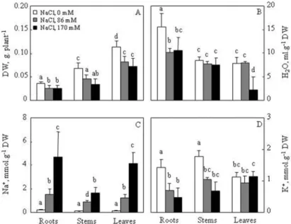
Control plants displayed a total dry weight of about 217 mg. This biomass was dis- tributed as follows: 52% in leaves, 31% in stems, and 17% in roots (Fig. 2A). Salt treatment significally decreased M. scutellata growth for the different organs: roots, stems and leaves at both, low (86 mM) and high (170 mM) NaCl concentrations For leaves, the decrease in biomass under salinity was due to a gradual reduction in whole area (Table 2). Moreover, water content was drastically reduced by salt concentration, the most pronounced effect was observed in leaves (–69% of control) and to a lower degree in roots (–44% of control) at NaCl 170 mM (Fig. 2B).
Medicago scutellata plants cultivated under increasing salinity showed a signifi- cantly higher Na+ accumulation in leaves and roots, as compared to stems (Fig. 2C).
At high salinity concentration (NaCl 170 mM) a main amount of Na+ content was more accumulated in roots and leaves than in stems, suggesting that M. scutellata was able to adopt an includer strategy. On the other hand, salt treatment induced a sig- nificant decrease of K+ content in roots and stems (Fig. 2D). Potassium content in leaves was not affected by salt. These results suggested that K+ was more allocated in leaves than stems and roots in the presence of salt.
Fig. 2. Effect of different NaCl concentrations (0, 86 and 170 mM) on organ biomass (A), water content (B), sodium content (C) and potassium (D) content of M. scutellata. Twenty-eight-day-old plants were grown for 7 days in the absence (control) or in the presence of NaCl. Data are the means of five replicates
± confidence interval. Means labeled with the same letter are not significantly different at p = 0.05 (ANOVA and mean comparison with Duncan’s test)
Effect of salt treatment on chlorophyll content
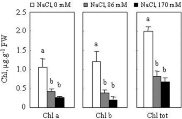
Salt treatment induced a large decrease in the chlorophyll of M. scutellata leaves (Fig. 3). The reduction was similar at both 86 and 170 mM NaCl. At high salinity, Chl a and Chl b contents diminished by 77% and 83%, respectively, in comparison to the control (Fig. 3).
Table 2
Effect of different NaCl concentrations on stem length, root length, total leaf area, number of stem shoots and on mesophyll thickness and vascular bundle diameter number of leaf of M. scutellata. Twenty-eight- day-old plants were grown for 7 days in the absence (control) or in the presence of NaCl. Stars indicate
significant differences between control and treatments
Parameters Treatment (NaCl, mM)
0 86 170
Stem length, cm 16 ± 1.0 13 ± 0.9* 11 ± 1.0*
Root length, cm 32 ± 7.0 26 ± 5.0 25 ± 4.0
Total leaf area, cm2 ∙ plant–1 7 ± 0.6 5 ± 0.7* 4 ± 0.3*
Number of stem shoots 7 ± 1.0 6 ± 0.0 5 ± 0.0
Mesophyll thickness, µm 6.117 – 5.072*
Vascular bundle diameter, µm 4.772 – 3.549*
Data are the means of five replicates ± confidence interval (p = 0.05).
Fig. 3. Effect of different NaCl concentrations (0, 86 and 170 mM) on chlorophyll contents of M. scutel- lata. Plants were grown and harvested as described in the legend of Fig. 2. Data are the means of four replicates ± confidence interval. Means sharing labeled with the same letter are not significantly different
at p = 0.05 (ANOVA and mean comparison with Duncan’s test)
Effect of salt treatment on membrane permeability
The extent of membrane damage was estimated by electrolyte leakage and MDA level in root and leaf tissues. These parameters increased by salt in both organs (Fig. 4). In roots and leaves, electrolyte leakage progressively were increased with NaCl concentration (Fig. 4A). In addition, increased MDA content in roots and leaves of M. scutellata indicated that the application of NaCl resulted in enhanced lipid peroxidation (Fig. 4B).
Fig. 4. Effect of different NaCl concentrations (0, 86 and 170 mM) on: electrolyte leakage and MDA content of M. scutellata. Plants were grown and harvested as described in the legend of Fig. 2. Data are the means of six replicates ± confidence interval. Means sharing labeled with the same letter are not sig-
nificantly different at p = 0.05 (ANOVA and mean comparison with Duncan’s test)
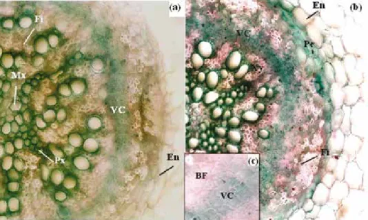
Fig. 5. Light micrographs showing root cross-sections of M. scutellata. Twenty-eight-day-old plants were grown for 7 days in the absence (a) or in the presence of NaCl 170 mM (b). (c) detail of (b) at the cam- bial zone. Roots were sectioned 2.5 cm from root-stem junction and stained using a mixture of alum carmine and methyl-green. Alum carmine was applied to reveal a cellulose cell wall component of phloem and methyl-green to reveal lignified and suberin in the cell walls. Enlargement ×400.
Abbreviations: BF – bast fibers; En – endodermis; Fi – fibers; Mx – metaxylem; Pe – pericycle; Px – protoxylem; VC – Vascular Cambium; Xy – xylem
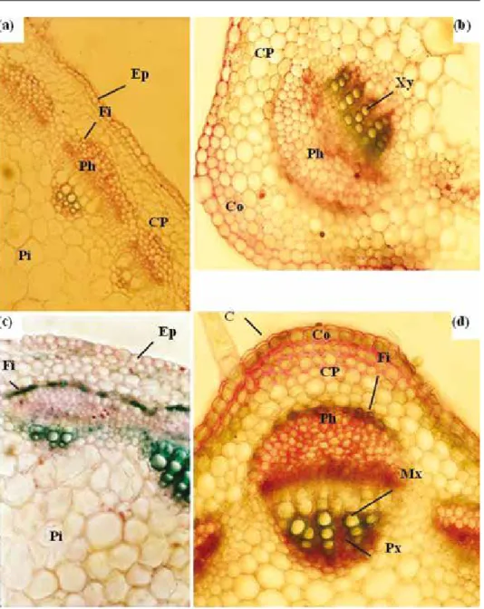
Fig. 6. Transverse sections of M. scutellata stems. Twenty-eight-day-old plants were grown for 7 days in the absence (a and b) or in the presence of 170 mM NaCl (c and d). Stems were sectioned at the fourth internode and stained using a mixture of alum carmine and methyl-green. Alum carmine was applied to reveal a cellulose cell wall component of phloem and methyl-green to reveal lignin and suberin contents of the cell walls. b detail of a; d detail of c. Enlargement: ×150 (a and c), ×200 (b and d). Abbreviations:
C – cuticle; Co – collenchyma; CP – cortical parenchyma; Ep – epidermis; Fi – fibers; Mx – metaxylem;
Ph – phloem; Pi – pith; Px – protoxylem; Xy – xylem
Acta Biologica Hungarica 69, 2018
Histological observations
Under NaCl stress (170 mM), reduction of root diameter was attributed to restricted central cylinder area (9.8, 3.6 and 3.8, for control plants and 7.1, 2.7 and 2.3 for treated plants, respectively, in root diameter, central cylinder diameter and thickness of cortex [mm]). Morphometric analyses (results not shown) confirm that NaCl increased the number of sclerenchyma fibers (Fi). Salt induced also a reduction in the number of tracheary elements, significant increase in lignified fibrous layer (Fi) and bast fiber (BF); at the cambial zone, it causes a significant lignification (Fig. 5).
After 7 days of NaCl treatment (170 mM), the stem diameter remained unchanged, however, the central cylinder exhibited a high lignification process (Fi) concomi- tantly with an increase in xylem number (Fig. 6). In the central cylinder of root sec- tions, it appeared clearly that the cell walls are much thicker and the pinkish colour is much more intense in the salt-treated than they were in the control seedlings, which is again consistent with an increased process of lignification under saline conditions (Fig. 6).
DISCUSSION
Soil salinity is a prevalent abiotic stress for plants, with retarded growth being a com- mon response to salinity. Plant growth is one of the most important agricultural indices of salt stress tolerance [27]. Salt stress can induce both a reduction in seed germination and a delay in the initial germination process in glycophytes and to a lesser extent in halophytes [11]. The increase in salinity does not only decrease germination, but also delays the initial germination rate [26]. In our present study, the latency for radicle emergence augmented with salt concentration, notably under 170 mM NaCl in M. scutellata seeds. It was hypothesized that the presence of NaCl, even at low con- centrations, could contribute to a decrease in the internal osmotic potential of germi- nating structures [18]. At 170 mM NaCl treatment, a significant decrease in the germi- nation percentage of M. scutellata was observed. Embryonic axis exhibited high reduction of DW and water content as compared to cotyledon especially at this con- centration of NaCl. This might be due to toxic effects of Na+ and/or Cl– in seed tissues as well as unbalanced nutrient uptake induced by salt. Toxic accumulation of salt in leaf cell cytoplasm is prevented by two strategies: by salt exclusion from leaves, or by efficient ion compartmentalization in vacuoles of include leaves. The excluder strategy is poorly efficient when soil salinity is high, and thus it is found only in salt-sensitive glycophytes. On the contrary, salt inclusion is typical of salt-tolerant glycophyte and halophytes. However, even in includers, Na+ transport to shoot is strictly controlled, and the efficiency of this control is related to salt tolerance [13].
In the present study, the application of NaCl (86 and 170 mM) in the hydroponic media caused a remarkable reduction in DW of the shoot (leaves and stems), and no salt effect was visible on roots. At this point, it seems that shoot was more sensitive to salt stress than root. These results are in good accordance with those reported by
da Silva et al. [8] who found that leaves and stems are more sensitive to salt stress than roots in Spondias tuberosa. In Pea, it has been shown that the application of 50 mM NaCl resulted in a drastic decrease in leaf growth [4]. Salt stress also reduced total leaf area of M. scutellata. These effects could be related to an inhibition of new leaf initiation and/or reduction of leaf expansion [5]. Our results also demonstrate a decrease of water content in roots and leaves (at 170 mM NaCl). In spite of the high concentration of NaCl, the stem hydration was maintained at the same value as in control plants. At 170 mM NaCl, Na+ accumulation is more important in roots and leaves than in stems. High Na+ content in roots and stems were associated with K+ limitation. These results, which are in agreement with those found in A. thaliana [3], M. sativa [23], and T. ammi [1], suggest that K+ is one of the most growth-limiting factors in M. scutellata under saline conditions.
Sensitivity of M. scutellata leaves to salt stress was probably due to an osmotic effect, as leaf tissue hydration was severely affected despite their high Na+ concentra- tions. However, some of these ions were accumulated in cytoplasm. According to Wenxue et al. [34], high Na+ concentrations were not associated with limitation of essential nutrient uptake like K+ and Ca2+, which were allocated to shoots.
When plants were grown in salty medium, chlorophyll concentration was largely diminished in M. scutellata. The loss of chlorophylls is considered as a marker of salt stress [5]. Thus, our results support the hypothesis that chloroplasts were severely altered by salt as well as leaf cell membrane, as attested by the magnitude of electro- lyte leakage. There is increasing evidence that membrane injury under salt stress is related to a higher production of toxic ROS [24]. Determining the MDA concentra- tion and hence, the extent of membrane lipid peroxidation, has often been used as a tool to assess the severity of oxidative stress [6]. Our data showed that, after seven days of NaCl treatment, membrane lipid peroxidation was induced and MDA con- tents in leaves and roots were much higher as compared to control. Our results are in agreement with those of Sairam and Srivastava [31], who reported enhanced concen- tration of MDA in salt-sensitive rice cultivar as compared to the tolerant cultivar, and in Lemna minor roots.
All results discussed above, demonstrate the effects of salt stress on some physi- ological and biochemical parameters in M. scutellata. This type of stress can also cause structural changes in plant organs. In Rangpur lime (C. limonia) and Etrog citron (C. medica) grown in the presence of 100 mM NaCl, the hypodermal cells developed lignified and suberized walls which blocked the plasmodesmata and resulted in degeneration of the cell contents [33]. According to these authors, the primary endodermal cells had lignified Casparian strips and plasmodesmata in other cell wall areas. These connections were blocked by secondary suberization except the endodermal passage cells opposite to protoxylem arcs. Zhong and Läuchli [35] have shown that 150 mM NaCl increased significantly cell wall uronic acid content in cot- ton seedlings, but reduced cellulose content on a per unit dry weight basis. These results reveal a relationship between the effects of high salinity on root growth and cell wall metabolism, particularly in regard to cellulose biosynthesis. In G. hirsutum, NaCl 200 mM induced the formation of an exodermis with Casparian bands and
Acta Biologica Hungarica 69, 2018
suberin lamellae close to the root base and in the transition zone to hypocotyl. The exodermis, which is absent in control roots, may play a role in protecting the root from water loss and/or leakage of solutes important for osmotic adjustment [29].
Contrary to the plants species mentioned previously, an increase of the number of wood fiber, bast fiber (which cover the liber) and suberization of the cambial zone was reported in M. scutellata. This phenomenon stiffens the organ and consequently decreases its growth. It may nevertheless be protected, at least in part against loss of water. In M. scutellata, the suberization of the cambium stiffens also the cell walls, reducing their elasticity and plasticity, and blocking the formation of secondary vas- cular elements (phloem and xylem) which contribute to exchange limitation and growth inhibition.
Besides suberization of tissues, M. scutellata reacts to salt by reducing mesophyll cell size and spongy parenchyma gaps which causes a narrowing of all cells and con- sequently a decrease in intercellular spaces and leaf thickness. This phenomenon causing a decrease in the rate of CO2 at the foliar parenchyma would lead to a disrup- tion of photosynthetic activities. Such a reduction was observed in other species under the effect of other types of stress. In bean, salinity caused a reduction in both mesophyll and epidermal cells [2]. In cucumber plants the salt stressed leaves had shorter palisade cells but the spongy mesophyll was thicker and had less air spaces compared to control plants [21]. Contrally, salinity significantly reduced the cross- sectional area, width, and radii of both epidermal and mesophyll cells along the leaf axis in wheat. The reduction was attributed to a decrease in the size and number of medium and small veins [17]. Reinoso et al. [30] showed that salinity induced ana- tomical changes in roots (young and mature zones) of Prosopis strombulifera. The diameters of the young zone of roots of plants grown in increasing salt concentrations were smaller than those of controls, with reduced number of cortex layers and reduced size of the vascular system. The roots from tolerant plants showed precocious suberization and (or) lignification of the endodermal cells and early activity of the pericycle. The stem diameter of young tolerant plants was notably diminished and less tissue lignification occurred. According to literature data, some water-soluble peroxidases have been hypothesised to have auxin oxidase activity (which might explain the effect observed on the root biomass), while the cell-wall peroxidases would be involved in lignification. Histochemical observation confirmed a more intense lignification in the root cells of the salt-tolerant species compared to the sensi- tive species in wheat seedlings, under the effect of NaCl [19].
All these anatomical changes, including the phenomenon of suberization and lig- nification refers to an early differentiation in tissues under saline conditions.
In conclusion, our results demonstrate that salinity inhibited germination and early seedling growth in M. scutellata. Content of Na+ in roots and leaves were higher than those in stems. Accumulation of Na+ resulted in a decrease of plant biomass, and chlorophyll content which presumably limited photosynthetic activity. Furthermore, salt induced membrane damages in both roots and leaves. Anatomical observations indicated a more intense suberization and lignification at the cambial zone of roots of M. scutellata under the effect of NaCl.
REFERENCES
1. Ashraf, M., Orooj, A. (2006) Salt stress effects on growth, ion accumulation and seed oil concentra- tion in an arid zone traditional medicinal plant ajwain (Trachyspermum ammi [L.] Sprague). J. Arid.
Environ. 64, 209–220.
2. Asish, K. P., Anath, B. D. (2004) Salt tolerance and salinity effects on plants. Ecotox. Environ. Safe.
60, 324–349.
3. Attia, H., Karray, N., Rabhi, M., Lachaâl, M. (2008) Salt-imposed restrictions on the uptake of mac- roelements by roots of Arabidopsis thaliana. Acta Physiol. Plant. 30, 723–727.
4. Attia, H., Nouaili, S., Soltani, A., Lachaâl, M. (2009) Comparison of the responses to NaCl stress of two pea cultivars using split-root system. Sci. Hort. 123, 164–169.
5. Attia, H., Ouhibi, C., Ellili, A., Msilini, N., Bouzaïen, G., Karray, N., Lachaâl, M. (2011) Analysis of salinity effects on basil leaf surface area, photosynthetic activity, and growth. Acta Physiol. Plant. 33, 823–833.
6. Ben Amor, N., Hamed, K. B., Debez, A., Grignon, C., Abdelly, C. (2005) Physiological and antioxi- dant responses of the perennial halophyte Crithmum maritimum to salinity. Plant Sci. 168, 889–899.
7. Chinnusamy, V., Jagendorf, A., Zhu, J. K. (2005) Understanding and improving salt tolerance in plants. Crop Sci. 45, 437–448.
8. da Silva, E. C., Nogueira, R. J. M. C., Araujo, F. P., Melo, N. F., de Azevedo Neto, A. D. (2008) Physiological responses to salt stress in young umbu plants. Environ. Exp. Bot. 63, 147–157.
9. Dionisio-Sese, M. L., Tobita, S. (1998) Antioxidant responses of rice seedlings to salinity stress. Plant Sci. 135, 1–9.
10. Duzan, H. M., Zhou, X., Souleimanov, A., Smith, D. L. (2004) Perception of Bradyrhizobium japoni- cum Nod factor by soybean [Glycine max (L.) Merr.] root hairs under abiotic stress conditions. J. Exp.
Bot. 55, 2641–2646.
11. El-Keblawy, A. (2004) Salinity effects on seed germination of the common desert range grass, Panicum turgidum. Seed Sci. Technol. 32, 943–948.
12. Fakhari, F., Sadeghi, H. (2016) Investigating the effects of pod elimination on salinity tolerance in annual Medic (Medicago scutellata L.). J. Rangeland Sci. 6, 232–241.
13. Garthwaite A. J., Millhollon, E. P., Lucas, M. C. (2005) Salt tolerance in wild Hordeum species is associated with restricted entry of Na+ and Cl– into the shoots. J. Exp. Bot. 56, 2365–2378.
14. Graham, P. H., Vance, C. P. (2003) Legumes: importance and constraints to greater use. Plant Physiol.
131, 872–877.
15. Heath, R. L., Packer, L. (1968) Photooxidation in isolated chloroplasts I: kinetics and stoichiometry of fatty acid peroxidation. Arch. Biochem. Biophys. 125, 189–198.
16. Hoagland, D. R., Arnon, D. I. (1950) The water culture method for growing plants without soil. Circ 347. Cal. Agri. Exp. Stat. Berkley.
17. Hu, Y., Fricke, W., Schmidhalter, U. (2005) Salinity and the growth of non-halophytic grass leaves:
the role of mineral nutrient distribution. Funct. Plant Biol. 32, 973–985.
18. Huang, W. L., Liu, F. L. (2002) Carbohydrate metabolism in rice during callus induction and shoot regeneration induced by osmotic stress. Bot. Bull. Acad. Sinica 43, 107–113.
19. Jbir, N., Chaïbi, W., Ammar, S., Jemmali, A., Ayadi, A. (2001) Root growth and lignification of two wheat species differing in their sensitivity to NaCl, in response to salt stress. C.R. Acad. Sci. Paris, Sciences de la vie / Life Sciences 324, 863–868.
20. Lazrek, F., Roussel, V., Ronfort, J., Cardinet, G., Chardon, F., Aouani, M. E., Huguet, T. (2009) The use of neutral and non-neutral SSRs to analyse the genetic structure of a Tunisian collection of Medicago truncatula lines and to reveal associations with eco-environmental variables. Genetica 135, 391–402.
21. Lechno, S., Zamski, E., Telor, E. (1997) Salt stress-induced responses in cucumber plants. J. Plant Physiol. 150, 206–211.
22. McKinney, G. (1941) Absorption of light by chlorophyll solutions. J. Biol. Chem. 140, 315–332.
Acta Biologica Hungarica 69, 2018 23. Mezni, M., Albouchi, A., Bizid, E., Hamza, M. (2002) Effet de la salinité des eaux d’irrigation sur la
nutrition minérale chez trois variétés de Luzerne pérenne (Medicago sativa). Agronomie 22, 283–291.
24. Mittler, R. (2002) Oxidative stress, antioxidants and stress tolerance. Trends Plant Sci. 7, 406–410.
25. Mtimet, A. (2001) Soils of Tunisia. In: Zdruli, P., Steduto, P., Lacirignola, C., Montanarella, L. (eds) Soil resources of southern and eastern mediterranean countries. Bari, Italy, pp. 243–262.
26. Nasri, N., Kaddour, R., Rabhi, M., Plassard, C., Lachaâl, M. (2011) Effect of salinity on germination, phytase activity and phytate content in lettuce seedling. Acta Physiol. Plant. 33, 935–942.
27. Parida, A. K., Das, A. B. (2005) Salt tolerance and salinity effects on plants. Ecotox. Environ. Safe.
60, 324–349.
28. Piano, E., Francis, C. M. (1992) The annual species of Medicago in the Mediterranean region: ecoge- ography and related aspects of plant introduction and breeding. In: Proceedings of the 10th Inter- national Conference of Eucarpia Medicago spp. (Group Lodi), Italy, pp. 373–385.
29. Reinhards, D. H., Rost, T. L. (1995) Salinity accelerates endodermal development and induces an exodermis in cotton seedling roots. Environ. Exp. Bot. 35, 563–574.
30. Reinoso, H., Sosa, L., Ramírez, L., Luna, V. (2004) Salt-induced changes in the vegetative anatomy of Prosopis strombulifera (Leguminosae). Rev. Can. Bot. 82, 618–628.
31. Sairam, R. K., Srivastava, G. C. (2002) Changes in antioxidant activity in sub-cellular fractions of tolerant and susceptible wheat genotypes in response to long term salt stress. Plant Sci. 162, 897–904.
32. Shalata, A., Neumann, P. M. (2001) Exogenous ascorbic acid (vitamin C) increases resistance to salt stress and reduces lipid peroxidation. J. Exp. Bot. 52, 2207–2211.
33. Walker, R. R., Sedgley, M., Blesing, M. A., Douglas, T. J. (1984) Anatomy, ultrastructure and assim- ilate concentrations of roots of citrus genotypes differing in ability for salt exclusion. J. Exp. Bot. 35, 1481–1494.
34. Wenxue, W., Bilsborrow, P. E., Hooley, P., Fincham, D. A., Lombi, E., Forster, B. P. (2003) Salinity induced differences in growth, ion distribution and partitioning in barley between the cultivar Maythorpe and its derived mutant Golden Promise. Plant Soil 250, 183–191.
35. Zhong, H., Läuchli, A. (1993) Changes of cell wall composition and polymer size in primary roots of cotton seedlings under salt stress. J. Exp. Bot. 44, 773–778.