http://prrssymposium.org/TopMenu/Proceedings.aspx.
Immunohistochemical characterization of type II pneumocyte proliferation after PRRSV (Type I) challenge
G. Balka1*, A. Ladinig2, M. Ritzmann2, A. Saalmüller3, W. Gerner3, T. Käser3, M. Rusvai1, H.
Weißenböck4
1Department of Pathology and Forensic Veterinary Medicine, Faculty of Veterinary Science, Szent István University
2Clinic for Swine, Department for Farm Animals and Veterinary Public Health, University of Veterinary Medicine Vienna
3Institute of Immunology, Department of Pathobiology, University of Veterinary Medicine Vienna
4Institute of Pathology and Forensic Veterinary Medicine, University of Veterinary Medicine Vienna
Abstract
The aim of the study was to characterize histologically and immunohistochemically the lung lesions after a challenge with a recently isolated PRRSV field strain in growing pigs 10 and 21 days post infection (DPI)
In the first phase of the study lung lesions were evaluated on routine HE stained slides.
The microscopic evaluation of the lung lesions was performed as a blinded analysis and the lesions were scored based on the following criteria: (1) pneumocyte hypertrophy and hyperplasia, (2) septal mononuclear infiltration, (3) intraalveolar necrotic debris, (4) intraalveolar inflammatory cell accumulation and (5) perivascular inflammatory cell accumulation.
For further characterization of the lung lesions, immunohistochemical stainings were performed using anti-cytokeratin, anti-Ki67, anti-TTF-1 (Thyroid Transcription Factor-1) and anti-myeloid receptor (MAC387) antibodies to identify alveolar epithelial cells, proliferating cells, type II pneumocytes, and macrophages, respectively. The evaluation of the immunohistochemical stainings revealed that humanized anti TTF-1 antibodies can succesfully identify type II pneumocytes in porcine lung tissues. Marked proliferation of these cells was confirmed by a significant (p<0.05) increase of TTF-1 positive cells in acute cases compared to the lungs of control pigs. Cytokeratin labeling marked the type I, and type II pneumocytes as well as bronchial epithelial cells, however this staining was not suitable for cell counting purposes.
When the routine histological scores were compared to the number of immunohistochemically positive cells, Ki67 cell counts were found to show positive correlation (p<0.05) with the overall severity of the lesions.
Introduction
Type II pneumocytes are cuboidal cells tipically located at the insertion of the alveolar septa. Their most important role is the production of pulmonary surfactant to reduce surface tension in the alveoli to prevent alveolar collapse during exspiration. The other important function of the type II pneumocytes is based on their ability to proliferate. After injury of type I pneumocytes, that are much more sensitive to harmful effects, type II pneumocytes will serve as progenitor cells to replace damaged and desquamated type I pneumocytes, and finally differentiate into the latter cell type. Clara cells are non-ciliated and non mucus secreting, progenitor cells that can proliferate, and replace ciliated and other non-ciliated cells in the terminal part of the bronchi (Caswell and Williams, 2007).
Thyroid transcription factor (TTF-1) is a 38 kDa homeodomain-containing nuclear protein, member of the Nkx2 transcription factor family. The protein was originally described
as a regulator of the thyroid-specific transcription of thyreoglobulin, thyreoperoxidase and thyrotropin receptor. In the lung tissues TTF-1 regulates surfactant gene and Clara cell secretory protein gene transcription (Bohinski et al. 1994; Zhou et al. 1996; Ray et al 1996).
As TTF-1 is exclusively expressed in the nuclei of type II pneumocytes and Clara cells in the lungs, it is widely used as a marker for the diagnostics of primary and metastatic lung cancer (Tan et al. 2003).
Porcine reproductive and respiratory syndrome virus is reported to cause interstitial pneumonia characterized by hyperplastic and hypertrophied type 2 pneumocytes, septal infiltration by mononuclear cells, and accumulation of necrotic alveolar exsudate (Halbur et al., 1996, Rossow, 1998)
Methods
Nine week-old PRRSV negative pigs were challenged with 2.2 × 10⁵ TCID50 of a Type 1, subtype 1 virulent PRRSV field isolate. Negative control pigs were inoculated with virus free cell culture supernatant. Animals were euthanized on 10 DPI (n=7) and 21 DPI (n=5). Lung lesions were compared to age matched pigs of the non-infected control group. All seven lung lobes were sampled, but only left middle lobes were included in this study to exclude the possible effect of different lesion distribution in the pig’s lung lobes (cranial and middle lobes had always more severe lesions compared to the caudal ones).
Severity (0-3) and distribution (0-3) of macroscopic and microscopic lung lesions were scored, and summarized based on the following criteria: (1) pneumocyte hypertrophy and hyperplasia, (2) septal mononuclear infiltration, (3) intraalveolar necrotic debris, (4) intraalveolar inflammatory cell accumulation and (5) perivascular inflammatory cell accumulation. The microscopical evaluation was performed as a blinded analysis.
For the immunohistochemical analyses anti-cytokeratin, anti-Ki67, anti-TTF-1 (Thyroid Transcription Factor-1) and anti-myeloid receptor (MAC387) antibodies were applied to identify alveolar epithelial cells, proliferating cells, type II pneumocytes, and macrophages, respectively. In case of Ki67, TTF-1 and MAC387 the labeled cells were counted in 50 non-overlapping and consecutively selected high magnification fields of 0.20 mm2. The results of immunohistochemical staining were compared to the overall HE histological score of the lung lobe to find out which antibody score correlates the most with the histological severity. SPSS software was applied to carry out the statistical analyses:
Student’s T-test was used for significance calculations, and Pearson's chi-square test for correlation analyses.
Results and discussion
As expected, the analysis of the HE stained slides revealed significant differences (microscopic lesions: p ≤ 0.0081) between challenged animals and the negative controls.
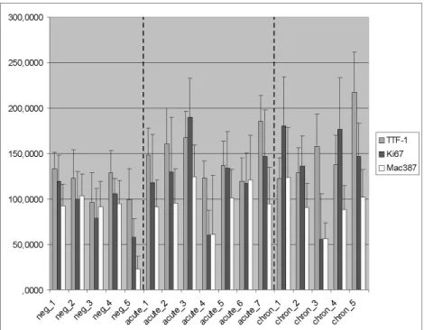
The analysis of the immunohistochemically stained slides revealed that humanized anti-TTF-1 antibodies successfully identify porcine type II pneumocytes, and Clara cells in the terminal bronchioli. Marked proliferation of these cells was confirmed by a significant (p<0.05) increase of TTF-1 positive cells in acute cases. Upregulation of Ki67 and MAC387 positive cells was also observed, however due to the relative low number of the sample animals and high values of standard deviation, the increase of these values were found not to be statistically significant (Figure 1.)
Cytokeratin labeling clearly identified the different epithelial cell types: type I, and type II pneumocytes as well as bronchial epithelial cells, however this staining was not suitable for cell counting purposes. Type I pneumocytes were flattened cells lining the alveolar spaces providing surface for gas exchange. In contrast type II pneumocytes were
rounded cells usually located at the insertion of the alveolar spaces in control lungs. In infected animals, as verified by TTF-1 staining as well, these rounded cells were found in markedly higher numbers.
Figure 1. Mean values and standard deviation of TTF-1, Ki67, and Mac387 labeled cells is shown in the figure. Results were calculated from the positive cell numbers counted in 50 non
overlapping microscopic fields. Dashed lines separate the three different (control, acute and chronic) groups.
When scores obtained from the routine HE stained histological slides were compared to the number of immunohistochemically positive cells by Pearson's chi-square test, from the three different antibodies Ki67 cell counts were found to show positive correlation (p<0.05) with the overall severity of the lesions.
The relatively high variation of the immunohistochemical mean values among the different animals of the PRRSV infected groups indicates great individual differences in the response to the infection both in terms of macrophage infiltration and pneumocyte proliferation. The high standard deviation values of the individual cases point the differences of the positive cell numbers in the different microscopic fields. This feature is in harmony with the routine histological findings, different areas within the same slide show great differences in the severity of the lesions.
Acknowledgements
This paper was supported by the János Bolyai Research Scholarship of the Hungarian Academy of Scienes and by the TÁMOP-4.2.2.B-10/1-2010-0011 „Development of a complex educational assistance/support system for talented students and prospective researchers at the Szent István University” project.
References
Bohinski, R.J., DiLauro, R., and Whitsett, J.A.: Lung-specific surfactant protein B gene promoter is a target for thyroid transcription factor 1 and hepatocyte nuclear factor 3 indicating common mechanisms for organ-specific gene expression along the foregut axis. Mol Cell Biol 14, 5671-5681. 1994.
Caswell, C. L. and Williams, K. J.: Respiratory system. In Jubb, Kennedy, and Palmer’s Pathology of Domestic Animals. Edited by Maxie, M. G. saunders, Elsevier. 2007.
Halbur, P.G., Paul, P.S., Meng, X-J., Lum, M.A, Andrews, J.J. and Rathje J.A.: Comparative pathogenicity of nine US porcine reproductive and respiratory syndrome virus (PRRSV) isolates in a five-week-old cesarean-derived, colostrum-deprived pig model.
J Vet Diagn Invest 8, 11-20. 1996.
Ray, M.K., Chen, C.Y., Schwarts, R.J., and DeMayo, F. J.: Transcription regulation of a mouse Clara cell-specific protein (mCC10) gene by the Nkx transcription factor family members thyroid transcription factor 1 and cardiac muscle–specific homeobox protein (CSX). Mol Cell Biol 16, 2056-2064. 1996.
Rossow, K.D.: Porcine reproductive and respiratory syndrome. Vet Pathol 35, 1-20. 1998.
Tan, D., Li, Q., Deeb, G., Ramnath, N., Slocum, H.K., Brooks J., Cheney R., Wiseman S., Anderson T., and Loewen G.. Thyroid transcription factor-1 expression prevalence and its clinical implications in non-small cell lung cancer: a high-throughput tissue microarray and immunohistochemistry study. Hum Pathol 34, 597-604. 2003.
Zhou, L., Lim, L., Costa, R.H., and Whitsett, J.A. Thyroid transcription factor-1 hepatocyte nuclear factor-3beta, surfactant protein B, C, and Clara cell secretory protein in developing mouse lung. J Histochem Cytochem 44, 1183-1193. 1996.