DOI: 10.1556/066.2018.47.2.10
PREPARATION, CHARACTERIZATION AND ANTI-AGEING ACTIVITY OF GASTRODIA ELATA BLUME POLYSACCHARIDE
L. CHENa,c,d,*, Y.P. ZHANGb and L.X. JINa,c,d
aJiangsu Marine Resources Development Research Institute, Huaihai Institute of Technology, 59 Cangwu Road, 222005, Haizhou. China
bShcool of Chemical Engineering, Dalian University of Technology, 2 Linggong Road, Ganjingzi, 116024, Dalian.
China
cJiangsu Key Laboratory of Marine Pharmaceutical Compound Screening, Huaihai Institute of Technology, 222005, Haizhou. China
dCo-Innovation Center of Jiangsu Marine Bio-industry Technology, Huaihai Institute of Technology, 222005, Haizhou. China
(Received: 16 September 2017; accepted: 25 January 2018)
Gastrodia elata Blume polysaccharide (GEP) was extracted and then chemically characterised. Its antioxidant activity was evaluated in vitro and in vivo. The results of the in vitro investigation show that GEP consists of glucose with molecular weight of 875 185 Da and exhibits high hydroxyl radical scavenging, as well as 2,2-diphenyl-1- picrylhydrazyl activity and reducing capacity. For antioxidant activity in vivo, D-galactose-induced-aged mice were orally administered with three different doses of GEP over a period of 6 weeks. The administration of GEP dose- dependently increased the body weight gain rates, liver and brain indices, superoxide dismutase and glutathione peroxidase activities, and malondialdehyde levels in the sera and brains of ageing mice. These results suggest that GEP exhibits high antioxidant activity and can retard human ageing associated with free radicals.
Keywords: Gastrodia elata Blume, polysaccharide, antioxidant activity, anti-ageing activity
Gastrodia elata Blume (G. elata Bl) belongs to the genus Gastrodia, family Compositae, and is a widely used traditional Chinese herb. G. elata Bl rhizomes are typically used as anticonvulsants, analgesics, sedative, and agent against general paralysis, epilepsy, vertigo, and tetanus (TANG & EISENBRAND, 1992; JIANG et al., 2010; KWON et al., 2014; LI et al., 2015).
G. elata Bl rhizomes have high water-soluble polysaccharide contents of up to 9.61%
(LIU et al., 2009). CHEN and co-workers (2011) extracted a water-soluble polysaccharide from G. elata Bl rhizomes by using α-(1→4) glucan with α-(1→4) glucosyl branches attached to the O-6 of branching points. MING and co-workers (2012) also prepared G. elata Bl polysaccharide mainly containing glucose molecules with (1→4)-α-D-glucan main chains that are occasionally branched with a-1,6 glycosidic linkages. G. elata Bl polysaccharides have many important biological activities, such as enhancing immunity, up-regulating protein expressions, disrupting pancreatic cancer cell growth, lowering fat levels, free radical scavenging, preventing hypertension, and protecting the retina (MIAO & SHEN, 2006; LIU et al., 2009; MING et al., 2012). However, the anti-ageing activities of the polysaccharide from G. elata Bl polysaccharide (GEP) are rarely reported.
In this study, GEP was extracted with hot water, and the anti-ageing activities of GEP in rats were investigated.
1. Materials and methods
1.1. Ethics statement
This study was approved by the ethics committee of Huahai Institute of Technology, China (ECHHIT20150528). All procedures were performed in compliance with relevant laws and institutional guidelines.
1.2. Materials
Dried G. elata Bl rhizomes were purchased from Shandong Dingli Rubber Industry Co., Ltd.
(Shandong, China). Standard monosaccharide samples (glucose, xylose, and mannose) were obtained from Sigma Chemical Co. (St. Louis, MO, USA). All other reagents were of analytical grade.
1.3. GEP preparation
Dried G. elata Bl rhizomes were pulverised and sifted through an 80-mesh sieve to yield fi ne powder. The obtained powder was soaked in distilled water under agitation at room temperature (~20 °C) for 1 h to produce a suspension with ~1% (w/v) concentration. The resulting suspension was placed in a thermostatic water bath at 90 °C for 4 h and then centrifuged at 5000 × g for 15 min. The proteins in the resulting supernatant were separated with the Sevag method, precipitated with three volumes of absolute ethanol, fi ltered using a Whatman GF/A fi lter paper, and fi nally freeze-dried.
1.4. GEP characterisation
The total sugar, protein, and ash contents in the products were determined with phenol–
sulphuric acid colorimetric method, Kjeldahl method, and the method described by HOU
(2004), respectively. The Fourier transform infrared (FTIR) spectra of the representative product samples were obtained from the KBr pellets by using a Nicolet Nexus FTIR 470 spectrophotometer over a wavelength range of 400–4000 cm–1. UV spectra were recorded on a UV spectrometer (Spectra Test, Germany). The molecular weights (MWs) of the GEP were determined by high performance gel fi ltration chromatography (LC-10A, Shimadzu, Japan) in which an Ultrahydrogel size exclusion column (LKB-Prodokter, AB, Bromma, Switzerland) and high-sensitive refractive index detector, Model ERC-7515 A (ERC Inc., Japan) were used. The GEP were eluted with 0.1 N NaNO3 at a fl ow rate of 0.9 ml min–1. Pullulan standards (P20–P800, JM Science, Inc., NY, USA) were used as molecular weight standard. The GEP was hydrolysed according to the methods described by SHENG and co-workers (2007). The monosaccharide compositions of the obtained AMP were assayed by and ion chromatograph (ICS-5000, Dionex, USA) in which a carbohydrate Column (CarboPac PA20, Dionex, USA) and pulse amperes detector (Dionex, USA) were used. The monosaccharides of AMP were eluted with mobile phase at a fl ow rate of 0.5 ml min–1. The composition and conditions of mobile phase are as follows: 0–21.1 min (97.4% of water and 2.6% of 250 mM NaOH);
21.1–30 min (92.4% of water, 2.6% of 250 mM NaOH, and 5.0% of NaAc); 30–50 min (20%
of water and 80% of 250 mM NaOH).
1.5. Animals and treatment
A total of 60 Kunming mice were purchased from Tumor Hospital Experimental Animal Center of Nanjing Medical University. The Kunming mice (30 males and 30 females) were 8 weeks old and had an average weight of 19–21 g. The mice were maintained under the following environmental conditions: temperature of 25 °C, humidity of 50%, and light conditions of 12:12 h light: dark cycle for 1 week before the experimental protocol. During the experimental period, the mice were provided with rodent laboratory chow and tap water ad libitum.
The mice were randomly divided into fi ve groups. In particular, 12 mice were included in the normal control group (NCG), 12 mice in the D-galactose (D-gal) model control group (MCG), and 12 mice each in the 1, 2, and 3 g kg–1 GEP-treated groups. For the establishment of the ageing model mice, D-gal saline (0.9%) solution with a dose of 100 g kg–1 body weight was injected into the back of each mouse once a day for 6 weeks. For the GEP-treated groups, the mice were intragastrically administrated with GEP with doses of 1, 2, or 3 g kg–1 BW day–1. For the NCG, the mice received subcutaneous injection of sterile saline instead of the same volume ofD-gal solution. For the MCG group, the mice received normal diet without GEP.
One day after the last drug administration, the mice were weighed and sacrifi ced through cervical dislocation. Blood samples from the retrobulbar venous plexus of the sacrifi ced mice were centrifuged at 10 000 × g and 4 °C for 15 min to yield blood sera. The sera were stored at −80 °C for further assay. The brains and livers were removed immediately, weighed and mixed with 0.9% ice-cold NaCl solution to yield 10% homogenates, which were centrifuged at 4000 × g and 4 °C for 15 min. The resulting supernatants were kept at −80 °C for biochemical analysis.
1.6. Antioxidant activities
The reducing capacity of GEP was determined according to the methods of QIAO and co- workers (2009). GEP (1 g) was mixed with 1.0 ml of 0.2 M phosphate buffer (pH 6.6) and 1.0 ml of 1% (w/v) potassium ferricyanide, and then incubated at 50 °C for 20 min before it was cooled at room temperature. Briefl y, 1 ml of trichloroacetic acid (10%, w/v) and 0.2 ml of fresh ferric trichloride (0.1%, w/v) were added to the reaction mixture. The resulting mixture was shaken, and its absorbance was assayed at 700 nm against a blank sample (water instead of GEP) after 10 min. The absorbance of the reaction mixture indicates the reducing capacity of the sample.
Reducing capacity = (A1 − A2) (1) where A1 is the absorbance of the sample and A2 is the absorbance of the sample under conditions similar to those of A1 except that water was used instead of ferric trichloride solution.
The hydroxyl radical scavenging activity (HRSA) of the GEP was determined according to the previously described methods (ANDREWS, 1986). The HRSA of GEP was calculated as follows:
A1 – A2
HRSA (%) = ________×100 (2) A – A
2,2-Diphenyl-β-picrylhydrazyl (DPPH) radical scavenging activity (DRSA) was assayed according to the method of QIAO and co-workers (2009). Briefl y, 0.2 ml of 400 μmol l–1 DPPH free radical (DPPH•) in dehydrated alcohol was added to 1.0 ml of GEP solution.
Afterwards, 2.0 ml of distilled water was added to the mixture. The resulting mixture was shaken and allowed to stand at room temperature in the dark for 30 min. The absorbance was assayed at 517 nm against a blank standard (distilled water instead of GEP and DPPH•
solutions). Low absorbance corresponded to high free-radical scavenging activity. The percentage of DRSA was calculated by using the following equation:
A0 – (A1 – A2)
DRSA (%)= _____________ × 100 (3) A0
where A0 is the absorbance of the control (distilled water instead of GEP), A1 is the absorbance of the sample, and A2 is the absorbance of the sample obtained under conditions similar to those in A1 except that distilled water was used instead of DPPH• solution.
1.7. Anti-ageing activities
The activities of superoxide dismutase (SOD) and glutathione peroxidase (GSH-Px) and level of malondialdehyde (MDA) in the supernatants of the tissue homogenates and sera were determined using commercially available kits. The assays were performed at 4 °C and according to the instructions provided by the manufacturers of the kits.
1.8. Statistical analysis
Data were presented as mean±SD, and ANOVA was performed to compare the means of the two groups. Statistical signifi cance at 95% probability level was set at P<0.05.
2. Results and discussion
2.1. Product characterisation
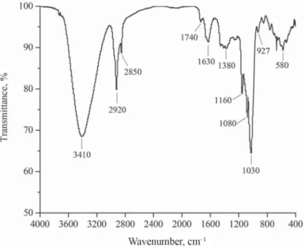
Total sugar, protein, and ash contents in the GEP product were 91.3%, 5.9%, and 2.7%, respectively, indicating that the GEP could be glycoproteins. The GEP samples were water- soluble white powders. The FTIR spectra of the GEP exhibited peaks at ~3410 cm–1 (O–H),
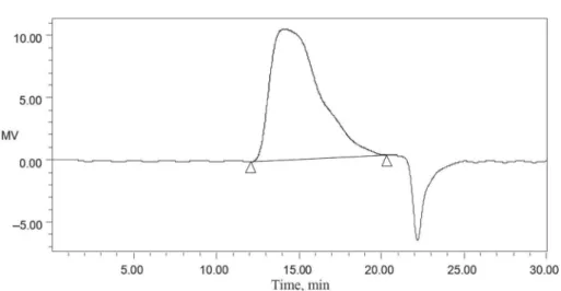
~1380 cm–1 (symmetrical deformations of –CH3 and –CH2), ~1030 cm–1 (stretching vibrations of the C–O–C molecules in the glucose circle), and ~1630 cm–1 (special absorbance peaks of the aldehydes in the GEP) (Fig. 1). The UV spectra of the GEP products exhibited two peaks at 200–280 nm (Fig. 2), indicating that the GEP product contains proteins. By contrast, the HPLC spectra of the GEP products displayed only one peak at 14.10 min, indicating that the GEP only contained one type of polysaccharide with MW of 107845 Da (Fig. 3). Furthermore, the result of monosaccharide composition analysis with gas chromatography indicated that the GEP only contained glucose (Fig. 4), and thus is consistent with the fi ndings of previous reports (CHEN et al., 2011; MING et al., 2012).
Fig. 1. FT-IR spectra of Gastrodia elata Blume polysaccharide
Fig. 3. HPLC spectra of Gastrodia elata Blume polysaccharide
Fig. 4. IC spectra of the sugar composition of Gastrodia elata Blume polysaccharide : sample; : standard
2.2. Antioxidant activity of GEP
Hydroxyl radical (HO•) has the highest activity among free radicals, and DPPH• is a relatively stable radical. Both types of free radicals can attack biological molecules, such as lipids, proteins, enzymes, DNA, and RNA, leading to cell or tissue injury (QIAO et al., 2009; ZHANG
et al., 2017). Furthermore, they are widely used to evaluate the scavenging activity of some antioxidants (YAO et al., 2013). The HRSA and DRSA of the GEP are shown in Figure 5A.
The scavenging activities of GEP increased dose-dependently. At 200 mg ml–1 concentration, the HRSA and DRSA of the GEP were 94.56% and 84.21%, respectively, indicating that the GEP had high radical-scavenging activities. Reducing capacity is positively correlated to antioxidant activity and is thus important in the evaluation of antioxidant activities (DUH et al., 1999). The reducing capacity of GEP is shown in Figure 5B. At concentrations of 6.25, 12.5, 25, 50, 100, and 200 mg ml–1, the absorbance values were 0.16, 0.44, 0.62, 0.77, and 0.87, respectively. These data suggested the role of reducing capacity in antioxidation.
0 20 40 60 80 100 120
6.25 12.5 25 50 100 200
HRSA, DRSA , %
Concentration , mg ml–1
0 0.2 0.4 0.6 0.8 1 1.2
6.25 12.5 25 50 100 200
Absorbance (700 nm)
Concentration, mg ml–1
B A
2.3. Anti-ageing activity of GEP
As shown in Table 1, the gains in body weights of and amounts of food consumed by mice in the intragastric D-gal administration group decreased signifi cantly compared with those in the normal control group (P<0.05). However, the intragastric administration of GEP dose- dependently increased body weight gain and food intake of each mouse (P<0.05). The intragastric administration of the D-gal group showed signifi cantly decreased liver and brain indices compared with the normal control group (P<0.05). Nevertheless, the liver and brain indices of mice in GEP-treated groups were higher than those in the MCG (P<0.05). Similarly, LI and co-workers (2016) reported that GEP up-regulated the expressions of brain-derived neurotrophic factor and stem cell factor protein in caudate putamen of focal cerebral ischemia rats. These fi ndings indicated that GEP can signifi cantly improve the liver and brain indices of mice.
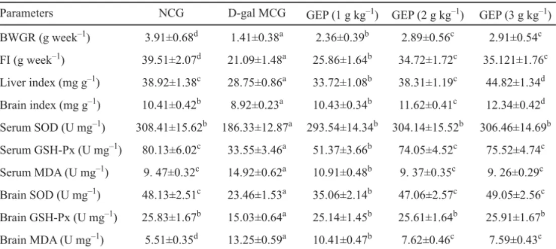
Table 1. Body weight gain rate (BWGR), food intake (FI), liver and brain indices, activities of SOD and GSH-Px, and level of MDA in the serum and brain of ageing mice for normal control group (NCG), D-gal model control
group (MCG) and Gastrodia elata polysaccharide (GEP).
Parameters NCG D-gal MCG GEP (1 g kg–1) GEP (2 g kg–1) GEP (3 g kg–1) BWGR (g week–1) 3.91±0.68d 1.41±0.38a 2.36±0.39b 2.89±0.56c 2.91±0.54c FI (g week–1) 39.51±2.07d 21.09±1.48a 25.86±1.64b 34.72±1.72c 35.121±1.76c Liver index (mg g–1) 38.92±1.38c 28.75±0.86a 33.72±1.08b 38.31±1.19c 44.82±1.34d Brain index (mg g–1) 10.41±0.42b 8.92±0.23a 10.43±0.34b 11.62±0.41c 12.34±0.42d Serum SOD (U mg–1) 308.41±15.62b 186.33±12.87a 293.54±14.34b 304.14±15.52b 306.46±14.69b Serum GSH-Px (U mg–1) 80.13±6.02c 33.55±3.46a 51.37±3.66b 74.05±4.52c 75.52±4.74c Serum MDA (U mg–1) 9. 47±0.32c 14.92±0.62a 10.91±0.48b 9. 37±0.35c 9. 26±0.29c Brain SOD (U mg–1) 48.13±2.51c 23.46±1.53a 35.06±2.14b 47.06±2.57c 49.05±2.56c Brain GSH-Px (U mg–1) 25.83±1.67b 15.03±0.64a 25.14±1.45b 25.61±1.64b 25.91±1.67b Brain MDA (U mg–1) 5.51±0.35d 13.25±0.59a 10.41±0.47b 7.62±0.46c 7.59±0.43c Values are expressed as mean±SD (n=3). Means with different superscripts within a column indicate signifi cant differences (P<0.05).
SOD catalyses the superoxide radicals to form hydrogen peroxide, which is subsequently decomposed to water and oxygen by GPx; this decomposition process prevents damage to membrane and biological structures (LV et al., 2007). The SOD and GSH-Px activities in organisms decrease irreversibly with ageing; therefore, both SOD and GSH-Px are widely used to evaluate ageing (MAHMOUD & EDENS, 2003).
MDA is one of the most known secondary products of lipid peroxidation, and typically used as indicator of oxidative damage and as ageing evaluation index; high MDA level indicates increased lipid peroxidation and oxidative damage (ROTRUCK et al., 1973; BAGCHI et al., 1995). The MDA levels in the sera and brains of the model mice signifi cantly increased, whereas the SOD and GSH-Px activities decreased signifi cantly compared with those of the normal group (Table 1, P<0.01). Intracellular free radicals in neurons accumulate with ageing, and this phenomenon decreases the ability of antioxidant defences, accelerates the
chain reaction of lipid oxidation and exacerbates neuroinfl ammation (WU et al., 2017). In the present study, the intragastricadministration of GEP signifi cantly reduced the MDA level but signifi cantly increased the SOD and GSH-Px activities in the sera and brains of D-gal induced ageing mice as compared with those of the model group (Table 1, P<0.05). These results suggested that GEP can effectively suppress oxidation-induced damage to the sera and brain tissues of D-gal-induced mice; this could be due to the antioxidant activities of GEP.
3. Conclusions
We prepared GEP through hot water extraction and investigated its antioxidant and anti- ageing activities. The results showed that GEP exhibits high antioxidant activity in vitro and anti-ageing activities in D-gal-induced mice. The present results verifi ed that GEP possesses high antioxidant and anti-ageing activities in D-gal-treated mice by facilitating antioxidant activities and attenuating lipid peroxidation.
*
This study was supported by the Open project of Jiangsu Marine Resources Development Research Institute (HY201601), Lianyungang science and technology planning project (JC1614), and the Priority Academic Program Development of Jiangsu Higher Education Institutions.
References
ANDREWS, A.T. (1986): Electrophoresis: Theory, techniques and biochemical and clinical applications. Clarendon, Oxford, pp. 53–75.
BAGCHI, D., BAGCHI, M., HASSOUN, E.A. & STOHS, S.J. (1995): In-vitro and in-vivo generation of reactive oxygen species DNA damage and lactate dehydrogenase leakage by selected pesticides. Toxicology, 104, 129–140.
CHEN, X., CAO, D., ZHOU, L., JIN, H., DONG, Q., YAO, J. & DING, K. (2011): Structure of a polysaccharide from Gastrodia elata Bl., and oligosaccharides prepared thereof with anti-pancreatic cancer cell growth activities.
Carbohydr. Polym., 86, 1300–1305.
DUH, P.D., DU, P.C. & YEN, G.C. (1999): Action of methanolic extract of mung bean hulls as inhibitors of lipid peroxidation and non-lipid oxidative damage. Food Chem. Toxicol., 37, 1055–1061.
HOU, M.L. (2004): Food analysis. Chemical Industry Press, Beijing, China. (in Chinese).
JIANG, Y.W., LEE, J.Y. & KIM, C.J. (2010): Anti-asthmatic activity of phenolic compounds from the roots of Gastrodia elata Bl. Int. Immunopharmacol., 10, 147–154.
KWON, J., KIM, N., LEE, D., HAN, A., LEE, J.W., SEO, E., LEE, J. & LEE, D. (2014): Metabolomics approach for the discrimination of raw and steamed Gastrodia elata using liquid chromatography quadrupole time-of-fl ight mass spectrometry. J. Pharm. Biomed. Anal., 94, 132–138.
LI, H.B., WU, F., MIAO, H.C. & XIONG, K.R. (2016): Effects of polysaccharide of Gastrodia elata Blume and electro- acupuncture on expressions of brain-derived neurotrophic factor and stem cell factor protein in caudate putamen of focal cerebral ischemia rats. Med. Sci. Monit. Basic. Res. 22, 175–180.
LI, Z.F., WANG, Y.W., OUYANG, H., LU, Y., QIU, Y., FENG, Y.L., JIANG, H.L., ZHOU, X. & YANG, S.L. (2015): A novel dereplication strategy for the identifi cation of two new trace compounds in the extract of Gastrodia elata using UHPLC/Q-TOF-MS/MS. J. Chromatogr. B., 988, 45–52.
LIU, M.X., LI, Q.F., LIU, Q., HUANG, Z.Q. & QIU, F. (2009): Study on extraction technology, structure and free radical scavenging activity of polysaccharides from Gastrodia elata B1. Food Sci., 30, 29–32. (in Chinese).
LV, L.S., GU, X.H., TANG, J. & HO, C.T. (2007): Antioxidant activity of stilbene glycoside from Polygonum multifl orum Thunb in-vivo. Food Chem., 104, 1678–1681.
MAHMOUD, K.Z. & EDENS, F.W. (2003): Infl uence of selenium sources on age-related and mild heat stress-related changes of blood and liver glutathione redox cycle in broiler chickens (Gallus domesticus). Comp. Biochem.
Phys. B., 136, 921–934.
MING, J., LIU, J., WU, S., GUO, X., CHEN, Z. & ZHAO, G. (2012): Structural characterization and hypolipidemic activity of a polysaccharide PGEB-3H from the fruiting bodies of Gastrodia elata Blume. Procedia Eng., 37, 169–173.
MIAO, H.C. & SHEN, Y.S. (2006): Antihypertensive effect of polysaccharides subtracted from Gastrodia elata Blume.
Chinese J. Hypertens., 14, 531–534. (in Chinese).
QIAO, D.L., KE, C.L., HU, B., LUO, J.G., YE, H., SUN, Y., YAN, X.Y. & ZENG, X.X. (2009): Antioxidant activities of polysaccharides from Hyriopsis cumingii. Carbohydr. Polym., 78, 199–204.
ROTRUCK, J.T., POPE, A.L., GANTHER, H.E., SWANSON, A.B., HAFEMAN, D.G. & HOEKSTRA, W.G. (1973): Selenium:
Biochemical role as a component of glutathione peroxidase. Science, 179, 588–590.
SHENG, J., YU, F., XIN, Z., ZHAO, L., ZHU, X. & HU, Q. (2007): Preparation, identifi cation and their antitumor activities in vitro of polysaccharides from Chlorella pyrenoidosa. Food Chem., 105, 533–539.
TANG, W. & EISENBRAND, G. (1992): Gastrodia elata Bl. -in: TANG, W. & EISENBRAND, G. (Eds) Chinese drugs of plant origin: Chemistry, pharmacology, and use in traditional and modern medicine. Springer, Berlin, pp. 545–547.
WU, S.J., LU, M.S. & WANG, S.J. (2017): Anti-aging activities of the water soluble chitosan from Clanis bilineata larvae. Int. J. Biol. Macromol., 102, 376–379.
YAO, X.C., CAO, Y. & WU, S.J. (2013): Antioxidant activity and antibacterial activity of peach gum derived oligosaccharides. Int. J. Biol. Macromol., 62, 1–3.
ZHANG, C., GAO, Z., HU, C.L., ZHANG, J.J., SUN, X.Y., RONG, C.B. & JIA, L. (2017): Antioxidant, antibacterial and anti-aging activities of intracellular zinc polysaccharides from Grifola frondosa SH-05. Int. J. Biol. Macromol., 95, 778–787.