724
Fructose-1,6-diphosphate Aldolase
Friedrich H. Bruns and Hans-Ulrich Bergmeyer
Aldolase catalyses the reaction ^:
(1) Fructose-1,6-diphosphate
v
' dihydroxyacetone phosphate + D-glyceraldehyde- 3-phosphate It also catalyses the reaction between dihydroxyacetone and the following aldehydes: acetaldehyde, D-glyceraldehyde, L-glyceraldehyde2
\ glycolaldehyde and glycolaldehyde-2-phosphate
3
), to give the corresponding phosphorylated derivatives, methyltetrose phosphate, D-fructose-1-phosphate, L-sor- bose-1-phosphate, D-xylulose-1-phosphate and D-xylulose-1,5-diphosphate, respectively. T h e cry
stalline enzyme obtained from bovine liver catalyses the cleavage of D-fructose-1-phosphate and the synthesis of erythrulose phosphate from dihydroxyacetone phosphate at about the same r a t e
4 )
, which contradicts the postulated existence of special enzymes for these r e a c t i o n s
5 - 7
) . Crystalline aldolase has been prepared from rat
8
) and r a b b i t
9
.
1 0
) muscle and also from y e a s t
1 1
) . In contrast to the enzyme from mammalian tissue the enzymes from yeast
8
), Clostridiumperfringens
12
) and Aspergillus niger
n
) require divalent metal ions.
Skeletal muscle has the highest concentration of aldolase (Table 1). In this tissue the enzyme accounts for ca. 1 0 % of the soluble protein.
Table 1. Aldolase activity in rat tissues
1 4
) (ul. fructose-1,6-diphosphate/g. fresh weight/hr. at 38° C)
Tissue Activity Tissue Activity
Skeletal muscle 74800 Parotid 3 800
Brain 15 800 Stomach 3 700
Heart muscle 15600 Bladder 3 2 0 0
Liver 12100 Placenta 3000
Bone marrow 9 5 0 0 Testicle 2 9 0 0
Adrenal 8 600 Lung 2800
Kidney 7 8 0 0 Uterus 2 1 0 0
Spleen 4 8 0 0 Erythrocytes 900
Thyroid 4 8 0 0 Pancreas 500
Thymus 4 7 0 0 Adipose tissue 400
Prostate 4 4 0 0 Serum 60
The activity of the enzyme in serum is comparatively low; particularly low values are found in human serum (Table 2).
D O. Meyerhof and K. Lohmann, Biochem. Z. 271, 89 [1934]; 273, 73, 413 [1934]; Naturwissenschaf
ten 22, 134, 220, 452 [1934].
2) O. Meyerhof, K. Lohmann and P. Schuster, Biochem. Z. 286, 301 [1936].
3
> W. L. Byrne and H. A. Lardy, Biochim. biophysica Acta 14, 495 [1954].
4
) R. J. Peanasky and H. A. Lardy, J. biol. Chemistry 233, 365, 371 [1958].
s) U. Kaletta-Gmiinder, H. P. Wolf and F. Leuthardt, Helv. chim. Acta 40, 1027 [1957].
6) F. Leuthard, E. Testa and H. P. Wolf, Helv. chim. Acta 36, 227 [1952].
Ti F. C. Charalampous and G. C. Mueller, J. biol. Chemistry 201, 161 [1953].
8) O. Warburg and W. Christian, Biochem. Z. 314, 149 [1943].
9
) /. F. Taylor, A. A. Green and G. T. Cori, J. biol. Chemistry 173, 591 [1948].
10) G. Beisenherz, H. J. Boltze, Th. Biicher, R. Czok, K. H. Garbade, E. Meyer-Arendt and G. Pflei
derer, Z. Naturforsch. 8b, 555 [1953].
") O. Warburg and K. Gawehn, Z. Naturforsch. 9b, 206 [1954].
12) R. C. Bard and /. C. Gunsalus, J. Bacteriol. 59, 387 [1950].
13) V. Jagannathan and K. Singh, Biochim. biophysica Acta 15, 138 [1954].
14
) /. A. Sibley and A. L. Lehninger, J. biol. Chemistry 777, 859 [1949].
11.1.a
Fructose-1,6-diphosphate Aldolase 725Table 2. Aldolase activity in the sera of different species
1 5
* (u,l. fructose- 1,6-diphosphate/ml. serum/hr. at 37° C)
Species Activity
Mean Range
Man D o g Horse Chicken Ox Sheep Guinea pig Rat Pig Rabbit Mouse
15 22 35 40 40 42 44 47 54 100
5.4 3.8
12 10 17 24 30 17 28 33 48 65
•8.0
•22 46 70 58 65 65 65 61 68 120
Four methods are available for the assay of enzyme activity by measuring the hydrolysis of fructose- 1.6-diphosphate:
1. determination of the alkali labile triose phosphate formed
1
*;
2. colorimetric estimation of the triose phosphate formed by the method originally described for l a c t a t e
1 6
.
1 7
) ;
3. measurement of activity by the spectrophotometric method of Warburg%>
[
°);
4. another colorimetric method in which the dinitrophenylhydrazones o f the free trioses are deter
mined 14, 19, 20).
The last two methods are described here.
A. Colorimetric Determination with 2,4-Dinitrophenylhydrazine
The triose phosphates (dihydroxyacetone phosphate and D-glyceraldehyde phosphate) formed from fructose-1,6-diphosphate by the action of aldolase are trapped with hydrazine. After deproteinization with trichloroacetic acid they are hydrolysed by N a O H . The free trioses are treated with 2,4-dinitro- phenylhydrazine, yielding a mixture of methylglyoxal-2,4-dinitrophenylosazone and pyruvic acid- 2,4-dinitrophenylhydrazone
1 8
). Both these compounds dissolve in alkali forming a red dye with an absorption maximum between 535 and 540 m\i.
The following d e s c r i p t i o n
1 9
, 20) j
s a
modification of the method of Sibley and Lehningerl4
K
Optimum Conditions for Measurements
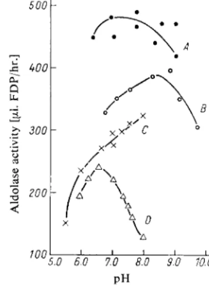
With collidine buffer the aldolase activity of serum, blood haemolysates and muscle homogenates has a broad p H optimum between 7 and 8, while with veronal buffer it is between 8.5 and 9. Phosphate and borate inhibit the enzyme; the latter reacts with the c/s-hydroxyl groups of the substrate
2 1
) (Fig. 1). In the presence of cyanide, crystalline aldolase from bovine liver has a p H optimum between 9.1 and 9.4 (glycylglycine-NaOH buffer) with fructose-1,6-diphosphate and between 8.1 and 8.4 with fructose-1-phosphate. However, it should be noted that the pH optimum measured in the pre-
15
> F. Bruns and Chr. Kirschner, Naturwissenschaften 41, 141 [1954].
16
) S. B. Barker and W. H. Summerson, J. biol. Chemistry 138, 535 [1941].
1 7 )
A. L. Dounce and G. Thannhauser-Beyer, J. biol. Chemistry 173 159 [1948].
i*> W. S. Beck, J. biol. Chemistry 212, 847 [1955].
19) F. H. Bruns, Biochem. Z. 325, 156 [1954].
20) F. H. Bruns and W. Puis, Klin. Wschr. 32, 656 [1954].
21) F. H. Bruns, Biochem. Z. 325, 429 [1954].
Principle
726 Section C: Measurement of Enzyme Activity
sence of ketone and aldehyde trapping agents is influenced by the dependence of the trapping reaction on p H . With fructose-1,6-diphosphate concentrations above 0.005 M the serum enzyme is saturated with substrate (KM = 0.75 X 1 0
-3
M). Even with greatly increased serum aldolase activity, as is found in certain pathological conditions, the rate of hydrolysis under the given assay conditions is directly proportional to the time and enzyme concentration (amount of serum). For animal sera and h o m o genates of liver, kidney, etc. this is only true after suitable dilution. M a x i m u m rates are obtained with serum at 46° C. A t 37° C the serum enzyme is not denatured, while crystalline muscle aldolase is partially inactivated above 3 0 ° C
1 7
> .
Reagents*)
1. Fructose-1,6-diphosphate, FDP
dibarium salt (ca. 6 5 % F D P- B a 2 ) ; commercial preparation, see p. 1014.
2. Sodium sulphate, anhydrous 3. Hydrochloric acid, A. R., 2 N 4. Sodium hydroxide, A. R., I N 5. Collidine (2,4,6-trimethylpyridine)
the liquid should be colourless; if not, it must be distilled; b. p. 171°C, sp. gr. 0.917.
6. Hydrazine sulphate 7. 2,4-Dinitrophenylhydrazine 8. Trichloroacetic acid Preparation of Solutions
I. Fructose-1,6-diphosphate (ca. 0.1 M FDP):
In a centrifuge tube suspend 3 g. fructose-1,6-diphosphate (dibarium salt) in ca. 20 ml.
distilled water and slowly add 2 N HC1 drop wise until complete solution is obtained.
Dissolve 2 g. sodium sulphate in 5 ml. distilled water and add to the FDP solution.
Stir and centrifuge off the precipitate of BaSC >4. Adjust the clear, usually yellowish
500
Fig. 1. pH-Activity curves for aldolase (human haemolysate 1 :10). Final concen
tration of the buffer: 0.033 M ; A) collidine buffer; B) veronal buffer; C) phosphate buffer; D ) borate buffer.
100
5.0 6.0 7.0 8.0 9.0 10.0 PH*) Complete reagent kits are commercially available (see p. 1036).
U.1.a Fructose-1,6-diphosphate Aldolase 727
supernatant to pH 7.4 by the dropwise addition of 1 N NaOH (pH-meter). Dilute the solution to 30 ml. with distilled water. This solution is approximately 0.1 M with respect to FDP.
II. Collidine buffer (0.1 M; pH 7.4):
Dissolve 4.84 g. ( = 5.28 ml.) collidine in distilled water and make up to 100 ml. To 25 ml. of this stock solution add 45 ml. 0.1 N HC1 and dilute to 100 ml. with distilled water. If necessary, adjust to pH 7.4 with dilute H Q or NaOH.
III. Hydrazine (0.56 M):
Dissolve 9.08 g. hydrazine sulphate in ca. 70 ml. distilled water and add ca. 5 g. solid NaOH. Add 10N NaOH until pH 7.4 is obtained (pH-meter) and dilute to 100 ml.
with distilled water.
IV. 2,4-Dinitrophenylhydrazine (0.1 % w/v):
Grind 250 mg. 2,4-dinitrophenylhydrazine in a mortar with small portions of 2 N HC1, filter and dilute to 250 ml. with 2 N HC1.
V. Trichloroacetic acid (10% w/v):
Dissolve 10 g. trichloroacetic acid in distilled water and make up to 100 ml.
VI. Sodium hydroxide (0.75 N):
Dissolve 3 g. (dry!) NaOH in distilled water and make up to 100 ml. Alternatively dilute 75 ml. 1 N NaOH to 100 ml. with distilled water.
VII. Collidine-hydrazine buffer:
Mix 100 parts collidine buffer (solution II) with 25 parts hydrazine solution (III) and 50 parts distilled water.
Stability of the solutions
Store all solutions, with the exception of IV and VI, in a refrigerator at 0 to 4° C. The collidine buffer will become acid with time due to evaporation of the collidine and therefore, if necessary, it must be re-adjusted to p H 7.4 with dilute N a O H .
Buffer, substrate and hydrazine solutions should be renewed after 2 to 3 weeks. Bacterial contamin
ation can be prevented by the addition of a few drops of chloroform or toluene.
Procedure
Use only fresh serum free from haemolysis.
Colorimetric m e a s u r e m e n t s
Wavelength: 540 mu. (absorption maximum of the colour) or an adjacent wavelength, e.g.
Zeiss filter S 53 E or 546 mu. Eppendorf photometer; light path: 1 cm.; final volume: 11 ml.;
temperature: 37°C. Measure against the control tube.
Pipette successively into test tubes:
Experimental Control
serum
collidine-hydrazine buffer (solution VII) FDP solution (I)
Incubate for 60 min. at 37° C (water bath). Add trichloroacetic acid
FDP solution (I)
1.00 ml.
1.75 ml.
0.25 ml.
1.00 ml.
1.75 ml.
3.00 ml. 3.00 ml.
0.25 ml.
728 Section C : Measurement of Enzyme Activity
Filter and mix
1.00 ml. filtrate with
0.75 ml. NaOH (solution VI).
Allow to stand for 10 min. at room temperature. The alkali labile triose phosphate is hydro
lysed. Then add
1.00 ml. 2,4-dinitrophenylhydrazine solution (IV) mix and incubate for 10 min. at 37°C in a water bath. Add
8.25 ml. NaOH (solution VI)
and mix. Between 3 and 15 min. measure the brown-red colour against the control with a colorimeter. After 15 min. the colour decreases. It is about 90% of its original value after 30 min. and about 50% after 2 hours.
Glyceraldehyde and dihydroxyacetone give different colour intensities. Hydrazine traps the two trioses to the same extent, so that the formation of a new equilibrium by the triose
phosphate isomerase of the serum is avoided.
Standard curve
Pure triose phosphate preparations are difficult to obtain. However, the method can be easily standardized by the determination of the alkali labile triose phosphate: the amount of inorganic phosphate formed on incubation of a portion of the trichloroacetic acid filtrate (1.0 ml.) with 1.0 ml. 1 N NaOH is determined. The NaOH must be free from S i 0 2 and should be freshly prepared from NaOH pellets. If the amount of alkali labile phosphate is plotted against the measured optical densities, a straight line is obtained
1 9 ).
Calculations
The aldolase activity is expressed as the amount of fructose-1,6-diphosphate ( F D P ) cleaved under the above conditions by 1.0 ml. serum. This amount is then given in u.1. F D P
1 4
>
1 9
) . 1 umole F D P = 340 pig. F D P - 22.4 u l F D P
1 u l F D P - 15.2 ug. F D P - 0.045 umoles F D P
1 umole F D P = 2 umoles triose phosphate = 62 u.g. orthophosphate 1 u.1. F D P = 2.77 u.g. inorganic phosphate.
According to Bruns
l9
\ if the measured optical densities are multiplied by 37, the aldolase activity in u l F D P / m l . serum/hr. at 37°C is obtained.
B. Determination with Enzymatic Auxiliary and Indicator Reactions (Spectrophotometric Assay)
1 0 )Principle
T o determine the activity of aldolase, the amount of dihydroxyacetone phosphate ( D A P ) and D-gly- ceraldehyde-3-phosphate (GAP) formed from fructose-1,6-diphosphate per unit time is measured.
The two triose phosphates are interconverted by triosephosphate isomerase (TIM):
TIM
(2) D A P G A P The equilibrium lies 9 6 % to the left.
The D A P is reduced with glycerol-l-phosphate dehydrogenase ( G D H ) and reduced diphosphopyri
dine nucleotide ( D P N H ) in the following indicator reaction which is quantitative:
(3) D A P + D P N H + H
+
> L-glycerol-1-phosphate + D P N +
H . l . a Fructose-1,6-diphosphate Aldolase 729
With an excess of T I M and G D H , reaction (1), p. 724, is rate limiting, and D A P and G A P are completely converted; for each mole of F D P 2 moles of D P N H are oxidized. The decrease of op
tical density per min. at 340 or 366 m[i is the measure of the activity.
Optimum Conditions for Measurements See p. 725.
Reagents**
1. Collidine (2,4,6-trimethylpyridine)
The liquid should be colourless; if not, it must be distilled; b. p. 1 7 l ° C , sp. gr. 0.917.
2. Hydrochloric acid, A. R., ca. 5 N 3. Mono-iodoacetic acid, sodium salt 4. Fructose-1,6-diphosphate
sodium salt, F D P - N a 3 H or crystalline cyclohexylammonium salt, F D P - ( C 6 H i
3
N ) 3 . Commercial preparation, see p. 1014.5. Reduced diphosphopyridine nucleotide, DPNH
disodium salt, D P N H- N a 2 . Commercial preparation, see p. 1011.
6. Sodium hydrogen carbonate, 1 % (w/v)
7. L-Glycerol-1-phosphate dehydrogenase, GDH**)
crystalline suspension in 2.0 M ammonium sulphate solution; commercial preparation, s e e p . 981.
8. Triosephosphate isomerase, TIM**)
crystalline suspension in 2.8 M ammonium sulphate solution; commercial preparation, s e e p . 998.
Purity of the e n z y m e preparations
The G D H and T I M preparations should contain < 0 . 0 l % aldolase, relative to their specific activity. The specific activity of the G D H should be at least 30 u n i t s
+
) / m g . and that of the T I M at least 1000 units+)/mg.
Preparation of Solutions
I. Buffer-iodoacetate-FDP solution (0.056 M collidine buffer, pH 7.4; 3 x 10⁻⁴ M iodo- acetate; ca. 2x 10~3 M FDP):
Dissolve 0.679 g. ( = 0.74 ml.) collidine, 6.2 mg. Na iodoacetate and 127.5 mg. FDP- (C 6 Hi 3 N) 3 or 100 mg. FDP-Na 3 H in ca. 90 ml. distilled water, adjust to pH 7.4 with ca. 0.6 ml. 5 N HC1 (glass electrode) and dilute to 100 ml. with distilled water.
II. Reduced diphosphopyridine nucleotide (ca. 1.5 x 10~
2
M (3-DPNH):
Dissolve 25 mg. DPNH-Na 2 in 2 ml. 1 % N a H C 0 3 solution.
III. Glycerol-1-phosphate dehydrogenase-triosephosphate isomerase, GDH-TIM (ca. 1.8mg.
GDH/ml.; ca. 0.2 mg. TIM/ml.):
Dilute the commercially available crystalline suspensions with 2.4 M ammonium sul
phate solution and mix; pH ca. 6. Protein ratio G D H : T I M ca. 10:1; activity ratio
ca.
1 : 3.
*) Complete reagent kits are available commercially, see p. 1036.
**) G D H and T I M are available commercially as a mixed crystalline suspension (C. F. Boehringer
& Soehne, G m b H , Mannheim, Germany), see p. 999.
+
) According to E. Racker et al.\ [jimole substrate/min., see p. 32, 33.
730 Section C : Measurement of Enzyme Activity
Stability of the solutions
Store all solutions at 0 to 4°C. The mixed enzyme suspension keeps for longer than a year; the D P N H solution for at least a week. Solution I becomes acid with time due to the evaporation of collidine and, if necessary, should be re-adjusted to pH 7.4 with dilute N a O H .
Procedure
Use only fresh serum free from haemolysis.
Spectrophotometric m e a s u r e m e n t s
Wavelength: 340 or 366 mu,; light path: 1 cm.; final volume: 3.0 ml.; temperature: 37°C (constant temperature cuvette holder). Measure against air or water. A control cuvette is not necessary.
Pipette successively into the cuvette:
2.74 ml. bufler-iodoacetate-FDP solution (I) 0.05 ml. DPNH solution (II).
Equilibrate for 5 — 10 min. and then mix in 0.01 ml. GDH-TIM suspension (III).
After ca. 1 min. any DAP and GAP contaminating the FDP preparation will have been reduced by the DPNH. Mix in
0.20 ml. serum.
The aldolase reaction now starts. After 1 —2 min. measure the optical density Eo and at the same time start a stopwatch. Read the optical densities E 2 , E4, E6 E 2 0 at 2 min. intervals for 20 min. The optical density changes per 2 min. should be similar; the reaction is usually linear with time. E 0 — E 2 0 = AE/20 min. is used for the calculations.
If no constant temperature cuvette holder is available or if the 20 min. incubation period is required for other work, equilibrate the reaction mixture without the serum in a test tube in constant temperature water bath (37° C), pour into a cuvette, mix in the serum and mea
sure EQ. Then pour back into the test tube, incubate for 20 min. at 37°C and after pouring into the cuvette again, measure E 2 0 .
With very high aldolase activity, (AE/20 min.) >0.250 at 366 mu,, dilute the serum 1:1 to 1:10 with solution (I).
Calculations According t o
1 9 )
an aldolase unit is the amount of enzyme which splits 1 u.1. F D P = 0.0446 umoles F D P per hour at 37° C. A t 366 mu. A E = 1.000 corresponds to a conversion o f 0.303 umoles D P N H / ml. or (V2) X 0.303x3 = 0.4545 umoles F D P in a 3 ml. assay mixture. Therefore if AE/hr. = 1.000/hr.
this equals
0.4545
= 10.2 units.
0.0446
So for measurements at 366 mu., with 0.2 ml. serum and an incubation time of 20 min.:
(AE/20 min.) X 1 0 . 2 x 6
0.2x20 (AE/20 min.)x 153 = aldolase units/ml. serum
H . l . a Fructose-1,6-diphosphate Aldolase 731 For measurements at 340 m[i it is necessary to divide by 1.89 because o f the ratio of the extinction coefficients of D P N H at 340 and 366 mjx (6.22 : 3.3).
( A E / 2 0 m i n . ) X l 5 3
= (AE/20 min.)X 81 = aldolase units/ml. serum.
1.89
For conversion to other units, see p. 33.
Example
Acute hepatitis; 0.2 ml. serum was analysed and the following optical densities were measured at 366 mu.:
E
0
= 0 . 5 0 5 E20
= 0.290AE/20 min. = 0.215/20 min.
0.215X 153 = 32.9 units/ml. serum.
Normal Values
The normal values lie between 3 and 8 units. Raised activity (over 20 units) is found particularly in h e p a t i t i s
1 9
*
2 0
* , progressive muscular dystrophy
2 2
* and myocardial infarction
2 3
*. For a review, s e e
2 4
) and also p. 703.
Stability of the Enzyme in the Sample
In the presence of large amounts of protein aldolase is relatively thermostable. Blood samples can be allowed to clot at r o o m temperature and the serum obtained by centrifuging. The activity in plasma is identical with that of serum. Leaving serum samples at r o o m temperature for several hours is not harmful. The serum activity remains unchanged on storage at 0 to 4 ° C for at least 3 to 4 days.
Interference with the activity assays by substances present in serum has so far not been observed.
Influence of Therapeutic Agents
Significant increases in serum aldolase activity have been observed after treatment with deoxycorti
costerone
2 5
*, cortisone and A C T H
2 6
* . The raised serum activity in prostatic carcinoma is decreased by treatment with oestrogens and this has been described as a test for the therapeutic activity
2 7
~
2 9
> .
22
) /. A. Sibley and A. L. Lehninger, J. nat. Cancer Inst. 9, 303 [1949].
2
3
) B. W. Volk, S. Losner and St. M. Aronson, Amer. J. med. Sci. 232, 38 [1956].
24) F. H. Bruns, Clin. chim. Acta 2, 257 [1957].
25) F. Schapira, C. R. Seances Soc. Biol. Filiales 150, 927 [1956].
26) F. Schapira, C. R. Seances Soc. Biol. Filiales 148, 1997 [1954].
27) R. Baker and D. Govan, Cancer Res. 13, 141 [1953].
28) R. Baker, J. Urology 69, 426 [1953].
2
9
> R. Baker, D. Govan, J. Huffer and / . Cason, J. clin. Endocrinol. 13, 383 [1953].