Cartilage Oligomeric Matrix Protein Negatively Influences Keratinocyte Proliferation via a 5 b 1-
Integrin: Potential Relevance of Altered
Cartilage Oligomeric Matrix Protein Expression in Psoriasis
Rena´ta Bozo´1, Edit Sze´l1, Judit Danis1,2, Barbara Guba´n1, Zsuzsanna Bata-Cso¨rg}o1,2, Korne´lia Szabo´1,2, Lajos Keme´ny1,2,3and Gergely Groma1,2
In psoriasis, nonlesional skin shows alterations at the dermaleepidermal junction compared with healthy skin.
Cartilage oligomeric matrix protein (COMP) is part of the papillary dermis of healthy skin, and its expression has not yet been studied in psoriatic skin. In this study, we found that COMP localization extended deeper into the dermis and formed a more continuous layer in psoriatic nonlesional skin compared with healthy skin, whereas in psoriatic lesions, COMP showed a partially discontinuous deposition at the dermaleepidermal junction. COMP and
b
1-integrin showed strong colocalization in nonlesional skin, where the laminin layer within the basement membrane is discontinuous. In in vitro models, the presence of exogenous COMP decreased the proliferation rate of keratinocytes, and this proliferation-suppressing effect was diminished by blockinga
5b
1-integrin. Our results suggest that COMP can interact witha
5b
1-integrin of basal keratinocytes through the disrupted basement membrane, and this interaction might stabilize the epidermis in the nonle- sional state by contributing to the suppression of keratinocyte proliferation. The antiproliferative effect of COMP is likely to be relevant to other skin diseases in which chronic nonhealing wounds are coupled with massive COMP accumulation.Journal of Investigative Dermatology(2020)140,1733e1742;doi:10.1016/j.jid.2019.12.037
INTRODUCTION
The pathogenesis of plaque-type psoriasis (Psoriasis vulgaris) is only partially understood, and only symptomatic treatment is currently available. In addition to hyperproliferation, altered keratinocyte differentiation, and massive immune cell infiltration, the dermal extracellular matrix (ECM) and the basement membrane (BM) are also affected in healthy- looking nonlesional skin of patients (Bata-Csorgo et al., 1998; Glinski et al., 1993; Mondello et al., 1996; Vaccaro et al., 2002). Nonlesional epidermal keratinocytes have been shown to represent a preactivated state for hyper- proliferation (Chen et al., 2001); these cells are more sensi- tive to stress (Szabo´ et al., 2014) and to proliferative signals (Bata-Csorgo et al., 1995). Alterations of the ECM that are
already present in nonlesional skin also affect the cell attachment modulator fibronectin (FN), which is differentially expressed in nonlesional skin. Previously, we found that fi- broblasts as well as basal keratinocytes express high levels of the FN splice variant that contains the extra domain A (EDAþ FN) in nonlesional skin (Guba´n et al., 2016; Sze´ll et al., 2004). Moreover, some integrins, including the FN- interactinga5b1-integrin, also exhibit an increased expres- sion (Bata-Csorgo et al., 1998; Guba´n et al., 2016) in kera- tinocytes at the dermaleepidermal junction (DEJ). The enhanced EDAþFN anda5b1-integrin production that we observed in psoriatic nonlesional skin may contribute to the induction of keratinocyte proliferation (Bata-Csorgo et al., 1998, 1995; Sze´ll et al., 2004). Furthermore, at the DEJ in nonlesional skin, the laminin layer of the BM is discontinuous and the connection of keratinocytes to the BM is also altered (McFadden and Kimber, 2016; Mondello et al., 1996).
Cartilage oligomeric matrix protein (COMP) is a non- collagenous glycoprotein component of the ECM. The flex- ible structure of the COMP homopentamer allows simultaneous interactions with multiple cellular and extra- cellular components (Malashkevich et al., 1996; Mo¨rgelin et al., 1992). COMP is mainly deposited in cartilage, but it is also located in tendons, ligaments, synovium, and skin. In addition, it is expressed in vascular smooth muscle cells, cardiomyocytes, and activated platelets (Mu¨ller et al., 1998;
Posey et al., 2018; Tan and Lawler, 2009; Wang et al.,
1Department of Dermatology and Allergology University of Szeged, Szeged, Hungary;2MTA-SZTE Dermatological Research Group, Szeged, Hungary; and3HCEMM-SZTE Skin Research Group, Szeged, Hungary Correspondence: Gergely Groma, Department of Dermatology and Allergology, Kora´nyi Fasor 6, Szeged, Hungary H-6720. E-mail:groma.
gergely@med.u-szeged.huorgroma.gergo@gmail.com
Abbreviations: BM, basement membrane; COMP, cartilage oligomeric ma- trix protein; DEJ, dermaleepidermal junction; ECM, extracellular matrix;
EDAþFN, fibronectin splice variant containing extra domain A; FN, fibro- nectin; KRT17, keratin 17; rhCOMP, recombinant human COMP
Received 11 March 2019; revised 27 November 2019; accepted 6 December 2019; accepted manuscript published online 11 February 2020; corrected proof published online 13 March 2020
2010). In healthy skin, COMP is primarily produced by fi- broblasts (Dodge et al., 1998) and localizes to the papillary dermis, where it is believed to take part in ECM stabilization and provide cohesion between the anchoring plaques of the upper dermis and the BM (Agarwal et al., 2012; Farina et al., 2006). Although COMP accumulation in the dermis is elevated in various fibrotic skin disorders (Agarwal et al., 2013; Inui et al., 2011), COMP has not been investigated previously in the context of psoriasis.
COMP modulates cellular behavior via direct interactions with cell surface proteins, including the a5b1 (Chen et al., 2005),a7b1, and avb3 (Rock et al., 2010) members of the integrin family. a5b1-integrin modulates processes in psori- asis pathogenesis, including inflammatory responses and keratinocyte proliferation (Bata-Csorgo et al., 1998; Chen et al., 2001; Pellegrini et al., 1992).
Here we show that the COMP level is elevated in nonle- sional psoriatic skin, where it colocalizes witha5b1-integrin and EDAþFN and has a suppressive effect on keratinocyte proliferation, which is likely mediated througha5b1-integrin.
In this way, COMP can override the proliferation-promoting effect of increased EDA þ FN and a5b1-integrin, which is associated with the disrupted laminin layer. (Bata-Csorgo
et al., 1998; Mondello et al., 1996). These results indicate a crucial role for COMP in the pathomechanism of psoriasis.
RESULTS
COMP level is elevated in psoriatic nonlesional skin
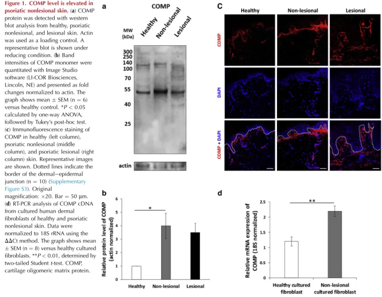
Nonlesional skin carries several known alterations of the ECM in the papillary dermis (Bos et al., 1983; Ting et al., 2000). Because COMP previously has been reported to also be present in the papillary dermis, COMP protein accumu- lation was characterized in nonlesional and lesional skin from patients with psoriasis and skin from healthy in- dividuals. COMP protein was detected with western blot analysis under reducing (Figure 1a and b andSupplementary Figure S1) and nonreducing (Supplementary Figure S2) con- ditions. Under reducing conditions, we detected elevated COMP monomer and fragment levels in psoriatic nonlesional protein extracts compared with healthy skin (Figure 1a and b andSupplementary Figure S1).
Subsequently, the distribution of COMP in tissues was analyzed using immunofluorescence staining. In line with previous reports, COMP was detected in the papillary dermis of healthy skin (Farina et al., 2006). In psoriatic nonlesional samples, COMP deposition extended deeper into the dermis
Figure 1. COMP level is elevated in psoriatic nonlesional skin.(a) COMP protein was detected with western blot analysis from healthy, psoriatic nonlesional, and lesional skin. Actin was used as a loading control. A representative blot is shown under reducing condition. (b) Band intensities of COMP monomer were quantitated with Image Studio software (LI-COR Biosciences, Lincoln, NE) and presented as fold changes normalized to actin. The graph shows meanSEM (n¼6) versus healthy control. *P<0.05 calculated by one-way ANOVA, followed by Tukey’s post-hoc test.
(c) Immunofluorescence staining of COMP in healthy (left column), psoriatic nonlesional (middle column), and psoriatic lesional (right column) skin. Representative images are shown. Dotted lines indicate the border of the dermaleepidermal junction (n¼10) (Supplementary Figure S3). Original
magnification:20. Bar¼50mm.
(d) RT-PCR analysis of COMP cDNA from cultured human dermal fibroblasts of healthy and psoriatic nonlesional skin. Data were normalized to 18S rRNA using the DDCt method. The graph shows mean SEM (n¼8) versus healthy cultured fibroblasts. **P<0.01, determined by two-tailed Studentt-test. COMP, cartilage oligomeric matrix protein.
and formed a more even and continuous layer than observed in healthy samples (Figure 1c andSupplementary Figure S3).
In contrast, COMP deposition in lesional skin extended to the upper part of the reticular dermis and exhibited a discontin- uously scattered distribution (Figure 1c and Supplementary Figure S3).
Because skin fibroblasts are the major producers of COMP protein (Dodge et al., 1998), we examined the mRNA expression of COMP in primary dermal fibroblasts derived from healthy and psoriatic nonlesional skin and detected elevated COMP mRNA expression in nonlesional fibroblasts (Figure 1d).
COMP colocalization with ITGB1 of basal keratinocytes and EDADFN is increased and with LAMA1 is decreased in nonlesional psoriatic skin
COMP is known to interact with several members of the integrin cell-surface receptor family, includinga5b1-integrin (Chen et al., 2005), whose expression increases together with EDA þFN in nonlesional skin, possibly owing to damaged BM (Bata-Csorgo et al., 1998; Mondello et al., 1996; Ting et al., 2000). To investigate the possible interactions of COMP with proteins in the DEJ that have been altered, confocal microscopic analysis with dual immunofluores- cence staining was applied and consecutive sections of the appropriate area were analyzed.
To determine whether COMP accumulation at the DEJ al- lows interaction with basal epidermal keratinocytes, COMP and b1-integrin (ITGB1) co-immunofluorescence staining was applied. In the papillary dermis, COMP staining partially colocalized with the ITGB1 from basal keratinocytes in healthy and nonlesional skin (Figure 2a and Supplementary Figure S4). However, the colocalization of the two proteins was most prominent in psoriatic nonlesional skin (Figure 2d).
LAMA1 is a component of the BM laminin layer, which is fragmented and occasionally completely missing in nonle- sional psoriatic skin (Mondello et al., 1996; Vaccaro et al., 2002). Therefore, to examine whether the damaged BM of nonlesional skin allows the interaction of COMP and ITGB1, LAMA1eCOMP dual immunostaining was performed. In nonlesional skin, COMPeITGB1 double-positive regions exhibited a discontinuous LAMA1 staining pattern (Figure 2b), and the co-occurrence of COMP and LAMA1 was significantly lower in nonlesional skin compared with healthy skin (Figure 2e).
In addition, FN has also been reported to interact with COMP (Di Cesare et al., 2002); therefore, confocal micro- scopic analysis was also applied to COMP and EDAþFN. In psoriatic nonlesional skin, in which colocalization of COMP and ITGB1 was apparent, partial colocalization of COMP and EDA þ FN was observed (Figure 2c), and the intensity of colocalization was significantly higher in nonlesional skin relative to healthy skin (Figure 2f).
COMP negatively influences keratinocyte proliferation via a5b1-integrin in vitro
To investigate whether the possible interaction between COMP and ITGB1 influences keratinocyte cellular behavior, we first performed an impedance measurementebased, real- time cellular analysis of the HPV-KER immortalized kerati- nocyte cell line. When the culturing plate was precoated with
recombinant human COMP (rhCOMP), cells exhibited reduced cell index values in a manner that was dependent on COMP concentration compared with cells grown on un- coated surfaces (Figure 3a). Cell index is influenced by changes in cell proliferation, viability, morphology, and adhesion (Dickhuth et al., 2015). To investigate whether the proliferation rate of HPV-KER cells was affected, a BrdU cell proliferation assay was performed. Precoating the surface with a high concentration (10mg/ml) of rhCOMP resulted in significantly reduced proliferation rates at 24 and 72 hours compared with cells grown on an uncoated surface. Cell proliferation of primary normal human epidermal keratino- cytes was also reduced when the surface was coated with 10 mg/ml rhCOMP (Figure 3c).
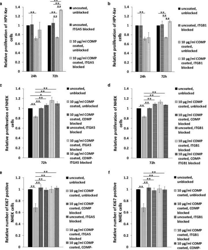
To test whether integrins mediate the observed negative effect of COMP on cell proliferation, blocking experiments using anti-ITGA5 and anti-ITGB1 polyclonal antibodies were performed. Blocking of either the ITGA5 or ITGB1 subunit in cells grown on a surface precoated with 10mg/ml rhCOMP abolished the negative effect of COMP on HPV-KER prolif- eration, whereas blocking either ITGA5 or ITGB1 alone had no negative effect on these cells (Figure 4a and b and Supplementary Figure S5). Similarly, the negative effect of COMP on the proliferation rate of primary normal human epidermal keratinocyte cells could also be abolished by blocking COMP, ITGA5, or ITGB1, as determined with the BrdU assay (Figure 4c and d) and Ki67 immunofluorescent staining (Figure 4e and f).
COMP has a negative effect on skin wound healing by attenuating keratinocyte proliferation and by compromising keratinocyte migration and activation in ex vivo wound models
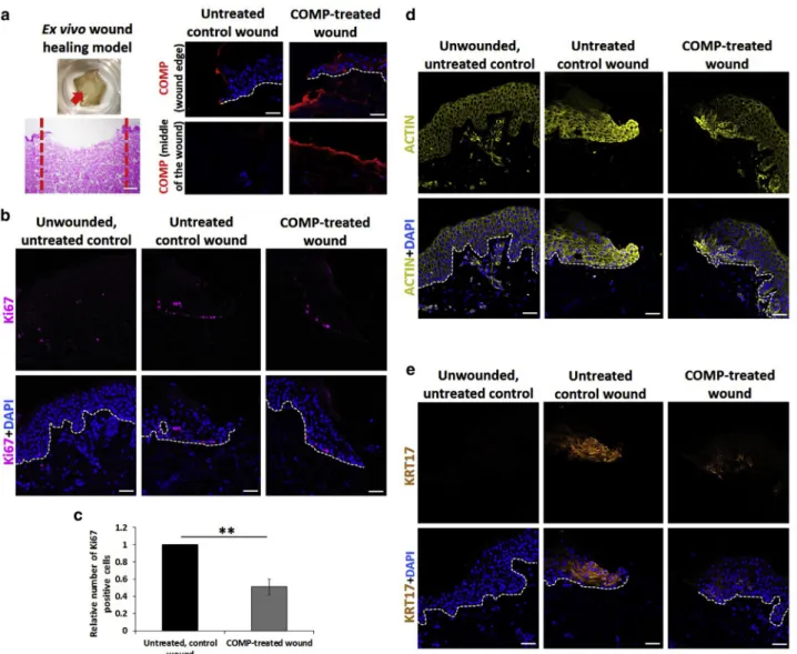
To further study the effect of COMP on keratinocytes, an ex vivo three-dimensional skin wound-healing model was applied. Standardized wounded skin samples, with or without rhCOMP treatment (10 mg/ml) (Figure 5a), and un- wounded controls were examined at 72 hours after wound- ing. Immunofluorescent staining revealed COMP localization on the dermal surface of the injured region in COMP-treated wounds, whereas COMP was not detected at the injured region of untreated wounds 72 hours after treatment (Figure 5a). By applying Ki67 immunofluorescent staining to detect proliferating cells, we found a markedly reduced number of Ki67-positive cells in COMP-treated wounds, indicating a decreased rate of proliferation (Figure 5b and c, Supplementary Figure S6a).
Cell migration processes at the wound edge during the closure of injuries require dynamic reorganization of the actin cytoskeleton in the keratinocytes located close to wound margins. To visualize these cells, immunofluores- cence staining for actin was applied. In wounds not exposed to COMP, we found that keratinocytes exhibited high levels of actin expression at wound edges, whereas actin expression at wound edges was markedly decreased in COMP-treated wounds, indicating that actin expression was compromised, possibly resulting in a reduction of active cell migration (Figure 5d).
Keratin 17 (KRT17) expression is known to be induced in keratinocytes at wound edges during healing (Mazzalupo
Figure 2. COMP colocalization with basal keratinocyteb1-integrin and EDADFN increases and with LAMA1 decreases in nonlesional psoriatic skin.Confocal microscopic immunofluorescence analysis of (a) COMP andb1-integrin, (b) COMP and LAMA1, and (c) COMP and EDAþFN colocalization in healthy (first row) and psoriatic nonlesional (second row) skin. Representative images are shown. Dotted lines indicate the enlarged regions. Colocalized pixels of the indicated proteins were calculated with ImageJ software (n¼5). Original magnification:63.
Bar¼10mm. The extent of colocalization of (d) COMP and b1-integrin, (e) COMP and LAMA1, and (f) COMP and EDAþFN was calculated using ImageJ/Fiji software.
The graphs show mean Pearson’s correlation coefficient RSEM (n¼5) versus healthy control. *P<0.05,
**P<0.01 determined by two-tailed Studentt-test. EDAþFN, fibronectin splice variant containing extra domain A.
COMP, cartilage oligomeric matrix protein.
et al., 2003). Therefore, we performed KRT17 immunoflu- orescence staining to further investigate the effect of COMP in the ex vivo wound healing model. In rhCOMP- treated wounds, KRT17 expression and re-epithelization were reduced and restricted to a smaller proportion of keratinocytes, compared with untreated control wounds (Figure 5e and Supplementary Figure S6b and c). This suggests that the presence of COMP compromised kerati- nocyte activation.
DISCUSSION
In psoriasis, the nonlesional skin contains ECM alterations compared with healthy skin. COMP is localized to the papillary dermis of healthy skin (Farina et al., 2006) and, through its interactions with type XII and XIV collagens, contributes to the stabilization of the DEJ (Agarwal et al., 2012). We found that, in psoriatic nonlesional skin, COMP is localized to the papillary dermis and, in contrast to healthy skin, it forms a continuous, more compact, linear layer beneath the basal keratinocytes. Apart from this altered localization, COMP expression was also elevated in dermal fibroblasts from psoriatic samples.
Psoriatic nonlesional skin is more sensitive to stress (Sonkoly et al., 2005; Sze´ll et al., 2016), and abnormalities at the DEJ and the BM are believed to be important in the development of the disease (Bos et al., 1983; Ting et al., 2000). Interruption of the BM (Mondello et al., 1996;
Vaccaro et al., 2002) may allow ECM components, nor- mally located directly below the BM, to come in direct contact with basal keratinocytes. COMP reportedly binds directly to the extracellular domain of ITGB1 of both car- diomyocytes (Huang et al., 2013) and cardiac fibroblasts,
resulting in the stabilization of ITGB1 by preventing its degradation and, subsequently, improving cellular survival (Huang et al., 2013; Posey et al., 2018). In cartilage, COMP mediates chondrocyte attachment and stabilization partially via a5b1-integrin (Tan et al., 2009). Our confocal micro- scopic analysis revealed a partial colocalization of papillary dermal COMP and ITGB1 in basal keratinocytes, which in- dicates the possibility of a direct interaction between these two proteins in vivo. In nonlesional skin, the a5-integrin (ITGA5) subunit is overexpressed in the basal layer of the epidermis, in contrast to healthy skin, where it is present at low levels or completely missing (Bata-Csorgo et al., 1998).
Our findings are in line with this observation, as COMP and ITGB1 strongly colocalize in psoriatic nonlesional epidermis and expression of both are upregulated in nonlesional skin.
Moreover, the BM is partially discontinuous in psoriatic nonlesional skin (Mondello et al., 1996; Vaccaro et al., 2002), allowing direct interaction. The possibility of this interaction is supported by our finding that, in areas where COMP and ITGB1 were found to have strong colocalization in psoriatic nonlesional skin, the expression of LAMA1, a member of the BM, is reduced or completely absent.
COMP also interacts witha7b1- andavb3-integrins (Chen et al., 2005; Rock et al., 2010). Of these proteins, onlya7b1 contains a ITGB1 subunit. There is currently no information available about a7b1-integrin expression in basal keratino- cytes. Thus, we assumed that, if COMP exhibits a strong interaction with ITGB1, itsa-subunit is likely to be ITGA5.
In addition to binding to a5b1-integrin, an FN receptor, COMP might also bind to the FN protein itself (Di Cesare et al., 2002). Furthermore, a5b1-integrineassociated FN and EDA þ FN are known to play roles in psoriasis
Figure 3. COMP negatively influences keratinocyte cell proliferation.(a) CI measurement of HPV-KER cells cultured on surfaces that were uncoated or coated with rhCOMP (1 and 10mg/ml). CI was determined using real-time impedance measurementebased cellular analysis. The graph is representative of four independent experiments, all showing similar results. The graph shows mean CISEM of four technical replicas for each group, *P<0.05 versus uncoated control, #P<0.05 versus 1mg/ml rhCOMP-coated group calculated by one-way ANOVA, followed by Tukey’s post-hoc test. BrdU cell proliferation assay of (b) HPV-KER and (c) NHEK cells cultured on uncoated and rhCOMP protein (1 and 10mg/ml)-coated surfaces at 24 and 72 hours following seeding. The graphs show mean proliferationSEM (n¼3). **P<0.01, calculated by one-way ANOVA followed by Tukey’s post-hoc test. CI, cell index; COMP, cartilage oligomeric matrix protein; NHEK, normal human epidermal keratinocyte; rhCOMP, recombinant human COMP.
Figure 4. COMP influences keratinocyte proliferation viaa5b1 integrin.(a, b) BrdU cell proliferation assay of HPV-KER cells cultured on surfaces that were uncoated or coated with rhCOMP protein (10mg/ml) for 24 and 72 hours following seeding and treated with (a) antiea5-integrin or (b) antieb1-integrin subunit antibodies. (c, d) BrdU assay and (e, f) Ki67-positive proliferating cell number determination of NHEKs grown on surfaces that were uncoated or coated with rhCOMP protein (10mg/ml) for 72 hours following seeding and treated with (c, e) antiea5-integrin and (d, f) antieb1-integrin subunit antibody in a combination with COMP protein neutralization. The graphs show mean proliferation/mean number of Ki67-positive cellsSEM (n¼3) versus uncoated control, *P<0.05, **P<0.01 calculated by one-way ANOVA, followed by Tukey’s post-hoc test. COMP, cartilage oligomeric matrix protein; NHEK, normal human epidermal keratinocyte; rhCOMP, recombinant human COMP.
pathogenesis (Bata-Csorgo et al., 1998; Ting et al., 2000).
Enriched expression of a5b1-integrin and EDA þ FN in nonlesional skin is thought to be due to the incompleteness of the laminin layer (Mondello et al., 1996; Vaccaro et al., 2002). Our confocal microscopic analysis revealed partial colocalization of COMP and EDAþFN in nonlesional skin.
These results suggest that, in addition to interacting with EDA þ FN, COMP may also affect basal keratinocytes via interactions with both the EDAþFN and its receptor,a5b1- integrin.
Keratinocyte behavior is influenced by ECM proteins through interactions with different cell surface integrins (Hamill et al., 2012; Tjin et al., 2014), and connection of basal keratinocytes to the altered BM could enhance prolif- eration (Yang et al., 2016). We analyzed the biological rele- vance of the interaction of COMP and ITGB1 in basal
keratinocytes using HPV-KER and normal human epidermal keratinocyte cells in vitro. We found that the presence of COMP resulted in reduced keratinocyte proliferation in both cell types and that this affect was reversible by blocking COMP with a specific antibody.
We also analyzed whether the observed negative effect of COMP on keratinocyte proliferation involves interaction with a5b1-integrin. By partially blocking the function of the ITGB1 or ITGA5 subunits with specific antibodies, the negative effect of COMP on cell proliferation was abolished, suggesting that the negative influence of COMP on kerati- nocyte proliferation involvesa5b1-integrin.
Our in vitro findings were also validated in an ex vivo wound model; exogenous COMP treatment delayed healing of artificial wounds, and this affect was coupled with reduced keratinocyte proliferation and compromised actin expression,
Figure 5. COMP has a negative effect on keratinocyte proliferation and is involved in keratinocyte migration and activation in ex vivo wound models.
(a) Representative images of the ex vivo skin wound healing models (n¼3; magnification4; Bar¼250mm). Immunofluorescent staining for COMP at wound edges and at the middle of the wounds of untreated controls and wounds treated with rhCOMP (n¼3; magnification20; Bar¼50mm). Immunostaining for (b) Ki67, (d) actin, or (e) keratin-17 in the ex vivo unwounded skin (left column), and wound-healing skin models with (right column) and without (middle column) rhCOMP treatment. Representative images are shown. Dotted lines indicate the border of the dermaleepidermal junction (n¼3; magnification20; bar¼50mm). (c) Ki67- positive cells in wounds that were not treated and were treated with rhCOMP protein. The graph shows mean number of Ki67-positive cellsSEM (n¼3) versus uncoated, untreated control **P<0.01 determined by two-tailed Studentt-test. COMP, cartilage oligomeric matrix protein; rhCOMP, recombinant human COMP.
both important aspects of wound healing (Gurtner et al., 2008). In addition, keratinocyte KRT17 expression, consid- ered a hallmark of normal wound healing (Mazzalupo et al., 2003), was also decreased in the presence of COMP. These results suggest that COMP has a negative influence on ex vivo wound healing. In normally healing wounds of healthy do- nors, COMP was hardly detectable when re-epithelialization was complete (Agarwal et al., 2013). Similarly, in psoriatic lesions, in which keratinocyte proliferation is abnormally increased, COMP was found to be discontinuous or completely absent from the papillary dermis. Although there are no data regarding keratinocyte proliferation or migration in wounds of COMP-deficient mice (Schulz et al., 2016), in human nonhealing wounds, such as venous leg ulcers, the level of COMP is reported to be highly elevated (Agarwal et al., 2013). Our findings are in agreement with this observation.
In conclusion, our study shows that COMP is present at an elevated level in the papillary dermis of nonlesional psoriatic skin and that it possibly reduces keratinocyte proliferation via the a5b1-integrin. These aspects of COMP contribute to the maintenance of the nonlesional, non- hyperproliferative state of psoriatic nonlesional epidermis, despite the overexpression of EDAþFN and ITGA5. Similar interactions may also take place in other skin diseases in which nonhealing wounds are coupled with massive COMP accumulation.
MATERIALS AND METHODS Skin samples and ethics
Skin punch biopsies (diameter¼6 mm) were collected from healthy volunteers (n¼10; age 18e70 years,Supplementary Table S1), and from patients with psoriasis with moderate-to-severe chronic plaque- type psoriasis from lesional (n¼13) and nonlesional skin areas (n¼ 13; minimum of 6 cm from lesional region; age 18e70 years [Supplementary Table S1]). Patients with psoriasis did not receive local therapy for at least 4 weeks and had not been subjected to systemic therapy for at least 8 weeks. Skin biopsies were taken from areas of skin that were not exposed to sun. Tissue collection was obtained after written informed consent, in accordance with the rules of the Helsinki Declaration. The study was confirmed by the Human Investigation Review Board of the University of Szeged (PSO-EDAFN-002, 34/2015, 3517, 23 February 2015, Szeged, Hungary; PSO-ECMPR-002 IF-562-5/2016 and 157/2015-SZTE, 3638, 21 September 2015, Hungary).
Fluorescence microscopic analysis
Biopsies were frozen in a cryogenic matrix (Thermo Fisher Scien- tific, Waltham, MA) or were paraffin embedded and were subse- quently cut into 5-mm sections. For fixation and permeabilization, 4% paraformaldehyde followed by 0.25% TritonX-100 (Thermo Fisher Scientific) or commercially available staining buffer set (eBioscience, Santa Clara, CA) were used. For blocking, Tris- buffered saline containing 1% bovine serum albumin and 1%
normal goat serum (Sigma-Aldrich, St. Louis, MO) was used, and for frozen samples, which were digested with chondroitinase ABC (5U, 1:100; Sigma-Aldrich), 10% fetal bovine serum (EuroClone, Pero, Italy) and 5% normal goat serum (Sigma-Aldrich) were applied. Samples were incubated with the following primary anti- bodies: polyclonal rabbit anti-human COMP (1:250), a kind gift
from Mats Paulsson and Frank Zaucke from the University of Co- logne (Agarwal et al., 2012); mouse anti-human ITGB1 (clone:
JB1B, 1:100, Abcam, Cambridge, United Kingdom); rabbit anti- human actin (1:100, Sigma-Aldrich); mouse anti-human Ki67 (1:100, Beckton Dickinson, Franklin Lakes, NJ); mouse anti-human KRT17 (ready to use, Dako, Santa Clara, CA); mouse anti-human LAMA1 (clone: LAM-89, 1:100, R&D Systems, Minneapolis, MN);
and mouse anti-human FN (EDA þ FN, clone: IST-9, 1:500, Abcam). Isotype controls were the following: rabbit polyclonal IgG (Santa Cruz Biotechnology, Dallas, TX) and mouse IgG1 (Beckton Dickinson) antibodies. As secondary antibodies, Alexa Fluor 546econjugated anti-rabbit IgG and Alexa Fluor 647econjugated anti-mouse IgG (Life Technologies, Carlsbad, CA) were used.
Nuclei were visualized with DAPI (Sigma-Aldrich) staining. Zeiss LSM 880 or Zeiss Axio Imager Z1 microscopes (Carl Zeiss AG, Oberkochen, Germany) were used for visualization.
Pearson’s correlation coefficient, R, was calculated using ImageJ/
Fiji software.
Cell cultures and examination of cellular properties
Cell proliferation assay. To investigate the effect of COMP on keratinocyte proliferation, a BrdU cell proliferation colorimetric ELISA assay (Abcam) was performed. HPV-KER cells, a stable hu- man keratinocyte cell line that has been characterized in our lab- oratory (Danis et al., 2018; Erdei et al., 2018; Tax et al., 2016), and normal human epidermal keratinocyte cells were plated at a den- sity of 10,000 cells per well in 96-well plates (Corning, NY) that were uncoated or coated with low- (1mg/ml) or high-concentration (10 mg/ml) rhCOMP protein (R&D Systems), in three technical replicates. For the blocking of ITGA5 and ITGB1, the following antibodies (1 mg antibody for 106 cells) were used: mouse anti- human ITGA5 antibody (clone: IIA1, Beckton Dickinson) and mouse anti-human ITGB1 (clone: JB1B). Goat anti-human COMP antibody (1mg antibody for 10mg/ml rhCOMP protein, R&D Sys- tems) was applied to block the COMP protein. Integrin- and COMP-blocking was applied to cells grown on uncoated plates or plates coated with 10 mg/ml rhCOMP protein. BrdU assay was performed at 24 and 72 hours after blocking, according to the manufacturer’s instructions.
Ex vivo skin wound-healing assay
Healthy skin samples were collected for the ex vivo organotypic wound healing assay. Approximately 1-cm diameter skin pieces were cut and mildly wounded in the middle using a 4-mm punch biopsy blade (Steele Supply Company, St. Joseph, MI). Wounded skin samples and unwounded control samples were incubated for 72 hours at an aireliquid interface on the upper part of transwell cell culture inserts. The dermal part was in contact with DMEM F12 culture media (Lonza Group, Basel, Switzerland) supple- mented with 10% fetal bovine serum (EuroClone) and 1% anti- biotic/antimycotic solution (Sigma-Aldrich). The middle of the wounds was treated for 72 hours with high-concentration (10mg/
ml) rhCOMP (R&D Systems) diluted in PBS or PBS only as a control. Samples were fixed in formalin and embedded in paraffin for immunofluorescent staining. To determine the rate of prolifer- ation, 50 cells on each wound edge were counted and the pro- portion of Ki67-positive cells was determined. Re-epithelization of untreated, control (where only PBS was administered), and COMP- treated wounds were assessed by measuring the area using the ImageJ software.
Statistical analysis
For comparing only two groups, two-tailed Studentt-test was per- formed. One-way ANOVA with Tukey post-hoc test was used to compare more than two groups. Differences were considered sta- tistically significant at **P<0.01, *P<0.05. Data were analyzed using R-Studio software, version 3.2.2 (R-Studio, Boston, MA).
Further methods
More detailed information of the materials and methods regarding protein isolation and western blot analysis, cell culture experiments, H&E staining, RNA isolation, and real-time PCR are presented in the Supplementary Materials.
Data availability statement
No datasets were generated or analyzed during this study.
ORCIDs
Rena´ta Bozo´:https://orcid.org/0000-0003-4242-2474 Edit Sze´l:https://orcid.org/0000-0001-9102-5542 Judit Danis:https://orcid.org/0000-0002-0270-5309 Barbara Guba´n:https://orcid.org/0000-0002-9406-7489 Zsuzsanna Bata-Cso¨rg}o:https://orcid.org/0000-0002-3732-1743 Korne´lia Szabo´:https://orcid.org/0000-0002-6231-3251 Lajos Keme´ny:https://orcid.org/0000-0002-2119-9501 Gergely Groma:https://orcid.org/0000-0001-8487-0465
CONFLICT OF INTEREST
The authors state no conflict of interest.
ACKNOWLEDGMENTS
The authors wish to thank Mats Paulsson and. Frank Zaucke for the COMP antibody. The authors are grateful for Brigitta Ga´l, Krisztina V, and Ro´bert Kui for collecting tissue samples from patients. Furthermore, the authors wish to also thank Mo´nika Kohajda for her excellent technical assistance and Ma´te´
Manczinger and Bala´zs Koncz for their help in the statistical analysis. Our work was supported by grants from National Research, Development and Innovation Office, Hungary (former Hungarian Scientific Research Fund) PD116992, K111885 and GINOP-2.2.1-15-2016-00007 research grants and co-financed by the European Social Fund in the framework of TAMOP- 4.2.4.A/2-11-1/2012-0001 “National Excellence Program” A2-SZGYA-FOK- 13-0001, and European Union’s H2020 Grant Agreement No. 739593. KS is a recipient of the Ja´nos Bolyai Research Scholarship of the Hungarian Acad- emy of Sciences. KS was supported by the UNKP-18-4 and RB by the UNKP-18-3 of New National Excellence Program of the Ministry of Human Capacities. KS was supported by the U´ NKP-19-4 and RB by the U´NKP-19-3 of New National Excellence Program of the Ministry for Innovation and Technology. RB was also supported by the Gedeon Richter Talentum Foundation (H-1103 Budapest, Gyo¨mr}oi str. 19-21.). The study took place in Szeged, Hungary.
AUTHOR CONTRIBUTIONS
Conceptualization: GG; Formal Analysis: RB, KS, GG; Investigation: RB, ES, JD, BG; Methodology: RB, GG; Supervision: LK, ZBC, GG; Writing - Original Draft Preparation: RB, GG; Writing - Review and Editing: KS, ZBC, GG.
SUPPLEMENTARY MATERIAL
Supplementary material is linked to the online version of the paper atwww.
jidonline.organd athttps://doi.org/10.1016/j.jid.2019.12.037.
REFERENCES
Agarwal P, Schulz JN, Blumbach K, Andreasson K, Heinega˚rd D, Paulsson M, et al. Enhanced deposition of cartilage oligomeric matrix protein is a common feature in fibrotic skin pathologies. Matrix Biol 2013;32:325e31.
Agarwal P, Zwolanek D, Keene DR, Schulz JN, Blumbach K, Heinega˚rd D, et al. Collagen XII and XIV, new partners of cartilage oligomeric matrix protein in the skin extracellular matrix suprastructure. J Biol Chem 2012;287:22549e59.
Bata-Csorgo Z, Cooper KD, Ting KM, Voorhees JJ, Hammerberg C. Fibro- nectin and alpha 5 integrin regulate keratinocyte cell cycling. A mecha- nism for increased fibronectin potentiation of T cell lymphokine-driven keratinocyte hyperproliferation in psoriasis. J Clin Invest 1998;101:
1509e18.
Bata-Csorgo Z, Hammerberg C, Voorhees JJ, Cooper KD. Kinetics and regu- lation of human keratinocyte stem cell growth in short-term primary ex vivo culture. Cooperative growth factors from psoriatic lesional T lymphocytes stimulate proliferation among psoriatic uninvolved, but not normal, stem keratinocytes. J Clin Invest 1995;95:317e27.
Bos JD, Hulsebosch HJ, Krieg SR, Bakker PM, Cormane RH. Immunocom- petent cells in psoriasis. In situ immunophenotyping by monoclonal anti- bodies. Arch Dermatol Res 1983;275:181e9.
Chen FH, Thomas AO, Hecht JT, Goldring MB, Lawler J. Cartilage oligomeric matrix protein/thrombospondin 5 supports chondrocyte attachment through interaction with integrins. J Biol Chem 2005;280:32655e61.
Chen G, McCormick TS, Hammerberg C, Ryder-Diggs S, Stevens SR, Cooper KD. Basal keratinocytes from uninvolved psoriatic skin exhibit accelerated spreading and focal adhesion kinase responsiveness to fibro- nectin. J Invest Dermatol 2001;117:1538e45.
Danis J, Janova´k L, Guba´n B, Go¨blo¨s A, Szabo´ K, Keme´ny L, et al. Differential inflammatory-response kinetics of human keratinocytes upon cytosolic RNA- and DNA-fragment induction. Int J Mol Sci 2018;19:774.
Di Cesare PE, Chen FS, Moergelin M, Carlson CS, Leslie MP, Perris R, et al.
Matrix-matrix interaction of cartilage oligomeric matrix protein and fibro- nectin. Matrix Biol 2002;21:461e70.
Dickhuth J, Koerdt S, Kriegebaum U, Linz C, Mu¨ller-Richter UD, Ristow O, et al. In vitro study on proliferation kinetics of oral mucosal keratinocytes.
Oral Surg Oral Med Oral Pathol Oral Radiol 2015;120:429e35.
Dodge GR, Hawkins D, Boesler E, Sakai L, Jimenez SA. Production of carti- lage oligomeric matrix protein (COMP) by cultured human dermal and synovial fibroblasts. Osteoarthr Cartil 1998;6:435e40.
Erdei L, Bolla BS, Bozo´ R, Tax G, Urba´n E, Keme´ny L, et al. TNIP1 regulates Cutibacterium acnes-induced innate immune functions in epidermal ker- atinocytes. Front Immunol 2018;9:2155.
Farina G, Lemaire R, Korn JH, Widom RL. Cartilage oligomeric matrix protein is overexpressed by scleroderma dermal fibroblasts. Matrix Biol 2006;25:
213e22.
Glinski W, Stepien-Sopniewska B, Majewski S, Glinska-Ferenz M, Go´rski A.
Alterations of T-cell: extracellular matrix proteins interactions in psoriasis.
Immunol Lett 1993;35:153e7.
Guba´n B, Vas K, Balog Z, Manczinger M, Bebes A, Groma G, et al. Abnormal regulation of fibronectin production by fibroblasts in psoriasis. Br J Der- matol 2016;174:533e41.
Gurtner GC, Werner S, Barrandon Y, Longaker MT. Wound repair and regeneration. Nature 2008;453:314e21.
Hamill KJ, Hopkinson SB, Hoover P, Todorovic V, Green KJ, Jones JCR.
Fibronectin expression determines skin cell motile behavior. J Invest Der- matol 2012;132:448e57.
Huang Y, Xia J, Zheng J, Geng B, Liu P, Yu F, et al. Deficiency of cartilage oligomeric matrix protein causes dilated cardiomyopathy. Basic Res Car- diol 2013;108:374.
Inui S, Shono F, Nakajima T, Hosokawa K, Itami S. Identification and char- acterization of cartilage oligomeric matrix protein as a novel pathogenic factor in keloids. Am J Pathol 2011;179:1951e60.
Malashkevich VN, Kammerer RA, Efimov VP, Schulthess T, Engel J. The crystal structure of a five-stranded coiled coil in COMP: a prototype ion channel?
Science 1996;274:761e5.
Mazzalupo S, Wong P, Martin P, Coulombe PA. Role for keratins 6 and 17 during wound closure in embryonic mouse skin. Dev Dyn 2003;226:
356e65.
McFadden JP, Kimber I. A review on the potential role of basement membrane laminin in the pathogenesis of psoriasis. Scand J Immunol 2016;83:3e9.
Mondello MR, Magaudda L, Pergolizzi S, Santoro A, Vaccaro M, Califano L, et al. Behaviour of laminin 1 and type IV collagen in uninvolved psoriatic skin. Immunohistochemical study using confocal laser scanning micro- scopy. Arch Dermatol Res 1996;288:527e31.
Mo¨rgelin M, Heinega˚rd D, Engel J, Paulsson M. Electron microscopy of native cartilage oligomeric matrix protein purified from the Swarm rat chon- drosarcoma reveals a five-armed structure. J Biol Chem 1992;267:
6137e41.
Mu¨ller G, Michel A, Altenburg E. COMP (cartilage oligomeric matrix protein) is synthesized in ligament, tendon, meniscus, and articular cartilage.
Connect Tissue Res 1998;39:233e44.
Pellegrini G, De Luca M, Orecchia G, Balzac F, Cremona O, Savoia P, et al.
Expression, topography, and function of integrin receptors are severely altered in keratinocytes from involved and uninvolved psoriatic skin. J Clin Invest 1992;89:1783e95.
Posey KL, Coustry F, Hecht JT. Cartilage oligomeric matrix protein: COMPo- pathies and beyond. Matrix Biol 2018;71e72:161e73.
Rock MJ, Holden P, Horton WA, Cohn DH. Cartilage oligomeric matrix protein promotes cell attachment via two independent mechanisms involving CD47 and alphaVbeta3 integrin. Mol Cell Biochem 2010;338:
215e24.
Schulz JN, Nu¨chel J, Niehoff A, Bloch W, Scho¨nborn K, Hayashi S, et al.
COMP-assisted collagen secretion–a novel intracellular function required for fibrosis. J Cell. Sci 2016;129:706e16.
Sonkoly E, Bata-Csorgo Z, Pivarcsi A, Polyanka H, Kenderessy-Szabo A, Molnar G, et al. Identification and characterization of a novel, psoriasis susceptibility-related noncoding RNA gene, PRINS. J Biol Chem 2005;280:
24159e67.
Szabo´ K, Bata-Cso¨rg}o Z, Dallos A, Bebes A, Francziszti L, Dobozy A, et al.
Regulatory networks contributing to psoriasis susceptibility. Acta Derm Venereol 2014;94:380e5.
Sze´ll M, Bata-Cso¨rgo Z, Koreck A, Pivarcsi A, Polya´nka H, Szeg C, et al.
Proliferating keratinocytes are putative sources of the psoriasis susceptibility-related EDAþ (extra domain A of fibronectin) oncofetal fibronectin. J Invest Dermatol 2004;123:537e46.
Sze´ll M, Danis J, Bata-Cso¨rg}o Z, Keme´ny L. PRINS, a primate-specific long non-coding RNA, plays a role in the keratinocyte stress response and psoriasis pathogenesis. Pflugers Arch 2016;468:935e43.
Tan K, Duquette M, Joachimiak A, Lawler J. The crystal structure of the signature domain of cartilage oligomeric matrix protein: implications for collagen, glycosaminoglycan and integrin binding. FASEB J 2009;23:
2490e501.
Tan K, Lawler J. The interaction of thrombospondins with extracellular matrix proteins. J Cell Commun Signal 2009;3:177e87.
Tax G, Urba´n E, Palota´s Z, Puska´s R, Ko´nya Z, Bı´ro´ T, et al. Propionic acid produced by Propionibacterium acnes strains Contributes to their patho- genicity. Acta Derm Venereol 2016;96:43e9.
Ting KM, Rothaupt D, McCormick TS, Hammerberg C, Chen G, Gilliam AC, et al. Overexpression of the oncofetal Fn variant containing the EDA splice- in segment in the dermal-epidermal junction of psoriatic uninvolved skin.
J Invest Dermatol 2000;114:706e11.
Tjin MS, Chua AWC, Ma DR, Lee ST, Fong E. Human epidermal keratinocyte cell response on integrin-specific artificial extracellular matrix proteins.
Macromol Biosci 2014;14:1125e34.
Vaccaro M, Magaudda L, Cutroneo G, Trimarchi F, Barbuzza O, Guarneri F, et al. Changes in the distribution of laminin alpha 1 chain in psoriatic skin:
immunohistochemical study using confocal laser scanning microscopy. Br J Dermatol 2002;146:392e8.
Wang L, Zheng J, Du Y, Huang Y, Li J, Liu B, et al. Cartilage oligomeric matrix protein maintains the contractile phenotype of vascular smooth muscle cells by interacting with alpha(7)beta(1) integrin. Circ Res 2010;106:514e25.
Yang S, Sun Y, Geng Z, Ma K, Sun X, Fu X. Abnormalities in the basement membrane structure promote basal keratinocytes in the epidermis of hy- pertrophic scars to adopt a proliferative phenotype. Int J Mol Med 2016;37:
1263e73.
SUPPLEMENTARY MATERIALS AND METHODS Protein isolation and western blot analysis
For preparation of tissue protein extracts, skin biopsies (healthy, psoriatic nonlesional, and psoriatic lesional) were cut into small pieces with a razor blade. Guanidine hydro- chloride (6 M, Sigma-Aldrich, St. Louis, MO) solution was used as an extraction buffer for 24 hours at 4 C under continuous agitation. For protein precipitation, an ethanol- based method was applied. Protein concentrations were measured using Bradford assay (Bio-Rad Laboratories, Her- cules, CA). Protein extracts (25 mg) were separated on a 4e20% gradient SDS polyacrylamide gel under reducing or nonreducing conditions. Proteins were transferred to a nitrocellulose membrane (Bio-Rad Laboratories) and blocked in 5% nonfat milk powder containing Tris-buffered saline for 60 minutes at room temperature. Membranes were incubated for overnight at 4 C with goat anti-human COMP primary antibody (1:2,000, R&D Systems, Minneapolis, MN), and rabbit anti-human actin primary antibody (1:2,000, Sigma- Aldrich). Subsequently, membranes were incubated with horseradish peroxidaseeconjugated anti-goat (Thermo Fisher Scientific, Waltham, MA), and anti-rabbit (Southern Biotech, Birmingham, AL) secondary antibodies, both diluted 1:2,000 for 60 minutes at room temperature. Signal was visualized with Clarity Max Western ECL Substrate (Bio-Rad Labora- tories) on a C-digit blot scanner (LI-COR Biosciences, Lincoln, NE).
H&E staining and light microscopic analysis
To visualize the tissue structure of ex vivo wound model samples, H&E (Leica Biosystems, Wetzlar, Germany) staining was performed according to the manufacturer’s instructions in a Leica ST5020 Multistainer device (Leica Biosystems). The stained samples were visualized with a Nikon eclipse TS100 microscope (Nikon, Minato, Tokyo, Japan).
Cell cultures and examination of cellular properties
Cell cultures. Primary dermal fibroblasts were isolated from healthy and psoriatic nonlesional skin biopsies, and normal human epidermal keratinocytes (NHEKs) were iso- lated from healthy skin samples. The epidermis was separated from the dermis with overnight incubation at 4C in Dispase II (neutral protease, grade II, 2 U/ml, Roche Diagnostics, Basel, Switzerland) (Szabad et al., 2007) solution. Keratino- cytes were obtained from the epidermal part after trypsin digestion for 10 minutes at 37 C (Sigma-Aldrich). NHEK cells were then grown in keratinocyte serum-free medium (Life Technologies, Carlsbad, CA) supplemented with 1%
antibiotic/antimycotic solution (Sigma-Aldrich), brain pitui- tary extract (50 mg/ml, Life Technologies), and epidermal growth factor (5 ng/ml, Life Technologies).
Fibroblasts were obtained from the dermal part after digestion for 120 minutes at 37 C. The medium, DMEM supplemented with 1 g/liter glucose (Lonza Group, Basel, Switzerland), also contained collagenase (from Clostridium histolyticum, 2.7 mg/ml, Sigma-Aldrich), deoxyribonuclease I (from bovine pancreas, 0.1 mg/ml, Sigma-Aldrich), hyal- uronidase (from bovine testes, 1.25 mg/ml, Sigma-Aldrich), and fetal bovine serum (2.5%, EuroClone, Pero, Italy) (Guba´n et al., 2016). Fibroblasts were cultured in DMEM
with 1 g/liter glucose (Lonza Group), supplemented with 5%
fetal bovine serum (EuroClone), 1% antibiotic/antimycotic solution (Sigma-Aldrich), and 1% L-glutamine (PAA Labora- tories GmbH, Pasching, Austria).
The human immortalized keratinocyte cell line HPV-KER was also used for our experiments. HPV-KER is a stable cell line, generated from NHEKs transfected with the HPV16/E6 oncogene in a pCMV vector. It was established by continuous culturing (Tax et al., 2016). It shows similar responses to NHEK cells in various immune activation protocols (Danis et al., 2018; Erdei et al., 2018). Culture conditions of HPV- KER cells are the same as NHEK cells. Each cell type was cultured at 37C in a humidified atmosphere with 5% v/v CO2.
Real-time, label-free cellular analysis of HPV-KER cells using the xCELLigence system. xCELLigence (ACEA Bio- sciences, San Diego, CA) is a real-time, impedance measurementebased cellular analysis system, where dimensionless cell index value is calculated (cell index ¼ [impedance at time point neimpedance in the absence of cells]/nominal impedance value). Differences in cell index values could be due to altered cell proliferation rate, viability, morphology, and adhesion (Dickhuth et al., 2015). This sys- tem was used to investigate the effect of the COMP protein on keratinocytes. HPV-KER cells were plated at a density of 10,000 cells per well in uncoated 96-well E-plates (ACEA Biosciences) or wells that were coated with low- concentration (1 mg/ml) or high-concentration (10 mg/ml) recombinant human COMP protein (R&D Systems). Imped- ance measurement was performed every 15 minutes for 140 hours, and a dimension-free cell index value was calculated for every time point. Four technical replicates were performed.
Further investigation of keratinocyte cell proliferation. To further investigate the effect of COMP on the proliferation of NHEK cells, Ki67 immunofluorescent staining (mouse anti- human Ki67 antibody, 1:100, Beckton Dickinson, Franklin Lakes, NJ) was applied, using integrin and COMP-blocking as described in the main text. Cells were plated at a density of 20,000 cells per well in 8-well chamber slides (SPL Life Sciences, Naechon-Myeon, Pocheon-si, Korea) that were uncoated or coated with high-concentration (10 mg/ml) re- combinant human COMP (R&D Systems) in three biological replicates. Ki67-positive cells were counted on three randomly selected areas per group, and statistical analysis was performed.
RNA isolation and real-time PCR. Total RNA was isolated from primary fibroblasts from healthy and psoriatic nonle- sional skin cultured in 75-cm2 cell culture flasks (Corning, NY) and collected at the fifth passage using TRI-Reagent (Molecular Research Center, Cincinnati, OH) as described by the manufacturer. The iScript cDNA Synthesis kit (Bio-Rad Laboratories) was used for cDNA synthesis, and 0.5mg total RNA was reverse transcribed. RT-PCR was performed on a C1000 Touch Thermal Cycler (Bio-Rad Laboratories) with the Universal Probe Library system (Roche Diagnostics) using qPCRBIO Probe Mix Lo-ROX (PCR Biosystem Ltd., London, United Kingdom) and the following primers:
COMP FWD: CACCGACGTCAACGAGTG, COMP REV:
TGGTGTTGATACAGCGGACT; 18S rRNA FWD: CGCTCCAC CAACTAAGAACG, 18SrRNA REV: CTCAACACGGGAAACC TCAC. The expression of COMP was normalized to 18S rRNA expression using theDDCt method.
SUPPLEMENTARY REFERENCES
Danis J, Janova´k L, Guba´n B, Go¨blo¨s A, Szabo´ K, Keme´ny L, et al. Dif- ferential inflammatory-response kinetics of human keratinocytes upon cytosolic RNA- and DNA- fragment induction. Int J Mol Sci 2018;19:
774.
Dickhuth J, Koerdt S, Kriegebaum U, Linz C, Mu¨ller-Richter UD, Ristow O, et al. In vitro study on proliferation kinetics of oral mucosal
keratinocytes. Oral Surg Oral Med Oral Pathol Oral Radiol 2015;120:
429e35.
Erdei L, Bolla BS, Bozo´ R, Tax G, Urba´n E, Keme´ny L, et al. TNIP1 regulates Cutibacterium acnes-induced innate immune functions in epidermal ker- atinocytes. Front Immunol 2018;9:2155.
Guba´n B, Vas K, Balog Z, Manczinger M, Bebes A, Groma G, et al. Abnormal regulation of fibronectin production by fibroblasts in psoriasis. Br J Der- matol 2016;174:533e41.
Szabad G, Kormos B, Pivarcsi A, Sze´ll M, Kis K, Kenderessy Szabo´ A, et al.
Human adult epidermal melanocytes cultured without chemical mitogens express the EGF receptor and respond to EGF. Arch Dermatol Res 2007;299:191e200.
Tax G, Urba´n E, Palota´s Z, Puska´s R, Ko´nya Z, Bı´ro´ T, et al. Propionic acid produced by Propionibacterium acnes strains contributes to their patho- genicity. Acta Derm Venereol 2016;96:43e9.
Supplementary Figure S1. COMP monomer and fragment level is elevated in psoriatic nonlesional skin under reducing conditions.COMP monomers and fragments were detected with western blot analysis from healthy, psoriatic nonlesional, and lesional skin under reducing conditions (n¼3).
The band intensities of (a) COMP monomer and (b) COMP fragment separately, as well as (c) the level of monomer and fragment together were analyzed with Image Studio software (LI-COR Biosciences) and presented as fold changes normalized to actin. The graph shows meanSEM (n¼3),
*P<0.05 versus healthy control, calculated by one-way ANOVA followed by Tukey’s post-hoc test. COMP, cartilage oligomeric matrix protein.
Supplementary Figure S2. COMP protein detection under nonreducing conditions.COMP protein was detected with western blot analysis from healthy, psoriatic nonlesional, and lesional skin under nonreducing conditions (n¼3). A representative blot is shown. COMP, cartilage oligomeric matrix protein; H, healthy; L, lesional; NL, nonlesional.
Supplementary Figure S3.
Characterization of COMP deposition in healthy, nonlesional, and lesional skin.Immunofluorescence staining for COMP in (a) healthy, (b) psoriatic nonlesional, and (c) psoriatic lesional skin (n¼10). Zeiss Axio Imager Z1 original magnification,20. Bar¼50 mm. COMP, cartilage oligomeric matrix protein.
Supplementary Figure S4. Enhanced colocalization of COMP withb1- integrin in nonlesional psoriatic skin.
Confocal microscopic immunofluorescence analysis of COMP andb1-integrin in (a) healthy and (b) psoriatic nonlesional skin using z-stack pictures. Dotted lines indicate the borders of enlarged regions. Colocalized pixels of COMP andb1-integrin were calculated by ImageJ software (n¼5). Original magnification,63. Bar¼10mm.
COMP, cartilage oligomeric matrix protein.
Supplementary Figure S5. COMP negatively influences keratinocyte CI viaa5b1-integrin.CI measurement of HPV-KER cells cultured on uncoated and rhCOMP-coated (10mg/ml) surfaces. CI was determined using real-time impedance measuremente based cellular analysis. The graph is representative of three independent experiments, all showing similar results. Mean CISEM of four technical replicas for each group. CI, cell index; COMP, cartilage oligomeric matrix protein; rhCOMP, recombinant human COMP.
Supplementary Figure S6. Reduced proliferation and re-epithelization in the presence of COMP during ex vivo skin wound healing.(a) Reduced number of Ki67-positive cells was detected in wound samples treated with rhCOMP (10mg/ml) compared with untreated control wounds. The graph shows meanSEM (n¼3),
*P<0.05 versus untreated control, calculated by two-tailed Studentt-test.
(b) Re-epithelization of artificial untreated and rhCOMP-treated (10 mg/ml) wounds on H&E-stained sections (n¼3). Original magnification,20. Bar¼50mm.
Representative pictures are shown.
Arrowheads indicate the newly synthetized areas. (c) Re- epithelization of untreated and control- and rhCOMP-treated wounds was measured using area
measurement of ImageJ software. The graph shows meanSEM (n¼3),
*P<0.05 versus untreated control, calculated by two-tailed Studentt-test.
COMP, cartilage oligomeric matrix protein; rhCOMP, recombinant human COMP.
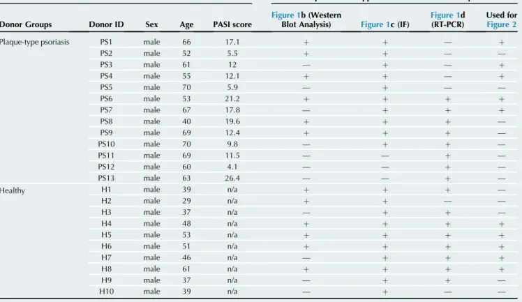
Supplementary Table S1. Donor Data and Experiments Applied to Donor Samples
Donor Information Experimental Application List of Donor Samples
Donor Groups Donor ID Sex Age PASI score
Figure 1b (Western
Blot Analysis) Figure 1c (IF)
Figure 1d (RT-PCR)
Used for Figure 2
Plaque-type psoriasis PS1 male 66 17.1 þ þ — þ
PS2 male 52 5.5 þ þ — —
PS3 male 61 12 — þ — þ
PS4 male 55 12.1 þ þ — þ
PS5 male 70 5.9 — þ — —
PS6 male 53 21.2 þ þ þ þ
PS7 male 67 17.8 — þ þ þ
PS8 male 40 19.6 þ þ þ —
PS9 male 69 12.4 þ þ þ —
PS10 male 70 9.8 — þ þ —
PS11 male 69 11.5 — — þ —
PS12 male 60 4.1 — — þ —
PS13 male 63 26.4 — — þ —
Healthy H1 male 39 n/a þ þ þ —
H2 male 29 n/a þ þ — —
H3 male 37 n/a — þ þ —
H4 male 48 n/a þ þ þ þ
H5 male 53 n/a þ þ þ þ
H6 male 51 n/a þ þ þ þ
H7 male 46 n/a — þ þ þ
H8 male 61 n/a þ þ þ þ
H9 male 37 n/a — þ þ —
H10 male 39 n/a — þ — —
Abbreviations: IF, immunofluorescence; n/a, not applicable.