royalsocietypublishing.org/journal/rsob
Research
Cite this article:
Climent-Cantó P, Carbonell A, Tamirisa S, Henn L, Pérez-Montero S, Boros IM, Azorín F. 2021 The tumour suppressor brain tumour (Brat) regulates linker histone dBigH1 expression in the
Drosophilafemale germline and the early embryo.
Open Biol.11:200408.
https://doi.org/10.1098/rsob.200408
Received: 21 December 2020 Accepted: 8 April 2021
Subject Area:
molecular biology/cellular biology
Keywords:linker histone H1, dBigH1, Brat, oogenesis, embryogenesis,
DrosophilaAuthors for correspondence:
Albert Carbonell
e-mail: albert.carbonell@irbbarcelona.org Fernando Azorín
e-mail: fambmc@ibmb.csic.es
†
These authors contributed equally to this study.
‡
Present address: National Heart and Lung Institute, Imperial College London, London, UK.
Electronic supplementary material is available online at https://doi.org/10.6084/m9.figshare.
c.5409175.
The tumour suppressor brain tumour (Brat) regulates linker histone dBigH1 expression in the Drosophila female germline and the early embryo
Paula Climent-Cantó
1,2,†, Albert Carbonell
1,2,†, Srividya Tamirisa
1,2,†, Laszlo Henn
3, Salvador Pérez-Montero
1,2,‡, Imre M. Boros
3,4and Fernando Azorín
1,21Institute of Molecular Biology of Barcelona, CSIC, Barcelona 08028, Spain
2Institute for Research in Biomedicine, IRB Barcelona, The Barcelona Institute for Science and Technology, Barcelona 08028, Spain
3Institute of Biochemistry, Biological Research Centre of Szeged, Szeged 6726, Hungary
4Department of Biochemistry and Molecular Biology, Faculty of Science and Informatics, University of Szeged, Szeged 6726, Hungary
AC, 0000-0002-1949-232X; FA, 0000-0002-8426-7858
Linker histones H1 are essential chromatin components that exist as multiple developmentally regulated variants. In metazoans, specific H1s are expressed during germline development in a tightly regulated manner. However, the mechanisms governing their stage-dependent expression are poorly under- stood. Here, we address this question in Drosophila, which encodes for a single germline-specific dBigH1 linker histone. We show that during female germline lineage differentiation, dBigH1 is expressed in germ stem cells and cystoblasts, becomes silenced during transit-amplifying (TA) cystocytes div- isions to resume expression after proliferation stops and differentiation starts, when it progressively accumulates in the oocyte. We find that dBigH1 silencing during TA divisions is post-transcriptional and depends on the tumour suppressor Brain tumour (Brat), an essential RNA-binding protein that regulates mRNA translation and stability. Like other oocyte-specific var- iants, dBigH1 is maternally expressed during early embryogenesis until it is replaced by somatic dH1 at the maternal-to-zygotic transition (MZT). Brat also mediates dBigH1 silencing at MZT. Finally, we discuss the situation in testes, where Brat is not expressed, but dBigH1 is translationally silenced too.
1. Introduction
Linker histones H1 constitute a conserved family of chromosomal proteins that bind nucleosomes and play central roles in the regulation of chromatin structure and function. Metazoan species usually contain multiple H1 variants that show differential patterns of expression during development and differentiation.
In this regard, a conserved feature in metazoans is the presence of germline- specific variants that replace somatic H1s in germ cells (reviewed in [1]). In many cases, female- and male-specific variants have been described. For instance, mammals usually contain three testis-specific H1s (H1T, HILS1 and H1T2) [2–7]
and one female-specific variant (H1oo) [8]. The presence of female- and male- specific H1s has also been reported inXenopus(B4 and H1fx) [9,10] and the sea urchin (Cs-H1 and SpH1) [11–13], while inCaenorhabditis elegans, the situation is more complex since the H1.1/HIS-24 variant is present in both the female and male germline, but it is also detected in somatic cells [14,15]. Female-specific variants have also been described in the zebrafish (H1M) [16,17] and echiura
© 2021 The Authors. Published by the Royal Society under the terms of the Creative Commons Attribution License http://creativecommons.org/licenses/by/4.0/, which permits unrestricted use, provided the original author and source are credited.
Downloaded from https://royalsocietypublishing.org/ on 03 February 2022
(H1M) [18]. Instead,Drosophilaencodes for a single germline- specific linker histone, dBigH1, that is expressed in both the male and the female germline [19]. Female-specific H1s usually persist during embryo development until the zygotic genome is activated at MZT [8,10–12,16–21].
In general, the patterns of expression of germline H1s are tightly regulated during lineage differentiation. Testis-specific variants are generally detected only after spermatogonia stop proliferation and differentiate to spermatocytes. In mam- mals, H1T is the first variant to be expressed in meiotic spermatocytes and, depending on the species, is also detected during spermatids differentiation [22–25]. On the other hand, expression of the other two mammalian male variants HILS1 and H1T2 is restricted to spermatids [4–7]. A similar situation is observed inDrosophila, where dBigH1 is detected in sper- matocytes, but not in the proliferating spermatogonia and upon spermatids differentiation [19]. dBigH1 is also detected in the male germ stem cell (GSC). Regarding female-specific H1s, their expression is mostly restricted to the oocyte and the early stages of embryo development. In humans and mice, H1oo expression is restricted to the growing/maturing oocyte entering meiosis and, after fertilization, it rapidly decays during the first mitotic divisions, becoming undetect- able at the 2–4 cells blastula stage when it is replaced by the somatic H1 variants [8,21,26,27]. In the sea urchin, the female-specific Cs-H1 variant is also replaced by somatic H1s at early cleavage stages [11,12]. However, the female- specific variants of Drosophila (dBigH1), zebrafish (H1M) andXenopus(B4) persist longer during embryo development, being replaced by the somatic H1s only after 14, 10 and 13 cleavages, respectively [10,16,17,19]. Translational regulat- ory mechanisms appear to play a central role in the regulation of germline H1s expression. InXenopus, translation of B4 mRNA is regulated by CPEBs proteins that bind at the 30UTR and, upon phosphorylation, promote polyadenylation [28–30]. Mamma- lian H1oo mRNA also contains several functional CPEs at the 30UTR [31,32]. In addition, in testes, several 5’UTR regulatory elements regulate HILS1 expression [4,33], and inDrosophila the translational repressor Bam is required to silence dBigH1 expression during spermatogonia proliferation [34]. However, little else is known about the mechanisms that govern stage- specific expression of germline H1s. Here, we address this question in theDrosophilafemale germline.
In Drosophila, the early stages of gametogenesis share remarkable similarities in females and males (reviewed in [35–39]). In both ovaries and testes, GSCs localize anterior, anchored to a niche of somatic cells, and divide asymmetri- cally for self-renewal and to produce daughter progenitor cells (cystoblasts (CBs) in females and gonioblasts (GBs) in males), which start the complex differentiation programme that, ultimately, leads to the production of functional gametes. Daughter cells undergo four successive rounds of transit-amplifying (TA) divisions with incomplete cytokinesis to produce a cyst of 16 sister germ cells (GCs) (spermatogonia in males and cystocytes in females) that remain intercon- nected and are surrounded by a somatic cells layer. Then, the pathways diverge; female cysts develop to produce a single egg, whereas male cysts differentiate to spermatocytes and undergo two meiotic divisions to produce 64 spermatids that develop to mature sperm cells. Our results show that, similar to males, dBigH1 is expressed in the female GSCs and CBs, is silenced in the proliferating TA cystocytes to resume expression upon oocyte differentiation. We report
that dBigH1 silencing in cystocytes depends on the tumour suppressor Brain tumour (Brat), a post-transcriptional regula- tor that is expressed in cystocytes and represses translation of GSC maintenance factors [40,41]. We also show that, during embryogenesis, Brat silences dBigH1 expression at MZT.
Altogether these results unveil the importance of post- transcriptional regulation in setting the patterns of expression of germline-specific H1 variants.
2. Material and methods
2.1. Fly stocks and genetic procedures
w1118, nos-Gal4::VP16[42] andDf(2 L)TE37C-7were obtained from Bloomington Drosophila Stock Center (BDSC). bratRNAi andbamRNAicorrespond to stocks 28 590 (y[1] v[1]; P{y[+t7.7]
v[+t1.8] = TRiP.HM05078}attP2) and 33 631 (y[1] v[1]; P{y[+
t7.7] v[+t1.8] = TRiP.HMS00029}attP2) from BDSC, respect- ively. dBigH1NTSOP CRISPR/CAS9 mutant is described in [43]. bamP-bam::GFP and bamP-GFP [44] were a gift from Dr M. Buszczak. bratK06028 was a gift from Dr J. Knoblich.
vasa-EGFP construct [45] was a gift from Dr A. Nakamura.
Transgenic lines carrying the various constructs described in figures 4 and 7 and electronic supplementary material, figure S4 were obtained by specific site-directed integration into ZH-86Fb and ZH-58A att line [46]. AllDrosophila stocks were maintained at 25°C on standard media. For RNAi knockdown, crosses were set up at 25°C.
2.2. Antibodies
Rabbit polyclonalαdBigH1 is described in [19]. Rat polyclonal αdBigH1 was raised as described in [19]. RabbitαdH1 was a gift from Dr J. Kadonaga and is described in [47]. Guinea pig polyclonalαTj was a gift from Dr D. Godt and is described in [48]. Rabbit αBrat was a gift from Dr J. Knoblich and is described in [49]. All other antibodies used in these experiments were commercially available: mouse monoclonalαadd (DSHB, 1B1), rat monoclonalαHA (Sigma, 3F10), mouse monoclonal αFasciclin III (DSHB, 7G10) and rat monoclonal αVasa (DSHB, 1ea).
2.3. Immunostaining experiments
Ovaries and testes were dissected in PBS, fixed in PBS with 4%
paraformaldehyde for 20 min, and then washed with PBS three times for 10 min each. The samples were first incubated in blocking solution, 2% bovine serum albumin diluted in PBT (PBS and 0.3%Triton X-100), for 1 h and then with the appro- priate primary antibodies diluted in blocking solution at 4°C overnight. The samples were washed with PBT three times for 10 min each and then incubated with blocking solution for 30 min. Then, samples were incubated with the appropriate secondary antibodies at 25°C for 2 h and washed with PBT three times for 10 min. Samples were mounted in Mowiol (Cal- biochem-Navabiochem) containing 0.2 ng µl−1DAPI (Sigma) and visualized in a confocal microscope (Leica TCS SP2- AOBS). Quantitative analyses presented in figures 1band 4d were performed using ImageJ. In each germarium, DAPI was used to define the nuclear area of three different cells from each region (1–3). Three additional ROIs (same area and non-nuclear) were also defined as background. In figure 1b,
ro yalsocietypublishing.org/journal/rsob Open Biol. 11 : 200408
2
Downloaded from https://royalsocietypublishing.org/ on 03 February 2022
the nuclear signal ofαdBigH1 was determined using the Gray mean value, and the average signal in each region was normal- ized against the average background signal. In figure 4d, the GFP signal was determined using the same method, but normalization was performed using the average signal of region 2, since the background levels of direct fluorescence were extremely low.
Embryos were dechorionated in bleach and fixed for 25 min in 1 : 1 solution of formaldehyde and heptane. Embryos were devitellinized in methanol followed by rehydration with PBT and blocking in PBT–BSA (2%). Samples were incubated with primary antibodies diluted in PBT–BSA at 4°C overnight.
After washing three times with PBT, embryos were incubated with secondary antibody and stained with DAPI at room temp- erature for 2 h followed by three washes in PBT. Embryos were mounted in vectashield (Vector labs) and imaged using Zeiss LSM 780 confocal microscope.
Primary antibodies used for immunostaining were:αdBigH1 (1 : 400),αdH1 (1 : 4000),αadd (1 : 100),αHA (1 : 200),αFasciclin
III (1 : 20),αVasa (1 : 300),αTj (1 : 5000) andαBrat (1 : 100). Sec- ondary antibodies conjugated with Cy2 and Cy3 (Jackson Immuno Research) were used at a 1 : 300 dilution.
2.4. RT – qPCR analysis
For qRT–PCR experiments, RNA was prepared from embryos using trizol reagent and purified using qiagen RNeasy mini kit following the manufacturer’s instructions. Total RNA (1 µg) was used for complementary DNA (cDNA) synthesis.
Reverse transcription was performed using oligo dT supplied in the kit. qRT–PCRs were run in triplicate in two independent experiments. Expression data were normalized toAct5Cand analysed using theΔΔCt method. Primers used were: dBigH1fw 50-AATATGGGCGAAGAAGAGGA-30, dBigH1rv 50-GAGAT TATCTGTCTCGACCTC-30, Act5cfw 50-CACCAAATCTTACA AAATGTGTGAC-30 and Act5crv 50-CATCGTCTCCGGCAA ATC-30.
(a)
DAPI
germarium
oocyte
oocyte stage 2
stage 3
stage 4
stage 5 merge
dBigH1
dBigH1
DAPI Fasciclin III
DAPI/dBigH1/Fasciclin III
1 2 3
(b)
(c)
region 1 region 2 region 3
TA region stage 1
oocyte oocyte GSCs
fusome spectrosome
CBs
FCs
nc
dBigH1
oocyte nc
DAPI
oocyte
nc
oocyte merge
1.0 1.5 2.0
region 1 region 2 region 3 normalized adBigH1 fluorescence intensity
**
0.0988
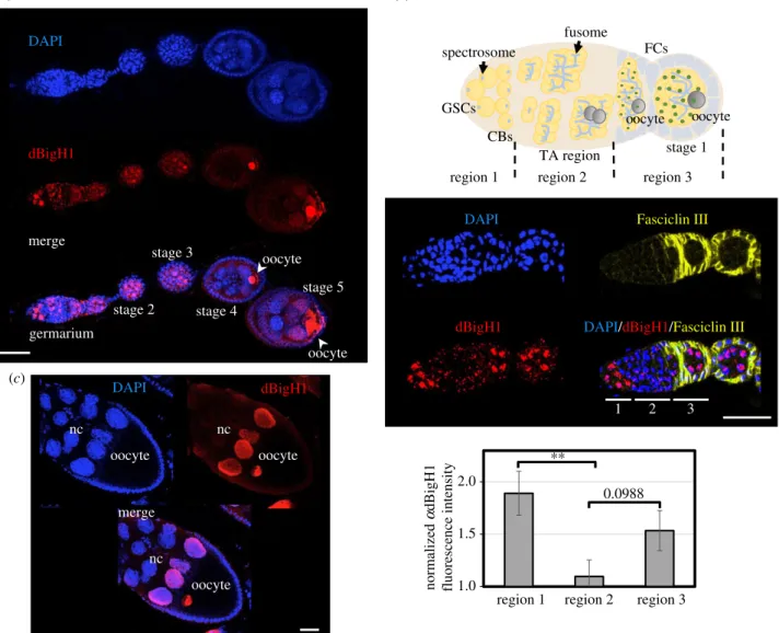
Figure 1.
The pattern of dBigH1 expression in
Drosophilaovaries. (a) Immunostaining with
αdBigH1 antibodies (in red) of an ovariole. The germarium and different stages of egg chamber development are indicated. The position of the oocyte is also indicated. DNA was stained with DAPI (in blue). Scale bar corresponds to 25 µm.
(b) Top: schematic of the germarium. Centre: immunostainings with
αdBigH1 (in red) and
αFasciclin III antibodies (in yellow), which label the somatic follicle cells (FC) surrounding the 16 cells cysts and the emerging egg chambers. Regions 1, 2 and 3 of the germarium are indicated. DNA was stained with DAPI (in blue). Scale bar corresponds to 25 µm. Quantitative analysis is shown at the bottom, where the intensity of
αdBigH1 immunostaining at regions 1, 2 and 3 is presented. (N = 3;
n
= 11; error bars are s.e.m.; two-tailed
t-Student,p-value: ** < 0.01.) (c) Immunostaining withαdBigH1 antibodies (in red) of a developing egg chamber (stage 10). The nurse cells (nc) and oocyte are indicated. DNA was stained with DAPI (in blue). Scale bar corresponds to 30 µm. See also electronic supplementary material, figures S1
–S3.
ro yalsocietypublishing.org/journal/rsob Open Biol. 11 : 200408
3
Downloaded from https://royalsocietypublishing.org/ on 03 February 2022
3. Results
3.1. dBigH1 expression is silenced during TA cystocytes divisions
Immunofluorescence (IF) experiments detected dBigH1 expression throughout oogenesis, from the germarium to the latest stages of egg chamber development (figure 1a). Female germline lineage differentiation begins at the germarium that, at the most anterior part, contains 2–3 female GSCs and the daughter CBs (region 1), which divide to generate developing cysts of increasing number of cystocytes (region 2). Then, at the 16-cell stage, cysts are surrounded by somatic epithelial follicle cells (FC), and bud off the germarium as individual egg chambers (region 3) [50] (figure 1b, top). In the germarium, intense nuclear αdBigH1 immunostaining was detected in region 1, being highly reduced to background levels during cystocytes proliferation in region 2, to reappear again in region 3 (figure 1b, centre and bottom). Cells showing
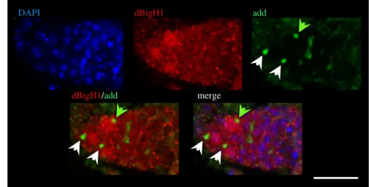
αdBigH1 immunostaining were positive for vasa, a specific germline marker [51,52], and negative for Traffic jam (Tj), a marker of somatic cells [48] (electronic supplementary material, figure S1), suggesting that dBigH1 expression was restricted to germ cells. We also analysed whether, in region 1, dBigH1 was expressed in both GSCs and CBs. For this pur- pose, we performed co-immunostaining experiments with αadd antibodies, which mark the spectrosome, a cytoskeleton structure that occupies an anterior position in GSCs, moves posterior in CBs and, later, grows and branches out to form the fusome that keeps cysts cells interconnected [53,54]
(figure 1b, top). In region 1, we detected nuclearαdBigH1 immunostaining in cells with anterior as well as posterior spec- trosomes (figure 2a), indicating that dBigH1 was expressed in both GSCs and CBs. In addition, silencing of dBigH1 expression during cystocytes proliferation was confirmed in flies carrying abamP-bam::GFPconstruct, which is specifically expressed in cystocytes [44], since nuclearαdBigH1 immuno- staining was not detected in cells expressing the reporter dBigH1
DAPI add
dBigH1/add merge
(a)
1 2 3
dBigH1 DAPI
GFP
dBigH1/GFP
merge bamP-bam::GFP (b)
Figure 2.
dBigH1 expression is silenced in TA cystocytes. (a) Immunostainings with
αdBigH1 (in red) and
αadd antibodies (in green), which label the spectrosome.
Only the tip region of the germarium, which contains the GSCs and CBs, is shown. Arrows indicate spectrosomes occupying an anterior (white) or a posterior (green) position. DNA was stained with DAPI (in blue). Scale bar corresponds to 15 µm. (b) The pattern of expression of a
bamP-bam::GFPreporter construct. dBigH1 was immunostained with
αdBigH1 antibodies (in red). GFP was direct fluorescence. Regions 1, 2 and 3 of the germarium are indicated. DNA was stained with DAPI (in blue). Scale bars correspond to 15 µm.
ro yalsocietypublishing.org/journal/rsob Open Biol. 11 : 200408
4
Downloaded from https://royalsocietypublishing.org/ on 03 February 2022
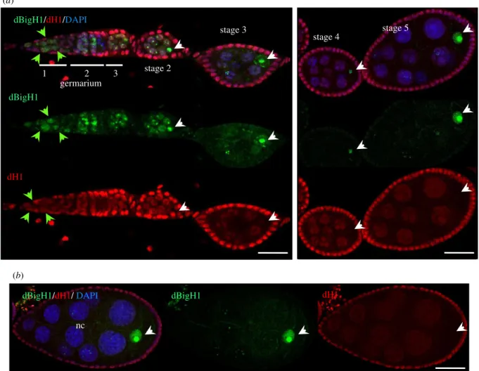
(figure 2b). After cystocytes stop proliferation and start to differentiate, dBigH1 expression resumed. In the budding cysts (region 3 of the germarium) and the early-developed egg chambers (stage 2), all nuclei were positive forαdBigH1 (figure 1a). Later, from stage 3 on, αdBigH1 immunostain- ing was progressively constrained to the oocyte nucleus (figure 1a,c), where it largely overlapped with DAPI at the condensed chromatin of the karyosome (electronic supple- mentary material, figure S2). A weak signal could also be detected in the nucleoplasm (electronic supplementary material, figure S2B; see also figure 3b), suggesting that a minor fraction of dBigH1 stays unbound and free in the nucleoplasm. At late developmental stages,αdBigH1 immu- nostaining was also detected in nuclei of the nurse cells (nc) proximal to the oocyte (figure 1a,c). This pattern of expression is very unusual and, interestingly, takes place around the stage when nc begin dumping of their content into the oocyte and, ultimately, die. In this regard, nc proximal to the oocyte are first in undergoing dumping. Though highly speculative, dBigH1 might regulate transcriptional activity in these cells during dumping. Of note, nuclear αdBigH1 immunostain- ing of germ cells was abolished in a null dBigH1NSTOP CRISPR/CAS9 mutant [43], showing its specificity (electronic supplementary material, figure S3). By contrast, background αdBigH1 immunostaining observed in the cytoplasm and somatic FC was also detected in the null dBigH1NSTOP
mutant, indicating it was unspecific (electronic supplementary material, figure S3).
We also analysed the pattern of expression in ovaries of the single somatic linker histone of Drosophila dH1 [55–57]. In addition to the somatic FC cells, which showed strongαdH1 immunostaining, we also detected dH1 expression in germline cells (figure 3a). In the germarium,αdH1 signal was detected in the GSCs/CBs, which express dBigH1, as well as in cystocytes, which lack dBigH1 (figure 3a). In the budding cysts and stage 2 egg chambers, dH1 expression was detected in both the nc and the oocyte that also contained dBigH1 (figure 3a). Later, when dBigH1 starts to accumulate in the oocyte (stages 3–5), dH1 expression decayed in the oocyte, becoming undetectable at stage 5 (figure 3a), while it was still detected in the nc (figure 3a). In the nc, dH1 expression also decreased upon development to almost undetectable levels (figure 3b).
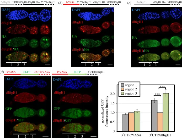
3.2. Silencing of dBigH1 in cystocytes is post- transcriptionally regulated
Results reported above suggest that dBigH1 expression is tightly regulated during early oogenesis, being silenced in proliferating cystocytes. This regulation is mainly post- transcriptional since expression of a ectopic dBigH1::HA construct, which carries thedBigH1regulatory elements and
dBigH1/dH1/ DAPI dBigH1 dH1
(a)
(b)
2 1
germarium
3 stage 2
stage 3 dBigH1/dH1/DAPI
stage 4
stage 5
dBigH1
dH1
nc
Figure 3.
The pattern of expression of somatic dH1 in
Drosophilaovaries. (a) Immunostainings with
αdBigH1 (in green) and
αdH1 (in red). The germarium and different stages of egg chamber development are indicated. Arrows indicate GSCs/CBs (green) and the oocyte nucleus (white). DNA was stained with DAPI (in blue).
Scale bar corresponds to 25 µm. (b) Immunostainings with
αdBigH1 (in green) and
αdH1 (in red) of an egg chamber at developmental stage 8. The nurse cells (nc) and oocyte nucleus (arrow) are indicated. DNA was stained with DAPI (in blue). Scale bars correspond to 25
μm.
ro yalsocietypublishing.org/journal/rsob Open Biol. 11 : 200408
5
Downloaded from https://royalsocietypublishing.org/ on 03 February 2022
largely mimics expression of endogenous dBigH1 (figure 4a), was not substantially altered when thedBigH1promoter was replaced by the germline-specificvasapromoter, which is ubi- quitously active in germline cells [45] (figure 4b) (see also figure 4d). We also observed that the deletion of thedBigH1 50UTR had no major effect on the pattern of dBigH1::HA expression in ovaries (figure 4c), suggesting that thedBigH1 30UTR is sufficient to silence dBigH1 expression in cystocytes.
In agreement, we observed that the 30UTR ofdBigH1silenced expression in cystocytes of a ubiquitously active vasa-EGFP reporter [45] (figure 4d). It must be noted that these ectopic dBigH1::HA constructs did not fully recapitulate dBigH1 silen- cing since we detected dBigH1::HA expression in cystocytes in approximately 25% of germaria. Noteworthy, the proportion of germaria showing ectopic dBigH1::HA expression in cysto- cytes tended to increase when the dBigH1 30UTR was replaced by that ofvasa(Fisher test,p-value: 0.227) (electronic supplementary material, figure S4A). Altogether these results suggest that elements within the dBigH1 30UTR mediate post-transcriptional silencing in cystocytes.
3.3. Brat regulates dBigH1 silencing in cystocytes
We noted that thedBigH13’UTR sequence contains two con- sensus binding sites for Brat [58] (electronic supplementary
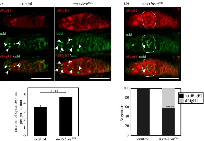
material, figure S5A), an important post-transcriptional regulator that is expressed in cystocytes (electronic sup- plementary material, figure S6) and represses translation of stem cell maintenance factors, promoting differentiation [40,41]. Interestingly, RNA immunoprecipitation experiments (RIP-Chip) performed in embryos showed that Brat interacts with thedBigH1mRNA [58]. Thus, we tested the possibility that Brat is involved in silencing dBigH1 expression in cysto- cytes. For this purpose, we performed RNAi-mediated depletion of Brat in ovaries using a nos-GAL4driver that is specifically expressed in the germline [42]. We observed that, in agreement with its role in promoting GSCs differentiation, Brat depletion increased the number of cells in which spectro- some structures were detected (figure 5a), suggesting an accumulation of GSCs/CBs. In addition, approximately 40%
of germaria showed detectable levels of dBigH1 expression in cyst cells interconnected by branched fusomes (figure 5b), suggesting that Brat is required to silence dBigH1 expression in cystocytes.
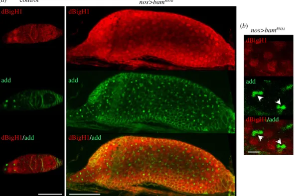
It was shown earlier that, in testes, dBigH1 expression is post-transcriptionally silenced during TA spermatogonial divisions by bag-of-marbles (Bam) [34]. Bam is also expressed in ovaries [59,60], where it represses translation of GSC main- tenance factors and induces differentiation [61–63]. In this regard, it is known that Bam is required for Brat expression dBigH1
HA
dBigH1/HA DAPI
1 2 3
(c)
dBigH1
HA
dBigH1/HA DAPI
1 2 3
dBigH1
HA
dBigH1/HA DAPI
1 2 3
PrdBigH1 5¢UTR/dBigH1 dBigH1::HA 3¢UTR/dBigH1
(a) (b) PrVASA 5¢UTR/dBigH1 dBigH1::HA 3¢UTR/dBigH1 PrdBigH1 dBigH1::HA 3¢UTR/dBigH1
PrVASA EGFP 3¢UTR/VASA PrVASA EGFP 3¢UTR/dBigH1
(d)
1 2 3
1 2 3
DAPI
GFP GFP
DAPI
dBigH1 dBigH1
dBigH1/GFP dBigH1/GFP
3¢UTR/dBigH1 0
0.5 1.0 1.5 2.0
3¢UTR/VASA normalized GFP fluorescence intensity
region 1 region 2
region 3 ***
***
Figure 4.
dBigH1 silencing in cystocytes depends on the 3
0UTR. (a
–c) The patterns of expression of ectopic dBigH1::HA constructs carrying the indicatedcis-regu-latory elements are presented. Immunostainings with
αdBigH1 (in red) and
αHA antibodies (in green) are shown. Regions 1, 2 and 3 of the germarium are indicated. DNA was stained with DAPI (in blue). Scales bar correspond to 15 µm. (d) The patterns of expression of EGFP constructs carrying the indicated
cis-regulatory elements are presented. GFP was direct fluorescence. DNA was stained with DAPI (in blue). Scale bars correspond to 15 µm. Quantitative analysis is shown in the right, where the intensity of GFP fluorescence in regions 1, 2 and 3 is presented for the indicated constructs. (N = 2;
n= 25; error bars are s.e.m.; two-tailed
t-Student,p-value: ***<0.001.) See also electronic supplementary material, figure S4A.ro yalsocietypublishing.org/journal/rsob Open Biol. 11 : 200408
6
Downloaded from https://royalsocietypublishing.org/ on 03 February 2022
in cystocytes since it represses the GSC maintenance factor nanos (nos) [63,64] that, in its turn, represses Brat [40].
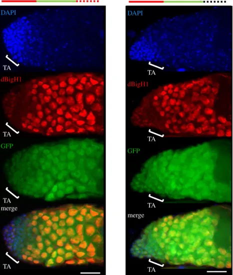
Thus, we anticipated that Bam would also regulated dBigH1 expression in cystocytes. We observed that the depletion of Bam in ovaries blocked early differentiation, giving rise to tumorous germaria that contained a large number of undifferentiated GSCs/CBs expressing dBigH1 (figure 6a). This strong phenotype, which was reported ear- lier in other Bam loss-of-function (LOF) mutations [59,65,66], made it challenging to determine the contribution of Bam to dBigH1 silencing in cystocytes. However, among the large number of GSCs/CBs observed upon Bam depletion, we detected dBigH1 expression in some early- developed cysts containing fusome-interconnected cells (figure 6b). These results suggest that, like in spermatogonia, Bam also regulates silencing of dBigH1 expression in cysto- cytes. Besides these similarities, the regulation of dBigH1 silencing in testes and ovaries shows some important differ- ences since, in contrast with what was observed in ovaries, the dBigH1 30UTR was not capable of silencing expression of vasa-EGFP in testes (figure 7) and replacement of the dBigH130UTR by the vasa30UTR did not affect silencing in spermatogonia of an ectopic dBigH1::HA construct (elec- tronic supplementary material, figure S4B). In this regard, we considered the possibility that alternative polyadenylation events could give rise to different 30UTRs in testes and
ovaries. However, RACE experiments showed the same dBigH1 30UTR in testes, ovaries and embryos (electronic supplementary material, figure S5A).
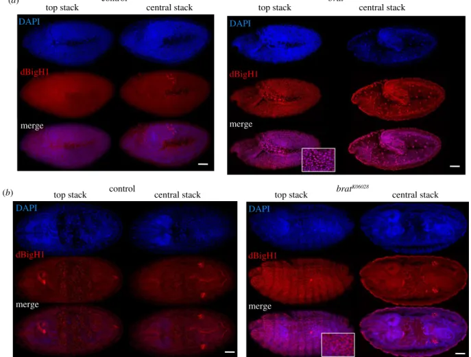
3.4. Brat also regulates dBigH1 silencing at MZT
It has been shown that Brat, which is maternally expressed during early embryogenesis, interacts with and silences a large subset of maternal mRNAs at MZT, among which the dBigH1 mRNA was identified [58,67]. Thus, we tested the possibility that Brat also silences dBigH1 expression at MZT.
For this purpose, we took advantage of the LOF mutation bratK06028, a recessive lethal P-element insertion allele that shows some defects in abdominal embryo segmentation and induces tumorous overgrowth in larval brains, where Brat is highly expressed [68,69]. Despite these defects, homozygous bratK06028mutants progress relatively normal through embry- ogenesis and larval development [58,67,69,70]. We observed that, like in control wild-type embryos, dBigH1 was ubiquitously expressed in homozygous bratK06028 embryos throughout blastoderm stages (electronic supplementary material, figure S7). However, while in control embryos, dBigH1 expression was constrained to the primordial germ cells (PGC) at gastrula stages (figure 8a,b, left panels), intense αdBigH1 immunostaining was detected in somatic cells in 46%
(N= 120) of homozygousbratK06028gastrula (figure 8a,b, right 0
1 2 3 4 5
control nos>bratRNAi
****
number of spectrosomes per germarium
0 20 40 60 80 100
control nos>bratRNAi
% germaria
no dBigH1 dBigH1
****
(a) (b)
dBigH1
add
dBigH1/add
dBigH1
add
dBigH1/add
control nos>bratRNAi
dBigH1
add
dBigH1/add
nos>bratRNAi
Figure 5.
dBigH1 silencing in cystocytes depends on Brat. (a) Top: immunostainings with
αdBigH1 (in red) and
αadd antibodies (in green), which label the spectrosome, of germaria from control wt and
bratRNAiflies in which Brat depletion was induced with a
nos-GAL4::VP16driver. Only the tip region of the germaria, which contains the GSCs and CBs, is shown. Arrows indicate spectrosomes. DNA was stained with DAPI (in blue). Scale bars correspond to 25 µm. Quantification of the results is shown below, where the number of spectrosomes per germarium is presented (N = 2;
n> 33; error bars are s.d.; two-tailed
t-Student,p-value: ****<0.0001). (b) Top: immunostainings with
αdBigH1 (in red) and
αadd antibodies (in green), which label the fusome, of a germarium from
bratRNAiflies in which Brat depletion was induced with a
nos-GAL4::VP16driver. A developing cyst showing dBigH1 expression is indicated by the circle. DNA was stained with DAPI (in blue).
Scale bar corresponds to 25 µm. Quantification of the results is shown below, where the proportion of germaria containing
αdBigH1-positive cysts is presented (N = 2;
n= 38; error bars are s.d.; Fisher test,
p-value: ****<0.0001). See also electronic supplementary material, figures S5 and S6.ro yalsocietypublishing.org/journal/rsob Open Biol. 11 : 200408
7
Downloaded from https://royalsocietypublishing.org/ on 03 February 2022
panels). Similar results were obtained in trans-heterozygous bratK06028/Df(2 L)TE37C-7embryos, which carried thebratK06028 mutation over the Df(2 L)TE37C-7 deficiency that uncovers Brat (58%,N= 108) (electronic supplementary material, figure S8). In addition to its role in translational regulation, Brat has been shown to regulate the stability of a subset of maternal tran- scripts at MZT, including the dBigH1 mRNA [58]. RT–qPCR experiments detected increased dBigH1 mRNA levels in bratK06028mutant embryos (electronic supplementary material, figure S5B), confirming the contribution of Brat to dBigH1 mRNA degradation/decay at MZT. Notably, in bratK06028 mutants, intenseαdH1 immunostaining was detected at gas- trula stages indicating that, under these conditions, dBigH1 and dH1 are both expressed (figure 9).
4. Discussion
Results reported here and elsewhere [34] show that the patterns of dBigH1 expression during the early stages of oogenesis and spermatogenesis are remarkably similar. In both cases, dBigH1 is expressed in GSCs and daughter progenitor cells, is silenced during TA divisions to resume expression when proliferation stops and differentiation begins. Moreover, translational regu- lation accounts for silencing of dBigH1 expression during TA divisions in both ovaries and testes. However, the actual mech- anisms involved show some important differences. Our results suggest that, in ovaries, Brat mediates translational dBigH1 silencing in cystocytes by directly binding thedBigH130UTR, which contains two Brat-binding sites. However, in testes, the situation must be different since Brat is not significantly expressed (see FlyAtlas and modENCODE tissue expression data in Flybase (https://flybase.org/reports/FBgn0010300)).
In good agreement, thedBigH130UTR is not required to silence
dBigH1 expression in spermatogonia and, along the same lines, it is not sufficient to silence vasa-EGFP expression.
Instead, in testes, dBigH1 silencing in spermatogonia is mediated by Bam [34]. Our results suggest that Bam is also required for dBigH1 silencing in cystocytes. However, in this case, its contribution might be indirect, through the activation of Brat expression [40]. Altogether these results suggest that, at least in part, the mechanisms governing translational regu- lation of dBigH1 expression are different in ovaries and testes.
We have also shown that Brat is required for dBigH1 silen- cing in gastrulated embryos. It has been reported that, during embryo development, Brat acts both as a translational repressor and a factor required for degradation/decay of maternal transcripts at MZT [58]. In this regard, sustained dBigH1 expression observed at gastrula stages inbratmutant embryos supports a contribution of Brat todBigH1mRNA stability at MZT. Instead, in early oogenesis, Brat probably acts as a repres- sor ofdBigH1mRNA translation since dBigH1 is silenced only transiently during TA divisions. Brat might also regulate the translation of maternaldBigH1transcripts in early embryo- genesis. The factors that control Brat action in repression or mRNA destabilization remain to be determined.
Our results challenge the usually accepted view that germ- line-specific H1s replace somatic variants, which implies that their patterns of expression do not generally overlap. Instead, we have shown that somatic dH1 is broadly expressed during oogenesis and, though it ends up being replaced by dBigH1 in the oocyte, the two variants largely coexist except during TA divisions, where dH1 is expressed, but dBigH1 is not. A similar situation was reported in testes, where dH1 coex- ists with dBigH1 in GSCs and GBs, but it is the only variant expressed in TA divisions [34]. However, in this case, once proliferation stops, dH1 expression is strongly silenced in spermatocytes, while dBigH1 is highly expressed [34]. Later, control
dBigH1
add
dBigH1/add dBigH1
add
dBigH1/add
nos>bamRNAi dBigH1
add
dBigH1/add (b)
(a) nos>bamRNAi
Figure 6.
The contribution of Bam to dBigH1 silencing in cystocytes. (a) Immunostainings with
αdBigH1 (in red) and
αadd antibodies (in green) of germaria from control wt and
bamRNAiflies in which Bam depletion was induced with a
nos-GAL4::VP16driver. Arrows indicate spectrosomes. Scale bars correspond to 25 µm.
(b) Enlarged image of immunostainings with
αdBigH1 (in red) and
αadd antibodies (in green) of early-developed cysts showing dBigH1 expression from
bamRNAiflies in which Bam depletion was induced with a
nos-GAL4::VP16driver. Arrows indicate growing fusomes. Scale bar corresponds to 5 µm.
ro yalsocietypublishing.org/journal/rsob Open Biol. 11 : 200408
8
Downloaded from https://royalsocietypublishing.org/ on 03 February 2022
dBigH1 is also silenced in spermatids [34]. Along the same lines, inbratmutant embryos, dBigH1 and dH1 also coexist at gastrula stages. Altogether, these results indicate that the pat- terns of expression of dH1 and dBigH1 are not necessarily exclusive. It is possible that dBigH1 and dH1 preferentially target different genomic loci since ectopic dBigH1 expression in S2 cells has shown that dBigH1 preferentially binds to and displaced dH1 from silent genomic regions with high dH1 con- tent [71]. On the other hand, recent results suggest a more complex situation since, in nullbigH1 mutants generated by CRISPR/CAS9, the lack of maternal dBigH1 is compensated by the expression of somatic dH1 from the earliest stages of embryo development [43,72], suggesting that dBigH1 represses dH1 expression in the early Drosophila embryo.
Further work is required to reach a better understanding of the actual link(s) between dBigH1 and dH1 expression and deposition.
Results reported here and elsewhere [19,34] show that the pattern of expression of dBigH1 is tightly regulated during germline lineage differentiation and embryogenesis, suggesting that dBigH1 plays specific functions in germline and embryo development. However, unveiling the functional contribution of dBigH1 is proving more difficult than antici- pated. Based on defects associated with a genetic mutation generated through imperfect excision of a 50UTR P-element insertion, dBigH1 was proposed to be essential during early
embryogenesis, contributing to the activation of the zygotic genome [19]. In addition, RNAi-mediated depletion of dBigH1 in testes induced strong developmental defects and reduced fertility [34]. However, CRISPR/CAS9 null bigH1 mutants turned out to be viable and fertile, progressing through embryogenesis likely due to the compensatory expression of somatic dH1 [43,72]. Moreover, CRISPR/CAS9 lines in which the CDS of dBigH1 was replaced by that of somatic dH1 are also viable, though showing DNA replication defects and altered chromatin condensation during early embryogenesis [73]. These observations suggest that to a large extent, dBigH1 and dH1 are functionally redundant, leaving the question of the possible specific functions of dBigH1 open.
In summary, our results show that the tumour suppressor Brat is crucial to silence dBigH1 expression in both ovaries and embryos. This regulatory mechanism might not be con- strained to the germline. In this regard, it was reported that, while dBigH1 is not detected in the normal larval brain (or any other somatic tissue), it becomes ectopically expressed in some Brat-induced brain tumours [74]. To what extent dBigH1 expression contributes to malignant growth in somatic tissues remains to be determined. It also remains to be determined if Brat orthologues in other species (such as human TRIM2,3, which are implicated in malignant glioma [75]) have similar effects in the expression of embryonic H1 linker histones.
PrVASA EGFP 3¢UTR/VASA
TA
TA
TA
TA
PrVASA EGFP 3¢UTR/dBigH1
TA
TA
TA
TA dBigH1
GFP
merge DAPI
dBigH1
GFP
merge DAPI
Figure 7.
The
dBigH1 30UTRdoes not silence gene expression in male TA-spermatogonia. The patterns of expression of EGFP constructs carrying the indicated
cis-regulatory elements are presented. The position of the TA region is indicated. GFP was direct fluorescence. DNA was stained with DAPI (in blue). Scale bars corre- spond to 25 µm. See also electronic supplementary material, figure S4B.
ro yalsocietypublishing.org/journal/rsob Open Biol. 11 : 200408
9
Downloaded from https://royalsocietypublishing.org/ on 03 February 2022
Data accessibility. Data and materials are available from the authors upon reasonable request.
Authors’contributions.P.C.-C., S.T. and S.P.-M. performed and designed the experiments and analysed the data. L.H. performed and designed the experiments, analysed the data, supervised the work and obtained financing. I.M.B. supervised the work and obtained
financing. A.C. performed and designed the experiments, analysed the data, directed and supervised the work, and wrote the paper. F.A. designed the experiments, directed and supervised the work, wrote the paper and obtained financing. All authors were involved in drafting and revising the article, and approving the article.
control (a)
dBigH1 DAPI
merge
bratK06028
(b)
dBigH1 DAPI
merge
dBigH1 DAPI
merge central stack
top stack top stack central stack
control bratK06028
central stack
top stack top stack central stack
DAPI
dBigH1
merge
Figure 8.
Brat regulates dBigH1 silencing in embryos. Immunostainings with
αdBigH1 antibodies (in red) of control wt and homozygous
bratK06028embryos at gastrula stages 9 (a) and 15 (b). Top and central stacks are presented. DNA was stained with DAPI (in blue). Scale bars correspond to 50 µm. For top stacks of homozygous
bratK06028embryos, enlarged views of merge images showing co-localization of
αdBigH1 signal with DAPI are also presented. See also electronic supplementary material, figures S5, S7 and S8.
dH1 DAPI
merge
bratK06028/Df(2L)TE37C-7 control
central stack
top stack top stack central stack
DAPI
dH1
merge
(a) (b)
Figure 9.
Sustained dBigH1 expression at gastrula stages does not affect dH1 expression. Immunostainings with
αdH1 antibodies (in red) of control wt and trans- heterozygous
bratK06028/Df(2 L)TE37C-7 embryos at gastrula stages 9 (a) and 15 (b). Top and central stacks are presented. DNA was stained with DAPI (in blue). Scale bars correspond to 50 µm. For top stacks, enlarged views of merge images are also presented.
ro yalsocietypublishing.org/journal/rsob Open Biol. 11 : 200408
10
Downloaded from https://royalsocietypublishing.org/ on 03 February 2022
Competing interests.The authors declare no conflicts of interest.
Funding. This work was supported by grants from MICINN (BFU2015-65082-P and PGC2018-094538-B-I00), the Generalitat de Catalunya (SGR2009-1023, SGR2014-204) and the European Community FEDER program to F.A., and from National Research, Development and Innovation Office (OTKA-116372) and Ministry for National Economy of Hungary (GINOP-2.3.2-15-2016-00032) to L.H./I.M.B.
Acknowledgements.We would like to thank Drs M. Buszczak, D. Godt, J. Kadonaga, J. Knoblich, M. Llimargas and A. Nakamura for kindly sharing fly stocks and antibodies. This work was carried out within the framework of the‘Centre de Referència en Biotecnologia’of the
‘Generalitat de Catalunya’. S.T. received funding from the European Union’s Horizon 2020 Research and Innovation Programme under the Marie Skłodowska-Curie grant agreement no. 754510. P.C.-C. and S.P.-M. acknowledge the receipt of FPI fellowships from MINECO.
References
1. Pérez-Montero S, Carbonell A, Azorín F. 2016 Germline-specific H1 variants: the‘sexy’linker histones.Chromosoma125, 1–13. (doi:10.1007/
s00412-015-0517-x)
2. Drabent B, Bode C, Doenecke D. 1993 Structure and expression of the mouse testicular H1 histone gene (H1t).Biochim. Biophys. Acta 1216, 311–333. (doi:10.1016/0167-4781(93) 90162-7)
3. Drabent B, Kardalinou E, Doenecke D. 1991 Structure and expression of the human gene encoding testicular H1 histone (H1t).Gene103, 263–268. (doi:10.1016/0378-1119(91)90284-I) 4. Iguchi N, Tanaka H, Yomogida K, Nishimune Y. 2003
Isolation and characterization of a novel cDNA encoding a DNA-binding protein (Hils1) specifically expressed in testicular haploid germ cells.
Int. J. Androl.26, 354–365. (doi:10.1046/j.0105- 6263.2003.00449.x)
5. Martianov I, Brancorsini S, Catena R, Gansmuller A, Kotaja N, Parvinen M, Sassone-Corsi P, Davidson I.
2005 Polar nuclear localization of H1T2, a histone H1 variant, required for spermatid elongation and DNA condensation during spermiogenesis.Proc. Natl Acad. Sci. USA102, 2808–2813. (doi:10.1073/pnas.
0406060102)
6. Tanaka Het al.2005 HANP1/H1T2, a novel histone H1-like protein involved in nuclear formation and sperm fertility.Mol. Cell Biol.25, 7107–7119.
(doi:10.1128/MCB.25.16.7107-7119.2005) 7. Yan W, Ma L, Burns KH, Matzuk MM. 2003 HILS1 is
a spermatid-specific linker histone H1-like protein implicated in chromatin remodeling during mammalian spermiogenesis.Proc. Natl Acad. Sci.
USA100, 10 546–10 551. (doi:10.1073/pnas.
1837812100)
8. Tanaka M, Hennebold JD, Macfarlane J, Adashi EY.
2001 A mammalian oocyte-specific linker histone gene H1oo: homology with the genes for the oocyte-specific cleavage stage histone (cs-H1) of sea urchin and the B4/H1 M histone of the frog.
Development128, 655–664.
9. Shechter D, Nicklay JJ, Chitta RK, Shabanowitz J, Hunt DF, Allis CD. 2009 Analysis of histones in Xenopus laevis. I. A distinct index of enriched variants and modifications exists in each cell type and is remodeled during developmental transitions.
J. Biol. Chem.284, 1064–1074. (doi:10.1074/jbc.
M807273200)
10. Smith RC, Dworkin-Rastl E, Dworkin MB. 1988 Expression of a histone H1-like protein is restricted
to earlyXenopusdevelopment.Genes Dev.2, 1284–1295. (doi:10.1101/gad.2.10.1284) 11. Brandt WF, Schwager SU, Rodrigues JA, Busslinger
M. 1997 Isolation and amino acid sequence analysis reveal an ancient evolutionary origin of the cleavage stage (CS) histones of the sea urchin.Eur. J.
Biochem.247, 784–791. (doi:10.1111/j.1432-1033.
1997.00784.x)
12. Mandl B, Brandt WF, Superti-Furga G, Graninger PG, Birnstiel ML, Busslinger M. 1997 The five cleavage- stage (CS) histones of the sea urchin are encoded by a maternally expressed family of replacement histone genes: functional equivalence of the CS H1 and frog H1 M (B4) proteins.Mol. Cell Biol.17, 1189–1200. (doi:10.1128/MCB.17.3.1189) 13. Strickland WN, Strickland M, Brandt WF, Von Holt C,
Lehmann A, Wittmann-Liebold B 1980 The primary structure of histone H1 from sperm of the sea urchinParechinus angulosus. 2. Sequence of the C-terminal CNBr peptide and the entire primary structure.Eur. J. Biochem.104, 567–578. (doi:10.
1111/j.1432-1033.1980.tb04460.x) 14. Jedrusik MA, Schulze E. 2003 Telomeric
position effect variegation inSaccharomyces cerevisiaebyCaenorhabditis eleganslinker histones suggests a mechanistic connection between germ line and telomeric silencing.Mol.
Cell Biol.23, 3681–3691. (doi:10.1128/MCB.23.10.
3681-3691.2003)
15. Jedrusik MA, Schulze E. 2007 Linker histone HIS-24 (H1.1) cytoplasmic retention promotes germ line development and influences histone H3 methylation inCaenorhabditis elegans.Mol. Cell Biol.27, 2229–2239. (doi:10.1128/MCB.01713-06) 16. Müller K, Thisse C, Thisse B, Raz E. 2002 Expression
of a linker histone-like gene in the primordial germ cells in zebrafish.Mech. Dev.117, 253–257.
(doi:10.1016/S0925-4773(02)00174-0) 17. Wibrand K, Olsen LC. 2002 Linker histone H1 M
transcripts mark the developing germ line in zebrafish.Mech. Dev.117, 249–252. (doi:10.1016/
S0925-4773(02)00173-9)
18. Franks RR, Davis FC. 1983 Regulation of histone synthesis during earlyUrechis caupo(Echiura) development.Dev. Biol.98, 101–109. (doi:10.1016/
0012-1606(83)90338-X)
19. Pérez-Montero S, Carbonell A, Morán T, Vaquero A, Azorín F. 2013 The embryonic linker histone H1 variant ofDrosophila, dBigH1, regulates zygotic genome activation.Dev. Cell26, 578–590. (doi:10.
1016/j.devcel.2013.08.011)
20. Newrock KM, Alfageme CR, Nardi RV, Cohen LH.
1978 Histone changes during chromatin remodeling in embryogenesis.Cold Spring Harb. Symp.
Quant. Biol.42, 421–431. (doi:10.1101/SQB.1978.
042.01.045)
21. Tanaka Y, Kato S, Tanaka M, Kuji N, Yoshimura Y.
2003 Structure and expression of the human oocyte-specific histone H1 gene elucidated by direct RT-nested PCR of a single oocyte.Biochem.
Biophys. Res. Commun.304, 351–357. (doi:10.1016/
S0006-291X(03)00610-7)
22. Drabent B, Bode C, Bramlage B, Doenecke D. 1996 Expression of the mouse testicular histone gene H1t during spermatogenesis.Histochem. Cell Biol.106, 247–251. (doi:10.1007/BF02484408)
23. Drabent B, Bode C, Miosge N, Herken R, Doenecke D. 1998 Expression of the mouse histone gene H1t begins at premeiotic stages of spermatogenesis.Cell Tissue Res.291, 127–132. (doi:10.1007/
s004410050986)
24. Seyedin SM, Kistler WS. 1980 Isolation and characterization of rat testis H1t. An H1 histone variant associated with spermatogenesis.J. Biol.
Chem.255, 5949–5954. (doi:10.1016/S0021- 9258(19)70722-4)
25. Steger K, Klonisch T, Gavenis K, Drabent B, Doenecke D, Bergmann M. 1998 Expression of mRNA and protein of nucleoproteins during human spermiogenesis.Mol. Hum. Reprod.4, 939–945.
(doi:10.1093/molehr/4.10.939)
26. Tanaka Met al.2005 H1FOO is coupled to the initiation of oocytic growth.Biol. Reprod.72, 135–142. (doi:10.1095/biolreprod.104.032474) 27. Mizusawa Y, Kuji N, Tanaka Y, Tanaka M, Ikeda E,
Komatsu S, Kato S, Yoshimura Y. 2010 Expression of human oocyte-specific linker histone protein and its incorporation into sperm chromatin during fertilization.Fertil. Steril.93, 1134–1141. (doi:10.
1016/j.fertnstert.2008.11.028)
28. Hake LE, Richter JD. 1994 CPEB is a specificity factor that mediates cytoplasmic polyadenylation during Xenopusoocyte maturation.Cell79, 617–627.
(doi:10.1016/0092-8674(94)90547-9)
29. Paris J, Swenson K, Piwnica-Worms H, Richter JD.
1991 Maturation-specific polyadenylation:in vitro activation by p34cdc2 and phosphorylation of a 58- kD CPE-binding protein.Genes Dev.5, 1697–1708.
(doi:10.1101/gad.5.9.1697)
30. Stebbins-Boaz B, Hake LE, Richter JD. 1996 CPEB controls the cytoplasmic polyadenylation of cyclin, Cdk2 and c-mos mRNAs and is necessary for oocyte
ro yalsocietypublishing.org/journal/rsob Open Biol. 11 : 200408
11
Downloaded from https://royalsocietypublishing.org/ on 03 February 2022
maturation inXenopus.EMBO J.15, 2582–2592.
(doi:10.1002/j.1460-2075.1996.tb00616.x) 31. Alizadeh Z, Kageyama S, Aoki F. 2005 Degradation
of maternal mRNA in mouse embryos: selective degradation of specific mRNAs after fertilization.
Mol. Reprod. Dev.72, 281–290. (doi:10.1002/mrd.
20340)
32. Racki WJ, Richter JD. 2006 CPEB controls oocyte growth and follicle development in the mouse.
Development133, 4527–4537. (doi:10.1242/dev.
02651)
33. Iguchi N, Tanaka H, Yamada S, Nishimura H, Nishimune Y. 2004 Control of mouse hils1 gene expression during spermatogenesis: identification of regulatory element by transgenic mouse.Biol.
Reprod.70, 1239–1245. (doi:10.1095/biolreprod.
103.024760)
34. Carbonell A, Pérez-Montero S, Climent-Cantó P, Reina O, Azorín F. 2017 The germline linker histone dBigH1 and the translational regulator Bam form a repressor loop essential for male germ stem cell differentiation.Cell Rep.21, 3178–3189. (doi:10.
1016/j.celrep.2017.11.060)
35. Kirilly D, Xie T. 2007 TheDrosophilaovary: an active stem cell community.Cell Res.17, 15–25. (doi:10.
1038/sj.cr.7310123)
36. Lehmann R. 2012 Germline stem cells: origin and destiny.Cell Stem Cell10, 729–739. (doi:10.1016/j.
stem.2012.05.016)
37. Spradling A, Fuller MT, Braun RE, Yoshida S. 2011 Germline stem cells.Cold Spring Harb. Perspect. Biol.
3, a002642. (doi:10.1101/cshperspect.a002642) 38. Xie T. 2012 Control of germline stem cell self-
renewal and differentiation in theDrosophilaovary:
concerted actions of niche signals and intrinsic factors.WIREs Dev. Biol.2, 261–273. (doi:10.1002/
wdev.60)
39. Yuan H, Yamashita YM. 2010 Germline stem cells:
stems of the next generation.Curr. Opin. Cell Biol.
22, 730–736. (doi:10.1016/j.ceb.2010.08.013) 40. Harris RE, Pargett M, Sutcliffe C, Umulis D, Ashe HL.
2011 Brat promotes stem cell differentiation via control of a bistable switch that restricts BMP signaling.Dev. Cell20, 72–83. (doi:10.1016/j.
devcel.2010.11.019)
41. Newton FG, Harris RE, Sutcliffe C, Ashe HL. 2015 Coordinate post-transcriptional repression of Dpp- dependent transcription factors attenuates signal range during development.Development142, 3362–3373. (doi:10.1242/dev.123273) 42. Van Doren M, Williamson AL, Lehmann R. 1998
Regulation of zygotic gene expression inDrosophila primordial germ cells.Curr. Biol8, 243–246.
(doi:10.1016/S0960-9822(98)70091-0)
43. Carbonell A, Henn L, Pérez-Roldán J, Tamirisa S, Szabó A, Boros IM, Azorín F. 2020 In response to Liet al.: Linker histones function inDrosophila embryogenesis.bioRxiv. (doi:10.1101/2020.03.21.
001529)
44. Chen D, McKearin DM. 2003 Dpp signaling silences bam transcription directly to establish asymmetric divisions of germline stem cells.Curr. Biol.13, 1786–1791. (doi:10.1016/j.cub.2003.09.033)
45. Sano H, Nakamura A, Kobayashi S. 2002 Identification of a transcriptional regulatory region for germline-specific expression of vasa gene in Drosophila melanogaster.Mech. Dev.112, 129–139.
(doi:10.1016/S0925-4773(01)00654-2)
46. Bischof J, Maeda RK, Hediger M, Karch F, Basler K.
2007 An optimized transgenesis system for Drosophilausing germ-line-specificwC31 integrases.Proc. Natl Acad. Sci. USA104, 3312–3317. (doi:10.1073/pnas.0611511104) 47. Bayona-Feliu A, Casas-Lamesa A, Reina O, Bernués
J, Azorín F. 2017 Linker histone H1 prevents R-loop accumulation and genome instability in heterochromatin.Nat. Commun.8, 283. (doi:10.
1038/s41467-017-00338-5)
48. Li MA, Alls JD, Avancini RM, Koo K, Godt D. 2003 The large Maf factor Traffic Jam controls gonad morphogenesis inDrosophila.Nat. Cell Biol.5, 994–1000. (doi:10.1038/ncb1058)
49. Betschinger J, Mechtler K, Knoblich JA. 2006 Asymmetric segregation of the tumor suppressor brat regulates self-renewal inDrosophilaneural stem cells.Cell124, 1241–1253. (doi:10.1016/j.cell.
2006.01.038)
50. Lin H, Spradling A. 1993 Germline stem cell division and egg chamber development in transplanted Drosophilagermaria.Dev Biol159, 140–152.
(doi:10.1006/dbio.1993.1228)
51. Lasko PF, Ashburner M. 1988 The product of the Drosophilagenevasais very similar to eukaryotic initiation factor-4A.Nature335, 611–617. (doi:10.
1038/335611a0)
52. Lasko PF, Ashburner M. 1990 Posterior localization of vasa protein correlates with, but is not sufficient for, pole cell development.Genes Dev.4, 905–921.
(doi:10.1101/gad.4.6.905)
53. de Cuevas M, Lee JK, Spradling AC. 1996 alpha- spectrin is required for germline cell division and differentiation in theDrosophilaovary.Development 122, 3959–3968.
54. Zaccai M, Lipshitz HD. 1996 Differential distributions of two adducin-like protein isoforms in the Drosophilaovary and early embryo.Zygote4, 159–166. (doi:10.1017/S096719940000304X) 55. Bayona-Feliu A, Casas-Lamesa A, Carbonell A,
Climent-Cantó P, Tatarski M, Pérez-Montero S, Azorín F, Bernués J. 2016 Histone H1: lessons from Drosophila.Biochim. Biophys. Acta1859, 526–532.
(doi:10.1016/j.bbagrm.2015.09.001)
56. Mariño-Ramírez L, Hsu B, Baxenavis AD, Landsman D. 2006 The histone database: a comprehensive resource for histones and histone fold-containing proteins.Proteins62, 838–842. (doi:10.1002/prot.
20814)
57. Nagel S, Grossbach U. 2000 Histone H1 genes and histone clusters in the genusDrosophila.
J. Mol. Evol.51, 286–298. (doi:10.1007/
s002390010090)
58. Laver JDet al.2015 Brain tumor is a sequence- specific RNA-binding protein that directs maternal mRNA clearance during theDrosophilamaternal-to- zygotic transition.Genome Biol.16, 94. (doi:10.
1186/s13059-015-0659-4)
59. McKearin D, Ohlstein B. 1995 A role for the Drosophilabag-of-marbles protein in the differentiation of cystoblasts from germline stem cells.Development121, 2937–2947.
60. Ohlstein B, McKearin D. 1997 Ectopic expression of theDrosophilaBam protein eliminates oogenic germline stem cells.Development124, 3651–3662.
61. Insco ML, Bailey AS, Kim J, Olivares GH, Wapinski OL, Tam CH, Fuller MT. 2012 A self-limiting switch based on translational control regulates the transition from proliferation to differentiation in an adult stem cell lineage.Cell Stem Cell11, 689–700.
(doi:10.1016/j.stem.2012.08.012)
62. Jin Z, Kirilly D, Weng C, Kawase E, Song X, Smith S, Schwartz J, Xie T. 2008 Differentiation-defective stem cells outcompete normal stem cells for niche occupancy in theDrosophilaovary.Cell Stem Cell2, 39–49. (doi:10.1016/j.stem.2007.10.021) 63. Li Y, Zhang Q, Carreira-Rosario A, Maines JZ,
McKearin DM, Buszczak M. 2013 Mei-p26 cooperates with Bam, Bgcn and Sxl to promote early germline development in theDrosophila ovary.PLoS ONE8, e58301. (doi:10.1371/journal.
pone.0058301)
64. Lavoie CA, Ohlstein B, McKearin DM. 1999 Localization and function of Bam protein require the benign gonial cell neoplasm gene product.Dev.
Biol.212, 405–413. (doi:10.1006/dbio.1999.9346) 65. McKearin DM, Spradling AC. 1990bag-of-marbles: a
Drosophilagene required to initiate both male and female gametogenesis.Genes Dev.4, 2242–2251.
(doi:10.1101/gad.4.12b.2242)
66. Ohlstein B, Lavoie CA, Vef O, Gateff E, McKearin DM.
2000 TheDrosophilacystoblast differentiation factor, benign gonial cell neoplasm, is related to DExH-box proteins and interacts genetically withbag-of- marbles.Genetics155, 1809–1819.
67. Sonoda J, Wharton RP. 2001Drosophilabrain tumor is a translational repressor.Genes Dev.15, 762–773.
(doi:10.1101/gad.870801)
68. Spradling AC, Stern D, Beaton A, Rhem EJ, Laverty T, Mozden N, Misra S, Rubin GM. 1999 The Berkeley Drosophilagenome project gene disruption project.
Single P-element insertions mutating 25% of vital Drosophilagenes.Genetics153, 135–177.
69. Arama E, Dickman D, Kimchie Z, Shearn A, Lev Z.
2000 Mutations in the beta-propeller domain of the Drosophilabrain tumor (brat) protein induce neoplasm in the larval brain.Oncogene19, 3706–3716. (doi:10.1038/sj.onc.1203706) 70. Reichardt I, Bonnay F, Steinmann V, Loedige I,
Burkard TR, Meister G, Knoblich JA. 2018 The tumor suppressor Brat controls neuronal stem cell lineages by inhibiting Deadpan and Zelda.EMBO Rep.19, 102–117. (doi:10.15252/embr.201744188) 71. Climent-Cantó P, Carbonell A, Tatarski M, Reina O,
Bujosa P, Font-Mateu J, Bernués J, Beato M, Azorín F. 2020 The embryonic linker histone dBigH1 alters the functional state of active chromatin.Nucleic Acids Res.48, 4147–4160. (doi:10.1093/nar/
gkaa122)
72. Li KK, Han D, Chen F, Li R, Zhou B-H, Bai Y, Yuan K, Rong YS. 2019 Compensatory replacement of the
ro yalsocietypublishing.org/journal/rsob Open Biol. 11 : 200408
12
Downloaded from https://royalsocietypublishing.org/ on 03 February 2022
BigH1 variant histone by canonical H1 supports normal embryonic development inDrosophila.
bioRxiv. (doi:10.1101/789735)
73. Henn L, Szabó A, Imre L, Román Á, Ábrahám A, Vedelek B, Nánási Jr P, Boros IM. 2020 Alternative linker histone permits fast paced
nuclear divisions in earlyDrosophilaembryo.Nucleic Acids Res.48, 9007–9018. (doi:10.1093/nar/
gkaa624)
74. Janic A, Mendizabal L, Llamazares S, Rossell R, Gonzalez C. 2010 Ectopic expression of germline genes drives malignant brain tumor growth in
Drosophila.Science330, 1824–1827. (doi:10.1126/
science.1195481)
75. Boulay JL, Stiefel U, Taylor E, Dolder B, Merlo A, Hirth F. 2009 Loss of heterozygosity of TRIM3 in malignant gliomas.BMC Cancer9, 71. (doi:10.1186/
1471-2407-9-71)
ro yalsocietypublishing.org/journal/rsob Open Biol. 11 : 200408
13