Methylation profiling of choroid plexus tumors reveals 3 clinically distinct subgroups
Christian Thomas
†, Martin Sill
†, Vincent Ruland, Anika Witten, Stefan Hartung, Uwe Kordes, Astrid Jeibmann, Rudi Beschorner, Kathy Keyvani, Markus Bergmann, Michel Mittelbronn, Torsten Pietsch, Jo¨rg Felsberg, Camelia M. Monoranu, Pascale Varlet, Peter Hauser, Adriana Olar, Richard G. Grundy, Johannes E. Wolff, Andrey Korshunov, David T. Jones, Melanie Bewerunge-Hudler, Volker Hovestadt, Andreas von Deimling, Stefan M. Pfister, Werner Paulus, David Capper
†, and Martin Hasselblatt
†Institute of Neuropathology, University Hospital Mu¨nster, Mu¨nster, Germany (C.T., V.R., A.J., W.P., M.H.); Division of Biostatistics, German Cancer Research Center (DKFZ), Heidelberg, Germany (M.S.); Core Facility Genomics of the Medical Faculty Mu¨nster, Mu¨nster, Germany (A.W.); Department of Pediatric Hematology and Oncology, University Medical Center Hamburg-Eppendorf, Hamburg, Germany (S.H., U.K.); Department of Neuropathology, Institute for Pathology and Neuropathology, University of Tu¨bingen, Tu¨bingen, Germany (R.B.); Faculty of Medicine, Institute of Neuropathology, University of Duisburg-Essen, Essen, Germany (K.K.); Department of Neuropathology, Klinikum Bremen-Mitte, Bremen, Germany (M.B.); Institute of Neurology (Edinger Institute), Goethe University, Frankfurt, Germany (M.M.); Institute of Neuropathology, University of Bonn Medical Center, Bonn, Germany (T.P.); Department of Neuropathology, Heinrich-Heine-University Du¨sseldorf, Du¨sseldorf, Germany (J.F.); Department of Neuropathology, Institute of Pathology, University of Wu¨rzburg, and Comprehensive Cancer Center Mainfranken (CCCM), Wu¨rzburg, Germany (C.M.M.); Department of Neuropathology, Sainte-Anne Hospital, Paris, France (P.V.); 2nd Department of Paediatrics, Semmelweis University, Budapest, Hungary (P.H.); Department of Hematopathology, Molecular Diagnostic Laboratory, The University of Texas MD Anderson Cancer Center, Houston, Texas (A.O.); Children’s Brain Tumour Research Centre, School of Medicine, Queen’s Medical Centre, University of Nottingham, Nottingham, UK (R.G.G.); Department of Pediatric Hematology Oncology and Blood and Marrow Transplantation, Cleveland Clinic Children’s Hospital, Cleveland, Ohio (J.E.W.); Department of Neuropathology, University Hospital Heidelberg, Heidelberg, Germany (A.K., A.v.D., D.C.); Clinical Cooperation Unit Neuropathology, German Research Center (DKFZ), Heidelberg, Germany (A.K., A.v.D., D.C.); German Cancer Consortium (DKTK), German Cancer Research Center (DKFZ), Heidelberg, Germany (A.K., D.T.J., A.v.D., S.M.P., D.C.); Division of Pediatric Neurooncology, German Cancer Research Center (DKFZ), Heidelberg, Germany (D.T.J., S.M.P.); Department of Pediatric Oncology and Hematology, University Hospital Heidelberg, Heidelberg 69120, Germany (D.T.J., S.M.P.); Microarray Unit, Genomics and Proteomics Core Facility, German Cancer Research Center (DKFZ), Heidelberg, Germany (M.B.-H.); Division of Molecular Genetics, German Cancer Research Center (DKFZ), Heidelberg, Germany (V.H.) Corresponding Author:Martin Hasselblatt, MD, Institute of Neuropathology, University Hospital Mu¨nster, Pottkamp 2, Mu¨nster 48149, Germany (hasselblatt@uni-muenster.de).
†C.T., M.S., D.C. and M.H. contributed equally to this work.
See the editorial by Segal and Karajannis, on pages 757–758.
Background.Choroid plexus tumors are intraventricular neoplasms derived from the choroid plexus epithelium. A better knowl- edge of molecular factors involved in choroid plexus tumor biology may aid in identifying patients at risk for recurrence.
Methods.Methylation profiles were examined in 29 choroid plexus papillomas (CPPs, WHO grade I), 32 atypical choroid plexus papillomas (aCPPs, WHO grade II), and 31 choroid plexus carcinomas (CPCs, WHO grade III) by Illumina Infinium HumanMethy- lation450 Bead Chip Array.
Results.Unsupervised hierarchical clustering identified 3 subgroups: methylation cluster 1 (pediatric CPP and aCPP of mainly supratentorial location), methylation cluster 2 (adult CPP and aCPP of mainly infratentorial location), and methylation cluster 3 (pediatric CPP, aCPP, and CPC of supratentorial location). In methylation cluster 3, progression-free survival (PFS) accounted for a mean of 72 months (CI, 55-89 mo), whereas only 1 of 42 tumors of methylation clusters 1 and 2 progressed (P,.001).
On stratification of outcome data according to WHO grade, all CPCs clustered within cluster 3 and were associated with shorter overall survival (mean, 105 mo [CI, 81-128 mo]) and PFS (mean, 55 mo [CI, 36-73 mo]). The aCPP of methylation cluster 3 also progressed frequently (mean, 69 mo [CI, 44-93 mo]), whereas no tumor progression was observed in aCPP of methylation clusters 1 and 2 (P,.05). Only 1 of 29 CPPs recurred.
Received 17 August 2015; accepted 15 December 2015
Published by Oxford University Press on behalf of the Society for Neuro-Oncology 2016. This work is written by (a) US Government employee(s) and is in the public domain in the US.
doi:10.1093/neuonc/nov322
Advance Access date 28 January 2016
Conclusions.Methylation profiling of choroid plexus tumors reveals 3 distinct subgroups (ie, pediatric low-risk choroid plexus tu- mors [cluster 1], adult low-risk choroid plexus tumors [cluster 2], and pediatric high-risk choroid plexus tumors [cluster 3]) and may provide useful prognostic information in addition to histopathology.
Keywords:atypical choroid plexus papilloma, choroid plexus carcinoma, copy-number alterations, epigenetics, prognosis.
Choroid plexus tumors are rare intraventricular neoplasms de- rived from the choroid plexus epithelium and represent 0.2% of all central nervous system neoplasms and 10%–20% of brain tumors arising during the first year of life.1,2According to the World Health Organization (WHO) classification, choroid plexus tumors can be divided into 3 entities based on histopathologi- cal features. Choroid plexus papilloma (CPP, WHO grade I) closely resembles normal choroid plexus tissue. The defining feature of atypical choroid plexus papilloma (aCPP, WHO grade II) is the presence of increased mitotic activity (≥2 mito- ses/10 high-power fields), while choroid plexus carcinoma (WHO grade III) is characterized by frank signs of malignancy including brisk mitotic activity, nuclear pleomorphism, high cel- lularity, blurring of the papillary growth pattern, and necrosis.3 In very young children, CPP and aCPP share molecular signa- tures and a comparable excellent outcome.4,5In contrast, aCPP is associated with recurrence in older children (≥3 y) and adults.6,7Underlying biological factors explaining these age- dependent prognostic differences in aCPP remain uncertain. In recent years, high-resolution molecular profiling led to the iden- tification of distinct molecular subgroups in a variety of tumor entities.8–10High-density DNA methylation arrays allow robust molecular subgrouping and copy number profiling from small amounts of archival formalin-fixed paraffin-embedded (FFPE) material.11Recently, methylation profiling has been reported to segregate CPC from CPP and aCPP.4Interestingly, methylation profiles of some CPPs and aCPPs in this series also clustered with CPCs, raising the possibility that methylation profiling could be used to identify patients at risk of tumor recurrence.
We aimed to characterize methylation profiles and clinical and histopathological features in a larger series of choroid plex- us tumors across all age groups. Here we show that methyla- tion profiling of choroid plexus tumors reveals 3 distinct molecular subgroups, which may aid in identifying patients at risk of recurrence.
Materials and Methods
Samples and PatientsFFPE samples of 92 choroid plexus tumors were collected from the archives of the Institutes of Neuropathology in Mu¨nster, Heidelberg, Bremen, Du¨sseldorf, Essen, Frankfurt, Tu¨bingen, and Wu¨rzburg as well as institutions that had previously sub- mitted cases for reference in context of the CPT-SIOP trials.
Two cases were retrieved from the Children’s Cancer and Leu- kemia Group Tissue Bank, Leicester, UK. Of these tumors, 19 had already been included in a previous series examining copy- number alterations using high-resolution arrays.12The histopa- thology of all samples was reviewed according to 2007 WHO criteria. Follow-up information could be retrieved from the
CPT-SIOP registry and treating physicians for 77 cases (84%).
Three non-neoplastic choroid plexus samples obtained from neuropathologically normal autopsy brains served as controls.
Investigations were approved by the Mu¨nster ethics committee (2007-420-f-S).
Immunohistochemistry
Staining for p53 was performed using a mouse monoclonal antibody (BP53 – 12; 1:100; citrate buffer pH 6.1 for antigen retrieval; Zytomed). The streptavidin-biotin method was used on an automated staining system (TechMate, DAKO). Expression of p53 was evaluated semiquantitatively by scoring the percent- age of stained nuclei. Strong nuclear staining in.10% of tumor cells was considered positive.
450k Methylation Array
Genomic DNA was isolated from FFPE material using the Max- well LEV FFPE Tissue DNA Purification Kit (Promega). DNA was subjected to quality analysis using the Infinium HD FFPE QC assay protocol (Illumina). Tumor samples were then subjected to bisulfite conversion using the EZ DNA Methylation-Gold-Kit (Zymo Research), followed by the Infinium HD FFPE restore pro- tocol. Samples were analyzed using the Illumina Infinium HumanMethylation450 Bead Chip according to the manufac- turer’s instructions at the Core Facility Genomics of the Medical Faculty Mu¨nster or the German Cancer Research Center (DKFZ) Genomics and Proteomics Core Facility. The following criteria were applied to filter the data: removal of probes targeting sex chromosomes, removal of probes containing a single nucle- otide polymorphism (dbSNP132 Common) within 5 base pairs of and including the targeted CpG-site (n¼24 536) and probes not mapping uniquely to the human reference genome (hg19) allowing for one mismatch (n¼9993). In total, 438 370 probes were kept for analysis.
To cluster samples and determine an optimal number of stable clusters, unsupervised consensus clustering13was applied. Cluster- ing was performed using the beta values of the 10 000 most variable methylated probes as measured by median absolute devi- ation. Within each of the 1000 resampling iterations of the consen- sus clustering, a hierarchical clustering using ward linkage as agglomeration method and Euclidean distance as distance measure was applied. A stable clustering forK¼3 clusters was de- termined by visually inspecting the resulting consensus matrices and delta-K criteria. To reorder the 10 000 most variable probes for the heat map visualization, probes were clustered by agglom- erative hierarchical clustering using the Euclidean distance as dis- tance measure and average linkage as agglomeration method.
Copy-number Analysis
Copy-number profiles were derived from intensity measures of the methylation probes as recently described10using the “con- umee” R package available through BioConductor (http://
bioconductor.org/packages/release/bioc/html/conumee.html).
A total of 72 tumors fulfilled the quality criteria for copy- number analysis.
Statistical Analysis
Patient characteristics were summarized using median and interquartile range (IQR) for continuous variables as well as fre- quency and percentage for categorical variables. Overall sur- vival (OS) time was defined as the time from the date of diagnosis to the date of death. Progression-free survival (PFS) time was defined as the time from the date of diagnosis to the date of death or relapse, whichever occurred first. Continu- ous and categorical variables were compared using the Mann-WhitneyUtest and the chi-square test, respectively.
For the comparison of genetic alterations, the chi-square test with Bonferroni correction for multiple comparisons was ap- plied. Survival analysis was performed using Kaplan-Meier esti- mation for survival curves and the log-rank test using IBM SPSS 22 software (release 22.0). In order to adjust for confounders among co-factors, a multivariate Cox proportional hazards model was fit. Forward stepwise model selection was per- formed by the Wald approach with the saturated model includ- ing WHO grade, age, location, copy-number alterations, and p53 status as co-factors. For all analyses,P,.05 was consid- ered to be significant.
Results
The 92 choroid plexus tumors comprised 29 CPPs (31%), 32 aCPPs (35%), and 31 CPCs (34%). As shown in Table1, the me- dian age of the 54 male and 38 female patients was 4 years (IQR, 1 –16 y). Fifty-nine tumors (64%) were located supraten- torially (ie, in the lateral or the third ventricle), whereas
27 tumors [29%] were of infratentorial location in the vicinity of the fourth ventricle. Data on tumor location were not avail- able for 6 tumors (7%). Overexpression of p53 protein was ob- served in 9 of 30 CPCs (37%) and 1 of 25 (4%) aCPPs that could be examined. For further details on patient characteristics and treatment, see Supplementary material, Table S1.
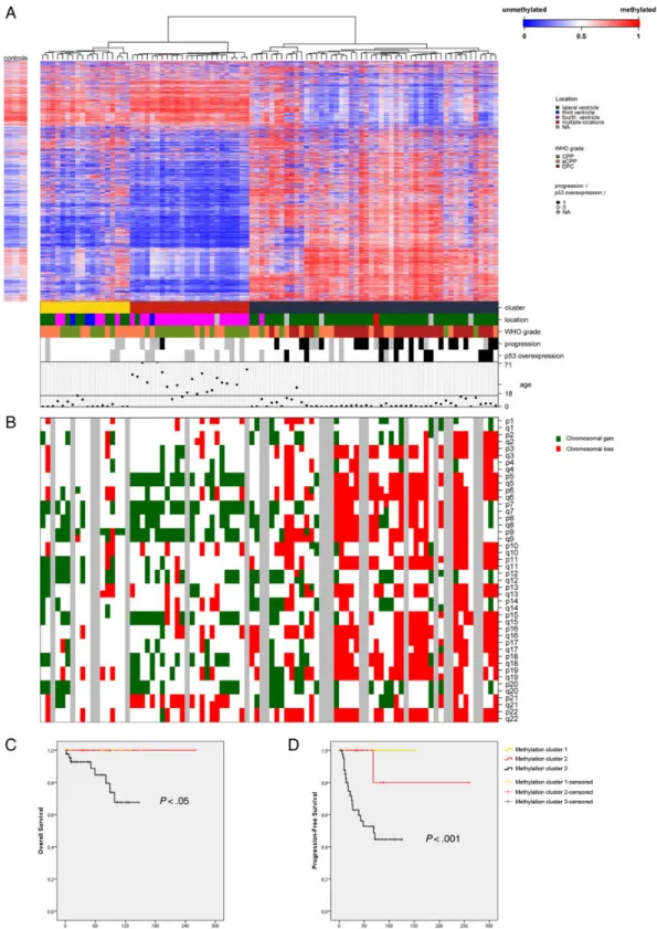
Unsupervised hierarchical clustering of DNA methylation patterns of the 92 tumors resulted in 3 clusters (Fig.1). Tumors of methylation cluster 1 comprised 8 CPPs (44%) and 10 aCPPs (56%) characterized by young age [median age, 1 y [IQR, 0–6 y)]
and mainly supratentorial location (72%). As shown in Fig.1B, frequent copy-number alterations observed in methylation cluster 1 included gains of chromosomes 12 (62%), 9p (62%), and 11 (54%). In contrast, tumors of methylation cluster 2 (16 CPPs [67%] and 8 aCPPs [33%]) were characterized by adult age (median age, 38 y [IQR, 25 –52 y]) and mainly infratentorial location (88%). Frequent copy-number alterations observed in methylation cluster 2 included gains of chromosomes 5 (82%), 7 (77%), and 9 (72%) as well as losses of chromosome 21q (64%). Tumors of methylation cluster 3 comprised CPCs (31 CPCs (62%) but also 14 aCPPs (28%) and 5 CPPs (10%). They were characterized by young age (median age, 2 y [IQR, 1 – 6 y]) and supratentorial location (88%). Recurrent copy-number alterations observed in methylation cluster 3 included chromo- somal losses such as 6q (73%), 6p, 22 (70%), 11p, 1, 6, and 19q (65%). Gains affecting chromosome 1p32 – 35.3, for which a role in the oncogenesis of CPC has been recently suggested,14 were present in 10 cases (20%) of methylation cluster 3 (8 CPCs and 2 aCPPs) but not in the other clusters (chi-squareP¼.02).
As shown in Supplementary material, Table S2, and summa- rized in Supplementary material, Figure S1, a number of genes was differentially methylated between clusters. Interest- ingly, MGMT was not among the differentially methylated genes but was unmethylated in all choroid plexus tumors examined.
The majority of CPPs and aCPPs were encountered within methylation cluster 1 and methylation cluster 2. Both of these clusters differed significantly by age and tumor location (P,.001). Both methylation clusters also differed on the
Table 1. Patient characteristics
All (N¼92) Cluster 1 (N¼18) Cluster 2 (N¼24) Cluster 3 (N¼50)
Sex (male/female) 54/38 11/7 11/13 32/18
Diagnosis
CPP 29 (31%) 8 (44%) 16 (67%) 5 (10%)
aCPP 32 (35%) 10 (56%) 8 (33%) 14 (28%)
CPC 31 (34%) 0 (0%) 0 (0%) 31 (62%)
Age (y) (median, IQR) 4 (1–16) 1 (0– 6) 38 (25–52) 2 (1–6)
Location
Supratentorial 59 (64%) 13 (72%) 2 (8%) 44 (88%)
Infratentorial 27 (29%) 5 (28%) 21 (88%) 1 (2%)
Unknown 6 (7%) 0 (0%) 1 (4%) 5 (10%)
Positive p53 status (nuclear accumulation of p53 protein in.10% of tumor cells)
10/75 (13%) 0/13 (0%) 0/16 (0%) 10/46 (22%)
Abbreviations: aCPP, atypical choroid plexus papilloma; CPC, atypical choroid plexus papilloma; CPP, choroid plexus papilloma; IQR, interquartile range.
Fig. 1. Methylation profiling of choroid plexus tumors. (A) Heat map of hierarchical clustering of methylation profiles as well as tumor location, neuropathological diagnosis, progression status, p53 status, and patient age. Note that choroid plexus tumors can be divided into 3 clusters:
methylation cluster 1 (young age and mainly supratentorial location), methylation cluster 2 (adult age and mainly infratentorial location), and methylation cluster 3 (young age and supratentorial location). Legend: aCPP, atypical choroid plexus papilloma; CPP, choroid plexus papilloma; CPC, choroid plexus carcinoma; NA, not available. (B) For each sample (N¼72) copy-number alterations (CNAs) are shown as indicated (green for chromosomal gain and red for chromosomal loss). (C) Kaplan Meier analysis showing that methylation cluster 3 is associated with shorter overall survival and progression-free survival as compared with methylation clusters 1 and 2. Log-rank testP,.05 andP,.001, respectively.
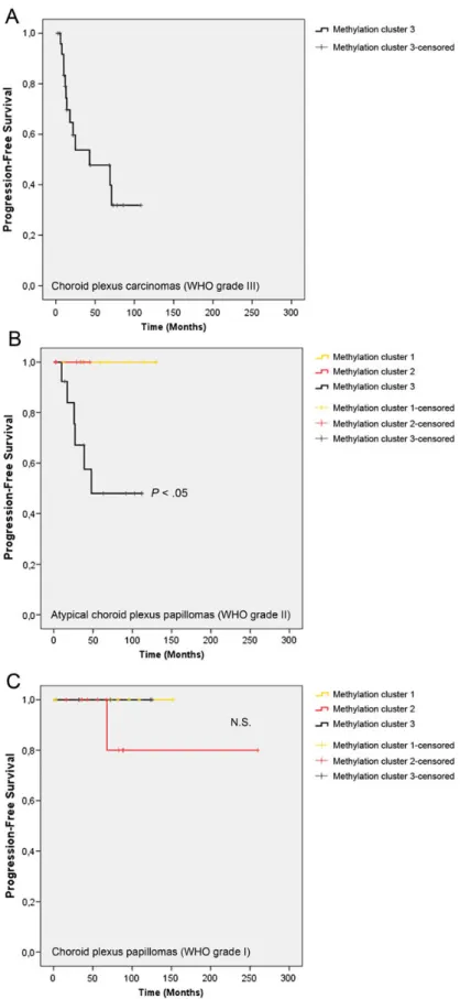
Fig. 2. Methylation profiling identifies molecular high-risk and low-risk clusters. Kaplan-Meier estimates of progression-free survival according to methylation clusters, examined (A) in choroid plexus carcinomas (WHO grade III,N¼26), (B) in atypical choroid plexus papillomas (WHO grade II, N¼27), and (C) choroid plexus papillomas (WHO grade I,N¼23). Note significant prognostic role of methylation clustering in atypical choroid plexus papillomas (log-rank testP,.05).
genetic level, with gains of chromosome 5 and losses of chromo- some 21q being more frequently encountered in methylation cluster 2 (P¼.0001 andP¼.02, respectively). Irrespective of these differences, outcome in methylation cluster 1 and meth- ylation cluster 2 was favorable: only one patient experienced tumor progression, and no patient succumbed to disease (me- dian follow-up, 78 mo). In contrast, the prognosis of patients in methylation cluster 3 was worse. In this cluster, which con- tained all CPCs and all p53-positive tumors examined, a total of 8 tumor-related deaths occurred, whereas none of the patients of methylation cluster 1 and methylation cluster 2 suc- cumbed to disease (Fig.1C, log-rank testP,.05). On Kaplan- Meier analysis (Fig.1D), estimated PFS accounted for a mean of only 72 months (CI, 55-89 mo) while only 1 of 42 tumors (2%) in methylation clusters 1 and 2 recurred (log-rank test P,.001). On multivariate analysis taking into account WHO grade, age, location, copy-number alterations, and p53 status as co-factors, however, WHO grade (but not methylation clus- tering) was the only independent predictor of OS, whereas WHO grade and p53 status remained the only predictors of PFS.
Next, the prognostic role of methylation clustering was examined according to WHO grade. As shown in Fig.2A, all CPCs (WHO grade III) clustered in cluster 3 and were associated with decreased OS (mean, 105 mo [CI, 81-128 mo]) and PFS (mean, 55 mo [CI, 36-73 mo]). Importantly, however, all recur- rences observed in aCPPs (WHO grade II) also occurred in this cluster (cluster 3; mean PFS, 69 mo [CI, 44-93 mo]), while none of the aCPPs of the low-risk clusters (cluster 1 and cluster 2) progressed (Fig.2B,P,.05). Only one CPP (WHO grade I) of the whole cohort recurred (cluster 2, Fig.2C). Taken together, these data demonstrate that methylation profiling might be especially useful for identifying aCPPs at increased risk of recurrence (ie, those aCPPs clustering within methylation cluster 3).
Discussion
Using methylation profiling of choroid plexus tumors across all age ranges and anatomical locations, 3 molecular clusters were identified. These clusters are of clinical importance: meth- ylation cluster 1 (young age and mainly supratentorial location) and methylation cluster 2 (adult age and mainly infratentorial location) are characterized by low risk of tumor progression, while methylation cluster 3 (young age and supratentorial lo- cation) contains choroid plexus tumors with a higher risk of progression.
Two recent studies highlighted molecular similarities of CPP and aCPP in young children based on copy-number profiling, DNA methylation, and gene expression signatures.4,5Our data confirm these findings, showing that some pediatric aCPPs with favorable prognosis indeed cluster together with CPPs (methyl- ation cluster 1). However, by taking advantage of a larger cohort of patients, our study also demonstrated that a signifi- cant proportion of pediatric CPPs and aCPPs cluster together with CPCs (methylation cluster 3) and are associated with a higher risk of tumor progression. Even though the possibility of sampling bias cannot be entirely excluded, none of these tumors met histopathological criteria for CPC on central review.
Our results show that methylation profiling can successfully supplement the histopathological examination for the identifi- cation of patients at risk of tumor progression. This holds especially true for patients harboring aCPPs, which may cluster with methylation clusters 1 and 2 (low risk of progression) or methylation cluster 3 (high risk of progression). Since increased mitotic activity (ie, the defining histopathological feature of aCPP) has recently been shown to be of limited prognostic relevance,7this finding is of clinical importance. In the present series, which represents the largest study on epigenetic patterns in choroid plexus tumors reported to date, consensus clustering resulted in 3 stable and clinically relevant subgroups. Neverthe- less, examination of even larger cohorts might potentially reveal additional subgroups (eg, within methylation cluster 3). Methyl- ation profiling is a robust and reliable high throughput method suitable for use on FFPE tissues and will be easy to implement into the work flow of international trials and registries.
The observation that adult choroid plexus tumors of infra- tentorial location also show unique methylation profiles is novel and further supports the notion that this group repre- sents a distinct biological subgroup. Methylation profiles of bio- logical subgroups associated with tumor location have also been reported in other tumor entities such as ependymal tu- mors8and glioblastoma.10Of note, copy-number alterations encountered in the adult patients of methylation cluster 2 were well in line with previous observations in older choroid plexus tumor patients.5,15A recent retrospective series suggest- ed that choroid plexus tumors in adults might be associated with less favorable outcome.16In the present study, however, adult choroid plexus tumors of methylation cluster 2 did not show worse outcome as compared with the pediatric choroid plexus tumors of methylation cluster 1.
In conclusion, methylation profiling reveals 3 clinically dis- tinct molecular subgroups of choroid plexus tumors: pediatric low-risk choroid plexus tumors (cluster 1), adult low-risk cho- roid plexus tumors (cluster 2), and pediatric high-risk choroid plexus tumors (cluster 3). Because aCPPs clustering within clus- ter 3 show an increased risk of progression as compared with aCPPs of clusters 1 and 2, methylation profiling may provide useful prognostic information in addition to histopathology in the diagnosis of choroid plexus tumors.
Supplementary Material
Supplementary material is available atNeuro-Oncology Journal online (http://neuro-oncology.oxfordjournals.org/).
Funding
This study was funded by Deutsche Krebshilfe (DKH110267) and IMF Mu¨nster (RU 2 1 14 04), Deutsche Kinderkrebsstiftung (DKS 2014.22), the German Cancer Research Center-Heidelberg Center for Personalized Oncology (DKFZ-HIPO), and the National Center for Tumor Diseases (NCT) Precision Oncology Program (POP). A.O. was supported by the National Institutes of Health/National Cancer Institute (T32CA163185). Further support came from a medical research grant from Illumina, Inc.
Acknowledgments
Susanne Peetz-Dienhart and Yvonne Crede provided expert technical assistance. We also acknowledge the excellent technical support of the Genomics and Proteomics Core Facility of the German Cancer Research Center (DKFZ) and the Core Facility Genomics of the Medical Faculty Mu¨nster. Joachim Gerß (Institute of Biostatistics and Clinical Research, University Hospital Mu¨nster, Mu¨nster, Germany) provided expert statistical advice.
Conflict of interest statement.None declared.
References
1. Ostrom QT, Gittleman H, Liao P, et al. CBTRUS statistical report:
primary brain and central nervous system tumors diagnosed in the United States in 2007-2011.Neuro Oncol. 2014;16(suppl.):
1– 63.
2. Rickert CH, Paulus W. Tumors of the choroid plexus.Microsc Res Tech. 2001;52(1):104– 111.
3. Paulus W, Brandner S. Choroid plexus tumours. In: Louis DN, Ohgaki H, Wiestler OD, Cavenee WK, eds.WHO Classification of Central Nervous System Tumours. Lyon: IARC Press; 2007:81–86.
4. Merino DM, Shlien A, Villani A, et al. Molecular characterization of choroid plexus tumors reveals novel clinically relevant subgroups.
Clin Cancer Res. 2015;21(1):184– 192.
5. Japp AS, Gessi M, Messing-Junger M, et al. High-resolution genomic analysis does not qualify atypical plexus papilloma as a separate entity among choroid plexus tumors.J Neuropathol Exp Neurol. 2015;74(2):110 –120.
6. Jeibmann A, Hasselblatt M, Gerss J, et al. Prognostic implications of atypical histologic features in choroid plexus papilloma.
J Neuropathol Exp Neurol. 2006;65(11):1069 – 1073.
plexus papilloma reconsidered: increased mitotic activity is prognostic only in older children.Acta Neuropathol. 2015;129(6):
925– 927.
8. Pajtler KW, Witt H, Sill M, et al. Molecular Classification of Ependymal Tumors across All CNS Compartments, Histopathological Grades, and Age Groups.Cancer Cell. 2015;27(5):728–743.
9. Taylor MD, Northcott PA, Korshunov A, et al. Molecular subgroups of medulloblastoma: the current consensus.Acta Neuropathol.
2012;123(4):465– 472.
10. Sturm D, Witt H, Hovestadt V, et al. Hotspot mutations in H3F3A and IDH1 define distinct epigenetic and biological subgroups of glioblastoma.Cancer Cell. 2012;22(4):425– 437.
11. Hovestadt V, Remke M, Kool M, et al. Robust molecular subgrouping and copy-number profiling of medulloblastoma from small amounts of archival tumour material using high-density DNA methylation arrays.Acta Neuropathol. 2013;125(6):913–916.
12. Ruland V, Hartung S, Kordes U, Wolff JE, Paulus W, Hasselblatt M.
Choroid plexus carcinomas are characterized by complex chromosomal alterations related to patient age and prognosis.
Genes Chromosomes Cancer. 2014;53(5):373 –380.
13. Monti S, Tamayo P, Mesirov J, Golub T. Consensus clustering: A resampling-based method for class discovery and visualization of gene expression microarray data.Machine Learning. 2003;
52(1-2):91 –118.
14. Tong Y, Merino D, Nimmervoll B, et al. Cross-Species Genomics Identifies TAF12, NFYC, and RAD54L as Choroid Plexus Carcinoma Oncogenes.Cancer Cell. 2015;27(5):712–727.
15. Rickert CH, Wiestler OD, Paulus W. Chromosomal imbalances in choroid plexus tumors.Am J Pathol. 2002;160(3):1105 – 1113.
16. Safaee M, Oh MC, Sughrue ME, et al. The relative patient benefit of gross total resection in adult choroid plexus papillomas.J Clin Neurosci. 2013;20(6):808 –812.