SEMMELWEIS EGYETEM DOKTORI ISKOLA
Ph.D. értekezések 2033.
MIRZAHOSSEINI ARASH
A gyógyszerészeti tudományok korszerű kutatási irányai című program
Programvezető: Dr. Antal István, egyetemi docens Témavezető: Dr. Noszál Béla, egyetemi tanár
1
CHARACTERIZATION OF THIOL-DISULFIDE ACID- BASE AND REDOX EQUILIBRIA WITH SPECIES-
SPECIFIC PARAMETERS
PhD thesis
Arash Mirzahosseini
Doctoral School of Pharmaceutical Sciences Semmelweis University
Supervisor: Béla Noszál, DSc Official reviewers:
Katalin Ősz, PhD György Ferenczy, DSc
Head of the Final Examination Committee:
Éva Szökő, DSc
Members of the Final Examination Committee:
Gábor Tóth, DSc Miklós Csala, DSc
Budapest, 2017
2
TABLE OF CONTENTS
Abbreviations 4
1. Introduction 7
1.1 Redox homeostasis and oxidative stress 7
1.2 Thiol chemistry 8
1.3 Species-specific chemical equilibria 12
2. Objectives 20
3. Methods 21
3.1 Materials 21
3.2 NMR spectroscopy measurements 21
3.3 pH-potentiometric titrations 21
3.4 UV spectroscopy measurements 22
3.5 Electrochemical measurements 22
3.6 Preparation of solutions for equilibrium constant determination 23
3.7 Mathematical analysis 23
3.8 TOF MS measurements 25
3.9 Synthetic protocols 25
3.9.1 Synthesis of ovothiol 25
3.9.2 General procedures 29
4. Results 34
4.1 The microscopic protonation constants of cysteamine 34
3
4.2 The microscopic protonation constants of cystamine 36 4.3 The microscopic protonation constants of cysteine, homocysteine,
and penicillamine 37
4.4 The microscopic protonation constants of cystine, homocystine,
and penicillamine disulfide 38
4.5 The microscopic protonation constants of glutathione 43 4.6 The microscopic protonation constants of glutathione disulfide 46 4.7 The microscopic protonation constants of ovothiol 48 4.8 The microscopic protonation constants of ovothiol disulfide 52 4.9 The microscopic protonation constants of lipoic acid and its
derivatives 55
4.10 Determining species-specific redox equilibrium constants and
standard redox potentials 58
5. Discussion 68
6. Conclusions 72
7. Summary 73
8. Összefoglalás 74
9. Bibliography 75
10. Publications 83
10.1 Publications pertaining to the doctoral thesis 83 10.2 Publications pertaining to separate subjects 84
Acknowledgement 85
4
ABBREVIATIONS
AMARES advanced method for accurate, robust, and efficient spectral fitting
Boc2O di-tert-butyl dicarbonate
Calcd calculated
Cys cysteine
CysASH cysteamine
CysASSCysA cystamine
CysSH cysteine
CysSSCys cystine
DHLA dihydrolipoic acid
DMF N,N-dimethylformamide
DNA deoxyribonucleic acid
DSS 4,4-dimethyl-4-silapentane-1-sulfonate Et3N triethylamine
G(OMe)2SH glutathione dimethyl ester
GLRX glutaredoxin
Glu glutamate
Gly glycine
GOMeGlySH glutathione methyl ester (methylated at the glycinyl residue)
GSH glutathione
5 GSSG glutathione disulfide
hCysSH homocysteine
HMBC heteronuclear multiple bond correlation HPLC high performance liquid chromatography HRMS high resolution mass spectrometry HSQC heteronuclear single quantum coherence MNA+ 1-methylnicotinamide
MNAH 1-methyldihydronicotinamide NAD+ nicotinamide adenine dinucleotide
NADH dihydronicotinamide adenine dinucleotide NADP+ nicotinamide adenine dinucleotide phosphate
NADPH dihydronicotinamide adenine dinucleotide phosphate NBS National Bureau of Standards
NMR nucleic magnetic resonance
Obs observed
Omm orders of magnitude minor OvOMeSSOv ovothiol A disulfide methyl ester OvOMeSSOvOMe ovothiol A disulfide dimethyl ester
OvSH ovothiol A
OvSHAmide ovothiol A amide
OvSMe S-methylovothiol A
OvSMeAmide S-methylovothiol A amide
6 OvSSOv ovothiol A disulfide
PenSH penicillamine
PenSSPen penicillamine disulfide
Ppm parts per million
Rm relatively minor
ROS reactive oxygen species
RSH thiol
RSSG glutathione heterodisulfide
RSSR homodisulfide
SAM S-adenosylmethionine
S.D. standard deviation TFA trifluoroacetic acid
TOFMS time-of-flight mass spectrometry
UV ultraviolet
UV/VIS ultraviolet/visible light
7
1. INTRODUCTION
1.1 Redox homeostasis and oxidative stress
It is through the decomposition of intricate biophysical chemical networks into elementary steps that the underlying mechanisms of (patho)biochemical processes can be well understood. The underlying fundamental reactions of metabolism, redox reactions, form reactive oxygen species (ROS) amid the natural respiration of every organism. Due to their highly reactive nature, these species are difficult to observe and therefore the knowledge regarding their biochemistry is hitherto developing. It is understood that these species play a key role in vital cellular regulation mechanisms, such as proliferation, cellular motility, transcription, transport, membrane integrity, immune responses, and programmed cell death [1]. The amount of ROS is regulated by a powerful antioxidant defense system within the cells to minimize their damage. The imbalance between the prooxidant and antioxidant pathways in the favor of the former however, may lead to oxidative stress. Prolonged oxidative damage exerted on DNA, membrane lipids, proteins, or the mitochondria has a pivotal role in the pathogenesis of various diseases, such as cancer, neurodegeneration (multiple sclerosis, Alzheimer’s disease, Parkinson’s disease, amyotrophic lateral sclerosis), cardiovascular diseases (atherosclerosis, cardiac ischemia, reperfusion), and pulmonary diseases (cystic fibrosis) to name a few [1]. The intracellular redox homeostasis is maintained mainly by oxidoreductase enzymes of the antioxidant system. The redox reactions occurring in biochemical processes, while diverse in character, are one- or two-electron transitions primarily taking the form of thiol-disulfide exchange. In fact, the thiol-disulfide pool is thought to be chiefly responsible for the intracellular redox homeostasis [2]. Enzymes play an undeniable role in governing these biochemical processes. However, it is vital that the characteristics, fine tuning, and optimization of non-enzymatic, ‘small molecule’ antioxidants are also understood, since the despondent fight against oxidative stress is in critical need of a potent antioxidant drug; an urgent unmet therapeutic need worldwide.
8 1.2 Thiol chemistry
The uptake and metabolism of sulfur is a sine qua non process for life. The eminent peptide- and protein-building sulfur-containing biomolecules are cysteine (CysSH) and methionine. However, numerous other biochemical processes owe their raison d’être to the intriguing chemistry of sulfur [3]. For instance, the high-energy thioester bond in acetyl (or malonyl) coenzyme A, assembled from the decarboxylated derivative of cysteine (cysteamine, CysASH), serves as the basis for the junction of almost all of eukaryote biometabolism. Iron-sulfur clusters [4] and sulfur oxygenation reactions [5]
are the initiators of radical chemistry. Methyltransferase and transsulfuration reactions, crucial for nucleic acid and protein production, make also use of S-adenosylmethionine (SAM) cycles [6]. One of the secondary metabolites of methionine metabolism is homocysteine (hCysSH), a candid risk factor of cardiovascular diseases [7-8]. Direct thiol-disulfide interchange is often the rate limiting step in protein folding processes.
The thiol-containing biomolecules in the redox homeostasis and signal transduction of cells bear highly promising therapeutic capacity [9-10]. Acting as a ubiquitous antioxidant, glutathione (γ-L-glutamyl-L-cysteinylglycine, GSH) protects cell organelles from reactive oxygen species, precluding thus neurodegenerative, cardiovascular and neoplastic pathobiochemical processes [11-12]. Nevertheless, an efficacious antioxidant or a set of selective antioxidants are urgent medical needs. In order to achieve these goals, a thorough understanding of thiol-disulfide equilibria is certainly required.
Glutathione is the single most important non-enzymatic intracellular antioxidant. As the prime molecule of the physiological defense system against reactive oxygen species, the agents of oxidative stress, GSH is oxidized to glutathione-disulfide (GSSG) as seen in Figure 1. Glutathione is a key factor in the maintenance of the redox homeostasis of biological systems, due partly to its cellular concentration exceeding that of other antioxidants. The concentration of GSH ranges between 0.5 and 10 mmol/L [13], and is largely maintained in its reduced form by glutathione reductase [14-15]. Glutathione is also responsible for the regulation of the thiol-disulfide balance in proteins and biogenic thiols [16-17]. In addition to its major role in oxidative stress and related pathophysiological conditions [18], glutathione is also the preeminent means of detoxification [19]. The ubiquitous and effective GSH-GSSG redox system is the most
9
thoroughly studied one [20], does not require enzyme catalysis to function, can be applied in aqueous media under a wide range of conditions. These advantages make the GSH-GSSG system a “gold standard” in thiol-disulfide biochemistry; hence every thiol- containing antioxidant is compared to glutathione [16]. The acid-base properties of GSH have long been in the focus of scientific interest [21-24], and even the species- specific protonation constants of GSH [25] and GSSG [26] have been studied.
Nevertheless, these values are only those of the major pathways.
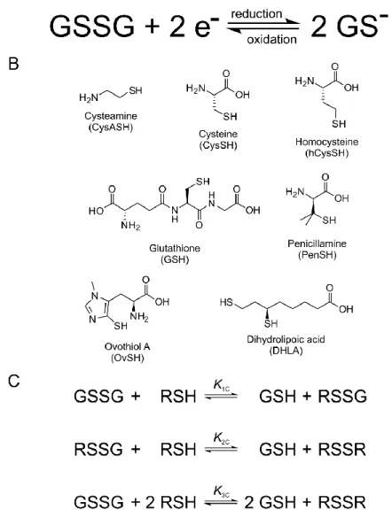
Figure 1. A: The thiol-disulfide redox half-reaction of glutathione. B: The structural formulae of the thiols studied. C: Scheme of the thiol-disulfide equilibria at the macroscopic level. RSH denotes the thiols; RSSR denotes their respective homodisulfides; RSSG denotes their respective heterodisulfides formed with glutathione.
10
It is a key feature of any thiol-disulfide redox system, that only the deprotonated thiol species are active in the redox process, i.e. only the anionic thiolate can be oxidized directly [27-30]. Since the deprotonated fraction of thiols depends on the solution pH, the oxidation-reduction potential of thiol-containing biomolecules is also pH-dependent.
These interwoven acid-base and redox processes are usually further influenced by overlapping protonation processes of multiple basic centers on the molecule in question.
Therefore, the redox potential of thiol-containing biomolecules is usually doubly pH- dependent. Primarily, the deprotonated fraction of every thiol depends on the pH of the solution, with strong dependence typically in the 6-11 pH range [16]. One clear exception to this typical acid-base behavior is ovothiol A with extremely low thiolate basicities [31], which extends the general basicity range for thiolates to 1-10 log units in terms of protonation constants. Secondarily, the thiolate oxidizability in most biomolecules is modulated by the adjacent basic groups, since protonation of any such group exerts an electron-withdrawing effect on the thiolate. In a molecule of n+1 basic sites the thiolate has 2n different, distinct oxidizabilities within a single, covalently unchanged molecular skeleton. Thus, macroscopic physico-chemical parameters cannot quantify the thiolate moiety specifically. A thorough characterization of the thiol- disulfide equilibria can be achieved by means of species-specific, so-called microscopic parameters, which are, in fact, well-established terms for acid-base systems [32-33].
Ovothiol A ((2S)-2-amino-3-(3-methyl-5-sulfanylimidazol-4-yl)propanoic acid, OvSH) is a naturally occurring 4-mercaptohistidine derivative [34-36] first observed in sea urchin (Strongylocentrotus purpuratus) eggs by Turner et al. [37]. Ovothiol A (henceforth referred to as simply ovothiol), B, and C differ in the level of methylation at the amino site: latters are mono- and dimethyl derivatives, respectively.
Mercaptohistidines also exist in marine algae [38], trout and salmon eggs [39] and related aquatic organisms. The function of ovothiols in marine invertebrate eggs is hypothesized to be defense against oxidative stress agents [39-40]. Ovothiol along with novel mercaptohistidines has been detected in many parasitic protozoa [41-45]. The molecular structure of ovothiol is certainly unique among naturally occurring small molecules: the number and especially the variety of functional groups relative to its low molecular mass greatly exceeds those of almost all other biomolecules. The uncommon positioning of the thiol group results in peculiar acid-base properties: the thiolate group
11
has an extremely low basicity [31,46]. The interaction between the imidazole ring and the thiol group also results in the significant elevation of the imidazole basicity compared to that of histidine. The observation that thiolate basicities and thiol-disulfide half-cell redox potentials are in correlation [16] supports the hypothesis [47-50] that ovothiol is one of the most potent natural antioxidants even in low pH media. The many interesting features of this molecule have prompted studies to find candidates for drug development. A new prospect has emerged in search of a non-synthetic antioxidant [51- 52], anticipating results in neurodegenerative therapy [53]. A different approach exploits the biological role of ovothiols to create prospective antitrypanosomal chemotherapeutic agents [54-55] by inhibiting the biosynthetic enzymes; since the synthesis of ovothiols and trypanothiones (not present in host cells) is essential for the survival of trypanosomatids.
The direct measurement of the redox potential for thiol-disulfide systems, by usual electrochemical methods is not feasible due to formation of stable metal-thiolate complexes at electrode surfaces [56]. Thus the redox potentials of GSH and other thiols can only be determined indirectly by measurement of equilibrium constants for their reaction with redox systems of known redox potentials [20]. However, until now only the apparent redox potential of GSH was determined, largely because the highly composite species-specific acid-base properties, which are in codependent interference with the redox behavior, were not known. The general scheme of thiol-disulfide redox equilibria (as depicted in Figure 1) is generally studied at the level of phenomena; the three conditional equilibrium constants are expressed by equations (1)-(3) and the Nernstian redox potential of a thiol-disulfide redox couple are expressed by equation (4).
1C
[GSH][RSSG]
[RSH][GSSG]
K (1)
2C
[GSH][RSSR]
[RSH][RSSG]
K (2)
12
2
3C 1C 2C 2
[GSH] [RSSR]
[RSH] [GSSG]
K K K (3)
2
[RSSR]
ln[RSH]
E E RT
zF (4)
1.3 Species-specific chemical microequilibria
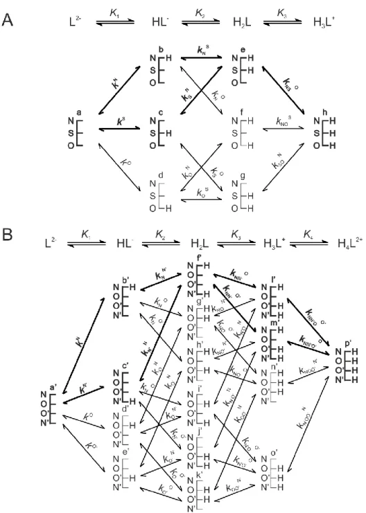
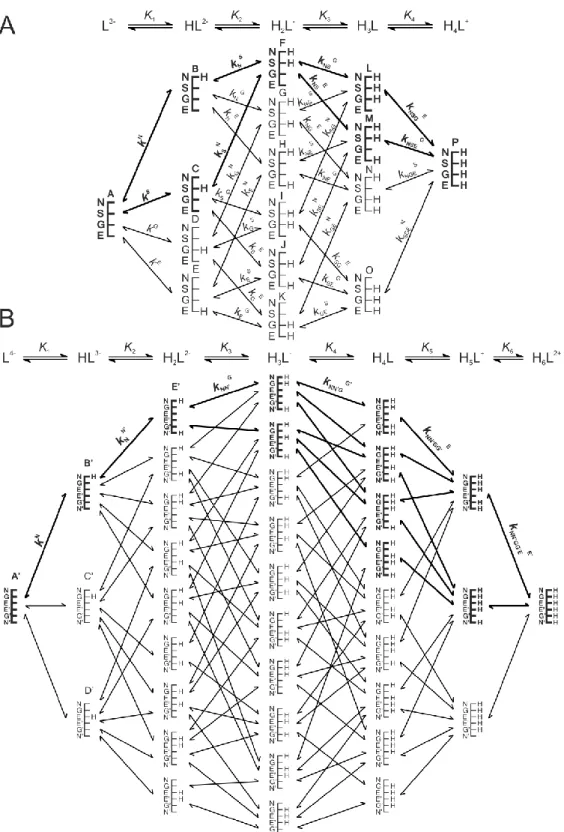
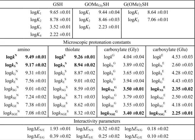
Figures 2, 3 and 4 represent the species-specific protonation scheme of a bidentate thiol (cysteamine) and its homodisulfide, a tridentate thiol (cysteine, homocysteine, penicillamine) and its homodisulfide, and a tetradentate thiol (glutathione, ovothiol) and its homodisulfide, respectively. Macroequilibria (top lines) indicate the stoichiometry of the successively protonated ligand and the stepwise macroscopic protonation constants.
In the microspeciation schemes, the different microspecies with their one-letter symbols (a, b, c …), and the microscopic protonation constants are depicted (kN, kNS …). The superscript at k for any microconstant indicates the protonating group while the subscript (if any) shows the site(s) already protonated. S, N, O, G and E symbolize the thiolate, amino, carboxylate (in cysteine and cystine), glycinyl carboxylate and glutamyl carboxylate (in GSH and GSSG after their one-letter symbol) sites, respectively. Some protonation constant examples for cysteine are shown below:
2 2
[H L]
[HL ][H ]
K (5)
3
3 1 2 3 2 3
[H L ] [L ][H ] K K K
(6)
N [b]
[a][H ]
k (7)
where K1, K2, K3, are successive macroconstants, β3 is one of the cumulative macroconstants, kN is the microconstant of cysteine involved in the production of
13
microspecies b. The concentrations of the various macrospecies comprise the sum of the concentration of those microspecies that contain the same number of protons, for example in GSH:
[H L ] [F] [G] [H] [I] [J] [K]2 (8)
Figure 2. The protonation macro- and microequilibrium schemes of CysASH (A) and CysASSCysA (B) in terms of stepwise macroscopic protonation constants (K1, K2), where L-, HL, etc. are the successively protonating ligands (top lines). Below are the species-specific protonation schemes in terms of microspecies (a, b, c, d) and microscopic protonation constants (kN, kNS …).
14
Figure 3. The protonation macro- and microequilibrium schemes of CysSH (A) and CysSSCys (B) in terms of stepwise macroscopic protonation constants (K1, K2, K3 …), where L2-, HL-, etc. are the successively protonating ligands (top lines). Below are the species-specific protonation schemes in terms of microspecies (a, b, c …) and microscopic protonation constants (kN, kNS …). The components of the major pathways are in bold. The above schemes are also representative of the other tridentate thiols, i.e.
homocysteine and penicillamine.
15
Figure 4. The protonation macro- and microequilibrium schemes of GSH (A) and GSSG (B) in terms of stepwise macroscopic protonation constants (K1, K2, K3 …), where L3-, HL2-, etc. are the successively protonating ligands (top lines). Below are the species-specific protonation schemes in terms of microspecies (A, B, C …) and microscopic protonation constants (kN, kNS …). For GSSG, only non-identical
16
microspecies and microconstants are shown, and only a few of the microconstants are depicted. The components of the major pathways are in bold. The protonation schemes of OvSH and OvSSOv are identical to the depicted schemes except Im (imidazole) and O (carboxylate) moieties are present instead of G and E.
The relationships between the macro- and microconstants defined first in the pioneer work of Niels Bjerrum [32] can be applied to cysteine:
N S O
1 k k k
(9)
N S N O S O
2 k kN k kN k kS
(10)
N S O 3 k k kN SN
(11)
Depending on the path of protonation, equations (10) and (11) can be written in 2 and 6 different, equivalent ways, respectively. Determination of protonation microconstants needs careful considerations and has been elaborated in detailed in a recent review [33].
The characterization of the microspeciation scheme necessitates the knowledge of the macroscopic protonation constants (accessible from titrimetric measurements) and the utilization of a species-specific spectroscopic assay or the introduction of auxiliary model compounds. The application of a spectroscopic method that delivers selective information regarding one or some of the basic sites in the molecule in question can in principle allow the determination of all microconstants. However, with decreasing size and an omni-interactive molecular skeleton the probability that a selective analyte signal can be found diminishes rapidly. Minor microspecies influence the analytical signals (NMR, UV, etc.) insignificantly, but they may be the reactive species in highly specific biochemical processes [57]. An alternative method for the elucidation of protonation microconstants is the deductive method; by way of this method model compounds are synthesized that are the closest possible mimics of the minor microspecies. The protonation constants of the simpler model compounds are regarded as constituent
17
elements of the microspeciation scheme. For the case of thiols, the model compounds were synthesized by methylating the thiol and carboxyl groups, thereby modeling the protonated form of these moieties. The site in which the methyl group is connected remains in neutral state, with practically identical electronic effects on the rest of the molecule as –COOH and –SH groups [58-59]. Using analogous equations as equations (9) and (11), and by introducing interactivity parameters, the calculation of leftover microconstants becomes straightforward. The interactivity parameter shows to what extent the protonation of site A reduces the basicity of site B, and vice versa. The interactivity parameter is generally considered to be the most invariant quantity in analogous moieties of different compounds and also in various protonation states of the neighboring moiety in the same molecule [60].
For the thiol-disulfide redox equilibria, only the apparent or conditional equilibrium constants (K3C) are directly available, by determining the equilibrium concentrations of RSH, GSSG, RSSR, and GSH in the reaction mixtures. In other words: these conditional equilibrium constants are pH-dependent parameters in terms of total concentrations of the thiol and disulfide species that are present, since the spectroscopic intensity of the analyte signal corresponds to the total concentration of a thiol reactant that is actually composed of the concentration of the variously protonated microspecies.
The concentration of the macrospecies can be written as the sum of the microspecies of the same number of bound protons, as shown in equation (8) for H2L- of GSH. The pH- dependent, apparent constants can be decomposed into pH-independent, species-specific equilibrium constants, the number of which is large, but definite. For example, the thiolate moiety in cysteine is adjacent to two other basic sites; the amino and the carboxylate groups. Therefore, the thiolate-bearing microspecies can exist in 4 different protonation states. Concurrently, the thiolates in the 4 different microspecies will be in different electrostatic environments, thus having 4 different oxidizabilities. Considering the opposite direction of the redox equilibrium, GSH with an amino and two carboxylate sites can have 8 different thiolate-bearing microspecies with 8 different electron densities on its sulfur. This leads to 4x8=32 different microscopic redox equilibria between cysteine and glutathione.
18
Since redox and acid-base reactions coexist, the observed apparent redox equilibrium constants, K1C, K2C, and K3C are pH-dependent. In order to get a clear insight into the redox equilibria, purified from the protonation effects, an improved evaluation method had to be introduced, including a set of species-specific, new type of redox equilibrium constants (k1, k2, k3). As equation (3) shows the complete redox transition includes two thiols and two disulfides. We therefore confined our efforts to the determination of the K3C macroconstants, and the related k3 microconstants, the number of which can certainly be numerous. This confinement overcomes the highly complex quantification of the heterodisulfides, which actually improves perspicuity. Keire et al. [16] have already demonstrated that a) there is correlation between the thiolate basicities and redox properties and b) purely redox thiol-disulfide equilibrium constants can only be obtained if these transitions can be decoupled from the acid-base processes by an appropriate evaluation method. Determination of the redox equilibrium constants requires all the species-specific protonation constants, including those of the minor microspecies; otherwise, redox processes could only be characterized at the level of phenomenon, especially for acidic media.
To epitomize the determination of microscopic redox equilibrium constants (k3) from the conditional equilibrium constants (K3C), the calculation of the k3bB for the reaction involving the b microspecies of cysteine, the corresponding f’ cystine microspecies, the B glutathione microspecies, and the corresponding E’ glutathione disulfide microspecies will be demonstrated. Note that b and B define that the disulfide microspecies are f’ and E’, since these are the only ones with identical protonation states in the side-chain. Superscripts b and B therefore unambiguously identify all the 4 microspecies in the microequilibrium in question. This example of the pH-independent, microscopic thiol-disulfide equilibrium constants is expressed by equation (12):
bB 2
3 2
[B] [f']
[b] [E']
k (12)
19
Quantification of redox equilibria in terms of such pH-independent, species-specific constants has long been a conceptual endeavor. The obvious reason why it has not so far been achieved is the fact that the microspecies concentrations cannot be measured. As shown below, the species-specific constants can be, however, obtained as the product of total species concentration and the relative abundance of the respective microspecies.
The relative abundance of the microspecies in turn, is a function of pH and the microscopic protonation constants. For the b cysteine microspecies, the concentration can be written as follows:
N
b CysSH CysSH 2 CysSH 3
1 2 3
[b] [CysSH] [CysSH] [H ]
1 [H ] [H ] [H ]
k
(13)
where [CysSH] is the total solution concentration of cysteine, xb is the relative abundance of microspecies b (i.e. xb = [b]/[CysSH]), kN is the microscopic protonation constant of cysteine involved in the formation of microspecies b. Substituting analogous equations as equation (13) for the concentrations of the microspecies in k3bB, one can write the equation of the microscopic equilibrium constant as follows:
2 2 2
bB 2 B f' B f'
3 2 2 2 3C 2
b E' b E'
[GSH] [CysSSCys]
[B] [f']
[b] [E'] [CysSH] [GSSG]
k K
(14)
Thus, if K3C, the apparent equilibrium constant and the 4 x values (as functions of the protonation constants and pH) have previously been determined, the k3bB value and all the analogous, pH-independent, species-specific redox microconstants can be calculated.
20
2. OBJECTIVES
The objectives of the doctoral thesis are the following:
- determination of the species-specific protonation constants of the most important biological thiols and their homodisulfides, namely: cysteamine, cysteine, homocysteine, penicillamine, glutathione, ovothiol, dihydrolipoic acid;
- determination of the species-specific redox equilibrium constants of the thiol- disulfude interchange reactions respective to glutathione;
- determination of the species-specific standard redox potential of glutathione using an indirect redox system for comparison, namely: 1-methylnicotinamide;
- determination of the species-specific standard redox potential of the most important biological thiols.
21 3. METHODS
3.1 Materials
All chemicals used for analytical and synthetic purposes were purchased from Sigma- Aldrich (Saint Louis, MO, USA) and used without further purification. Ovothiol and every model compound were synthesized according to the synthetic protocols described in section 3.9.
3.2 NMR spectroscopy measurements
NMR spectra were recorded on a Varian 600 MHz spectrometer at 298.2 ±0.1 K. The solvent in every case was an aqueous solution with H2O:D2O, 95:5, v/v (0.15 mol/L ionic strength), using DSS sodium (4,4-dimethyl-4-silapentane-1-sulfonate) as the reference compound. The sample volume was 600 μL, pH values were determined by internal indicator molecules optimized for NMR [61-62]. 1H NMR spectra were recorded with the presaturation solvent suppression sequence (number of transients=64, number of points=16384, acquisition time=851.968 ms, relaxation delay=15 s). For the
15N NMR measurements WET-HMBC sequence was used, with tetraethylammonium chloride (20 mmol/L, -316.7 ppm [63]) as a nitrogen standard. The solutions used to determine the protonation constants of the thiols also contained dithiothreitol to preclude oxidation.
3.3 pH-potentiometric titrations
A 716 DMS Titrino automatic titrator (Metrohm AG, Herisau, Switzerland) with a Metrohm 6.0204.100 combined pH glass electrode was used for the pH-potentiometric titrations, under automatic PC control. The electrode was calibrated by aqueous NBS standard buffer solutions. Constant temperature (298.2 ±0.1 K) was provided by a thermostated double-walled glass cell. Difference titrations were carried out in the absence (blank) and presence of ligands. First 1.5 mL of 0.1 mol/L HCl solutions were
22
titrated with 0.1 mol/L NaOH. Constant ionic strength of 0.15 mol/L was provided by the presence of KCl. Next, a ligand was added to the same volume of HCl solution and was subsequently titrated with NaOH. The initial concentration of the ligand was around 5 mmol/L in the titrations. Non-linear parameter fitting with Origin Pro 8 (OriginLab Corp., Northampton, MA, USA) provided the protonation constants from the interpolated volume differences.
3.4 UV spectroscopy measurements
Absorbance data were recorded on a Jasco V-550 UV/VIS spectrophotometer (serial-no:
C02951185, Jasco Intl. Co. Ltd., Tokyo, Japan). pH values were read on Metrohm 2.780.0010 precision pH meter with a 6.0258.600 Unitrode glass Pt 1000 electrode (Metrohm AG, Herisau, Switzerland), the pH-potentiometric system was calibrated using pH 1.68, 4.01, 6.87, 9.18 aqueous NBS standard buffer solutions. The samples were prepared by mixing an acidic and basic solution of the titrand (adjusted with HCl and NaOH, 0.15 mol/L ionic strength) in different ratios, the reference solution for the UV measurements was an aqueous solution adjusted to the same pH as the sample with HCl and NaOH.
3.5 Electrochemical measurements
The redox electrode (Radeklis OP-6123, Radeklis, Budapest, surface area ~2 cm2) used for electrode potential measurements was washed with 50 % nitric acid, then distilled water before each measurement and gently dried by touching onto tissue paper. The redox electrode was calibrated using ZoBell’s solution (2.64 g K4[Fe(CN)6].3H2O and 2.06 g K3[Fe(CN)6].H2O dissolved in 500 mL pH=7 0.15 mol/L Na2HPO4/KH2PO4
buffer). The electrode potential measurements were carried out using a Radelkis Laboratory Digital pH/mV meter OP-211/2 (Radelkis, Budapest) at 298 ±2 K in an 815- PGB glove box (Plas-Labs Ic., Lansing, MI, USA) under N2 atmosphere to preclude oxidation by air. All potentials were referred to saturated calomel electrode as reference
23
electrode. The pH values of the samples were determined using a Metrohm 6.0204.100 combined pH glass electrode, calibrated by aqueous NBS standard buffer solutions.
3.6 Preparation of solutions for equilibrium constant determination
Acidic (pH=0.85) and basic (pH=13.15) stock solutions containing the reagents were prepared in an 815-PGB glove box (Plas-Labs Ic., Lansing, MI, USA) under N2
atmosphere to preclude oxidation by air. The concentrations of the reagents were optimized for quantitative NMR, ca. 15 mmol/L. A series of solutions with different pH values were prepared by mixing the acidic with the basic stock solutions. D2O, DSS, glutathione reductase, and a pH indicator, which also served as a concentration standard, were added to the solutions. The samples were protected from sunlight and kept in the glove box under N2 atmosphere (298 ±2 K) for at most 7 days, until the reactions in all the samples had reached equilibrium.
3.7 Mathematical analysis
For the analysis of NMR titration curves of proton chemical shifts versus pH, the software Origin Pro 8 (OriginLab Corp., Northampton, MA, USA) was used. In 1H NMR-pH and potentiometric titrations the non-linear curve fitting regression analysis option was used with the following functions [64], respectively:
log pH H L
0
obs log pH
0
10 (pH)
10
i i
i
n i
i
n i
i
(15)log pH 0
log pH 0
10 (pH)
10
i
i
n i
i
n i
i
i
V A D
(16)24
where δHiL values stand for the chemical shifts of successively protonated ligands, n is the maximum number of protons that can bind to L, β is the cumulative protonation macroconstant, A is the NaOH volume corresponding to one unit of deprotonation, and D is the experimental correction fitting factor. The standard deviations of logβ values from the regression analyses were used to calculate the Gaussian propagation of uncertainty to the protonation microconstants derived in the Results chapter.
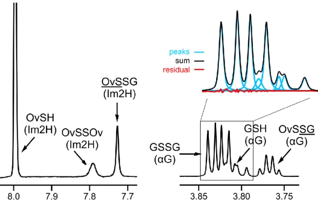
For the analysis of quantitative NMR measurements (sample spectrum in Figure 5), the AMARES [65] time domain fit algorithm (without apodization) of the jMRUI v. 5.1 software package [66] was used. For the regression analyses, the software Origin Pro 8 (OriginLab Corp., Northampton, MA, USA) was used. The standard deviations of the peak areas obtained by fitting Lorentzian peak shapes, the error of pH determination, and the standard errors of the microscopic redox equilibrium constants were used to calculate the Gaussian propagation of uncertainty to the standard redox potentials derived in the Results chapter.
Figure 5. The expanded 1H NMR spectrum of a sample containing ovothiol-glutathione mixture with example of peak fitting result (pH=9.20).
25 3.8 TOF MS measurements
The exact mass of the synthesized and isolated compounds was determined with an Agilent 6230 time-of-flight mass spectrometer equipped with a JetStream electrospray ion source in positive ion mode. JetStream parameters: drying gas (N2) flow and temperature: 10.0 L/min and 598 K; nebulizer gas (N2) pressure: 10 psi; capillary voltage: 4000 V; sheath gas flow and temperature: 598 K and 7.5 L/min. TOFMS parameters: fragmentor voltage: 170 V; skimmer potential: 170 V; OCT 1 RF Vpp: 750 V. Samples were introduced (0.1-0.3 µL) by the Agilent 1260 Infinity HPLC system (flow rate=0.5 mL/min, 70% methanol-water mixture 0.1% formic acid). Reference masses of m/z 121.050873 and 922.009798 were used to calibrate the mass axis during analysis. Mass spectra were acquired over the m/z range 100-1000 at an acquisition rate of 250 ms/spectrum and processed using Agilent MassHunter B.02.00 software.
3.9 Synthetic protocols 3.9.1 Synthesis of ovothiol
The total synthesis of ovothiol has been reported [67] and remains the preferred method of preparation, compared to the cumbersome task of gathering sea urchin eggs for extraction [37]. However the total synthesis reported by Holler et al. contains 9 steps, constructing the imidazole ring from scratch and has a total yield of 11%. A simpler method was therefore exploited, making use of histidine as the natural precursor to synthesize ovothiol in an enantiopure, convenient and cost-effective way (Figure 6).
The configuration of the end-product was verified with optical rotatory dispersion measurement of ovothiol disulfide.
26 Figure 6. The synthesis route of ovothiol.
(2S)-2-[(tert-Butoxycarbonyl)amino]-3-[1-(tert-butoxycarbonyl)-1H-imidazol-4-yl]- propanoic acid (2). To a suspension of L-histidine (1) (1.43 g, 9.2 mmol) in methanol (CH3OH) (30 mL), 3.24 g (21.2 mmol) of di-tert-butyl dicarbonate (Boc2O) and 1.8 mL (18.4 mmol) of triethylamine (Et3N) were added. The reaction mixture was stirred at room temperature overnight. The reaction mixture was then concentrated under vacuum and the crude product was purified on silica gel with column chromatography (CHCl3:CH3OH 9:1) to give 3.01 g (yield 92%) white solid: mp 346-348 K [68] (from chloroform, CHCl3); 1H NMR (600 MHz, DMSO-d6) δ 1.40 (9H, s, NαCO2C(CH3)3), 1.61 (9H, s, NImCO2C(CH3)3), 2.87 (1H, dd, J 8, 15 Hz, βH), 3.08 (1H, dd, J 4, 15 Hz, βH), 4.25 (1H, dd, J 4, 8 Hz, αH), 7.29 (1H, s, Im5H), 8.06 (1H, s, Im2H); 13C NMR (600 MHz, CD3OD) δ 28.1 (NαBoc-CH3), 28.8 (NImBoc-CH3), 31.1 (βC), 56.6 (αC), 80.1 (NαBoc-t-C), 86.9 (NImBoc-t-C), 116.1 (Im4C), 137.6 (Im5C), 141.1 (Im2C), 148.3 (NαBoc-C=O), 157.5 (NImBoc-C=O), 178.1 (COOH); HRMS m/z [M+H]+ Calcd:
356.1822, Found: 356.1824.
Methyl (2S)-2-[(tert-butoxycarbonyl)amino]-3-(1-methyl-1H-imidazol-5- yl)propanoate (3). To the solution of (2) (3.01 g, 8.5 mmol) in 20 mL CH3OH, 1.32
27
mL (21.3 mmol) of iodomethane (CH3I) was added at 273 K. The solution mixture was stirred overnight at 273 K. The reaction mixture was then extracted with ethyl acetate (after adjusting the pH between 7 and 8) and washed with brine. The resulting organic layer was dried over anhydrous sodium sulfate and concentrated under vacuum. The crude product was purified on silica gel with column chromatography (ethyl acetate:acetone 9:1) to give 1.97 g (yield 83%) white solid (the position of the methyl group on the imidazole ring was confirmed using NOESY [69] and 1H-15N HMBC [70]): mp 382-383 K (from ethyl acetate); 1H NMR (600 MHz, CD3OD) δ 1.40 (9H, s, CO2C(CH3)3), 2.98 (1H, dd, J 9, 15 Hz, βH), 3.14 (1H, dd, J 5, 15 Hz, βH), 3.66 (3H, s, NCH3), 3.74 (3H, s, OCH3), 4.42 (1H, dd, J 5, 9 Hz, αH), 6.78 (1H, s, Im5H), 7.59 (1H, s, Im2H); 13C NMR (600 MHz, CD3OD) δ 27.1 (βC), 28.7 (Boc-CH3), 31.8 (NCH3), 52.9 (OCH3), 54.2 (αC), 80.8 (Boc-t-C), 127.5 (Im5C), 129.4 (Im4C), 139.2 (Im2C), 157.7 (Boc-C=O), 173.4 (COO); HRMS m/z [M+H]+ Calcd: 284.1610, Found:
284.1602.
Methyl (2S)-3-(4-bromo-1-methyl-1H-imidazol-5-yl)-2-[(tert- butoxycarbonyl)amino]-propanoate (4). To the solution of (3) (1.02 g, 3.6 mmol) in 10 mL CH3OH, 0.71 g (4.0 mmol) of N-bromosuccinimide (NBS) was added in small amounts at 273 K. The reaction mixture was stirred at 273 K for 2 hours, and then the reaction mixture was concentrated under vacuum. The crude product was purified on silica gel with column chromatography (ethyl acetate:CHCl3 9:1) to give 0.98 g (yield 75%) white glassy solid: 1H NMR (600 MHz, CD3OD) δ 1.38 (9H, s, CO2C(CH3)3), 2.99 (1H, dd, J 9, 15 Hz, βH), 3.15 (1H, dd, J 6, 15 Hz, βH), 3.69 (3H, s, NCH3), 3.73 (3H, s, OCH3), 4.40 (1H, t, J 7 Hz, αH), 7.55 (1H, s, Im2H); 13C NMR (600 MHz, CD3OD) δ 27.1 (Boc-CH3), 28.6 (βC), 33.0 (NCH3), 49.8 (OCH3), 53.7 (αC), 79.4 (Boc-t-C), 115.5 (Im5C), 126.8 (Im4C), 138.9 (Im2C), 157.4 (Boc-C=O), 181.5 (COO);
HRMS m/z [M+H]+ Calcd: 362.0715, Found: 362.0720.
(2S)-2-[(tert-Butoxycarbonyl)amino]-3-{1-methyl-4-[(4-methylbenzyl)sulfanyl]-1H- imidazol-5-yl}propanoic acid (5). A solution of (4) (0.98 g, 2.7 mmol) and 1 mL (8.1 mmol) of (4-tolyl)methanethiol in 10 mL dry N,N-dimethylformamide (DMF) was added dropwise in nitrogen (N2) atmosphere to a stirred solution of 98 mg (4.1 mmol) sodium hydride (NaH) in 10 mL dry DMF. The reaction mixture was heated at 393 K in
28
an atmosphere of N2 for 3 hours. After cooling, the reaction mixture was concentrated in vacuo to leave a brown oil. The crude product was purified on silica gel with column chromatography (CH2Cl2:n-hexane 2:1) to give 0.81 g (yield 74%) brown oil: 1H NMR (600 MHz, CD3OD) δ 1.31 (9H, s, CO2C(CH3)3), 2.27 (3H, s, ArCH3), 2.63 (1H, dd, J 10, 15 Hz, βH), 2.82 (1H, dd, J 4, 15 Hz, βH), 3.67 (3H, s, NCH3), 3.81 (1H, d, J 13 Hz, SCH2), 3.86 (1H, d, J 13 Hz, SCH2), 4.09 (1H, dd, J 4, 10 Hz, αH), 6.97 (2H, d, J 8 Hz, o-ArH), 7,00 (2H, d, J 8 Hz, m-ArH), 7.56 (1H, s, Im2H); 13C NMR (600 MHz, CD3OD) δ 21.1 (ArCH3), 28.7 (Boc-CH3), 31.1 (βC), 32.7 (NCH3), 41.2 (SCH2), 55.9 (αC), 79.5 (Boc-t-C), 129.7 (o-Ar), 129.8 (Im5C), 129.9 (m-Ar), 134.5 (p-Ar), 137.5 (i- Ar), 139.3 (Im4C), 156.9 (Im2C), 161.4 (Boc-C=O), 178.0 (COOH); HRMS m/z [M+H]+ Calcd: 406.1800, Found: 406.1794.
(2S)-2-Amino-3-(1-methyl-4-sulfanyl-1H-imidazol-5-yl)propanoic acid, ovothiol (6). 82 mg (0.2 mmol) of (5) was dissolved in 0.5 mL anisole and cooled to 273 K.
Mercury(II) trifluoroacetate, 173 mg (0.4 mmol), was dissolved in 5 mL triflouroacetic acid (TFA) and added to the cooled solution. The mixture was stirred at 273 K for 2 h, concentrated in vacuo, taken up in 3 mL of water, and washed with 3x1 mL of diethyl ether. Hydrogen sulfide gas (H2S) was bubbled into the aqueous solution for 15 min, the suspension was filtered through Celite and the filtrate was concentrated in vacuo to give 67 mg (yield 78%) of the di(trifluoroacetate) salt of the title compound as a pale green glass: 1H NMR (600 MHz, H2O:D2O, 95:5, v/v) δ 3.51 (1H, dd, J 8, 16 Hz, βH), 3.67 (1H, dd, J 8, 16 Hz, βH), 4.51 (1H, dd, J 8 Hz, αH), 4.65 (3H, s, NCH3), 8.75 (1H, s, Im2H); 13C NMR (600 MHz, H2O:D2O, 95:5, v/v) δ 26.4 (βC), 36.8 (NCH3), 53.7 (αC), 122.0 (Im5C), 133.8 (Im4C), 139.0 (Im2C), 172.7 (COOH); UV (λmax, nm, H2O) 237, 278 (shoulder) [67]; HRMS m/z [M+H]+ Calcd: 202.0650, Found: 202.0654.
(2S,2'S)-3,3'-[Disulfanediylbis(1-methyl-1H-imidazol-4,5-diyl)]bis(2-
aminopropanoic acid), ovothiol disulfide (7). 50 mg (0.1 mmol) of compound (6) was taken up in 10 mL water, then air was bubbled through the solution for 1 h. NMR studies showed that the oxidation of (6) commenced quantitatively. Freeze-drying the solution yielded 49.4 mg (yield 99%) of the tetra(trifluoroacetate) salt of (7): 1H NMR (600 MHz, H2O:D2O, 95:5, v/v) δ 2.35 (1H, dd, J 7.5, 16 Hz, βH), 2.53 (1H, dd, J 7.5, 16 Hz, βH), 3.38 (1H, dd, J 7.5 Hz, αH), 3.63 (3H, s, NCH3), 7.72 (1H, s, Im2H); [α]D20
29
+74 (c 0.5, 0.1 mol/L HCl(aq)) [67]; UV (λmax, nm, 0.1 mol/L HCl(aq)) 257; HRMS m/z [M+H]+ Calcd: 400.0987, Found: 400.0988. Optical rotation was measured on a Jasco Model P-2000 polarimeter at the D line of sodium.
3.9.2 General procedures
Methyl ester formation was achieved by dissolving the reactant in 5 mL methanol and adding 1.5 equivalents of thionyl chloride at 273 K. The mixture was stirred at room temperature overnight, and then the solvent was evaporated in vacuo to give the title compound which was used in analytical experiments without purification. The synthesized products are itemized below.
Ovothiol disulfide dimethyl ester (8). 1H NMR (600 MHz, H2O:D2O, 95:5, v/v) δ 2.72 (2H, dd, J 8, 20 Hz, βH), 2.88 (2H, dd, J 8, 20 Hz, βH), 3.61 (6H, s, OCH3), 3.82 (2H, t, J 8 Hz, αH), 3.84 (6H, s, NCH3), 8.01 (2H, s, Im2H); HRMS m/z [M+H]+ Calcd:
401.1066, Found: 401.1052.
Glutathione methyl ester (Gly) (9). 1H NMR (600 MHz, H2O:D2O, 95:5, v/v) δ 1.91 (2H, m, βH(Glu)), 2.34 (2H, m, γH(Glu)), 2.75 (2H, m, βH(Cys)), 3.44 (1H, t, J 6 Hz, αH(Glu)), 3.61 (3H, s, OCH3(Gly)), 3.89 (2H, dd, J 5, 18 Hz, αH(Gly)), 4.22 (1H, dd, J 2.5, 5 Hz, αH(Cys)); HRMS m/z [M+H]+ Calcd: 322.1073, Found: 322.1012.
Glutathione dimethyl ester (10). 1H NMR (600 MHz, H2O:D2O, 95:5, v/v) δ 2.21 (2H, m, βH(Glu)), 2.58 (2H, m, γH(Glu)), 2.94 (2H, m, βH(Cys)), 3.75 (3H, s, OCH3(Gly)), 3.91 (3H, s, OCH3(Glu)), 3.97 (1H, t, J 6 Hz, αH(Glu)), 4.04 (2H, d, J 6 Hz, αH(Gly)), 4.55 (1H, t, J 6 Hz, αH(Cys)); HRMS m/z [M+H]+ Calcd: 336.1229, Found: 336.1574.
Cysteine methyl ester (11). 1H NMR (600 MHz, H2O:D2O, 95:5, v/v) δ 3.94 (1H, t, J 5.5 Hz, αH), 3.79 (3H, s, OCH3), 2.98 (1H, dd, J 6, 14 Hz, βH), 2.91 (1H, dd, J 5, 14 Hz, βH); HRMS m/z [M+H]+ Calcd: 136.0432, Found: 136.0416.
Cystine dimethyl ester (12). 1H NMR (600 MHz, H2O:D2O, 95:5, v/v) δ 4.40 (2H, dd, J 5, 7 Hz, αH), 3.85 (6H, s, OCH3), 3.34 (2H, dd, J 5, 15 Hz, βH), 3.28 (2H, dd, J 7, 15 Hz, βH); HRMS m/z [M+H]+ Calcd: 269.0630, Found: 269.0621.
30
Homocysteine methyl ester (13). 1H NMR (600 MHz, H2O:D2O, 95:5, v/v) δ 4.25 (1H, t, J 8 Hz, αH), 3.84 (3H, s, OCH3), 2.88 (2H, t, J 7 Hz, γH), 2.40 (2H, m, βH); HRMS m/z [M+H]+ Calcd: 150.0589, Found: 150.0593.
Methionine methyl ester (14). 1H NMR (600 MHz, H2O:D2O, 95:5, v/v) δ 4.30 (1H, t, J 6.5 Hz, αH), 3.85 (3H, s, OCH3), 2.69 (2H, t, J 8 Hz, γH), 2.30 (2H, m, βH), 2.13 (3H, s, SCH3); HRMS m/z [M+H]+ Calcd: 164.0745, Found: 164.0751.
Homocystine dimethyl ester (15). 1H NMR (600 MHz, H2O:D2O, 95:5, v/v) δ 4.31 (2H, t, J 7 Hz, αH), 3.86 (6H, s, OCH3), 2.90 (4H, t, J 7 Hz, γH), 2.42 (4H, m, βH);
HRMS m/z [M+H]+ Calcd: 297.0943, Found 297.0957.
Penicillamine methyl ester (16). 1H NMR (600 MHz, H2O:D2O, 95:5, v/v) δ 1.42 (3H, s, βCH3), 1.45 (3H, s, βCH3), 3.68 (1H, s, αH), 3.79 (3H, s, OCH3); HRMS m/z [M+H]+ Calcd: 164.0745, Found: 164.0724.
Penicillamine disulfide dimethyl ester (17). 1H NMR (600 MHz, H2O:D2O, 95:5, v/v) δ 1.50 (3H, s, βCH3), 1.57 (3H, s, βCH3), 3.66 (3H, s, OCH3), 4.19 (1H, s, αH); HRMS m/z [M+H]+ Calcd: 325.1256, Found: 325.1296.
Amide formation was achieved by dissolving the reactant in 5 mL methanol and adding 0.5 mL concentrated ammonia. The mixture was stirred at room temperature overnight;
subsequently the solvent was evaporated in vacuo to give the title compound which was used in analytical experiments without purification. The synthesized product is characterized below.
Ovothiol amide (18). 1H NMR (600 MHz, H2O:D2O, 95:5, v/v) δ 3.26 (1H, dd, J 8, 15 Hz, βH), 3.29 (1H, dd, J 8, 15 Hz, βH), 3.76 (3H, s, NCH3), 4.44 (1H, dd, J 7 Hz, αH), 8.26 (1H, s, Im2H); HRMS m/z [M+H]+ Calcd: 201.0810, Found: 201.0815.
S-methyl formation was achieved by dissolving the reactant in 5 mL of aqueous ammonia solution (pH=9) and adding 3 equivalents of CH3I. The solution was stirred at room temperature for 1 h under N2, and the excess ammonia was eliminated in vacuo.
31
The resulting aqueous solution was used in subsequent NMR experiments. The synthesized products are itemized below.
S-Methylovothiol (19). 1H NMR (600 MHz, H2O:D2O, 95:5, v/v) δ 2.37 (3H, s, SCH3), 3.19 (1H, dd, J 7, 15 Hz, βH), 3.25 (1H, dd, J 7, 15 Hz, βH), 3.78 (3H, s, NCH3), 3.95 (1H, dd, J 8 Hz, αH), 8.72 (1H, s, Im2H); HRMS m/z [M+H]+ Calcd: 216.0961, Found:
216.0960.
S-Methylovothiol amide (20). 1H NMR (600 MHz, H2O:D2O, 95:5, v/v) δ 3.26 (1H, dd, J 8, 15 Hz, βH), 3.28 (1H, dd, J 8, 15 Hz, βH), 3.82 (3H, s, NCH3), 4.25 (1H, dd, J 7 Hz, αH), 8.74 (1H, s, Im2H); UV (λmax, nm, H2O) 198, 225 (shoulder); HRMS m/z [M+H]+ Calcd: 215.0967, Found: 215.0959.
S-Methylcysteamine (21). 1H NMR (600 MHz, H2O:D2O, 95:5, v/v) δ 3.24 (2H, t, J 7 Hz, αH), 2.82 (2H, t, J 7 Hz, βH), 2.73 (3H, s, SCH3); HRMS m/z [M+H]+ Calcd:
92.0534, Found: 92.0567.
S-Methylcysteine (22). 1H NMR (600 MHz, H2O:D2O, 95:5, v/v) δ 3.94 (1H, dd, J 5, 8 Hz, αH), 3.09 (1H, dd, J 5, 15 Hz, βH), 2.99 (1H, dd, J 8, 15 Hz, βH), 2.15 (3H, s, SCH3); HRMS m/z [M+H]+ Calcd: 136.0432, Found: 136.0420.
S-Methylcysteine methyl ester (23). 1H NMR (600 MHz, H2O:D2O, 95:5, v/v) δ 4.22 (1H, dd, J 5, 7.5 Hz, αH), 3.83 (3H, s, OCH3), 3.11 (1H, dd, J 5, 15 Hz, βH), 3.00 (1H, dd, J 7.5, 15 Hz, βH), 2.14 (3H, s, SCH3); HRMS m/z [M+H]+ Calcd: 150.0589, Found:
150.0583.
S-Methylpenicillamine (24). 1H NMR (600 MHz, H2O:D2O, 95:5, v/v) δ 1.31 (3H, s, βCH3), 1.52 (3H, s, βCH3), 2.07 (3H, s, SCH3), 3.67 (1H, s, αH); HRMS m/z [M+H]+ Calcd: 164.0745, Found: 164.0687.
S-Methylpenicillamine methyl ester (25). 1H NMR (600 MHz, H2O:D2O, 95:5, v/v) δ 1.48 (3H, s, βCH3), 1.56 (3H, s, βCH3), 1.92 (3H, s, SCH3), 3.71 (3H, s, OCH3), 3.88 (1H, s, αH); HRMS m/z [M+H]+ Calcd: 178.0902, Found: 178.0930.
32
When necessary the thiol formation from a disulfide was achieved with reduction. The reactant was dissolved in 5 ml of methanol and 2.5 equivalents of sodium tetrahydroborate were added. After stirring at ambient temperature for 1 h, the reaction mixture was evaporated in vacuo to yield the title compound which was used in analytical experiments without purification. The synthesized product is characterized below.
Homocysteine (26). 1H NMR (600 MHz, H2O:D2O, 95:5, v/v) δ 3.85 (1H, t, J 7 Hz, αH), 2.83 (2H, t, J 8 Hz, γH), 2.27 (2H, m, βH); HRMS m/z [M+H]+ Calcd: 136.0432, Found: 136.0442.
Dihydrolipoic acid (27). 1H NMR (600 MHz, H2O:D2O, 95:5, v/v) δ 1.40 (1H, m, C4H2), 1.46 (1H, m, C4H2), 1.54 (1H, m, C5H2), 1.54 (2H, m, C3H2), 1.69 (1H, m, C5H2), 1.78 (1H, m, C7H2), 1.94 (1H, m, C7H2), 2.18 (2H, t, J 7.5 Hz, C2H2), 2.67 (1H, m, C8H2), 2.72 (1H, m, C8H2), 3.02 (1H, m, C6H); HRMS m/z [M+H]+ Calcd: 209.0670, Found: 209.0655.
Dihydrolipoamide (28). 1H NMR (600 MHz, H2O:D2O, 95:5, v/v) δ 1.44 (1H, m, C4H2), 1.51 (1H, m, C4H2), 1.60 (1H, m, C5H2), 1.60 (2H, m, C3H2), 1.68 (1H, m, C5H2), 1.78 (1H, m, C7H2), 1.93 (1H, m, C7H2), 2.28 (2H, t, J 7.7 Hz, C2H2), 2.67 (1H, m, C8H2), 2.71 (1H, m, C8H2), 3.01 (1H, m, C6H), 6.22 (1H, s, NH), 7.54 (1H, s, NH);
HRMS m/z [M+H]+ Calcd: 208.0830, Found: 208.0838.
When necessary the homodisulfide formation from a thiol was achieved with in situ oxidation. The thiol was dissolved in 2% aqueous hydrogen peroxide solution and the pH adjusted to 8.5. The solution was stirred for 30 minutes, and the resulting aqueous solution was used in subsequent NMR experiments. The synthesized products are itemized below.
Glutathione disulfide tetramethyl ester (29) 1H NMR (600 MHz, H2O:D2O, 95:5, v/v) δ 2.05 (4H, m, γH(Glu)), 2.18 (4H, m, βH(Glu)), 3.05 (4H, m, βH(Cys)), 3.68 (6H, s, OCH3(Gly)), 3.87 (6H, s, OCH3(Glu)), 3.45 (2H, t, J 6 Hz, αH(Glu)), 4.16 (4H, d, J 6
33
Hz, αH(Gly)), 4.81 (2H, t, J 6 Hz, αH(Cys)); HRMS m/z [M+H]+ Calcd: 669.2224, Found: 669.2222.
Glutathione disulfide dimethyl ester (Gly) (30) 1H NMR (600 MHz, H2O:D2O, 95:5, v/v) δ 2.04 (4H, m, βH(Glu)), 2.11 (4H, m, γH(Glu)), 3.11 (4H, m, βH(Cys)), 3.49 (2H, t, J 6 Hz, αH(Glu)), 3.68 (6H, s, OCH3(Gly)), 4.16 (4H, dd, J 5, 18 Hz, αH(Gly)), 4.81 (2H, dd, J 2.5, 5 Hz, αH(Cys)); HRMS m/z [M+H]+ Calcd: 641.1911, Found: 641.1912.
Cystamine (31). 1H NMR (600 MHz, H2O:D2O, 95:5, v/v) δ 3.40 (4H, t, J 7 Hz, αH), 3.02 (4H, t, J 7 Hz, βH); HRMS m/z [M+H]+ Calcd: 153.0520, Found: 153.0517.
Cystine (32). 1H NMR (600 MHz, H2O:D2O, 95:5, v/v) δ 4.23 (2H, dd, J 4, 9 Hz, αH), 3.41 (2H, dd, J 4, 15 Hz, βH), 3.22 (2H, dd, J 9, 15 Hz, βH); HRMS m/z [M+H]+ Calcd:
241.0317, Found: 241.0320.
Penicillamine disulfide (33). 1H NMR (600 MHz, H2O:D2O, 95:5, v/v) δ 1.44 (3H, s, βCH3), 1.55 (3H, s, βCH3), 4.01 (1H, s, αH); HRMS m/z [M+H]+ Calcd: 297.0943, Found: 297.1017.
34 4. RESULTS
The determination of species-specific protonation constants for every compound was performed based on case-tailored deduction methods, which nonetheless share a common underlying approach. The design of the deductive method is epitomized in detail for the simple case of cysteamine. The noteworthy alterations in the deductive method of the remaining compounds will be highlighted however, due to the intrinsic complexity of microspeciation of molecular entities with higher number of basic sites:
microspecies are coexisting, inseparable solution entities, with different location and/or number of protons at the basic sites on a definite covalent skeleton of the molecule, therefore their concentrations can only be measured indirectly [33]. Since the number of microspecies increases exponentially with the number of basic sites, complete, correct microspeciation for non-symmetrical, compact molecules has so far been reported for molecules of up to three basic sites. Ovothiol, for instance, has four distinct basic sites with an omni-interactive molecular structure, in which the protonation of any sites has an effect of a priori unknown magnitude on the basicity of every other site. Studies have shown [64] that complete microspeciation for molecules of four or more basic sites cannot be carried out without importing additional information, even though the protonation percentage is known for every site at each pH.
4.1 The microscopic protonation constants of cysteamine
Cysteamine was titrated under near physiological conditions and non-linear regression analysis of the 1H NMR chemical shift-pH profiles afforded the macroscopic protonation constants presented in Table 1. The relationships between the macroscopic and microscopic protonation constants (equivalent to equations (9) and (11)) actualized for cysteamine are:
N S
K1k k (17)
35
N S S N
2 k kN k kS
(18)
Thus, besides the macroconstants, the complete microspeciation of cysteamine needs at least one additional datum. This can be obtained via a deductive method, wherein the possible closest model of a minor microspecies is introduced and some appropriate protonation constant(s) of the model compound(s) is/are regarded as constituent protonation constants of the parent molecule. In the case of cysteamine, S- methylcysteamine was used to model the microspecies, in which the thiolate moiety is protonated. Therefore, the macroscopic protonation constant of S-methylcysteamine (Table 1) was equated to kSN of cysteamine. The remaining microscopic protonation constants were calculated using equations (17) and (18), and are presented in Table 1.
These data also yielded the interactivity parameter between the amino and thiolate groups:
N N S S
N/S S N
logE logk logk logk logk (19)
The interactivity parameter shows to what extent the protonation of site A reduces the basicity of site B, and vice versa. The interactivity parameter is generally considered to be the most invariant quantity in analogous moieties of different compounds and also in various protonation states of the neighboring moiety in the same molecule [60].
36
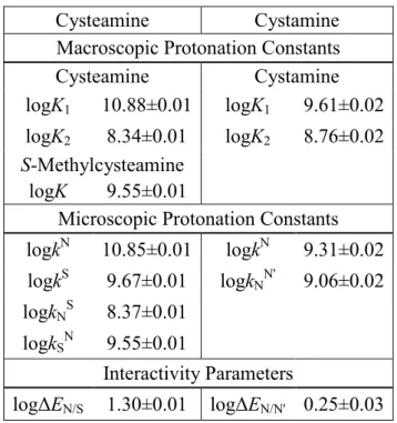
Table 1. The macroscopic and microscopic protonation constants (298 K, 0.15 mol/L ionic strength), and interactivity parameters of cysteamine, cystamine and their model compound in log units ±s.d.
Cysteamine Cystamine
Macroscopic Protonation Constants
Cysteamine Cystamine
logK1 10.88±0.01 logK1 9.61±0.02 logK2 8.34±0.01 logK2 8.76±0.02
S-Methylcysteamine
logK 9.55±0.01
Microscopic Protonation Constants logkN 10.85±0.01 logkN 9.31±0.02 logkS 9.67±0.01 logkNN' 9.06±0.02
logkNS 8.37±0.01
logkSN 9.55±0.01
Interactivity Parameters
logΔEN/S 1.30±0.01 logΔEN/N' 0.25±0.03
4.2 The microscopic protonation constants of cystamine
Cystamine, being the disulfide of cysteamine, is a symmetric molecule with two basic nitrogens, which can be arbitrarily named N and N’. Due to symmetry, their basicities are identical:
N N'
k k (20)
N N'
N' N
k k (21)
This reduces the multivariable problem to a system with two unknowns only, which can be resolved using relationships derived from equations (9) and (11):
37
N
logk logK1log 2 (22)
N'
N 2
logk logK log 2 (23)
Macroscopic protonation constants from 1H NMR-pH titration data and calculated microscopic protonation constants, along with the interactivity parameter between the two amino groups (logΔEN/N’) are presented in Table 1.
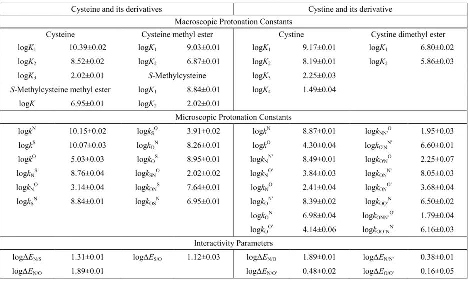
4.3 The microscopic protonation constants of cysteine, homocysteine, and penicillamine
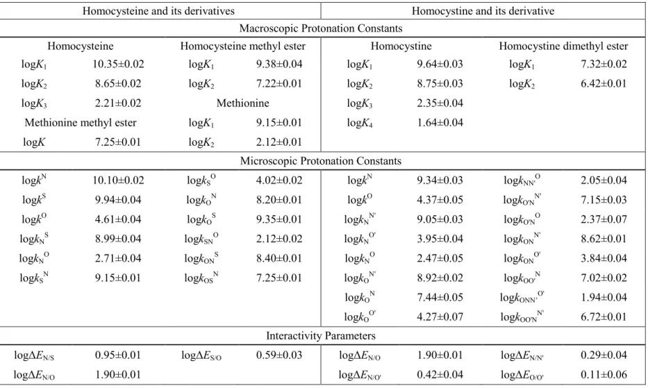
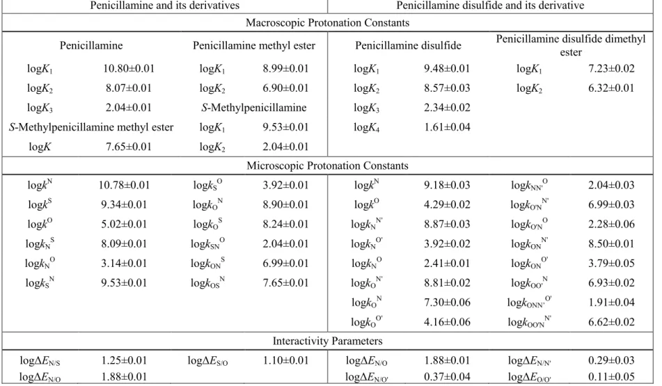
Cysteine, homocysteine, and penicillamine were titrated under near physiological conditions to afford the macroscopic protonation constants (Table 2, 3, 4). The microspeciation scheme of a tridendate molecule is a multivariable problem with 7 unknowns. Therefore, in addition to the three macroscopic protonation constants, four further quantities have to be introduced to elucidate all the microconstants. The kind and number of extra pieces of information to be introduced depend on the acid-base properties of the sites in the parent compounds. Chemical evidences clearly indicate that the inherent basicity decreases in the amino, thiolate, carboxylate order, in which the basicity of thiolate is “comparably lower”, whereas the basicity of the carboxylate is typically “orders of magnitude lower” than that of the amino group. Consequently, those microspecies in which the carboxylate site is protonated, but the amino is not, are
“orders of magnitude minor” (omm) ones, while those in which the thiolate is protonated, but the amino is not, are expected to be “relative minor” (rm) ones. Omm microspecies influence the analytical signals (NMR, UV, etc.) insignificantly, but they may be the reactive species in highly specific biochemical processes [57]. A complete microspeciation inevitably needs auxiliary derivative compounds that mimic the omm microspecies, and in many cases the rm microspecies too. The possible closest mimicry compounds of the d, c, and g cysteine microspecies are cysteine methyl ester, S- methylcysteine, and S-methylcysteine methyl ester, respectively, where the methyl moiety at the carboxylate or thiolate site keeps the site in question in its neutral form,