Full Terms & Conditions of access and use can be found at
https://www.tandfonline.com/action/journalInformation?journalCode=zjom20
Journal of Oral Microbiology
ISSN: (Print) (Online) Journal homepage: https://www.tandfonline.com/loi/zjom20
A case study of salivary microbiome in smokers and non-smokers in Hungary: analysis by shotgun metagenome sequencing
Roland Wirth , Gergely Maróti , Róbert Mihók , Donát Simon-Fiala , Márk Antal , Bernadett Pap , Anett Demcsák , Janos Minarovits & Kornél L. Kovács
To cite this article: Roland Wirth , Gergely Maróti , Róbert Mihók , Donát Simon-Fiala , Márk Antal , Bernadett Pap , Anett Demcsák , Janos Minarovits & Kornél L. Kovács (2020) A case study of salivary microbiome in smokers and non-smokers in Hungary: analysis by shotgun metagenome sequencing, Journal of Oral Microbiology, 12:1, 1773067, DOI: 10.1080/20002297.2020.1773067 To link to this article: https://doi.org/10.1080/20002297.2020.1773067
© 2020 The Author(s). Published by Informa UK Limited, trading as Taylor & Francis Group.
View supplementary material
Published online: 07 Jun 2020. Submit your article to this journal
Article views: 84 View related articles
View Crossmark data
A case study of salivary microbiome in smokers and non-smokers in Hungary:
analysis by shotgun metagenome sequencing
Roland Wirtha, Gergely Marótib, Róbert Mihókc, Donát Simon-Fialac, Márk Antalc, Bernadett Papb, Anett Demcsákd, Janos Minarovitsd and Kornél L. Kovács a,d
aDepartment of Biotechnology, University of Szeged, Szeged, Hungary; bInstitute of Plant Biology, Biological Research Center, Szeged, Hungary; cDepartment of Operative and Esthetic Dentistry, Faculty of Dentistry, University of Szeged, Szeged, Hungary; dDepartment of Oral Biology and Experimental Dental Research, Faculty of Dentistry, University of Szeged, Szeged, Hungary
ABSTRACT
Objective: To investigate the role of cigarette smoking in disease-development through altering the composition of the oral microbial community. Periodontitis and oral cancer are highly prevalent in Hungary; therefore, the salivary microbiome of smoker and non-smoker Hungarian adults was characterized.
Methods: Shotgun metagenome sequencing of salivary DNA samples from 22 individuals (11 non-smokers and 11 current smokers) was performed using the Ion Torrent PGMTM platform.
Quality-filtered reads were analysed by both alignment-based sequence similarity searches and genome-centric binning.
Results: Prevotella, Veillonella and Streptococcus were the predominant genera in the saliva of both groups. Although the overall composition and diversity of the microbiota were similar, Prevotella was significantly more abundant in salivary samples of current smokers compared to non-smokers. Members of the genus Prevotella were implicated in the development of inflammatory diseases and oral cancer. The abundance of the genus Megasphaera also increased in current smokers, whereas the genera Neisseria, Oribacterium, Capnocytophaga and Porphyromonas were significantly reduced. The data generated by read-based taxonomic classification and genome-centric binning mutually validated the two distinct metagenomic approaches.
Conclusion: Smoking-associated dysbiosis of the salivary microbiome in current cigarette smokers, especially increased abundance of Prevotella and Megasphaera genera, may facilitate disease development.
ARTICLE HISTORY Received 31 July 2019 Revised 6 April 2020 Accepted 13 April 2020 KEYWORDS
Smoking; saliva metagenome; read-based taxonomy; genome-centric binning; Prevotella;
Megasphaera; oral cancer
Introduction
The oral cavity of healthy humans harbors a diverse microbial community called the “normal flora”, which is composed of more than 700 bacterial species that regularly attach to and form biofilms on the surfaces of soft and hard tissues within the mouth [1–3].
Members of the oral biofilms are regularly shed into the saliva, which is bathing the oral mucosa [4]. Saliva is a complex biological fluid whose composition is affected both by local conditions in the oral cavity and systemic diseases [5–7]. Since saliva can be col- lected in a painless, non-invasive manner, substantial efforts have been made to identify disease-related sali- vary biomarkers, recently [8]. In addition to the bio- molecules accumulating in saliva during pathological processes, the oral microbiome may also be regarded as a new biomarker reservoir [9]. Thus, the changes of the salivary microbial community can also be exploited for the diagnosis and monitoring of oral and systemic diseases [10–18]. Smoking is an important risk factor for oral diseases, such as periodontitis and oral cancer,
and it is also associated with a wide variety of systemic diseases [19–21]. Although tobacco use, especially cigarette smoking, decreased in the last decades in Western countries, regular smoking is still a common habit in Central- and Eastern European countries [22,23], Asia, China, and North Africa (https://www.
who.int/gho/tobacco/use/en/). Tobacco smoke may contain more than 5,000 chemicals, among them toxic, mutagenic and carcinogenic substances [24].
These chemicals may initiate pathogenic alterations by interacting directly with various host cells and extracellular matrix components [25]. Nicotine, a major, highly addictive constituent of cigarette smoke modulates the immune responses [25,26].
Toxic compounds in tobacco smoke may cause cellular injury and cell death whereas carcinogens, including N-nitrosamines and polycyclic aromatic hydrocarbons may initiate tumorigenesis by forming DNA adducts and blocking DNA repair [27–31]. Chemicals in cigar- ette smoke may also contribute to disease development indirectly by changing the composition of the human
CONTACT Kornél L. Kovács kovacs.kornel@bio.u-szeged.hu Department of Biotechnology, University of Szeged, Szeged, Hungary Supplemental data for this article can be accessed here.
2020, VOL. 12, 1773067
https://doi.org/10.1080/20002297.2020.1773067
© 2020 The Author(s). Published by Informa UK Limited, trading as Taylor & Francis Group.
This is an Open Access article distributed under the terms of the Creative Commons Attribution License (http://creativecommons.org/licenses/by/4.0/), which permits unrestricted use, distribution, and reproduction in any medium, provided the original work is properly cited.
oral microbiome [32]. Alteration of the oral micro- biome in cigarette smokers may favour disease devel- opment by increasing the local density of bacterial pathogens or decreasing the prevalence of their com- petitors [12,33,34].
Cigarette smoking is one of the most important aspects in the development of oral diseases, including periodontitis and oral cancer, which are particularly prevalent in Hungary [35–37]. Smoking may influence disease progress by altering the microbial communities of the oral cavity; therefore,e in this cross-sectional study, we characterized the salivary microbiome of smoker and non-smoker Hungarian adults. We applied a metagenomic approach and used both alignment- based sequence similarity searches and genome- centric binning for the analysis of shotgun sequences generated by the Ion Torrent PGMTM platform [38–41].
Study design and recruitment of participants The study protocol was approved by the Institutional Review Board of the University of Szeged, Szeged, Hungary. Signed informed consent was obtained from each healthy adult participant enrolled into the study at the Department of Operative and Esthetic Dentistry, Faculty of Dentistry, University of Szeged, Hungary. Study participants were divided into two groups, non-smokers and current smokers, based on the data they provided regarding tobacco consump- tion (cigarette smoking). The smoking exposure of current smokers was calculated in pack-years. One pack contained 20 cigarettes.
Characteristics of study participants
A total of 11 healthy adult non-smokers (4 males and 7 females; mean age: 40 years; range: 26–46 years), including 8 never smokers and 11 healthy adult cur- rent smokers (8 males and 3 females; mean age:
41.5 years; range: 34–61 years) participated in this cross-sectional study. Three of the participants in the non-smoker group quit smoking 5.5, 5 and 1.5 years before the study, respectively. None of the participants suffered from known chronic illness and none were treated with antibiotics at least 6 months prior to sampling. In order to record the oral para- meters, all patients received a full mouth cariological and periodontal examination, performed by an experienced practitioner. The number of missing teeth (excluding third molars), Plaque Index (PI;
also known as the Silness-Löe Index), bleeding on probing (BOP; the presence or absence of bleeding within 15 sec after probing), probing depth (PD; in millimeters), and clinical attachment level (CAL; to describe the position of the soft tissue in relation to the cemento-enamel junction) were recorded. To describe the periodontal status of the patients,
a classification was used [42], which was proven to be reliable in our earlier works [43,44]. All patients with moderate or severe peridontitis were excluded from the study.
Methods
Measurement of exhaled carbon monoxide in healthy smokers and non-smokers
The level of exhaled carbon monoxide (CO) is a suitable indicator of smoking status [45,46]. We used a calibrated, portable CO monitor (piCO + Smokerlyzer, Breath CO monitor, Bedfont Scientific Ltd., Kent, UK) to assess the exhaled CO levels in the study groups of non-smokers and current smokers.
Participants were asked to exhale completely, inhale fully, and then hold their breath for as long as possible. Right after this, the participants were instructed to exhale slowly into the unit and exhale fully. This procedure was repeated three times and the mean value was calculated.
Saliva collection and DNA isolation from saliva Unstimulated whole saliva samples were collected from the participants by the simple drooling method, ali- quoted and stored at −80 C°. After thawing, saliva samples (3 ml, each) were centrifuged at 13 000 rpm for 5 min. DNA extractions were carried out by using the Macherey-Nagel NucleoSpin Soil DNA kit (Macherey-Nagel: 740,780.250). The lysis mixture con- tained 700 µL SL1 and 150 µL Enhancer SX lysis solu- tions. After lysis (bead beating), the kit protocol was followed. The quantity of DNA was determined in a NanoDrop ND-1000 spectrophotometer (NanoDrop Technologies, Wilmington, USA) and a Qubit 2.0 Fluorometer (Life Technologies, Carlsbad, USA).
DNA purity was tested by agarose gel electrophoresis and on an Agilent 2200 TapeStation instrument (Agilent Technologies, Santa Clara, USA).
Next-generationn sequencing and bioinformatics analysis
The recommendations of the Ion Torrent PGM™
sequencing platform were closely followed (Life Technologies, Thermo Fisher Scientific, USA). The sample libraries were prepared by Ion Xpress Plus Fragment Library Kit (Cat. No. 4471269; Thermo Fisher Scientific, USA) and quantified by Ion Library TaqMan® Quantitation Kit (Cat. No. 4468802; Thermo Fisher Scientific, USA) with the help of StepOne Real- Time PCR System (Applied Biosystems). Emulsion PCR was performed with OneTouch 2 and Ion OneTouch ES devices by using the Ion PGM Template OT2 200 kit (Cat. No. 4,480,974; Thermo
Fisher Scientific, USA). Barcoding was made by Ion Xpress Barcode Adapters 1–16 Kit (Cat. No.
4,471,250¸Thermo Fisher Scientific, USA). Sequencing was performed with Ion PGM 200 Sequencing kit (Cat.
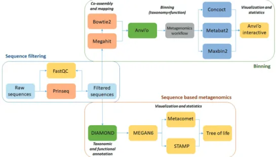
No. 4,474,004¸ Thermo Fisher Scientific, USA) on Ion Torrent PGM 316 chip. The raw data have been made publicly available at SRA accession: PRJNA553326 (Release date: 2019–07-09). The workflow of the sub- sequent data analysis is summarized in Figure 1.
Raw sequence filtering
Galaxy Europe server was employed to pre-process the raw sequences (www.usegalaxy.eu). Low-quality reads were filtered by Prinseq (min. length: 150 bp;
min. score: 15; quality score threshold to trim posi- tions: 20; sliding window used to calculate quality score: 1) [47]. Quality of raw and filtered sequences were checked with FastQC program.
Read-based metagenomics
Filtered high or moderate quality sequences were further analyzed by Diamond software, applying the LCA (Lowest Common Ancestor) algorithm [48].
Diamond parameters were set as follows: Blast Mode: BlastX, Reference database: NCBI nr database.
MEGAN6 was used to add taxon names to Diamond sequence classifications (Min Score: 80, Min support percent: 80, Min support: 15, Min complexity filter: 0.3, LCA algorithm: weighted) [49].
Statistical analysis of read – based metagenomics data
MEGAN6 was used to investigate microbial commu- nities and export data for statistic calculations.
UPGMA (Unweighted Pair-Group Method with Arithmetic Mean) with Bray–Curtis method was employed to cluster the samples (Figure 4(b)).
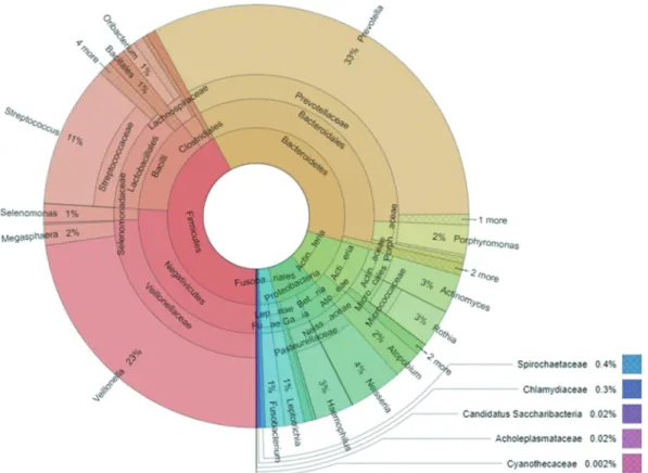
Rarefaction estimation was performed by MEGAN6 [49] (Supplementary figure 1). Krona program was used to visualize the average composition of micro- bial taxa [50] (Figure 2). The distribution of top 10 most abundant microbes between the two sets of samples was presented with Circos [51] (Figure 3).
For microbial core and diversity calculation MetaCoMET (Metagenomics Core Microbiome Exploration Tool), an interactive web tool, was used.
Shannon statistical method was performed to calcu- late alpha diversity (Figure 4(c)). Emperor program (integrated into MetaCoMET) carried out the princi- pal component analysis (Figure 4(a)). For core calcu- lation default parameter sets were fixed with the persistence Venn diagram type [52].
Statistical Analysis of Metagenomic Profiles (STAMP) was used to compute the abundance differ- ences in the case of whole microbiome. Dissimilar taxa were identified with two-sided t-test at 0.95 confidence intervals and the results with q-value (corrected p-value) of <0.05 were retained. In STAMP minimum difference between proportions was set to 0.3 and Storey FDR (False Discovery Rate) filtered out false positive significant differences [53,54] (Figure 5).
Genome-based evaluation of the sequencing data Filtered sequences produced by Prinseq were co- assembled with Megahit (Minimum contig length:
1000 bp, Minimum k-mer size: 21, Maximum k-mer size 141) [55]. After simplifying the header of contig FASTA file using the Anvi’o script, Bowtie2 was employed to map back the original sequences to the contigs [56]. Then, we used Anvi’o V5 to follow the
Figure 1. Summary of the data analysis workflow and the employed software packages. The main steps of the initial data filtering and bioinformatics steps to extract the read-based and genome-based metagenome data are boxed separately.
Figure 2. Overall composition of the salivary microbiome of study participants including the metagenomes of both smokers and non-smokers. Due to space limitations, only the most relevant bacterial genera, families, orders and phyla are indicated; the bacterial classes are not shown.
Figure 3. Relative abundance of the 10 most abundant bacterial genera in non-smokers and current smokers. Circos plot illustrating the most abundant bacterial genera listed in inset from 1 to 10. The widths of the bands are proportional to the abundance of the particular taxon in the two study groups.
‘metagenomics’ workflow [57]. Briefly, contig data- base was generated in the first step, where open read- ing frames were identified by Prodigal and contig k-mer frequencies were computed. Then, Hidden Markov Modell (HMM) of single-copy genes was aligned by HMMER [58–61]. We used InterProScan v5.31–70 and the metagenome classifier Kaiju to functional and taxonomic annotation of contigs [62–65]. The outputs were imported into the contig database. BAM files, made by Bowtie2, were used for profiling the contig database, this way we generated sample-specific information about the contigs (i.e.
mean coverage). These were merged together. Three automated binning programs, i.e. CONCOCT, METABAT2 and MAXBIN2, were employed to reconstruct microbial genomes from the contigs (Minimum length: 2,000 bp) [66–68]. We also used the Anvi’o human-guided binning and ‘anvi-refine’
options [57]. The binning results were incorporated to the contig database. Anvi’o interactive interface was employed to visualize and summarize the data in Figure 6.
Results
Lifetime tobacco exposure in current smokers Lifetime tobacco exposure of current smokers was calculated in pack-years by multiplying the average
number of packs of cigarettes smoked per day by the number of years the person had smoked. The mean smoking (tobacco) exposure of current smokers (N = 11) was 11.5 ± 8.2 pack-years (range: 0.6–23 pack-years) (Supplementary table 1).
The distribution of early periodontal lesions (mild gingivitis) was equal in both groups. The number of decayed teeth was higher in the group of smokers (4.18 SD: 0.78) compared to the group of non- smokers (2.45 SD: 0.85) but the difference between the group was not significant (P < 0.1499 Student’s test).
Exhaled carbon monoxide in healthy smokers and non-smokers
Data regarding exhaled CO levels were available for all non-smokers (N = 11; mean exhaled CO level:
1.7 ± 0.9 ppm) and for 6 of the 11 current smokers (mean exhaled CO level: 12.3 ± 12 ppm). The differ- ence between the exhaled CO level of non-smokers and current smokers was statistically significant (Studen’s t-test; P˂0.0001). The values found in our study were comparable to the data recorded for healthy adult non-smokers (1.5 ± 0.6 ppm) and healthy adult smokers (9.7 ± 5.7 ppm) in a recent independent study using the same type of CO moni- tor [69].
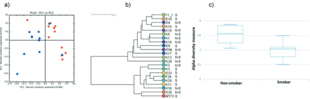
Figure 4. Diversity of the salivary microbiome in non-smokers and current smokers.
(a) Principal component analysis of salivary microbiomes of non-smokers and current smokers. Symbols: blue squares = non-smokers; red diamonds = current smokers (b) Hierarchical clustering analysis of salivary microbiomes of non-smokers and current smokers by UPGMA (unweighted pair group method with arithmetic mean). The two metagenome clusters characterizing the two very distinct study groups are boxed. Comparison of Shannon diversity indices. The alpha diversities are clearly distinct although statistically not rigorously different.
Figure 5. Bacterial genera which differ significantly in relative abundance between the salivary samples of non-smokers and current smokers. The statistical analysis was performed and visualized using the STAMP package. Mean abundance (mean proportion) and difference in mean proportion for genera showing significant difference in abundance are shown. The 95%
confidence intervals and statistical significance (corrected q value) are indicated as well.
Read-based characterization of the salivary microbiome in non-smokers and current smokers Shotgun metagenome sequencing of salivary DNA samples from 22 individuals (11 non-smokers and 11 current smokers) resulted in 256,567 quality- filtered reads; mean reads per sample: 11,662.1 (SD:
2,261.95). To monitor the efficiency of sequencing, rarefaction curves were computed (Supplementary figure 1). The rarefaction curves reached their asymp- totes around 5,000 reads, indicating that the sequen- cing depth was sufficient to cover almost all genera in the bacterial communities analyzed.
Based on the alignment of the quality-filtered reads with sequences in the NCBI nr database, 66 bacterial genera could be identified. The overall com- position of the oral microbiome of study participants (both non-smokers and current smokers) is shown in Figure 2. It is apparent that three genera, i.e.
Prevotella, Veilonella and Streptococcus, predomi- nated in the saliva of the Hungarian study
participants, although the mean relative abundance of 11 additional genera reached or exceeded 1%
(Supplementary table 2).
Comparison of the salivary microbiome of non- smokers and current smokers revealed that 48 bacter- ial genera were present in at least one specimen of both groups (Supplementary table 2). All of the 14 abundant genera (relative abundance 1% or higher) were shared by both microbiota. In addition, 34 rare genera (relative abundance ˂1%) were also shared by non-smokers and current smokers. In total, 8 rare genera were unique to non-smokers whereas 10 rare genera manifested themselves in current smokers only (Supplementary table 2).
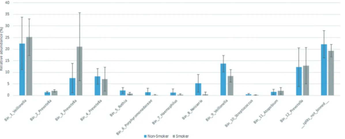
The relative distribution of the 10 most abundant genera is presented in Figure 3. It is noteworthy that the genera Prevotella, Veillonella and Streptococcus predominated in the saliva samples of current smo- kers, comprising about 90% of the total reads. In the non-smoker group, about 70% of all reads mapped to Figure 6. Analysis of shotgun sequences by genome-centric binning. Distribution of contigs built from filtered sequences of salivary bacterial communities. The grouping of contigs based on sequence-assignments of automated binning programs METABAT2, MAXBIN2 and CONCOCT as well as manually defined bins were visualised by the Anvi’o platform. SCG: single-copyy genes; GC: guanine-cytosine (GC) content.
these 3 genera. In accordance with PCA (principal component analysis) (Figure 4(a)), hierarchical clus- tering analysis by UPGMA (unweighted pair group method with arithmetic mean) (Figure 4(b)) also showed that the microbiomes of non-smokers and current smokers did not form two rigorously sepa- rated clusters although the tendencies with a few out- liers are clearly recognizable. We also noticed that a group of non-smokerss (K19, K5, K1, K18) and a group of current smokers (K3, D22, F26, K20, K11) were clearly located on two separate branches of the UPGMA tree (boxed in Figure 4(b)). This might be a sign of distinction albeit statistically non- significant. The Shannon index, which reflects both richness and evenness of microbial communities, was higher in case of the salivary microbiome of non- smokers, compared to that of the current smokers (Figure 4(c)). The difference, however, was again not statistically significant.
In spite of thee similarities in the composition and diversity of salivary microbial communities, the relative abundance of distinct genera differed significantly between non-smokers and current smokers. Two gen- era displayed pronounced changes. Prevotella (order Bacteroidales), the predominant genus in both groups, was more abundant in the saliva of current smokers (mean relative abundance: 36.8 ± 9.8%), compared to non-smokers (mean relative abundance: 26.0 ± 9.9%);
the difference between the groups was statistically sig- nificant (p = 0.044; Figure 5). In addition to Prevotella, the genus Megasphaera, a member of the order Negativicutes, belonging in the phylum Firmicutes, was also enriched in the saliva samples of current smokers (Figure 5). Although Megasphaera was a rare, low- abundance genus in the salivary bacterial communities of non-smokers, it reached a relative abundance above 1% in the salivary samples of current smokers. In con- trast, the salivary microbiome of non-smokers was
significantly enriched in the genera Neisseria (phylum:
Proteobacteria; order: Neisseriales), Oribacterium (phy- lum: Firmicutes; order: Clostridiales), Capnocytophaga (phylum: Bacteriodetes; order: Flavobacteriales) and Porphyromonas (phylum: Bacteriodetes; order: Bacter- iodales) (Figure 5).
Analysis of shotgun sequences by genome-centric binning
In addition to read-based taxonomic classification, the filtered sequences generated in our study were sepa- rately assembled in contigs, which were clustered into bins based on their inherent sequence features (gen- ome-centric binning). The workflow of both read-based classification and metagenomics binning is presented in Figure 1. A total of 12 bins were constructed, using the human-guided automated binning programs that rely on co-abundance of sequences as well as compositional information such as GC content, tetranucleotide fre- quencies and identification of single-copy genes (Figure 6). The putative genomes of the genera Prevotella and Veillonella each were represented by 4 and 2 separate bins, respectively, whereas the genera Rothia, Haemophilus, Neisseria, Streptococcus and Atopobium, as well as the family Porphyromonadaceae each occu- pied a single distinct bin. Based on genome-centric binning, Prevotella (bin 3) showed a significantly higher relative abundance in the salivary microbiome of cur- rent smokers, whereas the family Porphyromonadaceae (bin 6) and the genus Neisseria (bin 8) was significantly more abundant in the salivary microbiome of non- smokers (Figure 7). These data are compatible with the results of read-based taxonomic classification of salivary microbiomes from non-smokers and current smokers (see Figures 3 and 5) and therefore the two distinct metagenomic approaches mutually validate each other.
Figure 7. Relative abundance of bacterial genera identified by genome-centric binning. Prevotella (bin 3) showed a significantly higher relative abundance in the salivary microbiome of current smokers, whereas the family Porphyromonadaceae (bin 6) and the genus Neisseria (bin 8) were significantly more abundant in the salivary microbiome of non-smokers.
Discussion
Toxic and mutagenic chemicals as well as particulate matter in cigarette smoke may initiate oral diseases either via direct interactions with human cells in the oral cavity, or indirectly, by affecting the environ- ment of these cells such as the extracellular matrix and/or the oral microbiome [12,25,32,70,71].
Mutagens may cause genetic damage both in human cells and in bacterial cells inhabiting the oral cavity [31,70]. In addition, smoking may damage oral microcirculation and create an acidic, relatively hypoxic milieu in the oral cavity that may favour the growth of distinct members of the oral microbial community [72,73]. Establishment of a similar micro- environment may select for the local growth of dis- tinct anaerobic bacteria within neoplastic tissues, too [74,75].
Saliva is a complex biological fluid bathing the various anatomical structures of the oral cavity cov- ered by biofilm-forming microbial communities.
From mature biofilms located at intraoral surfaces, bacteria – including potential pathogens – are regu- larly shed into the saliva. In such a planktonic state, oral bacteria may be transmitted to new ecological niches [13,76].
Oral diseases and tobacco smoke exposure induce anatomical and physiological changes in the oral cav- ity, consequently the composition of surface-attached bacterial supragingival and subgingival biofilms and the structure of the salivary microbial community may be altered [17,77–81]. Smoking cessation may revert, however, the changes in the composition of oral microbiome [18,82]. Our study groups of current smokers and non-smokers comprised middle-aged d female and male healthy Hungarian citizens, having a cigarette smoking habit of various degrees. The tobacco consumption and the duration and extent of tobacco smoke exposure varied considerably (Supplementary table 1). The justification for select- ing such a diverse group of individuals was that we attempted to dissect a broad sample covering diverse individual variability in the environmental and life- style parameters of our subjects in order to track the general features of oral microbiome alterations caused by cigarette smoking. As expected, the price for this heterogenous sampling approach was sub- stantial individual deviation in microbial commu- nities within the two study groups. In spite of the dissimilarities in their individual case histories, the
“current smokers” and “non-smokers” demonstrated clear difference in their exhaled CO levels, a good indication of the substantial physiological influence caused by deleterious cigarette smoking regardless of other variables. The diversity of individual microbes affected by environmental and lifestyle conditions other than smoking habits masked several aspects of
oral microbiome rearrangements as being statistically not significant although indicating noticeable trends (Figure 4(a,b)). Accordingly, the difference was not rigorously significant, we found that the Shannon index which reflects both richness and evenness of microbial communities, was higher for the salivary microbiome of non-smokers compared to current smokers (Figure 4(c)). This may be related to a decreased evenness in the microbial community of current smokers which is characterized by an increased abundance of Prevotella (Figures 3 and 5).
Similarly, the microbiomes of non-smokers and cur- rent smokers did not form well separated clusters by principal component analysis (PCA) and hierarchical clustering (UPGMA). Nevertheless, location of a group of samples from non-smokers and another from current smokers on two separate branches of the UPGMA tree might be a sign of distinction between the two microbiomes (Figure 4(a,b)).
In the thorough shotgun full metagenomic study we found that all of the 14 abundant genera (relative abundance 1% or higher) and 34 rare genera (relative abundance ˂1%) were shared by the salivary micro- biomes of non-smokers and current smokers, whereas 8 rare genera were unique to non-smokers and 10 rare genera were unique to current smokers (Supplementary table 2). Remarkably, the read-based metagenomics data were supported by metagenomic binning sufficiently (Figures 6 and 7).
Both microbial communities were predominated by 3 genera, i.e. Prevotella, Veillonella and Streptococcus (Figure 3). Analysis of the complexity of salivary microbial communities in non-smokers and smokers gave variable results in previous studies. In most cases, analysis of oral wash samples or oral swabs did not reveal significant differences between the microbiomes of smokers and non-smokers [83–85]. To the contrary, a large study of oral wash samples revealed a significant difference in overall oral microbiome composition between current and non-current (former and never) smokers [18]. Further studies may resolve these apparent discrepancies.
We observed that although the overall composi- tion of the oral microbiome did not differ substan- tially between non-smokers and current smokers (Figures 3 and 4) the relative abundance of two dis- tinct genera, Prevotella and Megasphaera, was higher in salivary samples of current smokers (Figure 5).
Prevotella species are Gram-negative, anaerobic bac- teria which belong in the phylum Bacteriodetes.
Although in healthy humans the Prevotella genus is one of the dominant genera of the salivary micro- biome, distinct members of the genus Prevotella are associated with inflammatory diseases and may facil- itate carcinogenesis as well [86-88, reviewed by 89,90]. There are, however, contradicting observa- tions regarding the role of Prevotella in the
development of oral cancer [14,74,79,91–94]. Thus, it remains to be established whether members of the genus Prevotella are opportunistic inhabitants of malignant tumors or play a causative role in oral or colorectal carcinogenesis [95,96].
Megasphaera, the other genus of increased abun- dance in the saliva of current smokers belongs in the phylum Firmicutes. Megasphaera are Gram-negative anaerobic cocci, which reside in the upper digestive tract of adults, contributing to the microbial commu- nity of tongue dorsum, tonsils and saliva [86]. In line with our results, an increase in relative abundance of Megasphaera was observed in oropharyngeal samples and esophageal samples of smokers relative to non- smokers [97,98]. Perhaps the smoky environment may confer a growth advantage for Megasphaera [98]. Dysbiotic diseases, including periodontitis and bacterial vaginosis were also associated with higher relative abundance of Megasphaera species [99,100].
Moreover, the genus Megasphaera was associated with human papillomavirus (HPV) positive head and neck squamous carcinoma and lung cancer [101,102].
Our finding of an increased abundance of Prevotella and Megasphaera in the saliva of current smokers may facilitate the initiation and progression of various pathogenic processes within the oral cavity and may affect the composition of microbial commu- nities along the route of swallowed saliva as well, i.e.
on the surface of tonsils and throat, and possibly even the esophageal mucosa [86,98].
We observed that the salivary microbiome of cur- rent smokers was significantly depleted relative to non-smokers in the genera Neisseria (phylum:
Proteobacteria; order: Neisseriales), Oribacterium (phy- lum: Firmicutes; order: Clostridiales), Capnocytophaga (phylum: Bacteriodetes; order: Flavobacteriales) and Porphyromonas (phylum: Bacteriodetes; order:
Bacteriodales) (Figure 5). The decreased abundance of Neisseria in the saliva of current smokers could possi- bly be attributed to the selective toxicity of cigarette smoke for Neisseria species [103,104]. Compared to non-smokers, the abundance of Neisseria decreased in the oropharynx of smokers [97].
Increased abundance of Oribacterium parvum and other distinct bacterial species in prediagnostic oral wash samples was associated with a decreased risk for esophageal adenocarcinoma development [15]. An increased level of Oribacterium was also detected in oral rinse samples of patients with oral cavity carci- noma and oropharyngeal carcinoma [14,101].
Therefore, it is not easy to predict how the decreased salivary level of Oribacterium, observed in our cur- rent smoker study group, may affect pathological processes in the gastrointestinal tract.
Analysis of relative abundance of Capnocytophaga species in the oral cavity of smokers and non-smokers
also yielded apparently conflicting results in various laboratories. Thomas et al. observed an increased level of Capnocytophaga in oral swab samples of smokers, whereas Wu et al. found a decreased level in oral wash samples of smokers [18,84]. Our finding, based on saliva samples collected by the simple drooling method, is in accordance with the data of Wu et al.
[18]. One may assume that using oral swab samples may permit a more efficient collection of bacteria deeply embedded in biofilms at various surfaces com- pared to taking unstimulated saliva samples or oral wash samples.
Nicotine, an important component of cigarette smoke, inhibited the growth of Porphyromonas gingi- valis [105]. This observation may explain the decreased level of Porphyromonas species in saliva samples of current smokers, compared to non- smokers, as demonstrated in our current study, in accordance with previous findings [18,83].
In summary, we conclude that probably several environmental, personal health history and life style factors affect the alterations in the oral microbiota in smoker and non-smoker individuals. Our study did not address most of these potential factors, which may blur somewhat the picture. Nevertheless, the clear message of this study is that tobacco smoking brings about dysbiotic, deleterious changes in the oral microbiota and this may lead to severe oral and general unwanted health consequences for the smok- ing people. We also identified a few genera for diag- nostic purposes of the warning molecular taxonomy signs of the dangerous consequences for smokers.
Acknowledgments
The support of the grant GINOP-2.3.2-15-2016-00011 by the European Regional Development Fund to a project led by JM is kindly acknowledged. Additional projects GINOP- 2.2.1-15-2017-00081, GINOP-2.2.1-15-2017-00033, and EFOP-3.6.2-16-2017-00010 also contributed to this work.
The grants were managed by European Union Economic Development and Innovation Operational Programme, National Research, Development and Innovation Office (NKFIH), Hungary. RW and GM received support from the Hungarian NKFIH fund projects PD121085 and FK123899 financed under the PD16 and FK16 funding schemes. This work was also supported by the János Bolyai Research Scholarship (for GM) of the Hungarian Academy of Sciences.
Disclosure statement
No potential conflict of interest was reported by the authors.
Funding
This work was supported by the European Union Economic Development and Innovation Operational
Programme, National Research, Development and Innovation Office (NKFIH), Hungary [GINOP-2.3.2-15- 2016-00011]; European Union Economic Development and Innovation Operational Programme, National Research, Development and Innovation Office (NKFIH), Hungary [GINOP-2.3.2-15-2016-00011]; Hungarian NKFIH fund [FK123899]; European Union Economic Development and Innovation Operational Programme, National Research, Development and Innovation Office (NKFIH) [EFOP-3.6.2-16-2017-00010]; European Union Economic Development and Innovation Operational Programme, National Research, Development and Innovation Office (NKFIH), Hungary [2.2.1-15-2017- 00033]; European Union Economic Development and Innovation Operational Programme, National Research, Development and Innovation Office (NKFIH), Hungary [GINOP-2.3.2-15-2017-00811]; Hungarian NKFIH fund [PD121085].
ORCID
Kornél L. Kovács http://orcid.org/0000-0002-3926-0497
References
[1] Chen T, Yu WH, Izard J, et al. The human oral microbiome database: a web accessible resource for investigating oral microbe taxonomic and genomic information. Database (Oxford). 2010;bAQ3.
[2] Dewhirst FE, Chen T, Izard J, et al. The human oral microbiome. J Bacteriol. 2010;192(19):5002–5017.
[3] Human Microbiome Project Consortium. Structure, function and diversity of the healthy human microbiome. Nature. 2012;486(7402):207–214..
[4] Belstrøm D, Holmstrup P, Bardow A, et al.
Comparative analysis of bacterial profiles in unsti- mulated and stimulated saliva samples. J Oral Microbiol. 2016;8:30112.
[5] Javaid MA, Ahmed AS, Durand R, et al. Saliva as a diagnostic tool for oral and systemic diseases.
J Oral Biol Craniofac Res. 2016;6:66–75.
[6] Malamud D, Rodriguez-Chavez IR. Saliva as a diagnostic fluid. Dent Clin North Am. 2011;55 (1):159–178.
[7] Zhang CZ, Cheng XQ, Li JY, et al. Saliva in the diagnosis of diseases. Int J Oral Sci. 2016;8 (3):133–137.
[8] Yoshizawa JM, Schafer CA, Schafer JJ, et al. Salivary biomarkers: toward future clinical and diagnostic utilities. Clin Microbiol Rev. 2013;26(4):781–791.
[9] Lim Y, Totsika M, Morrison M, et al. Oral Microbiome: A new biomarker reservoir for oral and oropharyngeal cancers. Theranostics. 2017;7 (17):4313–4321.
[10] Ahn J, Chen CY, Hayes RB. Oral microbiome and oral and gastrointestinal cancer risk. Cancer Causes Control. 2012;23(3):399–404.
[11] Balan P, Chong YS, Umashankar S, et al. Keystone species in pregnancy gingivitis: a snapshot of oral microbiome during pregnancy and postpartum period. Front Microbiol. 2018;9:2360.
[12] Jiang Y, Zhou X, Cheng L, et al. The impact of smoking on subgingival microflora: from periodontal health to disease. Front Microbiol. 2020;11:66.
[13] Le Bars P, Matamoros S, Montassier E, et al. The oral cavity microbiota: between health, oral disease, and cancers of the aerodigestive tract. Can J Microbiol.
2017;63(6):475–492.
[14] Lim Y, Fukuma N, Totsika M, et al. The performance of an oral microbiome biomarker panel in predicting oral cavity and oropharyngeal cancers. Front Cell Infect Microbiol. 2018;8:267.
[15] Peters BA, Wu J, Pei Z, et al. Oral microbiome composition reflects prospective risk for esophageal cancers. Cancer Res. 2017;77(23):6777–6787.
[16] Yamashita Y, Takeshita T. The oral microbiome and human health. J Oral Sci. 2017;59(2):201–206.
[17] Wolf A, Moissl-Eichinger C, Perras A, et al. The salivary microbiome as an indicator of carcinogen- esis in patients with oropharyngeal squamous cell carcinoma: A pilot study. Sci Rep. 2017;7(1):5867.
[18] Wu J, Peters BA, Dominianni C, et al. Cigarette smoking and the oral microbiome in a large study of American adults. Isme J. 2016;10(10):2435–2446.
[19] Edwards R. The problem of tobacco smoking. BMJ.
2004;328(7433):217–219.
[20] Leite FRM, Nascimento GG, Scheutz F, et al. Effect of smoking on periodontitis: A systematic review and meta-regression. Am J Prev Med. 2018;54(6):831–841.
[21] Peterson LA, Bellile EL, Wolf GT, et al. University of Michigan head and neck specialized program of research excellence program. Cigarette use, comor- bidities, and prognosis in a prospective head and neck squamous cell carcinoma population. Head Neck. 2016;38(12):1810–1820.
[22] Eriksen M, Mackay J, Schlugar NW, et al. County fact sheet, Hungary. In: The tobacco atlas. fifth ed.
American Cancer Society; 2015. http://www.tobaccoa tlas.org/country-data/hungary/
[23] Ezzati M, Riboli E. Behavioral and dietary risk fac- tors for noncommunicable diseases. New Engl J Med. 2013;369(10):954–964.
[24] Talhout R, Schulz T, Florek E, et al. Hazardous compounds in tobacco smoke. Int J Environ Res Public Health. 2011;8(2):613–628.
[25] Mehta H, Nazzal K, Sadikot RT. Cigarette smoking and innate immunity. Inflamm Res. 2008;57(11):
497–503.
[26] Sopori ML, Kozak W. Immunomodulatory effects of cigarette smoke. J Neuroimmunol. 1998;83 (1–2):148–156.
[27] Ewa B, Danuta MŠ. Polycyclic aromatic hydrocar- bons and PAH-related DNA adducts. J Appl Genet.
2017;58(3):321–330.
[28] Law AD, Fischer C, Jack A, et al. Tobacco, microbes, and carcinogens: correlation between tobacco cure conditions, tobacco-specific nitrosamine content and cured leaf microbial community. Microb Ecol.
2016;72:120–129.
[29] Xue J, Yang S, Seng S. Mechanisms of cancer induc- tion by tobacco-specific NNK and NNN. Cancers (Basel). 2014;6(2):1138–1156.
[30] Yalcin E, de la Monte S. Tobacco nitrosamines as culprits in disease: mechanisms reviewed. J Physiol Biochem. 2016;72(1):107–120.
[31] Wiencke JK. DNA adduct burden and tobacco carcinogenesis. Oncogene. 2002;21(48):7376–7391.
[32] Roberts FA, Darveau RP. Microbial protection and virulence in periodontal tissue as a function of poly- microbial communities: symbiosis and dysbiosis.
Periodontol 2000. 2015;69(1):18–27.
[33] Brook I. The impact of smoking on oral and naso- pharyngeal bacterial flora. J Dent Res. 2011;90 (6):704–710.
[34] Qi F, Kreth J. Characterization of anti-competitor activities produced by oral bacteria. Methods Mol Biol. 2010;666:151–166.
[35] Ferlay J, Steliarova-Foucher E, Lortet-Tieulent J, et al. Cancer incidence and mortality patterns in Europe: estimates for 40 countries in 2012. Eur J Cancer. 2013;49(6):1374–1403.
[36] Hermann P, Gera I, Borbély J, et al. Periodontal health of an adult population in Hungary: findings of a national survey. J Clin Periodontol. 2009;36 (6):449–457.
[37] Hettmann A, Demcsák A, Á B, et al. Prevalence and genotypes of human papillomavirus in saliva and tumor samples of head and neck cancer patients in Hungary. Infect Genet Evol. 2018;59:99–106.
[38] Borozan I, Watt S, Ferretti V. Integrating alignment-based and alignment-free sequence simi- larity measures for biological sequence classification.
Bioinformatics. 2015;31(9):1396–1404.
[39] Dröge J, McHardy AC. Taxonomic binning of meta- genome samples generated by next-generation sequencing technologies. Brief Bioinform.
2012;6:646–655. DOI:10.1093/bib/bbs031
[40] Hilton SK, Castro-Nallar E, Pérez-Losada M, et al.
Metataxonomic and metagenomic approaches vs.
culture-based techniques for clinical pathology.
Front Microbiol. 2016;7:484.
[41] Marchesi JR, Ravel J. The vocabulary of microbiome research: a proposal. Microbiome. 2015;3:31.
[42] Fernandes JK, Wiegand RE, Salinas CF, et al.
Periodontal disease status in Gullah African Americans with type 2 diabetes living in South Carolina. J Periodontol. 2009;80:1062–1068.
[43] Antal M, Battancs E, Bocskai M, et al. An observa- tion on the severity of periodontal disease in past cigarette smokers suffering from rheumatoid arthri- tis – evidence for a long-term effect of cigarette smoke exposure? BMC Oral Health. 2018;18:82.
[44] Antal M, Braunitzer G, Mattheos N, et al. Smoking as a permissive factor of periodontal disease in psoriasis. PLoS ONE. 2014;9(3):e92333.
[45] Deveci SE, Deveci F, Açik Y, et al. The measurement of exhaled carbon monoxide in healthy smokers and non-smokers. Respir Med. 2004;98(6):551–556.
[46] Middleton ET, Morice AH. Breath carbon monoxide as an indication of smoking habit. Chest. 2000;117 (3):758–763.
[47] Schmieder R, Edwards R. Quality control and pre- processing of metagenomic datasets. Bioinformatics.
2011;27:863–864.
[48] Buchfink B, Xie C, Huson DH. Fast and sensitive protein alignment using DIAMOND. Nat Methods.
2015;12:59–60.
[49] Huson DH, Beier S, Flade I, et al. MEGAN commu- nity edition - interactive exploration and analysis of large-scale microbiome sequencing data. PLoS Comput Biol. 2016;12:1–12.
[50] Ondov BD, Bergman NH, Phillippy AM. Interactive metagenomic visualization in a Web browser. BMC Bioinformatics. 2011;12(1):385.
[51] Krzywinski M, Schein J, Birol I, et al. Circos: an information aesthetic for comparative genomics.
Genome Res. 2009;19:1639–1645.
[52] Wang Y, Xu L, Gu YQ, et al. MetaCoMET: A web platform for discovery and visualization of the core microbiome. Bioinformatics. 2016;32:3469–3470.
[53] Parks DH, Beiko RG. Identifying biologically rele- vant differences between metagenomic communities.
Bioinformatics. 2010;26:715–721.
[54] Parks DH, Tyson GW, Hugenholtz P, et al. STAMP:
statistical analysis of taxonomic and functional profiles. Bioinformatics. 2014;30(21):3123–3134.
[55] Dinghua L, Liu C-M, Luo R, et al. MEGAHIT: an ultra-fast single-node solution for large and complex metagenomics assembly via succinct de Bruijn graph.
Bioinformatics. 2015;31:1674–1676.
[56] Langmead B, Salzberg SL. Fast gapped-read align- ment with Bowtie 2. Nat Methods. 2012;9:357–359.
[57] Eren AM, ÖC E, Quince C, et al. Anvi’o: an advanced analysis and visualization platform for
‘omics data. Peer J. 2015;3:e1319.
[58] Campbell JH, O’Donoghue P, Campbell AG, et al.
UGA is an additional glycine codon in uncultured SR1 bacteria from the human microbiota. Proc Natl Acad Sci. 2013;110:5540–5545.
[59] Finn DR, Clements J, Eddy RS. HMMER web server:
interactive sequence similarity searching. Nucleic Acids Res. 2011;39:W29–W37.
[60] Rinke C, Schwientek P, Sczyrba A, et al. Insights into the phylogeny and coding potential of microbial dark matter. Nature. 2013;499:431–437.
[61] Simão FA, Waterhouse RM, Ioannidis P, et al. BUSCO:
assessing genome assembly and annotation complete- ness with single-copy orthologs. Bioinformatics.
2015;31:3210–3212.
[62] Agarwala R, Barrett T, Beck J, et al. Database resources of the national center for biotechnology information. Nucleic Acids Res. 2018;46:D8–D13.
[63] Finn RD, Bateman A, Clements J, et al. Pfam: the protein families database. Nucleic Acids Res.
2014;42:222–230.
[64] Jones P, Binns D, Chang HY, et al. InterProScan 5:
genome-scale protein function classification.
Bioinformatics. 2014;30:1236–1240.
[65] Menzel P, Ng KL, Krogh A. Fast and sensitive taxo- nomic classification for metagenomics with Kaiju.
Nat Commun. 2016;7:1–9.
[66] Alneberg J, Bjarnason BS, de Bruijn I, et al.
CONCOCT: clustering cONtigs on COverage and ComposiTion. arXiv: 13124038v1. 2013;1–28.
[67] Kang DD, Froula J, Egan R, et al. MetaBAT, an efficient tool for accurately reconstructing single genomes from complex microbial communities.
PeerJ. 2015;3:e1165.
[68] Wu YW, Simmons BA, Singer SW. MaxBin 2.0: an automated binning algorithm to recover genomes from multiple metagenomic datasets. Bioinformatics.
2015;32:605–607.
[69] Ejazi MA, Shameem M, Bhargava R, et al.
Correlation of exhaled carbon monoxide level with disease severity in chronic obstruction pulmonary disease. Lung India. 2018;35(5):401–406.
[70] De Flora S, Balansky R, Gasparini L, et al. Bacterial mutagenicity of cigarette smoke and its interaction with ethanol. Mutagenesis. 1995;10(1):47–52.
[71] Sapkota AR, Berger S, Vogel TM. Human patho- gens abundant in the bacterial metagenome of cigarettes. Environ Health Perspect. 2010;118(3):
351–356.
[72] Parvinen T. Stimulated salivary flow rate, pH and lac- tobacillus and yeast concentrations in non-smokers and smokers. Scand J Dent Res. 1984;92(4):315–318.
[73] Scardina GA, Messina M, Melilli D, et al.
Permanence of modifications in oral microcircula- tion in ex-smokers. Med Sci Monit. 2019;25:866–871.
[74] Hooper SJ, Crean SJ, Fardy MJ, et al. A molecular analysis of the bacteria present within oral squa- mous cell carcinoma. J Med Microbiol. 2007;56(Pt 12):1651–1659.
[75] Raghunand N, Gatenby RA, Gillies RJ.
Microenvironmental and cellular consequences of altered blood flow in tumours. Br J Radiol. 2003;76 Spec No 1:S11–22.
[76] Kaplan JB. Biofilm dispersal: mechanisms, clinical implications, and potential therapeutic uses. J Dent Res. 2010;89(3):205–218.
[77] Bagaitkar J, Demuth DR, Scott DA. Tobacco use increases susceptibility to bacterial infection. Tob Induc Dis. 2008 Dec;4:12.
[78] Guerrero-Preston R, White JR, Godoy-Vitorino F, et al. High-resolution microbiome profiling uncovers Fusobacterium nucleatum, Lactobacillus gasseri/john- sonii, and Lactobacillus vaginalis associated to oral and oropharyngeal cancer in saliva from HPV posi- tive and HPV negative patients treated with surgery and chemo-radiation. Oncotarget. 2017;8 (67):110931–110948.
[79] Mager DL, Haffajee AD, Devlin PM, et al. The sali- vary microbiota as a diagnostic indicator of oral cancer: a descriptive, non-randomized study of cancer-free and oral squamous cell carcinoma subjects. J Transl Med. 2005;3:27.
[80] Mason MR, Preshaw PM, Nagaraja HN, et al. The subgingival microbiome of clinically healthy current and never smokers. Isme J. 2015;9(1):268–272.
[81] Takeshita T, Kageyama S, Furuta M, et al. Bacterial diversity in saliva and oral health-related conditions:
the Hisayama study. Sci Rep. 2016;6:22164.
[82] Delima SL, McBride RK, Preshaw PM, et al.
Response of subgingival bacteria to smoking cessation. J Clin Microbiol. 2010;48(7):2344–2349.
[83] Morris A, Beck JM, Schloss PD, et al. Lung HIV microbiome project. Comparison of the respiratory microbiome in healthy nonsmokers and smokers.
Am J Respir Crit Care Med. 2013;187 (10):1067–1075.
[84] Thomas AM, Gleber-Netto FO, Fernandes GR, et al.
Alcohol and tobacco consumption affects bacterial richness in oral cavity mucosa biofilms. BMC Microbiol. 2014;14:250.
[85] Yu G, Phillips S, Gail MH, et al. The effect of cigar- ette smoking on the oral and nasal microbiota.
Microbiome. 2017;5(1):3.
[86] Segata N, Haake SK, Mannon P, et al.
Composition of the adult digestive tract bacterial microbiome based on seven mouth surfaces, ton- sils, throat and stool samples. Genome Biol.
2012;13(6):R42.
[87] Haffajee AD, Socransky SS. Relationship of cigarette smoking to the subgingival microbiota. J Clin Periodontol. 2001;28(5):377–388.
[88] Boutin S, Hagenfeld D, Zimmermann H, et al.
Clustering of subgingival microbiota reveals micro- bial disease ecotypes associated with clinical stages of
periodontitis in a cross-sectional study. Front Microbiol. 2017;8:340.
[89] Larsen JM. The immune response to Prevotella bac- teria in chronic inflammatory disease. Immunology.
2017;151(4):363–374.
[90] Mai X, Genco RJ, LaMonte MJ, et al. Periodontal pathogens and risk of incident cancer in postmeno- pausal females: the Buffalo OsteoPerio study.
J Periodontol. 2016;87(3):257–267.
[91] Ganly I, Yang L, Giese RA, et al. Periodontal pathogens are a risk factor of oral cavity squamous cell carcinoma, independent of tobacco and alcohol and human papillomavirus. Int J Cancer. 2019;145(3):775–784.
[92] Nagy KN, Sonkodi I, Szöke I, et al. The microflora associated with human oral carcinomas. Oral Oncol.
1998;34(4):304–308.
[93] Pushalkar S, Ji X, Li Y, et al. Comparison of oral microbiota in tumor and non-tumor tissues of patients with oral squamous cell carcinoma. BMC Microbiol. 2012;12:144.
[94] Schmidt BL, Kuczynski J, Bhattacharya A, et al.
Changes in abundance of oral microbiota associated with oral cancer. PLoS One. 2014;9(6):e98741.
[95] Bundgaard-Nielsen C, Baandrup UT, Nielsen LP, et al. The presence of bacteria varies between color- ectal adenocarcinomas, precursor lesions and non-malignant tissue. BMC Cancer. 2019;19(1):399.
[96] Cummins J, Tangney M. Bacteria and tumours: cau- sative agents or opportunistic inhabitants? Infect Agent Cancer. 2013;8(1):11.
[97] Charlson ES, Chen J, Custers-Allen R, et al.
Disordered microbial communities in the upper respiratory tract of cigarette smokers. PLoS One.
2010;5(12):e15216.
[98] Vogtmann E, Flores R, Yu G, et al. Association between tobacco use and the upper gastrointestinal microbiome among Chinese men. Cancer Causes Control. 2015;26(4):581–588.
[99] Kumar PS, Griffen AL, Moeschberger ML, et al.
Identification of candidate periodontal pathogens and beneficial species by quantitative 16S clonal analysis. J Clin Microbiol. 2005;43(8):3944–3955.
[100] Marconi C, Donders GG, Parada CM, et al. Do atopobium vaginae, Megasphaera sp. and Leptotrichia sp. change the local innate immune response and sialidase activity in bacterial vaginosis? Sex Transm Infect. 2013;89(2):167–173.
[101] Guerrero-Preston R, Godoy-Vitorino F, Jedlicka A, et al. 16S rRNA amplicon sequencing identifies microbiota associated with oral cancer, human papil- loma virus infection and surgical treatment.
Oncotarget. 2016;7(32):51320–51334.
[102] Lee SH, Sung JY, Yong D, et al. Characterization of microbiome in bronchoalveolar lavage fluid of patients with lung cancer comparing with benign mass like lesions. Lung Cancer. 2016;102:89–95.
[103] Bardell D. Viability of six species of normal orophar- yngeal bacteria after exposure to cigarette smoke in vitro. Microbios. 1981;32(127):7–13.
[104] Ertel A, Eng R, Smith SM. The differential effect of cigarette smoke on the growth of bacteria found in humans. Chest. 1991;100(3):628–630.
[105] Baek O, Zhu W, Kim HC, et al. Effects of nicotine on the growth and protein expression of Porphyromonas gingivalis. J Microbiol. 2012;50(1):143–148.