TOXOPLASMA GONDII GENOTYPES CIRCULATING IN DOMESTIC PIGS IN SERBIA
Ljiljana KURUCA1*, Aleksandra UZELAC2, Ivana KLUN2, Vesna LALOŠEVIĆ1 and Olgica DJURKOVIĆ-DJAKOVIĆ2
1Department of Veterinary Medicine, Faculty of Agriculture, University of Novi Sad, Trg Dositeja Obradovića 8, 21 000 Novi Sad, Serbia; 2National Reference Laboratory for Toxoplasmosis (NRLToxo), Centre of Excellence for Food- and Vector-borne Zoonoses,
Institute for Medical Research, University of Belgrade, Belgrade, Serbia (Received 17 January 2019; accepted 2 May 2019)
Consumption of undercooked or raw pork is considered a significant risk factor for human infection with Toxoplasma gondii. In this study, we investigated the genetic structure of 18 T. gondii strains obtained from slaughter pigs from Northern Serbia (mainly Vojvodina). The examined samples originated from eight pigs from large commercial farms, six backyard pigs and four free-range Man- galica pigs, all found to be positive for either viable T. gondii or T. gondii DNA.
Genotyping was attempted from both pig tissues and mouse brains from the bio- assays using a multiplex multilocus nested polymerase chain reaction–restriction fragment length polymorphism (Mn-PCR-RFLP) method with seven markers (GRA6, аlt. SAG2, PK-1, BTUB, C22-8, CS3 and Apico). Identification was achieved for nine T. gondii isolates. Seven isolates were classified as type II and two as type III. These results are consistent with previous studies on animal iso- lates from Serbia as well as with previous reports that type III is more frequently found in samples from Southern Europe than in those from other parts of the con- tinent.
Key words: Toxoplasma gondii, domestic pigs, Serbia, genotypes, Mn- PCR-RFLP
Toxoplasma gondii is a cosmopolitan zoonotic protozoan, clinically signif- icant for its detrimental effect on the developing fetus, as well as an opportunistic pathogen that causes a severe disease in immunocompromised individuals.
The consumption of raw or undercooked meat of infected animals has long been known as one of the main routes of human infection, and the consump- tion of such pork is considered highly hazardous (EFSA, 2011).
Despite sexual reproduction (genetic exchange) of T. gondii in its definite hosts (Felidae), isolates obtained from humans and animals throughout Europe and North America show a remarkably clonal structure, characterised by three
*Corresponding author; E-mail: pavicic.ljiljana@gmail.com; Phone: 00381 (21) 485-3350
genetic lineages referred to as types I, II and III (Howe and Sibley, 1995). Type II has been shown to be dominant in human samples, while both types II and III are commonly found in animals (Sibley et al., 2009). In North America, a fourth clonal lineage (also referred to as haplogroup 12 or type 12) has been described, primarily in wildlife (Khan et al., 2011). On the other hand, in Africa, Asia and South America, a much greater genetic diversity is present, with frequent atypi- cal and recombinant strains of T. gondii, probably due to a variety of wild felid species, which provide a fertile ground for genetic recombination (Dardé, 2008).
In Serbia, most isolates obtained from human samples have been identi- fied as type II, but one atypical strain has been detected (Djurković-Djaković et al., 2006; Štajner et al., 2013). In animals, genotyping of T. gondii strains isolat- ed from pigeons, sheep and horses revealed both type II (pigeons and sheep) and type III (pigeons and horses) (Marković et al., 2014; Klun et al., 2017). In this paper, we present the first data on the T. gondii genotypes in pigs from Serbia.
Materials and methods
Samples. Samples from 18 slaughter pigs in which either viable T. gondii or T. gondii DNA was detected by mouse bioassay and PCR, respectively (Ku- ruca et al., 2016, 2017), were used for strain genotyping. All pigs originated from the territory of Northern Serbia and all but one were from Vojvodina province.
Eight of these pigs were raised on large commercial farms, six were backyard pigs and four were free-range pigs of the Mangalica breed (autochthonous to the area). Genotyping was attempted from pig diaphragm digests (or heart, in the case of the Mangalica) as well as from brains of the mice from the bioassay.
Since only positive tissues (for either T. gondii cysts or DNA) were analysed, for some pigs only one type of sample (either pig tissue or mouse brain) was sub- jected to genotyping.
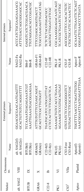
Molecular characterisation of T. gondii isolates. Genotyping was per- formed by the multiplex multilocus nested polymerase chain reaction–restriction fragment length polymorphism (Mn-PCR-RFLP) method (Su et al., 2010) using seven markers. Markers and sequences of the corresponding primers are summa- rised in Table 1.
Reaction mixtures were prepared according to Su et al. (2006), with few modifications such as use of the commercial 2X PCR Master Mix (Thermo Fischer Scientific, Waltham, MA, USA). Briefly, multiplex PCR reaction was performed in a final volume of 25 µl mixture containing 12.5 µl of 2X PCR Mas- ter Mix, 0.15 µM mixture of external forward (F) and reverse (R) primers for each marker, nuclease-free water and 1.5–3 µl of sample DNA. The primer mix- ture consisted of equal volumes of 14 external primers (seven F and seven R) previously diluted to 0.15 µM. For the nested PCR reaction, a 25-µl mixture was
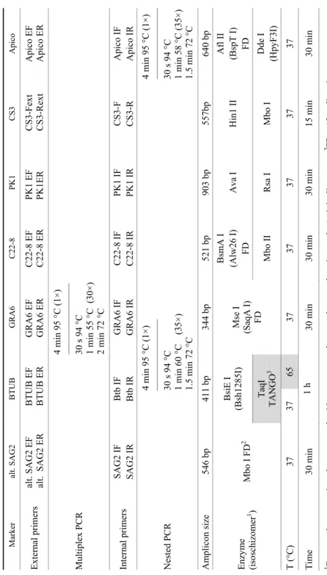
prepared separately for each marker and consisted of 12.5 µl of 2X PCR Master Mix, 0.30 µM of each internal primer, nuclease free water and 2–4 μl of amplifi- cation product of the first Mn-PCR reaction. Details of the Mn-PCR-RFLP pro- tocol, adapted to our choice of markers, are presented in Table 2.
After amplification, the PCR products were digested with appropriate re- striction enzymes. The digestion mixture consisted of 1–2 U of restriction enzyme, 1X FD buffer (Fast Digest, Thermo Fischer Scientific, Waltham, MA, USA), nu- clease-free water and 5 µl of Mn-PCR reaction product, in a 25-µl reaction vol- ume.
Mn-PCR-RFLP results were visualised by electrophoresis in 2.5% agarose gel stained with ethidium bromide and read against a 50-bp DNA ladder (Fer- mentas, Thermo Fischer Scientific, Waltham, MA, USA). RH (type I), Me49 (type II) and NED (type III) strains were used as positive controls and nuclease- free water as negative control.
Results
After performing Mn-PCR-RFLP on T. gondii strains from 18 pigs (a total of 29 samples, of which 14 were pig tissue digests and 15 mouse brains), identi- fication was achieved for nine isolates. Seven isolates were classified as type II and two as type III (Table 3).
Due to the variations in the efficacy of Mn-PCR-RFLP observed between pig diaphragms and mouse brains from the corresponding bioassay, results are presented for the tissue type that amplified the best. When efficacy was equiva- lent between the tissues, results obtained from pig tissues were chosen over the results from mouse brain.
Discussion
Genotyping of T. gondii performed in this study revealed the presence of both type II and type III strains in pigs from Northern Serbia. In some of the identified isolates, amplification of one or more markers failed, likely due to the insufficient amount of DNA in the sample (Vujanić, 2012). Lack of DNA was al- so the probable reason why direct genotyping from pig tissues was successful in only two cases (Table 3, pigs NF48 and BM17). The abundance of T. gondii in tissues of naturally infected pigs may be less than 1 cyst per 50 g (Dubey et al., 1996), and mouse bioassay may be necessary to increase the parasite load to a detectable level. In this study, the ‘boosting’ effect of the bioassay was particu- larly evident in at least one instance (sample 6BM/14, Table 3), where genotyp- ing from the pig tissue failed completely, whereas from the mouse brains, T.
gondii DNA was extracted in amounts sufficient for the amplification of all sev- en markers.
Table 1 Markers and primer sequences used in Mn-PCR-RFLP MarkerChromosome no.External primersInternal primers Name Sequence1 Name Sequence2 alt. SAG2 VIII alt. SAG2(f) alt. SAG2(r) GGAACGCGAACAATGAGTTT GCACTGTTGTCCAGGGTTTT SAG2-Fa SAG2-RaACCCATCTGCGAAGAAAACG ATTTCGACCAGCGGGAGCAC BTUB IX BTUB(f) BTUB(r) TCCAAAATGAGAGAAATCGT AAATTGAAATGACGGAAGAABtb-F Btb-R GAGGTCATCTCGGACGAACA TTGTAGGAACACCCGGACGC GRA6 X GRA6(f) GRA6(r)ATTTGTGTTTCCGAGCAGGT GCACCTTCGCTTGTGGTT GRA6-F1 GRA6-R1TTTCCGAGCAGGTGACCT TCGCCGAAGAGTTGACATAG C22-8 Ib C22-8(f) C22-8(r) TGATGCATCCATGCGTTTAT CCTCCACTTCTTCGGTCTCA C22-8F C22-8R TCTCTCTACGTGGACGCC AGGTGCTTGGATATTCGC PK1 VI PK1(f) PK1(r)GAAAGCTGTCCACCCTGAAA AGAAAGCTCCGTGCAGTGAT PK1-F PK1-RCGCAAAGGGAGACAATCAGT TCATCGCTGAATCTCATTGC CS33 VIIa CS3-Fext CS3-Rext GTGTATCTCCGAGGGGGTCT TGTGACTTCTTCGCATCGAC CS3-F CS3-R AGCGGATTTCCAACACTGTC CTGCTGCATTCACAAACTCC Apico PlastidApico(f) Apico(r)TGGTTTTAACCCTAGATTGTGG AAACGGAATTAATGAGATTTGAA Apico-F Apico-R GCAAATTCTTGAATTCTCAGTT GGGATTCGAACCCTTGATA 1 Su et al., 2006; 2 Su et al., 2010; 3 Pena et al., 2008; Wang et al., 2013
Table 2 Mn-PCR-RFLP protocol adapted from Su et al. (2010) Marker alt. SAG2 BTUBGRA6 C22-8 PK1 CS3 Apico External primers alt. SAG2 EF alt. SAG2 ER BTUB EF BTUB ERGRA6 EF GRA6 ER C22-8 EF C22-8 ERPK1 EF PK1ER CS3-Fext CS3-Rext Apico EF Apico ER Multiplex PCR
4 min 95 °C (1×) ––––––––––––––– 30 s 94 °C 1 min 55 °C (30×) 2 min 72 °C
Internal primersSAG2 IF SAG2 IRBtb IF Btb IRGRA6 IF GRA6 IR C22-8 IF C22-8 IRPK1 IF PK1 IR CS3-F CS3-R Apico IF Apico IR Nested PCR
4 min 95 °C (1×) ––––––––––––––– 30 s 94 °C 1 min 60 °C (35×) 1.5 min 72 °C
4 min 95 °C (1×) ––––––––––––––– 30 s 94 °C 1 min 58 °C (35×) 1.5 min 72 °C Amplicon size 546 bp411 bp344 bp521 bp903 bp557bp 640 bp Enzyme (isoschizomer1 ) Mbo I FD2BsiE I (Bsh1285I) Mse I (SaqA I) FD
BsmA I (Alw26 I) FDAva I Hin1 II Afl II (BspT I) FD TaqI TANGO3Mbo II Rsa IMbo I Dde I (HpyF3I) T (°C)37 37 65 37 37 37 37 37 Time 30 min 1 h30 min 30 min 30 min 15 min 30 min 1 Enzyme that recognises the same nucleotide sequence and cuts at the same location as the ‘original’ enzyme; 2 FD = ‘fast digest’ enzyme; 3 TANGO buffer (Thermo Scientific), specifically designed for double digestion with non-FD enzymes
Table 3
Toxoplasma gondii isolates successfully identified by MnPCR-RFLP analysis
No. Sample code
Markers T.
gondii Type
Pig
origin Tissuea GRA6 аlt.
SAG2 PK-1 BTUB C22-8 CS3 Apico
1 NF56/112 II II NA NA NA NA II II F brain
2 NO4/145 III III III III III NA III III B brain
3 NF47/105 II II II II II II II II F brain
4 NF48/59 III III NA III II NA III III F diaphragm
5 NF51/66 II II II II II NA II II F brain
6 NF52/108 II II II II II NA II II F brain
7 BM17/113 NA II NA II II NA II II М diaphragm
8 NF57/62 II II II II II NA II II F brain
9 6BM/14 II II I/II II II II II II М brain
F = farm, B = backyard/household, М = Mangalica; NA = not amplified; aBrain tissues originated from mice, whereas diaphragms originated from pigs
The predominance of type II over type III in pigs from Serbia is in accord- ance with the results of other studies from Europe, including most recent data from the Czech Republic (Slany et al., 2016), France (Djokic et al., 2016) and It- aly (Bacci et al., 2015; Vergara et al., 2018). The results of this study are also consistent with those previously obtained for other animals from Serbia. Using the conventional PCR-RFLP method and a set of six genetic markers (SAG1, 5'SAG2, 3'SAG2, GRA6, 5'GRA7 and 3'GRA7), two pigeon isolates, as well as one sheep isolate, were classified as type II, whereas one pigeon isolate was identified as type III (Marković et al., 2014). Type III has recently also been de- tected in tissues of two slaughter horses, using microsatellite analysis with 15 markers (Klun et al., 2017). The detection of type III isolates in two pigs in this study supports earlier findings of a greater representation of type III in the coun- tries of Southern Europe (De Sousa et al., 2006; Dubey et al., 2006; Vergara et al., 2018). Due to the region’s (relative) geographical vicinity to Africa, it has been suggested that this may be a consequence of the spread (e.g. through bird migrations, transportation and trade of animals and goods etc.) of type III from Africa and/or other countries in which this type is frequent (Mercier et al., 2010;
Shwab et al., 2014; Klun et al., 2017).
It is interesting that only type II strains were isolated from the free-range Mangalica pigs, although such a production provides greater opportunity of com- ing in contact with T. gondii, especially since they all originated from a special nature reserve, characterised by high biodiversity of flora and fauna. On the other hand, this finding may be due to the limited number of Mangalica pigs examined in this study and further research, involving more animals, is needed to better understand the T. gondii population structure in these pigs.
Another interesting observation is that one pig (NF48) from which type III was isolated originated from the same farm as the five pigs in which type II iso- lates were identified, which suggests the circulation of two different types of T.
gondii in the farm surroundings. Detection of different T. gondii lineages in pigs from the same farm has recently been reported by Vergara et al. (2018).
In conclusion, this study presents the first data on the molecular character- isation of T. gondii strains circulating in domestic pigs in Serbia. Detection of both type II and type III strains corroborates the findings of previous studies on animal isolates from this country. The presented results also add to the existing body of data that shows the predominance of type II over type III in animals from Europe, but also the more frequent detection of type III in Southern Europe, compared to other parts of the continent.
Acknowledgement
This study was supported by the Ministry of Education, Science and Technologi- cal Development of the Republic of Serbia (project grants No. TR 31034 and III 41019).
References
Bacci, C., Vismarra, A., Mangia, C., Bonardi, S., Bruini, I., Genchi, M., Kramer, L. and Brindani, F. (2015): Detection of Toxoplasma gondii in free-range, organic pigs in Italy using sero- logical and molecular methods. Int. J. Food Microbiol. 202, 54–56.
Dardé, M.-L. (2008): Toxoplasma gondii, ‘new’ genotypes and virulence. Parasite. 15, 366–371.
De Sousa, S., Ajzenberg, D., Canada, N., Freire, L., Da Costa, J. M. C., Darde, M. L., Thulliez, P.
and Dubey, J. P. (2006): Biologic and molecular characterization of Toxoplasma gondii isolates from pigs from Portugal. Vet. Parasitol. 135, 133–136.
Djokic, V., Blaga, R., Aubert, D., Durand, B., Perret, C., Geers, R., Ducry, T., Vallee, I., Djurković- Djaković, O., Mzabi, A., Villena, I. and Boireau, P. (2016): Toxoplasma gondii infection in pork produced in France. Parasitol. 143, 557–567.
Djurković-Djaković, O., Klun, I., Khan, A., Nikolić, A., Knežević-Ušaj, S., Bobić, B. and Sibley, L. D. (2006): A human origin type II strain of Toxoplasma gondii causing severe encepha- litis in mice. Microb. Infect. 8, 2206–2212.
Dubey, J. P., Lunney, J. K., Shen, S. K., Kwok, O. C., Ashford, D. and Thulliez, P. (1996): Infec- tivity of low numbers of Toxoplasma gondii oocysts to pigs. J. Parasitol. 82, 438–443.
Dubey, J. P., Vianna, M. C., Sousa, S., Canada, N., Meireles, S., Correia da Costa, J. M., Marcet, P. L., Lehmann, T., Dardé, M. L. and Thulliez, P. (2006): Characterization of Toxoplasma gondii isolates in free-range chickens from Portugal. J. Parasitol. 92, 184–186.
EFSA (2011): Scientific Opinion on the public health hazards to be covered by inspection of meat (swine). EFSA J. 9, 1–198.
Howe, D. K. and Sibley, L. D. (1995): Toxoplasma gondii comprises three clonal lineages : corre- lation of parasite genotype with human disease. J. Infect. Dis. 172, 1561–1566.
Khan, A., Dubey, J. P., Su, C., Ajioka, J. W., Rosenthal, B. M. and Sibley, L. D. (2011): Genetic analyses of atypical Toxoplasma gondii strains reveal a fourth clonal lineage in North America. Int. J. Parasitol. 41, 645–655.
Klun, I., Uzelac, A., Villena, I., Mercier, A., Bobić, B., Nikolić, A., Rajnpreht, I., Opsteegh, M., Aubert, D., Blaga, R., Van der Giessen, J. and Djurković-Djaković, O. (2017): The first isolation and molecular characterization of Toxoplasma gondii from horses in Serbia. Para- sit. Vectors 10, 167.
Kuruca, L., Klun, I., Uzelac, A., Nikolić, A., Bobić, B., Simin, S., Djurković-Djaković, O. and Lalošević, V. (2016): Detection of viable Toxoplasma gondii in free-range pigs from the Special Nature Reserve of Zasavica. Contemp. Agric. 65, 1–6.
Kuruca, L., Klun, I., Uzelac, A., Nikolić, A., Bobić, B., Simin, S., Lalošević, D., Lalošević, V. and Djurković-Djaković, O. (2017): Detection of Toxoplasma gondii in naturally infected do- mestic pigs in Northern Serbia. Parasitol. Res. 116, 3117–3123.
Marković, M., Ivović, V., Štajner, T., Djokić, V., Klun, I., Bobić, B., Nikolić, A. and Djurković- Djaković, O. (2014): Evidence for genetic diversity of Toxoplasma gondii in selected in- termediate hosts in Serbia. Comp. Immunol. Microbiol. Infect. Dis. 37, 173–179.
Mercier, A., Devillard, S., Ngoubangoye, B., Bonnabau, H., Bañuls, A. L., Durand, P., Salle, B., Ajzenberg, D. and Dardé, M. L. (2010): Additional haplogroups of Toxoplasma gondii out of Africa: Population structure and mouse-virulence of strains from Gabon. PLoS Negl.
Trop. Dis. 4, e876.
Pena, H. F. J., Gennari, S. M., Dubey, J. P. and Su, C. (2008): Population structure and mouse- virulence of Toxoplasma gondii in Brazil. Int. J. Parasitol. 38, 561–569.
Shwab, E. K., Zhu, Xi.-Q., Majumdar, D., Pena, H. F. J., Gennari, S. M., Dubey, J. P. and Su, C.
(2014): Geographical patterns of Toxoplasma gondii genetic diversity revealed by multi- locus PCR-RFLP genotyping. Parasitol. 141, 453–461.
Sibley, L. D., Khan, A., Ajioka, J. W. and Rosenthal, B. M. (2009): Genetic diversity of Toxoplasma gondii in animals and humans. Philos. Trans. R. Soc. Lond. B Biol. Sci. 364, 2749–2761.
Slany, M., Reslova, N., Babak, V. and Lorencova, A. (2016): Molecular characterization of Toxo- plasma gondii in pork meat from different production systems in the Czech Republic. Int.
J. Food Microbiol. 238, 252–255.
Štajner, T., Vasiljević, Z., Vujić, D., Marković, M., Ristić, G., Mićić, D., Pašić, S., Ivović, V., Ajzenberg, D. and Djurković-Djaković, O. (2013): Atypical strain of Toxoplasma gondii causing fatal reactivation after hematopoietic stem cell transplantation in a patient with an underlying immunological deficiency. J. Clin. Microbiol. 51, 2686–2690.
Su, C., Shwab, E. K., Zhou, P., Zhu, X. Q. and Dubey, J. P. (2010): Moving towards an integrated approach to molecular detection and identification of Toxoplasma gondii. Parasitol. 137, 1–11.
Su, C., Zhang, X. and Dubey, J. P. (2006): Genotyping of Toxoplasma gondii by multilocus PCR- RFLP markers: A high resolution and simple method for identification of parasites. Int. J.
Parasitol. 36, 841–848.
Vergara, A., Marangi, M., Caradonna, T., Pennisi, L., Paludi, D., Papini, R., Ianieri, A., Giangas- pero, A. and Normanno, G. (2018): Toxoplasma gondii lineages circulating in slaughtered industrial pigs and potential risk for consumers. J. Food Prot. 81, 1373–1378.
Vujanić, М. (2012): Molecular detection and genotyping of Toxooplasma gondii strains isolated in Serbia. PhD Thesis. Faculty of Biology, University of Belgrade, Belgrade, Serbia.
Wang, L., Cheng, H., Huang, K., Xu, Y., Li Y., Du, J., Yu, L., Luo, Q., Wei, W., Jiang, L. and Shen, J. (2013): Toxoplasma gondii prevalence in food animals and rodents in different re- gions of China: isolation, genotyping and mouse pathogenicity. Parasit. Vectors 6, 273.