ANTIMICROBIAL SUSCEPTIBILITY OF PASTEURELLA MULTOCIDA ISOLATED FROM SHEEP AND PIGS IN SPAIN –
SHORT COMMUNICATION
Dolores CID1, José Francisco FERNÁNDEZ-GARAYZÁBAL1,2, Chris PINTO1,3, Lucas DOMÍNGUEZ1,2 and Ana Isabel VELA1,2*
1Animal Health Department, Veterinary School, Universidad Complutense de Madrid, Av. Puerta de Hierro, s/n, 28040 Madrid, Spain; 2Centro de Vigilancia Sanitaria Veterinaria (VISAVET), Universidad Complutense, Madrid, Spain; 3London School of
Hygiene and Tropical Medicine/ Royal Veterinary College, University of London, London, United Kingdom
(Received 20 May 2019; accepted 25 September 2019)
Pasteurella multocida is responsible for economically important diseases in sheep and pigs. Antimicrobial susceptibility studies are essential for initiating rational and effective empirical therapy of P. multocida infections. In this study we investigated the antimicrobial susceptibility to 18 antimicrobial agents of 156 clinical isolates of P. multocida from sheep (n = 87) and pigs (n = 69) using the microdilution method. Both sheep and pig isolates exhibited low levels of re- sistance (≤ 15%) to ceftiofur, gentamicin, neomycin, spectinomycin, chlortetracy- cline, tulathromycin, florfenicol, danofloxacin, and enrofloxacin and trime- thoprim/sulphamethoxazole, high resistance rates (> 15% up to 50%) to oxytetra- cycline, tilmicosin, and tiamulin, and very high resistance rates (> 50%) to tylosin tartrate, clindamycin, and sulphadimethoxine. However, sheep isolates exhibited significantly lower percentages of resistance and lower MIC90 values (P < 0.05) than pig isolates for most of the antimicrobials tested. In addition, sheep isolates exhibited also significantly lower phenotypic antimicrobial resistance diversity (8 resistotypes vs. 30 resistotypes). LAC-LIN-SUL-MAC was the resistotype most frequently detected in sheep (39.1%) and LIN-SUL-MAC in pig isolates (26.1%). The differences in susceptibility patterns could be influenced by the lower use of antimicrobials in the small ruminant industry compared with the pig farming industry.
Key words: Antimicrobial resistance, Pasteurella multocida, pigs, sheep, production system
Pasteurella multocida is an important pathogen responsible for a diversity of diseases with an economic impact in different livestock species, including sheep and pigs. (Wilson and Ho, 2013). Although this pathogen is usually sus- ceptible to several antimicrobials, resistance in different animals has been report-
*Corresponding author; E-mail address: avela@ucm.es; Phone: 0034 (913) 943-3709
ed (San Millan et al., 2009; Tang et al., 2009), which represents a threat regard- ing treatment options. Therefore, monitoring antimicrobial susceptibility is es- sential to acquire regional information and help veterinarians to initiate rational and effective empirical therapy in acute situations. Antimicrobial susceptibility studies in P. multocida have mainly been performed in isolates from cattle, poul- try, or pigs (Post et al., 1991; Lizarazo et al., 2006; Kumar et al., 2009; Furian et al., 2016). However, similar studies including isolates from sheep are scarce (Berge et al., 2006; Sarangi et al., 2015; Cucco et al., 2017). In Spain, infor- mation on the antimicrobial susceptibility of porcine P. multocida isolates has been reported (Lizarazo et al., 2006), but no similar studies have been conducted in sheep. Thus, the aim of this study was to assess the antimicrobial susceptibil- ity of clinical P. multocida isolates from sheep and pigs, providing information on their antimicrobial resistance patterns. This study provides the first data about the antimicrobial susceptibility of ovine P. multocida isolates in Spain.
One hundred and fifty-six P. multocida isolates obtained between 2001 and 2009 from sheep (n = 87) and pigs (n = 69) were included in this study. All porcine isolates were recovered from clinical cases of pneumonia, septicaemia, and arthritis, and most of the ovine isolates from cases of pneumonia (García- Alvarez et al., 2017). Bacteria were isolated from samples on Columbia blood agar plates (bioMérieux) incubated at 37 °C for 24 h. Isolates were biochemical- ly identified by the commercial identification system API 20E strips (bioMé- rieux, S.A.) and further confirmed by a species-specific PCR assay (Townsend et al., 1998). Capsular types and sequence types (STs) were determined previously (García-Alvarez et al., 2017).
Antimicrobial susceptibility was determined by the microdilution method using a commercially prepared, dehydrated 96-well microtitre MIC panel (BOPO6F, Sensititre; Trek Diagnostic Systems Inc., UK). The antimicrobial agents used and their respective dilution ranges are indicated in Tables 1 and 2.
Inocula were prepared from a 24-h Columbia blood agar plate by suspending four colonies in 5 mL of sterile distilled water. The inoculum was adjusted to 0.5 McFarland standard and further diluted 1/100 in 10 mL of Muller-Hinton broth.
Fifty microlitres of the adjusted inoculum was deposited in each well of the mi- croplate panel. Microdilution panels were sealed and further incubated at 37 °C for 24 h. The breakpoints used are shown in Tables 1 and 2. Staphylococcus au- reus ATCC 29213 and Escherichia coli ATCC 25922 were included as quality controls with each batch of organisms tested. In this study, resistance rates were classified into three categories: low rates, percentage of resistant strains < 15%;
high rates, 15–50%; and very high rates, > 50%. Multidrug-resistance (MDR) was defined as acquired non-susceptibility to at least one agent in three or more antimicrobial categories. For the purpose of this study, a bacterial isolate was considered resistant to an antimicrobial category when it was resistant to at least one agent in that category. The association between the host origin, clinical
origin, capsular type or sequence type of P. multocida isolates and antimicrobial susceptibility was determined using the Chi-square test, with P < 0.05 considered significant. Data were analysed using the Epi InfoTM 7 program of the Centers for Disease Control and Prevention (CDC).
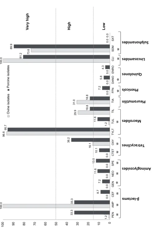
Antimicrobial resistance data of P. multocida have been commonly gener- ated using different methodologies and different antimicrobials as well as using different breakpoints, which can hamper comparison of our results. Despite these drawbacks and using the breakpoints indicated in Tables 1 and 2, we detected in both sheep and pig isolates low levels of resistance to CEF, GEN, NEO, SPE, CTET, TUL, FFN, DANO, ENRO and SxT (≤ 15%; Tables 1 and 2, Fig. 1), which agrees with most previous reports both for pig (Yoshimura et al., 2001;
Tang et al., 2009; Sellyei et al., 2009; Nedbalcová and Kučerová, 2013; Dayao et al., 2014; de Jong et al., 2014; El Garch et al., 2016; Cucco et al., 2017) and sheep isolates (Sarangi et al., 2015; Cucco et al., 2017). On the other hand, high resistance rates (> 15–50%) to OXY, TIL and TIA and very high resistance rates (> 50%) to TYLT, CLI, and SDM were identified (Tables 1 and 2, Fig. 1). Over- all, similar high or very high levels of resistance to these antimicrobials have also been reported previously (Lizarazo et al., 2006; Tang et al., 2009; Nedbalcová and Kučerová, 2013; de Jong et al., 2014; Tahamtan and Hayati, 2014; El Garch et al., 2016; Cucco et al., 2017). Based on these high levels in the resistance in vitro, these antimicrobials should not be used empirically to treat P. multocida infections or be used with caution under field conditions. Of special concern are the very high levels of resistance to TYLT and SDM found in this study both in pig and sheep isolates, as these antimicrobials are considered critically important and highly important, respectively, for human medicine (World Health Organiza- tion, 2019).
It was unexpected that all strains of P. multocida from sheep were re- sistant to ampicillin (Table 1), compared with the low level of resistance (1.2%) detected for penicillin. As no CLSI-defined breakpoints for ampicillin are availa- ble for ovine P. multocida isolates, in this study we used the breakpoint recom- mended in the Clinical and Laboratory Standard Institute Vet08 guideline for cat- tle isolates (CLSI, 2018; ≥ 0.25 µg/ml) which is much higher than that recom- mended for swine isolates (≥ 2 µg/ml). Similarly, using the breakpoint recom- mended by the European Committee on Antimicrobial Susceptibility Testing for P. multocida (EUCAST, 2019; ≥ 1 µg/ml), all sheep isolates would have been considered susceptible. Therefore, the high percentage of resistance to ampicillin among sheep isolates is likely biased and overestimated by the breakpoint used in this study and point out the necessity for establishing specific breakpoints for ovine isolates.
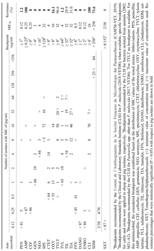
Table 1 Minimum inhibitory concentrations (MICs) for 18 antimicrobial agents of ovine Pasteurella multocida isolates (n = 87) Anti- microbial
Number of isolates with MIC of (μg/ml) Breakpoint (µg/ml)MIC90Resistance (%)0.120.250.5 1 2 4 8 163264128256>256 PEN < 815 1 > ≥ 1b 0.121.2 AMP< 87 > ≥ 0.25b 0.25100 CEF< 861 > ≥ 8b 0.250 GEN < 6619 2 > > 4a 2 0 NEO < 843 > > 16a 4 0 SPE < 5 72 10 > ≥ 128b 32 0 CTET < 646 2 14 1 > 8a 4 0 OXY < 5513 5 14 > 8a 16 16.1 TYLT < 1 2 8 50 24 >2 ≥ 8e 32 96.6 TUL< 6510 4 5 1 1 1 >≥ 64b 4 1.2 TIL < 545 2 19 7 >≥ 32d 32 29.9 TIA < 2 12 46 27 >≥ 32c 32 31.0 FFN < 4541 1 > ≥ 8b 0.50 DANO < 87 > ≥ 1b 0.120 ENRO< 86 1 > ≥ 2b 0.120 CLI < 2 8 70 7 >≥ 4c 16 100 SDM < 23 >64> 256a > 25673.6 2/38 4/76 SXT< 87 > > 8/152a 2/38 0 a Breakpoints recommended by the Comité de l’Antibiogramme de la Société Française de Microbiologie for Pasteurellaceae (CASFM-VET, 2019). b Breakpoints recommended by the Clinical and Laboratory Standards Institute (CLSI) for P. multocida (2018-VET08); when available, specific breakpoints for cattle and swine were applied to sheep and pig isolates, respectively.c Breakpoints recommended by the CLSI for bacteria isolated from animals (2002; M31-A2). d Breakpoints recommended by the CLSI for Pasteurella spp. other than P. multocida (2017; VET06). e For TYLT no breakpoint is available. For this antimicrobial, the considered breakpoint was established based on the distribution of MIC values of the tested strains. Abbreviations: PEN, penicillin; AMP, ampicillin; CEF, ceftiofur; GEN, gentamicin; NEO, neomycin; SPE, spectinomycin; CTET, chlortetracycline; OXY, oxytetracycline; TYLT, tylosin tartrate; TUL, tulathromycin; TIL, tilmicosin; TIA, tiamulin; FFN, florfenicol; DANO, danofloxacin; ENRO, enrofloxacin; CLI, clindamycin; SDM, sul- phadimethoxine; SXT; Trimethoprim/Sulphamethoxazole. (<): minimum value of concentration used; (>): maximum value of concentration used. Re- sistance percentages that were statistically significant (P < 0.05) with respect to pig isolates are shown in bold
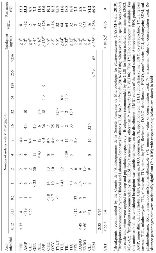
Table 2 Minimum inhibitory concentrations (MICs) for 18 antimicrobial agents of porcine Pasteurella multocida isolates (n = 69) Anti- microbial
Number of isolates with MIC of (μg/ml) Breakpoint (µg/ml)MIC90Resistance (%)0.120.250.5 1 2 4 8 163264128256>256 PEN < 354 7 5 2 2 14 >≥ 1b 8 33.3 AMP< 391 6 4 4 1 4 >10 ≥ 2b > 3233.3 CEF< 551 5 2 6 >≥ 8b 2 8.7 GEN < 2330 11 1 4 > > 4a 4 7.2 NEO < 4312 6 8 >> 16a 32 11.6 SPE < 5 36 18 1 >9 ≥ 128d > 12813.0 CTET< 1814 13 8 9 >7 > 8a 8 10.1 OXY < 1710 10 3 4 >25 > 8a > 1636.2 TYLT < 3 6 28 32 >≥ 8e 32 95.7 TUL < 4312 4 1 1 8 >≥ 64b 64 11.6 TIL < 3915 2 2 11 >≥ 32b 64 18.8 TIA < 7 3 5 8 33 13 >≥ 32c 32 18.8 FFN < 1237 9 6 5 >≥ 8b 2 7.2 DANO < 49 6 9 1 > 4 ≥ 2d 0.55.8 ENRO < 603 3 2 1 > ≥ 1b 0.254.3 CLI < 1 3 9 8 16 32 >≥ 4c 16 81.2 SDM < 7 >62> 256a > 25689.9 2/38 4/76 SXT< 55 >14 > 8/152a 4/76 0 a Breakpoints recommended by the Comité de l’Antibiogramme de la Société Française de Microbiologie for Pasteurellaceae (CASFM-VET, 2019). b Breakpoints recommended by the Clinical and Laboratory Standards Institute (CLSI) for P. multocida (2018-VET08); when available, specific breakpoints for cattle and swine were applied to sheep and pig isolates, respectively.c Breakpoints recommended by the CLSI for bacteria isolated from animals (2002; M31-A2). d Breakpoints recommended by the CLSI for Pasteurella spp. other than P. multocida (2017; VET06). e For TYLT no breakpoint is available. For this antimicrobial, the considered breakpoint was established based on the distribution of MIC values of the tested strains. Abbreviations: PEN, penicillin; AMP, ampicillin; CEF, ceftiofur; GEN, gentamicin; NEO, neomycin; SPE, spectinomycin; CTET, chlortetracycline; OXY, oxytetracycline; TYLT, tylosin tartrate; TUL, tulathromycin; TIL, tilmicosin; TIA, tiamulin; FFN, florfenicol; DANO, danofloxacin; ENRO, enrofloxacin; CLI, clindamycin; SDM, sul- phadimethoxine; SXT, Trimethoprim/Sulphamethoxazole. (<): minimum value of concentration used; (>): maximum value of concentration used. Re- sistance percentages that were statistically significant (P < 0.05) with respect to pig isolates are shown in bold
Fig. 1. Antimicrobial resistance to 18 antimicrobials tested of P. multocida isolates obtained from sheep and pigs. * Statistically significant differences (P < 0.05) between sheep and pig isolates
100 90 80 70 60 50 40 30 20 10 0 1.20.00.00.00.00.01.2
8.7 7.211.613.0 10.1
16.1
36.2 11.6
33.333.3
100.0 29.9 18.818.8
31.0 0.0
7.2 0.00.00.00.0
5.8 4.3
96.6 95.7100.0 81.2
89.9 73.6
Ovine isolates Porcine isolates Very high High Low PENAMP CEFGENNEOSPECTETOXY TUL TYLTTIL TIAFFN DANOENROCLI SDMSXT
Aminoglyco sides
Tetracyclines Macrolides
Pleuromutili n
Phenicols Quinolones
Linco samid es
Sulpho namide
s
β-lactams
The resistotypes identified in this study and their distribution among sheep and pig isolates are shown in Table 3. None of the ovine or porcine P. multocida isolates was susceptible to the nine antimicrobial categories tested, with the resis- totype LAC-LIN-SUL-MAC being the most frequently detected among sheep isolates (39.1%) and the resistotype LIN-SUL-MAC among pig isolates (26.1%;
Table 3). No statistically significant differences were detected between percent- ages of sheep and pig MDR isolates (97.7% vs. 92.8%) but sheep isolates exhib- ited lower phenotypic antimicrobial resistance diversity (8 resistotypes vs. 30 re- sistotypes) than pig isolates (Table 3). More than half (55.8%) of the P. multo- cida isolates of this study were genetically characterised by MLST by García- Alvarez et al. (2017), with most of the ovine and porcine isolates belonging to a limited number of sequence types (ST50 and ST19 among ovine isolates and ST3, ST11 and ST62 among porcine isolates). A comparison of the antimicrobial resistance patterns and STs of the isolates included in both studies did not detect any association between resistance patterns and prevalent genotypes (P > 0.05;
data not shown). Therefore, unlike in other pathogens (Klugman, 2003; Durante- Mangoni and Zarrilli, 2011; Edelstein et al., 2013), the differences in the antimi- crobial resistance between sheep and pig isolates should not be related to the presence of particular resistant genotypes within the population of P. multocida.
Moreover, capsular types A and D were the most frequent in P. multocida iso- lates in both animal species as determined previously (García-Alvarez et al., 2017). No associations were identified between resistance patterns of P. multo- cida with capsular types or with the clinical origin of the isolates (data not shown).
Sheep isolates exhibited significantly lower percentages of resistance (P <
0.05) than pig isolates for 12 antimicrobials (Tables 1 and 2, Fig. 1). Moreover, the MIC90 values for most antimicrobials were also lower in sheep than in pig isolates (Tables 1 and 2). The differences in the level of antimicrobial suscepti- bility between sheep and pig isolates could be associated with the different amount of antimicrobials used in the two farming sectors. In fact, pig farming is one of the livestock activities with the highest antimicrobial use (Moreno, 2014), while the use of antimicrobials is minimal in the small ruminant industry (Santman-Berends et al., 2014). Therefore, the higher resistance rates observed among pig isolates might reflect the selective pressure related to the higher use of antimicrobials in pig farming, at least for some antimicrobials.
Furthermore, the resistance rates and MIC90 values observed in this study among porcine P. multocida isolates for most antimicrobials were higher than those observed in a similar study carried out in Spain (Lizarazo et al., 2006), suggesting a shift in the resistance to these antimicrobials in Spanish P. multo- cida isolates from pigs. These results point out the need for active surveillance programmes to monitor the antimicrobial resistance patterns of P. multocida.
Table 3
Resistance phenotypes (resistotypes) of Pasteurella multocida isolates according to host species
Resistance to Ovine Porcine Total n % n % n %
LAC-LIN 2 2.3 2 1.3
LAC-SUL 3 4.3 3 1.9
LIN-MAC 2 2.9 2 1.3
LAC-LIN-MAC 15 17.2 1 1.4 16 10.3
LAC-LIN-PLE 1 1.4 1 0.6
LAC-SUL-MAC 1 1.1 1 0.6
LIN-SUL-MAC 18 26.1 18 11.5
LIN-MAC-TET 2 2.9 2 1.3
SUL-MAC-ANF 1 1.4 1 0.6
SUL-MAC-AMI 1 1.4 1 0.6
MAC-PLE-TET 1 1.4 1 0.6
LAC-LIN-MAC-TET 1 1.4 1 0.6
LAC-LIN-MAC-PLE 5 5.7 5 3.2
LAC-LIN-SUL-MAC 34 39.1 6 8.7 40 25.6
LAC-SUL-MAC-TET 3 4.3 3 1.9
LIN-SUL-MAC-PLE 4 5.8 4 2.6
LIN-SUL-MAC-TET 3 4.3 3 1.9
SUL-MAC-TET-AMI 1 1.4 1 0.6
SUL-MAC-TET-QUIN 1 1.4 1 0.6
LIN-MAC-SUL-TET 2 2.9 2 1.3
LIN-SUL-MAC-AMI 4 5.8 4 2.6
LAC-LIN-SUL-MAC-TET 11 12.6 2 2.9 13 8.3
LAC-LIN-SUL-MAC-AMI 1 1.4 1 0.6
LAC-LIN-SUL-MAC-PLE 16 18.4 1 1.4 17 10.9
LIN-MAC-PLE-TET-AMI 1 1.4 1 0.6
LIN-MAC-SUL-PLE-TET 1 1.4 1 0.6
LAC-LIN-SUL-MAC-AMI-QUIN 1 1.4 1 0.6
LAC-LIN-SUL-MAC-TET-AMI 1 1.4 1 0.6
LAC-LIN-SUL-MAC-PLE-TET 3 3.4 1 1.4 4 2.6
LIN-SUL-MAC-TET-AMI-QUIN 1 1.4 1 0.6
LAC-SUL-MAC-PLE-TET-ANF-AMI 1 1.4 1 0.6
LAC-LIN-SUL-MAC-PLE-TET-ANF-AMI 1 1.4 1 0.6
LAC-LIN-SUL-MAC-PLE-TET-ANF-AMI-QUIN 2 2.9 2 1.3
Total 87 100.0 69 100.0 156 100.0
Abbreviations: LIN: lincosamides; SUL: sulphonamides; MAC: macrolides; TET: tetracyclines;
LAC: β-Lactams; QUIN: quinolones; AMI: aminoglycosides; ANF: phenicols; PLE: pleuromu- tilins
Acknowledgements
The authors thank Almudena Casamayor and Sherezade Leao for their invaluable technical assistance. This study was supported by GR/SAL/0580/2004 (Community of
Madrid, Spain) and AGL2009-10136 (Ministerio de Ciencia e Innovación, Spain). Chris Pinto was supported by the Programme Alban, the European Union Programme of High Level Scholarships for Latin America, scholarship No. E07D404011PE.
References
Berge, A. C., Sischo, W. M. and Craigmill, A. L. (2006): Antimicrobial susceptibility patterns of res- piratory tract pathogens from sheep and goats. J. Am. Vet. Med. Assoc. 229, 1279–1281.
Clinical and Laboratory Standards Institute (2002): Performance Standards for Antimicrobial Disk and Dilution Susceptibility Tests for Bacteria Isolated from Animals – Approved Standard, 2nd edition. Document M31-A2, CLSI/NCCLS, Wayne, Pennsylvania, USA.
Clinical and Laboratory Standards Institute (2017): Methods for Antimicrobial Dilution and Disk Susceptibility Testing of Infrequently Isolated or Fastidious Bacteria Isolated from Ani- mals. Document VET06. 1st edition. Wayne, Pennsylvania, USA.
Clinical and Laboratory Standard Institute (2018): Performance Standards for Antimicrobial Disk and Dilution Susceptibility Tests for Bacteria Isolated from Animals. Document VET08, Approved Standard, 4th edition. Wayne, Pennsylvania, USA.
Comité de l’Antibiogramme de la Société Française de Microbiologie. Recommandations Vétérinaires (2019): Paris, France: Société Française de Microbiologie. https://www.sfm-microbiologie.
org/2019/07/09/casfm-veterinaire-2019/.
Cucco, L., Massacci, F. R., Sebastiani, C., Mangili, P., Bano, L., Cocchi, M., Luppi, A., Ortenzi, R., Pezzotti, G. and Magistrali, C. F. (2017): Molecular characterization and antimicrobial susceptibility of Pasteurella multocida strains isolated from hosts affected by various dis- eases in Italy. Vet. Ital. 57, 21–17.
Dayao, D. A. E., Gibson, J. S., Blackall, P. J. and Turni, C. (2014): Antimicrobial resistance in bacte- ria associated with porcine respiratory disease in Australia. Vet. Microbiol. 171, 232–235.
de Jong, A., Thomas, V., Simjee, S., Moyaert, H., El Garch, F., Maher, K., Morrissey, I., Butty, P., Klein, U., Marion, H., Rigaut, D. and Vallé, M. (2014): Antimicrobial susceptibility moni- toring of respiratory tract pathogens isolated from diseased cattle and pigs across Europe:
The VetPath study. Vet. Microbiol. 172, 202–215.
Durante-Mangoni, E. and Zarrilli, R. (2011): Global spread of drug-resistant Acinetobacter bau- mannii: molecular epidemiology and management of antimicrobial resistance. Future Mi- crobiol. 6, 407–422.
Edelstein, M. V., Skleenova, E. N., Shevchenko, O. V., D’Souza, J. W., Tapalski, D. V., Azizov, I.
S., Sukhorukova, M. V., Pavlukov, R. A., Kozlov, R. S., Toleman, M. A. and Walsh, T. R.
(2013): Spread of extensively resistant VIM-2-positive ST235 Pseudomonas aeruginosa in Belarus, Kazakhstan, and Russia: a longitudinal epidemiological and clinical study. Lancet Infect. Dis. 13, 867–876.
El Garch, F., de Jong, A., Simjee, S., Moyaert, H., Klein, U., Ludwig, C., Marion, H., Haag- Diergarten, S., Richard-Mazet, A., Thomas, V. and Siegwart, E. (2016): Monitoring of an- timicrobial susceptibility of respiratory tract pathogens isolated from diseased cattle and pigs across Europe, 2009–2012: VetPath results. Vet. Microbiol. 194, 11–22.
European Committee on Antimicrobial Susceptibility Testing (2019): Breakpoint Tables for Inter- pretation of MICs and Zone Diameters, Version 9.0, http://www.eucast.org.
Furian, T. Q., Borges, K. A., Laviniki, V., Rocha, S. L., de Almeida, C. N., do Nascimento, V. P., Salle, C. T. and Morales, H. L. (2016): Virulence genes and antimicrobial resistance of Pasteurella multocida isolated from poultry and swine. Braz. J. Microbiol. 47, 210–216.
García-Alvarez, A., Vela, A. I., San Martín, E., Chaves, F., Fernández-Garayzábal, J. F., Lucas, D.
and Cid, D. (2017): Characterization of Pasteurella multocida associated with ovine pneumonia using multi-locus sequence typing (MLST) and virulence-associated gene pro- file analysis and comparison with porcine isolates. Vet. Microbiol. 204, 180–187.
Klugman, K. P. (2003): The role of clonality in the global spread of fluoroquinolone-resistant bac- teria. Clin. Infect. Dis. 36, 783–785.
Kumar, P., Singh, V. P., Agrawal, R. K. and Singh, S. (2009): Identification of Pasteurella multo- cida isolates of ruminant origin using polymerase chain reaction and their antibiogram study. Trop. Anim. Health Prod. 41, 573–578.
Lizarazo, Y. A. V., Ferri, E. F. R., de la Fuente, A. J. M. and Martin, C. B. G. (2006): Evaluation of changes in antimicrobial susceptibility patterns of Pasteurella multocida subsp. multocida isolates from pigs in Spain in 1987–1988 and 2003–2004. Am. J. Vet. Res. 67, 663–668.
Moreno, M. A. (2014): Survey of quantitative antimicrobial consumption per production stage in farrow-to-finish pig farms in Spain. Vet. Rec. Open 13, e000002.
Nedbalcová, K. and Kučerová, Z. (2013): Antimicrobial susceptibility of Pasteurella multocida and Haemophilus parasuis isolates associated with porcine pneumonia. Acta Vet. Brno 82, 3–7.
Post, K. W., Cole, N. A. and Raleigh, R. H. (1991): In vitro antimicrobial susceptibility of Pas- teurella haemolytica and Pasteurella multocida recovered from cattle with bovine respira- tory disease complex. J. Vet. Diagn. Invest. 3, 124–126.
San Millan, A., Escudero, J. A., Gutierrez, B., Hidalgo, L., Garcia, N., Llagostera, M., Dominguez, L. and Gonzalez-Zorn, B. (2009): Multiresistance in Pasteurella multocida is mediated by coexistence of small plasmids. Antimicrob. Agents Chemother. 53, 3399–3404.
Santman-Berends, I., Luttikholt, S., Van den Brom, R., Van Schaik, G., Gonggrijp M., Hage, H.
and Vellema, P. (2014): Estimation of the use of antibiotics in the small ruminant industry in The Netherlands in 2011 and 2012. PLoS One 9, e105052.
Sarangi, L. N., Thomas, P., Gupta, S. K., Priyadarshini, A., Kumar, S., Nagaleekar, V. K., Kumar, A. and Singh, V. P. (2015): Virulence gene profiling and antibiotic resistance pattern of In- dian isolates of Pasteurella multocida of small ruminant origin. Comp. Immunol. Microbi- ol. Infect. Dis. 38, 33–39.
Sellyei, B., Varga, Z., Szentesi-Samu, K., Kaszanyitzky, E. and Magyar, T. (2009): Antimicrobial susceptibility of Pasteurella multocida isolated from swine and poultry. Acta Vet. Hung.
57, 357–367.
Tang, X., Zhao, Z., Hu, J., Wu, B., Cai, X., He, Q. and Chen, H. (2009): Isolation, antimicrobial re- sistance, and virulence genes of Pasteurella multocida strains from swine in China. J. Clin.
Microbiol. 47, 951–958.
Tahamtan, Y. and Hayati, M. (2014): Multi drug resistance of Pasteurella spp. isolated from sheep and goats in Iran. Res. J. Microbiol. 9, 51–58.
Townsend, K. M., Frost, A. J., Lee, C. W., Papadimitriou, J. M. and Dawkins, H. J. (1998): Devel- opment of PCR assays for species- and type-specific identification of Pasteurella multo- cida isolates. J. Clin. Microbiol. 36, 1096–1100.
Wilson, B. A. and Ho, M. (2013): Pasteurella multocida: from zoonosis to cellular microbiology.
Clin. Microbiol. Rev. 26, 631–655.
World Health Organization (2019): Critically Important Antimicrobials for Human Medicine. 6th rev. ed., Geneva. Licence: CC BY-NC-SA 3.0 IGO; https://creativecommons.org/licenses/
by-nc-sa/3.0/igo.
Yoshimura, H., Ishimaru, M., Endoh, Y. S. and Kojima, A. (2001): Antimicrobial susceptibility of Pasteurella multocida isolated from cattle and pigs. J. Vet. Med. B: Infect. Dis. Vet. Public Health 48, 555–560.