ORIGINAL ARTICLE
GABA
Areceptor subunit deregulation in the hippocampus of human foetuses with Down syndrome
Ivan Milenkovic1,2 •Tamara Stojanovic2•Eleonora Aronica3,4•Livia Fu¨lo¨p5•Zsolt Bozso´5•
Zolta´n Ma´te´6•Yuchio Yanagawa7•Homa Adle-Biassette8• Gert Lubec9•Ga´bor Szabo´6•Tibor Harkany10,11• Ga´bor G. Kova´cs2• Erik Keimpema10
Received: 23 May 2017 / Accepted: 5 November 2017 / Published online: 22 November 2017 ÓThe Author(s) 2017. This article is an open access publication
Abstract
The function, regulation and cellular distribution of GABAAreceptor subunits have been extensively documented in the adult rodent brain and are linked to numerous neurological disorders. However, there is a surprising lack of knowledge on the cellular (sub-) distribution of GABAAreceptor subunits and of their expressional regulation in developing healthy and diseased foetal human brains. To propose a role for GABAAreceptor subunits in neurodevelopmental disorders, we studied the developing hippocampus of normal and Down syndrome foetuses. Among thea1–3 andc2 subunits probed, we find significantly altered expression profiles of thea1,a3 andc2 subunits in developing Down syndrome hippocampi, with the a3 subunit being most affected.a3 subunits were selectively down-regulated in all hippocampal subfields and develop- mental periods tested in Down syndrome foetuses, presenting a developmental mismatch by their adult-like distribution in early foetal development. We hypothesized that increased levels of the amyloid precursor protein (APP), and particularly its neurotoxicb-amyloid (1–42) fragment, could disrupta3 gene expression, likely by facilitating premature neuronal differentiation. Indeed, we find increased APP content in the hippocampi of the Down foetuses. In a corresponding cellular model, soluble b-amyloid (1–42) administered to cultured SH-SY5Y neuroblastoma cells, augmented by retinoic acid- induced differentiation towards a neuronal phenotype, displayed a reduction ina3 subunit levels. In sum, this study charts a comprehensive regional and subcellular map of key GABAAreceptor subunits in identified neuronal populations in the hippocampus of healthy and Down syndrome foetuses and associates increasedb-amyloid load with discordant down- regulation ofa3 subunits.
Keywords InterneuronDifferentiation Pyramidal cellCell migrationNeurodegeneration
Introduction
c-Aminobutyric acid (GABA) is the main inhibitory neu- rotransmitter in the adult brain, which signals through GABAA and GABAB receptors (GABAA/B-Rs) (Johnston et al. 1978; Wilkin et al. 1981). GABAA-Rs are ligand- gated ion channels organized by pentameric assembly from
a panoply of nineteen subunits (Sieghart 1995). Although theoretically, many arrangements are possible to form a pentameric receptor, only a limited number of combina- tions seem to confer functionality in vivo (Olsen and Sie- ghart2008). By virtue of their distinct subunit composition, GABAA-Rs show substantial diversity in their biophysical and pharmacological properties, as well as distribution throughout the brain (Belelli et al. 2009; Eyre et al.2012;
Hortnagl et al.2013; Pirker et al.2000; Ramerstorfer et al.
2011; Sieghart 1995; Sieghart et al. 2012; Varagic et al.
2013a,b). Their regional diversity in the adult brain allows GABAA-Rs to drive region- and cell-type-specific inhibi- tion, underlying, e.g., sensory and motor processing, sleep- wakefulness, emotional control, learning, memory and cognition (Fritschy and Panzanelli2014).
Electronic supplementary material The online version of this article (https://doi.org/10.1007/s00429-017-1563-3) contains supplementary material, which is available to authorized users.
& Ivan Milenkovic
ivan.milenkovic@meduniwien.ac.at
& Erik Keimpema
erik.keimpema@meduniwien.ac.at
Extended author information available on the last page of the article https://doi.org/10.1007/s00429-017-1563-3(0123456789().,-volV)(0123456789().,-volV)
A pivotal role for GABAA-Rs during embryonic brain development has been proposed because of their gradual enrichment from early foetal development (Cobas et al.
1991; Fiszman et al.1993; Stojanovic et al. 2016) to gate the GABA-mediated control of cell proliferation (Marti- nez-Cue et al. 2013), migration (Behar et al.2000; Heck et al. 2007) and differentiation (Cuzon et al. 2006), par- ticularly for cortical interneurons and pyramidal cells.
Since impaired GABA signaling in development and adulthood is strongly correlated with pathological states associated with excess excitation, including epilepsy (Pavlov et al.2011), anxiety and depression (Kalueff and Nutt2007), a possible pathological contribution of altered GABAA-R-mediated signalling has been posited in neu- rodevelopmental disorders such as Rett syndrome (Blue et al. 1999; Yamashita et al. 1998), fragile X syndrome (D’Hulst et al. 2006), Prader-Willi/Angelman syndrome (Braat and Kooy 2015) and Down syndrome (Braudeau et al.2011; Martinez-Cue et al.2014; Potier et al.2014).
Down syndrome (trisomy 21) is the most common congenital cause of mental retardation with an incidence of approximately 1 in 750 births (Gardiner et al. 2010; Lott and Dierssen2010; Parker et al.2010). Histological studies showed that disrupted proliferation and migration of neu- rons and glial cells in human foetal brains with Down syndrome is associated with reduced hippocampal and cortical volume, delamination and delayed myelination (Abraham et al.2012; Contestabile et al.2007; Golden and Hyman 1994; Guidi et al. 2008; Kanaumi et al. 2013;
Larsen et al. 2008). Recent studies in mouse models of Down syndrome showed that excess inhibition through GABAA-Rs could underlie morphological deficits and the ensuing cognitive decline (Braudeau et al.2011; Fernandez et al. 2007; Martinez-Cue et al. 2013). As such, RO4938581, a GABAA-R a5 subunit-specific negative allosteric modulator, rescued these learning and memory deficits (Martinez-Cue et al.2013,2014). However, if the developmentally altered GABAA-R subunit expression profiles could underlie phenotypic deficits of specific neuronal contingents in Down syndrome remain unknown.
Here we sought to investigate the distribution ofa1,a2, a3 andc2 GABAA-R subunits at the network, cellular and subcellular levels in the developing human hippocampal formation from healthy subjects and age-matched cases with Down syndrome. These particular subunits were chosen, since a2 and a3 are predominantly expressed during in-utero development in rodents with their substi- tution fora1 subunits timed for around birth (Fritschy et al.
1994). Together with their widespread pairing with the c2 subunit (Hortnagl et al. 2013), we probed possible alter- ations to the spatiotemporal hippocampal distribution in human Down syndrome foetuses and dissected cell-type- specific enrichment in mouse hippocampi. We find that the
spatiotemporal expression of the a3 subunit is especially disrupted in Down syndrome, seen as a premature down- regulation in the CA1–CA3 subfields and dentate gyrus, mimicking an adult-like phenotype at early developmental stages. Since b-amyloid deposits are correlated with the cognitive decline of the ageing brain (Rodrigue et al.
2009), and Down syndrome patients have elevated b- amyloid(1–42) levels in plasma (Obeid et al. 2016) and present early b-amyloid plaque formation (Motte and Williams 1989), we hypothesized that soluble b-amy- loid(1–42), produced from an increased pool of amyloid precursor protein (APP) encoded on chromosome 21 (Korenberg et al. 1989) and linked to cellular growth responses (Freude et al. 2011; Wang et al. 2009), is responsible for the adult-like phenotype observed in late- gestational foetuses. Indeed, in our cell culture models, including human SH-SY5Y neuroblastoma cells, we find that soluble b-amyloid(1–42) promotes the progression of differentiating neuroblasts towards a neuron-like pheno- type with coincident down-regulation of a3 subunits. This suggests that b-amyloid(1–42) might partake in imposing premature maturation events in the brains of Down syn- drome subjects.
Methods
Human tissue collection and cataloguing
We collected 28 cases with diagnosed Down syndrome (by karyotyping) and 24 age-matched cases of normal brain development and adulthood at the Brain Bank of the Institute of Neurology, Medical University of Vienna, Austria (Nr. 1316/2012). Foetal brain tissue was obtained from spontaneous or medically induced abortions with their parameters published previously (Milenkovic et al.
2017). Only brains of foetuses [15–34 gestational weeks (GW)], infants (up to 6 months of the early postnatal per- iod) and adults (15–62 years) whose cause of death was unrelated to other genetic disorders, head injury, neuro- logical diseases or other known diseases (e.g. infections) were included. Exclusion criteria were other chromosome aberrations, major CNS malformations, as well as brains with post-mortem autolysis, severe hypoxic/ischemic encephalopathy, intraventricular haemorrhages, severe hydrocephalus, meningitis or ventriculitis (Supplementary Table 1). Biological periods were defined according to paediatric guidelines (Kanaumi et al. 2013): period 1 included early pregnancy period [14–16 gestational weeks (GW)], period 2 spanned the middle pregnancy period (16–28 GW), period 3 corresponded to the late pregnancy period (28 GW onwards, newborns and babies) and adults (15–62 years). Adult cases were used for photographic
illustrations, but excluded from our statistical evaluation.
Tissue was obtained and used according to the Declaration of Helsinki and compatible institutional guidelines (Kanaumi et al.2013).
Foetal tissue processing for immunohistochemistry
Foetal tissues were kept at 4° for 24–48 h due to local regulations before immersion fixation in formalin (10%).
Tissues were kept in formalin on average for 34–37 days before embedding in paraffin. Formalin-fixed, paraffin- embedded tissue blocks containing the hippocampus and temporal and insular cortices were cut in the coronal plane at 3-lm thickness and mounted on gelatin pre-coated glass slides (Star Frost). Anatomical regions were delineated according to the atlas of human central nervous system development (Bayer and Altman 2005) on hematoxylin–
eosin-stained specimens. The immunohistochemical stain- ing methods used here were described previously (Alpar et al. 2014; Kanaumi et al. 2013). In brief, following deparaffinization and rehydration, sections were preincu- bated either in low pH EnVisionTMFLEX antigen retrieval solution at 98°C for 20 min (BD24 anda3) or with high pH (a2 and c2), and subsequently manually stained over- night with antibody concentrations listed in Supplementary Table 3. DAKO’s EnVision detection kit was used to visualize the horseradish peroxidase/3,30-diaminobenzidine (DAB) reaction with H2O2 substrate (0.01%; DAKO).
Sections were counterstained with hematoxylin–eosin, dehydrated in ascending concentrations of ethanol, cleared with xylene and covered with Consil-Mount (Thermo Scientific). Sections were inspected on a Nikon Eclipse E400 microscope.
For mouse tissues, sections were blocked with 5% nor- mal donkey serum (NDS; Jackson), 2% bovine serum albumin (BSA; Sigma) and 0.3% Triton X-100 (Sigma) in PBS. Next, sections were incubated with select combina- tions of primary antibodies (Supplementary Table 3) in 2%
NDS, 0.1% BSA and 0.3% Triton X-100 in PBS at 4°C for 72 h. Secondary antibodies were applied at a concentration of 1:300 at 22–24°C for 2 h with 2% BSA in PBS. Sec- tions were routinely counterstained with Hoechst 33,342 (Sigma), a nuclear marker, before being coverslipped with Entellan (in toluene; Merck).
Animal tissue
Tissue collection from live animals conformed to the 2010/63/EU directive and was approved by the Austrian Ministry of Science and Research (66.009/0145-WF/II/3b/
2014). Particular care was taken to minimize the number and suffering of experimental subjects. Adult mice
[C57BL/6J and GAD67gfp/?(Tamamaki et al.2003)] were sedated with isoflurane (5%) and subsequently humanely killed by cervical dislocation for fresh foetal tissue collection or transcardially perfused with 4%
paraformaldehyde (PFA) in 0.1 M PB (pH 7.4) for immunohistochemistry (n =2–3 per experiment). The brains were dissected, post-fixed in 4% PFA overnight and subsequently cryoprotected in 30% sucrose for at least 48 h before being cryosectioned (Leica CM 1850 UV) at 50lm thickness (free-floating sections) in phosphate-buffered saline (0.01 M PBS; pH 7.4) and processed for immuno- histochemistry (Alpar et al.2014). Neonatal mice [C57BL/
6J and CCKBAC/DsRed::GAD67gfp/? (Calvigioni et al.
2017)] were decapitated with their brains’ immersion fixed in 4% PFA overnight. Brains were cryoprotected in 30%
sucrose and freeze–thaw cryosectioned at 20lm thickness onto electrically charged glass slides (SuperFrost Plus).
Primary neuronal and SH-SY5Y cultures
Foetal hippocampal neurons were obtained (Alpar et al.
2014) by harvesting embryos from C57BL/6NRj pregnant mice on embryonic day (E)18.5. Tissues were dissociated in 0.1% trypsin (Gibco) and DNAse Type 1 (Sigma) in DMEM (Invitrogen) for 5 min at 37 °C. Dissociated cells were washed with 0.4% BSA in DMEM followed by repeated washes in Neurobasal medium containing 100 U/
mL penicillin and 100lg/mL streptomycin (Gibco), 1 mM Glutamax (Gibco) and 19 B27 supplement (Gibco). Pri- mary neurons were grown on poly-D-lysine (PDL; Sigma)- coated glass coverslips at 37 °C for 2, 4 and 7 days in vitro (DIV), and fixed with 4% PFA in PBS on ice for 15 min before being processed for immunohistochemistry.
SH-SY5Y human neuroblastoma cells were maintained in DMEM/F12 (1:1) containing 10% foetal bovine serum (FBS; Gibco), 1 mM sodium pyruvate (Gibco), 1 mM Glutamax and penicillin–streptomycin (as above). For differentiation, SH-SY5Y cells were seeded on PDL- coated glass coverslips in full growth medium for[3 h.
Subsequently, their differentiation was initiated with a growth medium containing 1% FBS and 10lM all-trans retinoic acid (Sigma) for 7 DIV (Cheung et al. 2009).
Differentiated cells were treated once (starting on day 7 and for 48 h) with syntheticb-amyloid (1–42), from 25 nM to 5lM, custom-synthesized at the Department of Medical Chemistry, Szeged, Hungary (Bozso et al. 2010). Purified peptides were freshly dissolved in distilled water at room temperature, sonicated and stored at-20°C until use to prevent peptide aggregation (Bozso et al.2010) at a stock concentration of 0.5 mM. b-Amyloid(1–42) was then freshly used at concentrations ranging from 25 nM to 5lM (Lee et al.2013). Cell density, to mark cell survival, was monitored on an EVOS XL Core microscope (Thermo
Fisher), and quantified (209magnification, 3–5 fields per condition) using Imaris964 (Bitplane, 8.3.0). Thereafter, SH-SY5Y cells were either fixed in 4% PFA in PBS and processed for immunocytochemistry or lysed for western blotting or qPCR.
Gene expression analysis
mRNA extraction was performed from fresh frozen tissues and cultured cells using a SPLIT RNA extraction kit (Lexogen). Onelg mRNA was converted to cDNA using a High Capacity cDNA Reverse Transcription kit (Thermo Fisher) on a T100 thermal cycler (Bio-Rad) and PCR amplified, as applicable, by mouse or human specific pri- mers (Supplementary Table 2). PCR products were resolved on a 1.5% agarose gel and imaged on a ChemiDoc XRS?system (Bio-Rad).
Western blotting
Brain tissues and cultured cells were collected in lysis buffer containing (in mM): 25 HEPES (Sigma), 1 EDTA (Sigma), 6 MgCl2(Sigma), 1 DTT (Sigma) and 1x protease inhibitor cocktail (EDTA Free; Roche) and disrupted by ultrasonication (5 pulses, 50% intensity; Bandelin Elec- tronic). Protein concentrations were measured on a Nan- odrop 2000 spectrophotometer (Thermo Fisher) and diluted, if necessary, to 1lg/lL. Samples were resolved on a 13% SDS-containing gel cartridge (20lg protein load) in an Amersham WB system (GE Healthcare). Primary anti- body concentrations are referred to in Supplementary Table 3. Secondary anti-rabbit and anti-mouse antibodies were used at a concentration of 1:2500. Protein prevalence was analysed with the Amersham WB software package with total protein labelled by carbocyanine-5 in bulk as loading control. Fresh–frozen hippocampal tissue from adult subjects for antibody validation was provided by the Brain Bank of the Institute of Neurology, Medical University of Vienna, Austria, and processed similarly.
Antibody generation, characterization and specificity
The GABAA receptor subunit-specific antibodies used in this study were generated earlier in the laboratory of Dr.
W. Sieghart (Hortnagl et al.2013; Milenkovic et al.2013;
Sieghart1995; Sieghart and Sperk2002; Stojanovic et al.
2016). They were characterized extensively for specificity on knock-out brain lysates with western blot (Milenkovic et al. 2013), on knock-out brain sections with immuno- histochemistry (Pirker et al. 2003; Zimprich et al. 1991) and displayed similar staining patterns as reported by others using alternative antibody sources (Waldvogel et al.
1999,2004,2008). Polyclonal antibodies against GABAA receptor subunitsa2,a3 andc2 were raised in rabbits using maltose-binding protein (MBP) fusion proteins produced in Escherichia coli as previously described (Kasugai et al.
2010; Pirker et al.2000; Sperk et al.1997). Antisera were purified by affinity chromatography using the respective GST-subunit fusion proteins (Mossier et al. 1994). All three polyclonal antibodies were raised against sequences identical for rats and humans (C-terminal epitope ofa2 and N-terminal epitope of a3) and showed strong homology between rat and human sequences (loop epitope ofc2). The commercially available monoclonal anti-a1 antibody (BD24; Millipore, 1:100) showed a similar cellular hip- pocampal staining pattern as previously reported (Wald- vogel et al.1999,2004).
Image analysis
For chromogenic immunostainings, images of entire glass slides were captured with a high-resolution digital slide scanner (NanoZoomer 2.0-HT: C9600-13, Hamamatsu Photonics). Single images were exported from digitalized slides using the compatible viewer software (NDP.view, NanoZoomer Digital Pathology Image) containing hip- pocampal structures: the dentate gyrus (DG), cornus ammonis 1–4 (CA1–4) subfields and subiculum. Individual layers within subfields (molecular layer (ML), granule cell layer (GL) and polymorph layer (PL) of DG; deep poly- morph layer (dPL) of CA4; ventricular zone (VZ), inter- mediate zone (IZ), stratum oriens (SR), pyramidal cell layer (PyL), stratum radiatum (SR) and stratum lucidum (SL) of CA3; VZ, IZ, SO, PyL and SR for CA2 and CA1;
VZ, IZ, PyL and ML for subiculum) were densitometri- cally analysed in detail. Cases of early development, where the CA1 subfield could not be separated from the subicu- lum, were used only to describe immunoreactivity distri- bution but were excluded from our statistical analysis. In addition, a white matter structure (internal capsule (IC)) was imaged to normalize signal density values. For each receptor subunit, three images at 409magnification were exported from the CA1 and subiculum, while other smaller subregions in CA2, CA3 and the internal and external limbs of the DG were cropped out using a free-hand tool at 109 magnification. Quantitative analysis of immunoreac- tivity was performed in ImageJ (NIH, 1.50 g). After colour deconvolution to spectrally unmix the hematoxylin pig- ment (Colour Deconvolution Plugin), images were con- verted to 8 bit. The threshold defining immunopositivity was set uniformly for all images. Coverage of immunopositive structures was calculated for all exported images and expressed as percentage of the total surface area. Thus, potential technical bias due to different fixation and variable enzymatic DAB reactions were minimized.
For immunofluorescence, images were captured with a Zeiss LSM880 laser-scanning microscope and linearly adjusted using the ZEN2010 software (Zeiss, Jena, Germany).
Statistical analysis
Data were tested for outliers (boxplots), normal distribution (Lilliefors test) and homogeneity of variance (Levene’s test). ANOVA univariate general linear model (GLM) was applied to ranked values of each subunit (a1, a2,a3, and c2) in all structures to determine differences between Down syndrome and control cases within individual age periods. Subsequently, post hoc analysis with Tukey’s range test (if equal variances and equal sample sizes were assumed) or Games–Howell test (if equal variances and equal sample size could not be assumed) was performed. A p value of\0.05 was considered statistically significant.
Values fora3 were not normalized, as this subunit is pre- dominantly expressed in the white matter during foetal development (see Fig.4), thus excluding the collection of tissue ‘‘background’’. Statistical analyses were performed by using IBM SPSS version 21.0.
Results
General remarks
In this study, we investigated the distribution of develop- mentally prevalent c2, a1, a2 and a3 GABAA receptor subunits in the hippocampal CA regions (CA1–4), subiculum and dentate gyrus during development in human foetuses, infants and adults diagnosed with Down syn- drome and age-matched controls (Supplementary Table 1).
Since the migratory transition of neurons from the CA1 to the subiculum occurs gradually in period 1 (for a descrip- tion of developmental periods see above), these two regions were designated as CA1/subiculum for that time (Arnold and Trojanowski 1996). To prevent stochastic staining variability (‘edge effects’), we divided the dentate gyrus into inner (DGi; continuation of CA3) and outer limbs (DGo; flanking the CA4).
GABAARc2
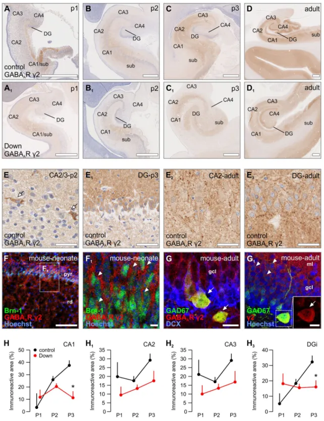
In periods 1 and 2, moderate c2 immunoreactivity was noted throughout the human hippocampus, with the most notable signal localized to the CA1/subiculum (Fig.1a, b).
Although the immunoreactivity comprised mainly of neu- ropil labelling, we observed numerous cell populations that expressed c2 subunits already early during development (period 2: n=16 cases): cellular immunoreactivity was
first detected in the CA1/subiculum [including pyramidal cells and interneurons (Fig.1e)], later attained by other hippocampal regions. In period 3 (n =5 cases), many pyramidal cells in the subiculum and CA regions, as well as granule cells in the dentate gyrus were positive forc2 subunits (Fig.1e1). The increase in c2 immunoreactivity peaked in adulthood (Fig.1d), with the most prominent increase noted in the CA1–3 regions (Fig.1e2). c2 Immunoreactivity was also seen across all layers of the adult dentate gyrus and comprised both neuropil and cytoplasmic labelling (Fig.1e3).
To verify the cellular identify of the observed c2 staining patterns, we testedc2 expression in the developing mouse hippocampus by means of multiple fluorescence immunohistochemistry. In neonates (n=2), c2 immunoreactivity was mainly localized to the pyramidal layer in the CA1 and co-localized with Brn-1, a tran- scription factor transiently expressed in developing pyra- midal cells (Alvarez-Bolado et al.1995) (Fig. 1f, f1). In the adult dentate gyrus (n=2), c2 immunoreactivity was mainly seen as neuropil labelling in the molecular layer and perisomatically in the granule cell layer. Intermingled interneurons, genetically co-labelled for GAD67 (Gad1) by in-frame GFP knock-in (Tamamaki et al. 2003), exhibited somaticc2 immunoreactivity (n=2; Fig.1g, g1).
To further examine c2’s subcellular localization, we cultured primary mouse neurons to identify its spatiotem- poral distribution pattern during the developmental period encompassing neurite outgrowth, growth cone motility and synaptogenesis. Up to 4DIV, c2 immunoreactivity was found indiscriminately on the perikarya, neurites and growth cones of pyramidal-like cells (Supplementary Fig. 1A, A1). In more mature cultures (7DIV) with pre- formed neuronal networks, c2-positive pearl–lace-like punctae were found on neuronal perikarya and processes, and were opposed by parallel-running processes, likely axons, positive for vesicle associated membrane protein 2 (VAMP2), which participates in the docking and fusion of synaptic vesicles (Washbourne et al.1995), as well as the vesicular GABA transporter (VGAT) (McIntire et al.1997) (Supplementary Fig. 1D–D1). Occasional VGAT? interneurons were found expressing somatodendritic c2 subunits (Supplementary Fig. 1D2) in a punctate fashion.
The c2 expression pattern in early developmental peri- ods of Down syndrome cases, periods 1 (n=4) and 2 (n =17), was comparable to that observed in age-matched controls (Fig.1a, b vs. a1, b1). However, the amount ofc2 immunoreactivity was generally reduced in the hip- pocampus. A significant loss of c2 immunoreactivity was found in the subiculum (F(1,37)=6.627, p=0.014), the CA1 (F(1,38)=15.008, p=0.000), and both the internal limb (F(1,30)=7.181, p =0.012) and the external limb (F(1,30)=5.337,p =0.028) of the dentate gyrus (Fig. 1h,
Fig. 1 Distribution of GABAARc2 in normal vs. Down syndrome cases.a–d1Overview images of foetal and adult hippocampi from representative Down syndrome cases and age-matched controls.e–e3 GABAAR c2 immuno-labelling was found on pyramidal cells and interneurons (arrows,e–e1), with the most prominent staining of the neuropil in the dentate gyrus in adulthood (e2, e3). f, f1 Young pyramidal cells (arrowheads, Brn-1?) are positive forc2 subunits in
the neonatal mouse.g,g1Interneurons (arrows, GAD67?) negative for double cortin (DCX) in the granular cell layer harbourc2 subunits.
h–h3Analysis of immunoreactivity in the CA1, CA2, CA3 and inner dentate gyrus region, of GABAARc2 subunits throughout develop- ment in Down syndrome cases. *p\0.05. Scalebars=1 mm (a,a1, b,b1,c,c1) 500lm (d,d1,f); 100lm (e,e2); 50lm (e); 30lm (e3, f1,g); 15lm (g1)
h3; Supplementary Fig. 3E, E1) of Down syndrome cases (n=6 control vs. n=5 Down syndrome) around birth (period 3). In turn, c2 immunoreactivity in the CA2 and CA3 subfields was comparable between diseased and heathy cases (Fig.1f1, f2).
GABAARa1
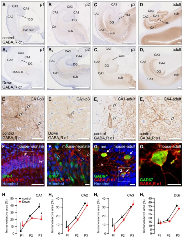
In the hippocampi of human foetuses examined for periods 1 and 2, the a1 subunit was rarely detected (Fig.2a, b).
Instead, there was a prominent increase in a1 subunit immunoreactivity in period 3 (around birth) in most hip- pocampal subregions during normal development, except for CA3, with further enrichment until adulthood (Fig.2c, d). In striking contrast to weak neuropila1 immunoreac- tivity in periods 1 and 2, period 3 was characterized bya1 labelling in cell bodies and processes of the CA1–3 pyra- midal layers, as well as the granule layer of the dentate gyrus (Supplementary Fig. 3A, A1). Towards adulthood, single cells reminiscent of pyramidal cells were decorated bya1 subunits in the pyramidal layer, predominantly in a patch-like manner along their plasmalemma. The other major group of cells positive for a1 had densely labelled cytoplasm and processes and was located in the outer part of the pyramidal layer just beneath the stratum oriens, resembling basket cells (arrowheads, Fig.2d, e–e2). In the stratum radiatum, interneuron-like cells were found scat- tered among a1 immunoreactivity processes (Fig.2e3). A similar pattern was observed in the subiculum (data not shown). The dentate gyrus exhibited strong neuropil immunoreactivity in its molecular layer and to a lesser extent in its granule cell layer in period 3 and particularly in adulthood (Supplementary Fig. 1B2, 1B3, 4A). Similar to other hippocampal regions and ages, strong immunore- activity was mostly noted in interneuron-like cells (Sup- plementary Fig. 1B2, 1B3, 4A1).
Immunohistochemistry for the a1 subunit in neonatal mouse hippocampus showed that pyramidal cells positive fora1 in the pyramidal layer were mostly negative forBrn- 1(Fig.2f, f1). These cells layered aboveBrn-1?cells and are either maturing (down-regulatedBrn-1) or a subpopu- lation positive for other transcription factors, such asBrn-2 (Alvarez-Bolado et al. 1995). Interneurons defined by the expression of GAD67 in the CA1 and dentate gyrus of the mouse hippocampus were positive for thea1 subunit, being comparable to humans (Fig.1g, g1).
The cellular distribution ofa1 subunits was determined in mouse primary neuronal cultures. Labelling fora1 was not detected in DIV4 pyramidal-like neurons except min- imal immunoreactivity on the proximal segment of their neurites (Supplementary Fig. 1B, B1).a1 labelling was not seen in growth cones either (Supplementary Fig. 1B1). At 7DIV, densely packed post-synaptica1 immunoreactivity
puncta apposed VAMP2 and VGAT immunoreactivities along somas and dendrite-like processes of pyramidal- shaped neurons (Supplementary Fig. 1e, e1). Interneurons (with VGAT? axons) were occasionally found decorated by a1 puncta along their somatodendritic axis (Supple- mentary Fig. 1e2). This pattern is similar to that of thec2 subunit, reminiscent of extrasynaptic receptors involved in tonic GABA signalling (Milenkovic et al. 2013).
Although the layer-specific and region-specific distri- bution ofa1 subunits in the hippocampus of subjects with Down syndrome was comparable to those of normal foe- tuses, we noted an overall reduction in a1 immunoreac- tivity (Fig.2a–d vs. a1–d1). This reduction was mainly due to the loss of neuropil immunoreactivity, while neuronal perikarya retained labelling with an intensity equalling control cases (Fig.2e1). Interestingly, morphologically identified interneurons seemed to be the least affected by this reduction in period 3 (Fig.2e1) with statistically sig- nificant decreases observed only in the neuropil of the CA1 subfield (F(1,40)=5.494,p =0.024) (Fig.2f). Indeed, the majority of Down syndrome cases in period 3 showed a significant decrease in a1 immunoreactivity in the CA1 (Fig.2c1) which persisted until adulthood (Fig.2d1). In other CA and DG regions, a1 immunoreactivity was in general weaker (Supplementary Fig. 3F, F1), but not sta- tistically significant in any of the investigated periods (Fig.2h–h3).
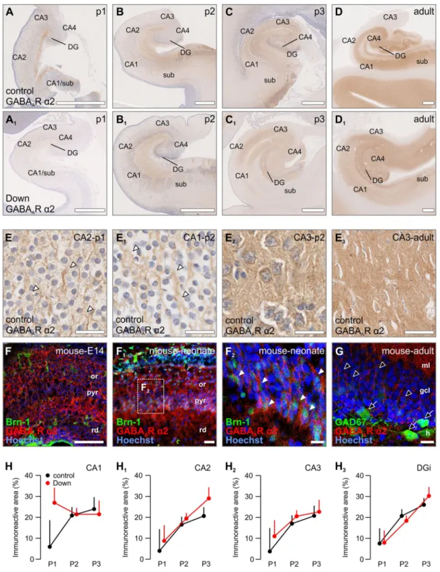
GABAAR a2
In period 1, thea2 subunit was predominantly observed on a fine network of fibres throughout all subregions studied in the human hippocampus (Fig.3a). For instance, a2 immunoreactivity was enriched in wavy processes passing along cells from period 1 onward in the early pyramidal layer (Fig. 3a–d, e, e1). During period 2, an increasing number of a2-expressing cells with neuron-like morphol- ogy was noted in the dentate gyrus, CA layers and the subiculum, and exhibited cytoplasmica2 immunoreactivity (Fig.3b). In the developing mouse, this cytoplasmic immunoreactivity was seen at E14 throughout the entire primordial hippocampus (Fig.3f). Processes coursing between cells, comparable to the human foetus (Fig.3e, e1), appeared in the neonatal mouse emanating fromBrn-1- positive cells, suggesting immature pyramidal neurons (Fig.1f2).
During successive stages of human development, immunoreactivity on processes became gradually reduced (Fig.3e1), leaving a strong neuropil labelling, as well as numerous cells (including pyramidal-like and granule-like cells), in the adult CA regions and dentate gyrus (Fig.3e2, e3). Notably, a2 immunoreactivity seemed to be more pronounced in the inner 1/3 of the molecular layer of the
Fig. 2 Distribution of GABAAR a1 in normal vs. Down syndrome cases.a–d1Overview images of foetal and adult hippocampi from representative Down syndrome cases and age-matched controls.e–e3
Labelled pyramidal cells and interneurons (arrows) were found from period 3 onwards in hippocampal structures. Note the decrease in somatic and neuropil staining in Down syndrome (e1). f, f1 a1
subunits were found on Brn-1- pyramidal neurons (open arrow- heads). g,g1Interneurons in the dentate gyrus expressa1 subunits (arrows).h–h3Quantifications revealed a significant loss of GABAAR a1 subunits in the CA1 region of period 3 only. *p\0.05.
Scalebars=1 mm (a, a1,b, b1, c, c1) 500lm (d, d1, f); 100lm (e,e2,e3); 50lm (e1); 30lm (f1,g,g1)
adult DG (Supplementary Fig. 3B, B1). In the adult mouse, a2 immunoreactivity was similarly decreased in the pyra- midal layer, leaving a perisomatic staining pattern on pyramidal cells, but was not found expressed by GFP- containing interneurons on a GAD67gfp/? background (Fig.3g).
Similar to the patterns found in vivo,a2 subunits were seen on perikarya, processes and growth cones of cultured mouse pyramidal-like neurons at 4DIV (Supplementary Fig. 1C, C1). By 7DIV in maturing cultures, a2 immunoreactivity was mainly restricted to postsynapse-like structures on pyramidal-like neurons apposing VGAT and VAMP2 boutons (Supplementary Fig. 2E–E2) with mini- mal IMMUNOREACTIVITY remaining on the somato- dendritic compartment relative to earlier time points.
The overall distribution of a2 subunits in Down syn- drome was similar compared to the control cases (Fig.3a–
d vs. a1–d1). The majority of hippocampal subregions (the dentate gyrus, CA2, CA3, CA4) presented neither signifi- cant differences in immunoreactivity nor cellular distribu- tion (Fig.3h–h3). The initial higher neuropil immunoreactivity in Down cases reached levels compara- ble to those in control cases in period 2 (Fig.3b, b1, f). For all other areas (Supplementary Fig. 3G, G1), the immunoreactivity measured was generally weaker in Down syndrome albeit not reaching statistical significance.
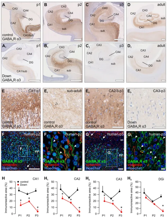
GABAARa3
In contrast to the distribution ofa1 anda2 subunits, thea3 subunit was found at higher levels in the developing human hippocampus and parahippocampal gyrus (Fig.4a–d) than in adults. From period 1 on, the a3 subunit was indis- criminately expressed in all subfields. These included: (1) processes in the pyramidal layer, as well as the neuropil, of the subiculum and CA regions (Fig.4e; Supplementary Fig. 3D–D3), (2) pyramidal-like and interneuron-like somata in the subiculum and CA regions (Fig.4e1, e2) and (3) granule cells in the dentate gyrus (Supplementary Fig. 3C, C1). Neurons expressing a3 subunits showed somatic immunoreactivity, which continued in wavy neu- rites that were associated with vimentin-positive processes, a marker for radial glia (Dahl et al.1981), suggestive of prospective axons during pathfinding (Fig.4f, f1). Bulk immunoreactivity progressively decreased in the subicu- lum, CA2, CA3 and CA4 with advancing gestational age (Fig.4c, d). This was primarily due to reduced a3 immunoreactivity in processes, whereas strong and often punctuate somatic a3 immunoreactivity was retained in pyramidal cells in period 3 (Fig.4g, g1). Distinct from other developmental ages, the dentate gyrus exhibited one of the strongest a3 immunoreactivity in adults, with immunolabeling in the inner 1/3 of its molecular layer,
corresponding to input fields of the local inhibitory cir- cuitry. Furthermore, the majority of granule cells co-ex- presseda3 subunits, as well (Supplementary Fig. 3D, D1).
These data suggest a role for a3 subunits in developmen- tally regulated GABA signalling in emergent neuronal networks during human foetal development.
Throughout development in Down syndrome, we noted substantially lower levels of both a3 immunoreactivity intensity and its more restricted layer distribution (Fig.4a–
d vs. a1–d1) relative to controls. In period 1, a3 immunoreactivity was significantly lower in the CA1 of Down syndrome cases (F(1,39)=4.582, p=0.039) (Fig.4h). In period 2, levels of immunoreactivity were still significantly lower than in control cases in the subiculum (F(1,39)=19.118, p\0.001), CA1 (F(1,39)=12.121, p =0.001), CA2 (F(1,34)=6.478, p=0.016) and CA3 (F(1,32)=4.985, p =0.03) (Fig.4h–h3; Supplementary Fig. 3H). Furthermore, the adjacent intermediate and marginal zones in the CA regions exhibited a reduction in expression patterns, too. In period 3, we observed a sig- nificant and subtotal loss ofa3 immunoreactivity, leaving barely any immunoreactivity detectable throughout the hippocampus in Down syndrome cases (Fig.4c1); the subiculum (F(1,39)=19.919, p\0.001), CA1 (F(1,39)=31.205, p\0.001), CA2 (F(1,34)=43.841, p\0.001), CA3 (F(1,32)=24. 975,p\0.001), the inter- nal limb of the dentate gyrus (F(1,34)=20.741,p\0.001) and external limb of the dentate gyrus (F(1,34)=18.879, p\0.001) (Supplementary Fig. 4D–D1, H–H3). Notewor- thy,a3 immunoreactivity on interneuron-like cells seemed more robust than pyramidal cells, which were devoid ofa3 immunoreactivity around periods 1 and 2 (Fig.4b, b1, e2, e3). In contrast,a3 expression in interneurons was reduced at much later developmental stages (Fig.4e1), while lower a3 levels on interneurons were observed in adult Down syndrome cases (Fig. 4d, d1).
a3 Subunits in neurochemically defined hippocampal neurons
Since the a3 subunit’s expression and distribution were most affected in Down’s syndrome and this subunit is the least described during brain development, we next pursued its (sub)-cellular localization by multi-colour fluorescence immunohistochemistry and high-resolution confocal laser- scanning microscopy in foetal and adult mouse tissues.
Similar to its human developmental pattern (Fig.1g–h2), a3 subunits were mostly found in the neonatal (comparable to period 3) pyramidal layer (Fig.5a). Here, a3 subunits were found on the somata and proximal processes in close proximity to radial glia fibres (RC2?) of neurons that co- labelled for Brn-1 (Fig.5b, b1), indicating that young
Fig. 3 Distribution of GABAAR a2 in normal vs. Down syndrome cases.a–d1Overview images of foetal and adult hippocampi from representative Down syndrome cases and age-matched controls.e–e3
GABAAR a2 immunoreactivity was found throughout all ages, labelling wavy processes (arrowheads,e,e1) and neuropil (e2,e3).f–f2 a2 Subunits were found throughout the embryonic (E14) and neonatal hippocampus on Brn-1? pyramidal neurons (arrowheads, f2).
gInterneurons and granule cells in the dentate gyrus were negative fora1 subunits (open arrows and arrowheads, respectively).h–h3A trend towards an increase of GABAARa2 immunoreactivity was only found in the CA1 region during period 1. Scalebars=1 mm (a,a1,b, b1,c,c1) 500lm (d,d1); 200lm (f); 50lm (e3,f1); 30lm (e,e1,e2, g); 15lm (f2)
Fig. 4 Distribution of GABAAR a3 in normal vs. Down syndrome cases.a–f1 Overview images of foetal and adult hippocampi from representative Down syndrome cases and age-matched controls.e–e3
Labelling for GABAAR a3 was found as early as period 1 in immature pyramidal-like neurons in the pyramidal layer of the CA1 (e). Immunoreactivity was gradually lost into adulthood, with interneurons remaining positively labelled (arrowhead, e1). The overall neuropil and pyramidal layer staining of the hippocampus in
Down syndrome was generally lower (e2vs. e3). f, f1a3 Subunits were found in the pyramidal layer of the human CA1 (period 2) coursing along vimentin? radial glia processes. g, g1 a3 Subunit immunoreactivity was reduced to a somatic pattern in later periods (period 3).h–h3Quantifications reveal a significant loss of GABAAR a3 subunits in Down syndrome over all ages and all structures measured. *p\0.05. Scalebars=1 mm (a,a1,b,b1,c,c1) 500lm (d,d1,f); 100lm (e,e1,g); 50lm (e2,e3); 30lm (e3); 15lm (f1,g1)
pyramidal cells can harbour a3 expression during their developmental trajectory.
In addition, a3 subunits were found on bipolar cells [negative for the neuronal migration marker doublecortin (DCX) (Gleeson et al.1998)], surrounded by a meshwork of VGAT? puncta (Fig.5c–e1). To validate if these bipolar cells are either GAD67? and/or cholecystokinin (CCK)-containing interneurons (Klausberger and Somogyi 2008), we utilized a novel CCKBAC/DsRed::GAD67gfp/?
dual-colour reporter mouse (Calvigioni et al. 2017).
Almost alla3?cell bodies co-localized with GAD67, with a subpopulation being also positive for CCK (Fig.5f–g1), reinforcing that morphologically differentiating interneu- rons can express a3. Comparable to its human develop- mental distribution (Fig.3d), a3 immunoreactivity was reduced in the adult mouse hippocampus: residual punc- tate labelling in the pyramidal layer and mainly GFP? interneurons were noted (Fig.5h–j1). These data suggest that interneurons retain their a3 expression, whereas pyramidal cells down-regulate this GABAAsubunit during hippocampal development.
We then further dissecteda3 subcellular distribution in cultured mouse primary neurons with an emphasis on its redistribution and/or down-regulation during neuronal maturation. At 2DIV, a3 immunoreactivity was resolved along the soma, processes and growth cones of pyramidal- like cells (Fig.6a–a2). After 4DIV, a3 immunoreactivity was still retained in actin-rich growth cones, fostering a concept on a3 subunit contributions to GABA-mediated axon guidance (Fig.6b–b2) (Ageta-Ishihara et al.2009). At 7DIV, when neuronal networks first appeared, a3 immunoreactivity was only sparsely detected on somas and processes (Fig.6c–c2). Yet, a3 subunits concentrated in apposition to VGAT? presynapses, which also contained VAMP2 (Fig.6d–e2), thus marking bona fide synapses.
Moreover, a3 IMMUNOREACTIVITY was occasionally seen in cells adopting glia-like morphology (Supplemen- tary Fig. 2A–A2). These results imply that a3 subunits in developing neurons are either progressively down-regu- lated or recruited to post-synaptic sites. Interneurons are recognized as the sole cellular component of the cortical circuitry to retain a3 expression (Fig.5f–h1) perisomati- cally until adulthood.
Cellular exposure tob-amyloid leads to down- regulation ofa3 subunits
To seek a more mechanistic view on the down-regulation ofa3 subunit expression in Down syndrome, we selected amyloid precursor protein 1 (APP) as a candidate for upstream regulation due to its presence on chromosome 21 and for it being implicated in driving neuronal
differentiation and synaptogenesis (Korenberg et al.1989;
Wang et al.2009). In addition, inhibition ofc-secretase, a multiprotein complex with enzymatic activity to cleave APP and generate full-length b-amyloid (1–42), restores neurogenesis and synaptogenesis in a mouse model of Down syndrome (Giacomini et al.2015).
We found that APP was significantly up-regulated in the stratum oriens of the pyramidal layer of the CA1 region during periods 1/2 (p\0.01; n =5 control vs. n=5 Down cases), with a coincidently marked increase in the stratum radiatum in cases with Down syndrome (Fig.7a–
b2). Since plasmab-amyloid (1–42) levels are increased in young adults with Down syndrome and associate with accelerated ageing in these patients (Obeid et al.2016), we hypothesized that an increase of soluble b-amyloid, pro- duced by c-secretase-mediated proteolytic cleavage of excess APP, might disrupt the morphogenesis of develop- ing neurons (Freude et al.2011) and precipitate regulatory changes to dictate a3 subunit availability. Since cultured primary neurons express low amounts of a3 subunits, especially when neuronal networks are formed (Fig.6b, c), we relied on human SH-SY5Y neuroblastoma cells as a cellular model to test our hypothesis. SH-SY5Y cells express a3 subunits at high mRNA and protein levels (Supplementary Fig. 4A, A1) and undergo morphological changes when exposed to b-amyloid (J. Mulder and T.
Harkany, unpublished data).
Differentiation of SH-SY5Y cells was initiated by application of retinoic acid (Cheung et al. 2009) and verified by their up-regulation of b-III-tubulin (TUJ1), a cytoskeletal marker of immature neurons (Fig.7c–d1; n =3 coverslips). Coincidently, a3 subunits appeared in processes of SH-SY5Y cells undergoing retinoic acid-in- duced differentiation (Fig. 7d, d1). After application of fresh b-amyloid(1–42) at concentrations (100–250 nM) that did not affect the survival of cells undergoing retinoic acid-induced differentiation in our culture paradigm (Supplementary Fig. 4B–C2), TUJ1 levels significantly increased relative to untreated yet differentiated controls (Fig.7e; n=3 each). This response was anti-parallel with a3 subunit levels (that decreased; Fig.7e). When applying lM concentrations of b-amyloid(1–42), we noted cytotoxicity (Harkany et al. 2000a, b) (Supple- mentary Fig. 4B–C2), indicating that there is a narrow developmental window in which b-amyloid(1–42) might exert morphogenic, perhaps even differentiation-promot- ing effects on developing neurons, if a tandem of TUJ1 and a3 are seen as read-outs. In sum, our data suggests that b-amyloid, a pathogenic outcome of increased APP levels, in Down syndrome could contribute to the remodelling of GABA synapses by altering their subunit composition (Fig.7f).
Discussion
In this study, we present the first comprehensive descrip- tion of the expression pattern ofa1,a2,a3 andc2 GABAA
receptor subunits in the human hippocampus of Down
syndrome cases and age-matched controls along a broad developmental trajectory. Although the existence of func- tional GABAAreceptors in primate and human brains was demonstrated by visualization of benzodiazepine binding sites (Reichelt et al. 1991; Shaw et al. 1991), there is a Fig. 5 GABAARa3 localizes to
pyramidal cells and
interneurons in the foetal mouse brain.a–a2In the pyramidal layer, GABAARa3 subunits were found on somata (asterisks) and processes (arrowheads) adjacent to radial glia (RC-2?), indicative of young pyramidal cells.b,b1a3 Subunits were expressed on Brn-1 containing pyramidal cells (arrowhead), but not on interneuron-like cells (open arrowheads).cInterneuron-like cells were negative for the migration marker DCX (open arrowheads).d–e1In the CA1, GABAARa3 immunoreactivity was found on migrating interneuron-like cells embedded in a VGAT?meshwork (arrowheads,e–e1).f–g1The dual-transgenic mouse GAD67- GFP::CCK-DsRed revealed that these cells are indeed
interneurons (GAD67?; open arrowheads), with a
subpopulation of CCK-positive cells (solid arrowheads).h–j1In the adult, GABAARa3 staining was limited to somata of interneurons (arrowheads) in the CA1 region (h,h1) and the dentate gyrus (i–j1). Occasional cells were GAD67-negative, most likely CCK-containing interneurons (arrows).
Scalebars=500lm (f);
100lm (d,h,i); 50lm (e,j);
30lm (a–c); 15lm (g)
surprising gap of knowledge at the protein level due to the lack of appropriate tools. Therefore, we applied novel knock-out-tested antibodies against GABAAR subunits to address this issue.
We find differential maturation ofa1 anda2 subunits in the hippocampus. Similar to data from animal models,a1 seems to have late expressional onset in the human brain (Fritschy et al. 1994). In contrast, an abundance of a3 subunits throughout the developmental hippocampus was observed with significantly diminished levels in the adult.
GABAARs have been implicated in the migration of post- mitotic neurons, including both pyramidal cells (Behar et al.2000) and interneurons (Cuzon et al. 2006), postu- lating a central role for this subunit to control migration and differentiation. Our finding that a3 subunits are robustly expressed during development on both interneu- rons and pyramidal cells in vivo, as well as on actin-rich motile growth domains in vitro, reinforces a non-synaptic trophic and/or guidance role for GABA signalling through a3 subunits in the developing hippocampus. In addition, thea3-subunit seems to be exchanged perinatally to other subunits (i.e.a1 anda2) in the human foetal brain, which might account for the change in electrophysiological properties that GABAAR signalling undergoes during
normal brain development (Owens et al.1999). While, the c2 subunit showed widespread expression in the hip- pocampus, we observed a striking similarity in expression pattern between a3 and c2 subunits in the CA1 and subiculum, suggesting their probable functional co-occur- rence. The temporal co-expression of these two subunits suggests that these possible complexes are destined for synaptic terminals, since c2 is classified as the post-sy- naptic subunit and is involved in the trafficking of the complex towards the synapse (Vithlani et al. 2011).
Noteworthy, we did not detect significant amounts ofa3 in the DG, suggesting different assembly partners for c2 in this region.
In the hippocampus of foetal Down syndrome subjects, our data indicates complex expression profiles coincidently affecting a1, a3 andc2 subunits. For instance, a1 andc2 subunits showed selective depletion in the CA1 and subiculum, whereas in other hippocampal areas these changes were not as obvious. Interestingly, the distribution of a2 subunits in the hippocampus bore a striking resem- blance to thec2 subunit, both of them exhibiting a subtotal depletion in the CA1 and subiculum in period 3. In con- trast, thea3 subunit demonstrated the most robust changes in Down syndrome cases. A strong reduction was observed Fig. 6 GABAARa3 redistributes during mouse neuronal differenti-
ation. a–b2 In cultured mouse primary hippocampal neurons, GABAAR a3 subunits are localized to somata, neurites and growth cones (f-actin-positive), up to 4 days in culture (arrowheads).c–c2
When neuronal networks start to form, GABAAR a3 subunits redistribute with a punctate pattern on neurites and somatas (arrow- heads).d–e2GABAAR a3 subunits are found adjacent to VGAT?
puncta (d1,d2) and the presynaptic vesicular marker VAMP2 (e1,e2), indicating that GABAAR a3 relocalizes to neurotransmitter release sites upon network formation.f–f2Intermittent interneurons, VGAT?, were found decorated with GABAAR a3 subunits (arrowheads).
Scalebars=50lm (e,j); 30lm (a,a2,b,b2,c,c2,e,f,f2); 15lm (d,d2,e2)
throughout all investigated time periods with an overall lower expression and an almost complete loss from period 3 onwards. Most adult animal studies focused on pharma- cological manipulation ofa5 subunit containing GABAA- Rs (Braudeau et al.2011; Fernandez et al.2007; Martinez- Cue et al. 2013), due to their involvement in cognition (Redrobe et al. 2012; Rudolph and Mohler 2014; Wang et al. 2012). We were unable to reliably probe the a5 subunit with the antibodies available to us in develop- mental tissues, revealing only a weak punctate staining that we deemed unspecific due to a lack of regional and tem- poral patterns (data not shown). Therefore, we only focussed on GABAAR subunits whose consistent staining pattern we deemed specific.
Given the fact that GABAARs are thought to provide a stop signal for migrating cells in the cortex (Behar et al.
2000), a disturbance in GABAAR subunits would suggest alterations to neuronal migration in Down syndrome.
Indeed, a change in neurogenesis and reduced neuronal number in the cortex have been described in Down syn- drome before (Becker et al. 1991; Golden and Hyman 1994; Ross et al. 1984; Wisniewski et al.1984). Since we localized a3 subunits to motile growth domains on both pyramidal cells and interneurons, our data suggest that a premature down-regulation ofa3 subunits might contribute
to those observed changes in neuronal migration, especially if this reduction leads to altered GABAAR-mediated cur- rents or second messenger signalling systems (Luscher et al. 2011). Thus, a premature redistribution of subunits could eventually lead to improper neuronal placement and differentiation, and disturb prenatal giant depolarizing potentials which are important for the maturation of synapses (Ben-Ari et al. 2007) overall, resulting in cogni- tive deficits.
A concurrent comorbidity in Down syndrome is the early development ofb-amyloid plaques in the adult brain and Alzheimer’s-like neurological abnormalities (Masters et al.1985; Motte and Williams1989). Due to the position of APP on chromosome 21, APP is thought to elevate levels of full-length b-amyloid, leading to early plaque deposits. In addition, inhibitors of c-secretase, restored neurogenesis and synaptogenesis in a mouse model of Down syndrome (Giacomini et al. 2015). However, b/c secretase cleavage of APP can generate bioactive peptides other than b-amyloid(1–42) itself, including small C-ter- minal fragments that interact with signalling proteins (van der Kant and Goldstein 2015) and its intracellular domain (AICD50) retained aftercsecretase cleavage which can act as a transcription factor (Konietzko 2012). Recent data demonstrate that AICD50 overexpression affects the levels of Sox2, a key transcription factor in the regulation of stem cell maintenance and lineage commitment (Sarlak et al.
2016). Since the physiological role of these cleavage products are still poorly understood, we focussed on b- amyloid as it has been correlated with cellular differenti- ation (Freude et al. 2011; Wang et al. 2009) and the peculiarity of accelerated ageing in Down syndrome (Obeid et al.2016). We thus posit an increase of APP, and elevated levels of solubleb-amyloid (1–42) as a factor in promoting early brain maturation.
In our developmental Down syndrome cohort, we were not able to find b-amyloid plaques, suggesting that any pathological effect of b-amyloid is not due to its aggre- gation in foetal brains. Instead, we observed the first signs of plaque formation in tissues of a 15-year-old patient from a separate cohort, with more substantial deposits in patients[30 years old. Asb-amyloid oligomers are more toxic than un-aggregated peptide (Zhao et al. 2012), an increase in the soluble un-aggregated form could promote cellular morphological changes without compromising cell survival. In our foetal Down syndrome cases, we find elevated quantities of APP, and experimental challenges with external b-amyloid (1–42) delivery triggered the down-regulation of a3 subunits similarly to what is observed in human foetal brain. This observation is in line with our previous results, showing that developmental physiologic tau phosphorylation is disturbed in individuals with Down syndrome (Milenkovic et al. 2017), most Fig. 7 Beta amyloid is implicated in the premature differentiation of
human neuronal-like cells.a–b2APP levels in the hippocampus are significantly increased in Down syndrome patients (period 1/2, n=5–6). c, c1 In undifferentiated human SH-SY5Y cells, a3 subunits are localized to the cytoplasma (arrowheads).d,d1Upon differentiation, TUJ1 levels increase and a3 subunits appear on elongating TUJ1? processes (arrowheads). e Non-toxic concentra- tions of beta amyloid(1–42) down-regulate a3 subunits while increasing TUJ1 levels.fProposed mechanism of premature matu- ration of brain circuitry. *p\0.05. Scalebars=200lm (a,a1,a2);
50lm (c1,d1)