RESEARCH ARTICLE
Exercise and Cardiac Remodeling in Normal and Athletic States
Exercise-mitigated sex-based differences in aging: from genetic alterations to heart performance
Denise B€orzsei,1* Dániel Priksz,2* Renáta Szabo,1,3Mariann Bombicz,2Zoltán Karácsonyi,4 Lászlo G. Puskás,5,6Liliána Z. Feher,5Zsolt Radák,7Krisztina Kupai,1 Aniko Magyarine Berko,1 Csaba Varga,1Bela Juhász,2and Aniko Posa1,3
1Department of Physiology, Anatomy and Neuroscience, Faculty of Science and Informatics, University of Szeged, Szeged, Hungary;2Department of Pharmacology and Pharmacotherapy, Faculty of Medicine, University of Debrecen, Debrecen, Hungary;3Department of Physiology, Anatomy and Neuroscience, Interdisciplinary Excellence Centre, University of Szeged, Szeged, Hungary;4Department of Orthopedics, Faculty of Medicine, University of Debrecen, Debrecen, Hungary;5Avidin Limited, Szeged, Hungary;6Laboratory of Functional Genomics, Biological Research Centre, Hungarian Academy of Sciences, Szeged, Hungary; and7Research Institute of Sport Science, University of Physical Education, Budapest, Hungary
Abstract
The prevalence of cardiovascular diseases dramatically increases with age; therefore, striving to maintain a physiological heart function is particularly important. Our aim was to study the voluntary exercise–evoked cardioprotective effects in aged male and female rats, from genetic alterations to changes in heart performance. We divided 20-month-old female and male Wistar rats to control and running groups. After the 12-wk-long experimental period, echocardiographic measurements were performed.
Afterwards, hearts were either removed for biochemical measurements or mounted into a Langendorff-perfusion system to detect infarct size. The following genes and their proteins were analyzed from heart: catechol-O-methyltransferase (Comt), endo- thelin-1 (Esm1), Purkinje cell protein-4 (Pcp4), and osteoglycin (Ogn). Recreational exercise caused functional improvements; how- ever, changes were more prominent in males. Cardiac expression of Comt and Ogn was reduced as a result of exercise in aged males, whereas Pcp4 and Esm1 showed a marked overexpression, along with a markedly improved diastolic function. The key result of this study is that exercise enhanced the expression of the Pcp4 gene and protein, a recently described regulator of cal- cium balance in cardiomyocytes, and suppressed Comt and Ogn gene expression, which has been associated with impaired car- diac function. In addition, as a result of exercise, a significant improvement was observed in the size of infarct elicited by left anterior descending coronary artery occlusion. Our results clearly show that age and sex-dependent changes were both appa- rent in key proteins linked to cardiovascular physiology. Exercise-moderated fundamental genetic alterations may have contrib- uted to the functional adaptation of the heart.
NEW & NOTEWORTHYVoluntary exercise has proved to be an effective therapeutic tool to improve cardiac function in aged rats with clearly visible sex differences. Long-term exercise is associated with decreased Ogn and Comt expression and enhanced presence of Pcp4 and Esm1 genes. Sex-dependent changes were also observed in the expression of the cardiovascu- lar key proteins. Fundamental alterations in gene and protein expression may contribute to the improvement of cardiac performance.
exercise; aging; echocardiography; gene expression; Pcp4
INTRODUCTION
Cardiovascular diseases (CVDs) are the leading cause of morbidity and mortality worldwide and have a pronounced prevalence with age (1,2). Longevity and aging are the two sides of the coin as they depend on environmental and life- style factors, sex differences, and genetic markers as well.
Aging implies numerous pathological alterations in the car- diovascular system, among others. Cardiac aging is first characterized by molecular and cellular changes, which then lead to structural, morphological and functional abnormal- ities. Besides, older individuals tend to suffer from chronic inflammation called“inflamm-aging”that manifests itself in the appearance of inflammatory cytokines and oxidative
* D. B€orzsei and D. Priksz contributed equally to this paper.
Correspondence: B. Juhász (juhasz.bela@med.unideb.hu).
Submitted 9 August 2020 / Revised 14 December 2020 / Accepted 15 December 2020
stress. Although developing inflammation is a powerful risk factor in itself, cardiac remodeling further increases the chances of CVDs and the vulnerability of the heart (3, 4).
Cardiac remodeling process is a compensatory mechanism to the loss of cardiomyocytes which affects extracellular ma- trix (ECM) structure (5). Cardiovascular homeostasis greatly depends on the structure of the ECM; thus, imbalanced col- lagen accumulation caused ECM disruption contributes to the appearance of pathological cardiovascular lesions (6).
These adverse changes play an important part in the devel- opment of CVDs by deteriorating elastic properties in left ventricle (LV) and eventually causing diastolic dysfunction (7). Petretto et al. (8) demonstrated that LV mass (LVM) is partly influenced by genetic factors as well; they have found that osteoglycin (Ogn) is an effective regulator of LVM in human and animal studies. Namely, its increased expression was found to be associated with elevated LVM. In line with these observations, elevated Ogn expression was found in the case of myocardial infarction and coronary heart dis- eases in human studies as well (9). Besides Ogn, other princi- pal genes might also be informative in the pathogenesis of CVD. Xie et al. (10) proved that the upregulation of the en- dogenous calmodulin regulator, Purkinje cell protein-4 (Pcp4), contributes to a compensatory mechanism against cardiac hypertrophy and arrhythmia. As Pcp4 is able to reduce the release of Ca2þ from the sarcoplasmic reticulum of cardiac cells, it is presumable that it has a prominent role in cardioprotection. Intracellular Ca2þ level in cardiomyo- cytes is affiliated with catechol-O-methyltransferase (Comt) enzyme function as well. Increased amount of catechol- amines causes intracellular Ca2þ overload in myocytes and leads to cardiac dysfunction (11). Consequently, Comt, the enzyme that catalyzes catecholamine metabolism, has been also linked to CVD. It is related to a higher risk of adverse cardiovascular events, such as elevated systolic blood pres- sure and atherosclerotic disease (12); however, long-term physical exercise has been shown to be effective in reducing Comt activity in human studies (13).
The above-mentioned lifestyle factors have always been exceedingly important in maintaining cardiovascular health.
Sedentary lifestyle has long been linked to unfavorable car- diovascular events and shortened life expectancy (14).
According to today’s perception, it is the leading contributor to the development and progression of CVDs (15). It is well documented that people with active lifestyles are less threat- ened by coronary artery diseases than sedentary individuals (16). Dozens of studies discuss that regular exercise contrib- utes to the improvement of cardiovascular function by nor- malizing blood pressure, resting heart rate, and myocardial perfusion (17,18). Regular exercise is able to lower the degree of lipid peroxidation and improve antioxidant properties in adult and in aged individuals as well (19). Moreover, endo- thelium and vascular network also indicate complex adaptive changes in response to exercise. Physical exercise is associated with increased vascularization and angiogen- esis in intact and injured heart as well (20). Although the underlying mechanisms are still unclear, a recent study by Rocha et al. (21) proved that endothelin (Esm1) plays an important role in angiogenesis through the modulation of vascular endothelial growth factor (VEGF) in endothelial cells.
CVDs are responsible for approximately 17.9 million deaths per year (22); therefore, it should be considered as a priority health issue. The characterization of molecular and genetic markers in connection with these pathophysiological abnormalities is pivotal for a deeper understanding of car- diac aging. Extensive data highlight the prominent part of exercise in the therapeutic options focused on cardiovascu- lar health (18,22,23). Taken together, we hypothesized that recreational exercise may improve not only the functional properties of the heart and moderate the non-functional part of the infarcted myocardium but might also cause modifica- tions in the expression of Comt, Ogn, Pcp4, and Esm1 genes in aged rats in a sex-dependent way.
METHODS
Experimental ProtocolIn this current study, 20-month-old male and female Wistar rats were used. Animals were kept at a standard 20– 23C temperature, and appropriate light/dark cycle was pro- vided; all circumstances were accomplished according to Directive 2010/63/EU. At the beginning of the study, female and male rats were divided into two main groups: control (aging male, aging female) and running animals (aging male R, aging female R). Control rats were placed into standard cages, whereas running animals were provided with a run- ning wheel–equipped cage. This protocol was defined as a voluntary exercise model; animals were not forced to exer- cise but had free access to the wheel 24 h a day. At the end of the 12-wk experimental period, echocardiographic measure- ments were performed under anesthesia. Following this pro- cedure, all rats were euthanized and their hearts were excised. Hearts were randomly divided into two subgroups:
1) stored at 80C until biochemical measurements or 2) subjected immediately to ex vivo isolated heart experiments.
To detect the effects of voluntary exercise in the case of an acute injury, left anterior descending coronary artery (LAD) occlusion was applied ex vivo, via Langendorff system. The detailed experimental protocol is shown inFig. 1. All proce- dures were approved by the National Scientific Ethical Committee on Animal Experimentation (XX/4801/2015, 25/
2013DEMÁB) and complied with the ARRIVE guidelines.
Transthoracic Echocardiography
Echocardiographic measurements of rats were carried out under intramuscular anesthesia (ketamine 50 mg/kg, xyla- zine 5 mg/kg) at the end point of the study. The chest of each animal was shaved, and the rats were positioned in a dorsal decubitus position. Data acquisition was performed using a Vivid E9 sonograph (GE Healthcare, Chicago, IL), with an i13L linear array probe at 14 MHz. Two-dimensional and M- mode measurements at parasternal long axis and short axis views were carried out as recommended by the American Society of Echocardiography (24). Electrocardiograms were recorded during all measurements. M-mode tracings were performed at the midpapillary muscle level, in either para- sternal long or short axis views. Echocardiographic meas- urements included interventricular and left ventricular free-wall thickness in diastole and systole (IVSs, IVSd), left ventricular internal diameter at end-diastole (LVIDd) and
end-systole (LVIDs), aortic root diameter (Ao) and left atrial diameter (LA). Fractional shortening was computed by using the equation [(LVIDdLVIDs)/LVIDd]100%, and the ejec- tion fraction (EF) was calculated by the software of the sono- graph. Relative wall thickness was calculated as 2LVPWd/
LVIDd. Mitral annular peak systolic excursion (MAPSE) and tricuspid annular peak systolic excursion (TAPSE) were assessed with M-mode, measuring the distance of mitral or tricuspid annular movement between end-diastole to end- systole. All measurements were averaged over three tofive consecutive cardiac cycles. Doppler (PW) imaging of the mi- tral, tricuspid, and aortic valves was obtained from the apical three-, four-, andfive-chamber views. From the mitral inflow velocity image, the following measurements were obtained:
left ventricular peak E and peak A waves (mitral early and latefilling velocities) and theE- toA- ratio (E/A). Mitral valve closure to opening time (MCOT), ejection time (ET), isovolu- mic contraction and relaxation time (IVCT and IVRT, respec- tively) were measured, and myocardial performance index (MPI or Tei-index) was calculated. Left ventricular outflow tract (LVOT) parameters (velocity and pressure gradients) were also obtained: LVOT Vmax, Vmean; LVOT maxPG, meanPG; and VTI. Pulsed wave Doppler measurements were also carried out on the right side of hearts, and RV E, RV A waves, as well as the E/A ratio of the right ventricle, were also determined. Tissue velocity imaging (TVI) meas- urements were analyzed from the apical four-chamber view and from the parasternal long axis and short axis views as well. A 2-mm tissue sampling volume was obtained at the mitral annulus from both septal and lateral walls. From the acquired images, the following functional parameters were measured: e0and a0wave velocities, sep- tal e0/a0ratio, E/e0(early diastolic mitral inflow velocity di- vided by average value of septal tissue Doppler early diastolic velocity) (25).
EchoPAC PC software (GE Healthcare, Chicago, IL) was used to analyze the recorded measurements by a blinded reader. Shapiro-Wilk normality test was used to estimate Gaussian distribution, and Student’s t test was applied to estimate differences between groups.
Determination of Gene Expression
RNA extraction and quantitative real-time PCR.
Total RNA was isolated from rat right and left ventricle tis- sues with Qiagen miRNeasy Mini Kit according to the manu- facturer’s protocol (Qiagen, Hilden, Germany). On-column DNase digestion was carried out with the RNase-Free DNase Set (Qiagen GmbH). The quality and quantity of the isolated RNA were measured with NanoDrop1000 Version 3.8.1.
(Thermo Fisher Scientific, Waltham, MA). RNA samples were stored at80C until use. Reverse transcription from 2mg of total RNA was performed with the High-Capacity cDNA Archive Kit (Applied Biosystems, Foster City, CA) in a total volume of 20mL according to the manufacturer’s protocol.
After dilution with 80μL of PCR-grade water (Roche Applied Science, Mannheim, Germany), 1μL of the diluted reaction mix was used as a template in the quantitative real- time PCR (qRT-PCR) with FastStart Essential DNA Green Master (Roche Applied Science, Mannheim, Germany) using the following protocol: 10 min at 95C followed by 45 cycles of 95C for 15 sec., 60C for 25 s and 72C for 25 s. Thefluores- cence intensity of SYBR Green dye was detected after each amplification step. Melting temperature analysis was done after each reaction to check the quality of the products.
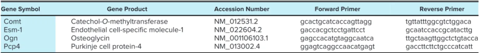
Primers were designed using the online Roche Universal Probe Library Assay Design Center. The quality of the pri- mers was verified by MS analysis provided by Bioneer (Daejeon, Korea). Primer sequences and accession numbers are listed in Table 1. Relative expression ratios were calcu- lated as normalized ratios to rat Hprt1 and Gusb genes. After amplification, the melting curve was checked to verify the specificity of the PCRs. The presented relative gene expres- sion ratios wereDDCT values (log2).
LightCycler 1536 system.
Identical reaction volumes were prepared by the Agilent Bravo Liquid Handling Platform (Agilent Technologies) in a 24 64-well design two times according to Agilent and Roche’s recommendations. Each 2μL reaction mixture Figure 1.Detailed experimental protocol. LAD, left anterior
descending artery; Ogn, osteoglycin; Comt, catechol-O- methyltransferase; Pcp4, Purkinje cell protein-4; Esm1, endothelin 1. Thisfigure was created using modified Servier Medical Art templates, which are licensed under a Creative Commons Attribution 3.0 License;https://smart.servier.com
contained 8 ng cDNA, 10 pmole gene-specific primers, and 1μL 2 qPCRBIO SyGreen Mix (PCR Byosystems, London, UK). The following amplification protocol was used: 95C for 2 min (activation), 60 cycles of 95C for 10 s, and 60C for 10 s, followed by 40C for 10 s final cooling. Amplification was performed on the LightCycler 1536 System (Roche Applied Science) using 42 gene-specific primers and 2 refer- ence genes. Data were collected and processed using the LightCycler 1536 SW 1.0 software. Curves were analyzed by using dynamic tube and slope correction methods. Relative expression of the analyzed genes was normalized to the mean.
Determination of Cardiac Comt, Esm1, Ogn, Pcp4 Concentrations
Right and left ventricle tissues were homogenized in phos- phate buffer (PBS) (pH 7.4) for 20 s and placed into the centri- fuge for 20 min at 3,000 rpm, at 4C. Centrifuged supernatants were collected and used for enzyme linked immune sorbent assay (ELISA; GenAsia, Shanghai). Standard solutions were prepared and added into the wells together with 50ml strepta- vidin-HRP. Into the sample wells, 40ml of supernatants, 10ml antibodies, and 50ml streptavidin-HRP were added. The plate was covered with a seal plate membrane and incubated for Table 1. Primers used in qRT-PCR analysis
Gene Symbol Gene Product Accession Number Forward Primer Reverse Primer
Comt Catechol-O-methyltransferase NM_012531.2 gcactgcatcaccagttagg tgttatttggcgtctggaca
Esm-1 Endothelial cell-specific molecule-1 NM_022604.2 gaccacgctcctgattcct gcaatccaccgcatacttg
Ogn Osteoglycin NM_001106103.1 gagccacatgtaggcaatca ttgctaagttggctctgtacca
Pcp4 Purkinje cell protein-4 NM_013002.4 ggagtcaggccaacatgagt gaccttcttctgcccatcatt
qRT-PCR, Quantitative real time polymerase chain reaction, Comt, catechol-O-methyltransferase; Esm1, endothelin-1; Ogn, osteogly- cinl Pcp4, Purkinje cell protein-4.
Table 2. Echocardiographic outcomes
Aging Male Aging Female Aging Male Running Aging Female Running
mean mean ± SE mean mean ± SE vs. aging
male mean mean ± SE vs. aging
male mean mean ± SE vs. aging
female
vs. aging male R
LA/Ao 0.8583 0.0514 0.8859 0.0697 ns 0.9776 0.0446 ns 0.8667 0.0602 ns ns
RWT% 0.4331 0.0355 0.3467 0.0217 ns 0.4217 0.0228 ns 0.3792 0.0412 ns ns
LV mass, g 0.9652 0.0546 0.6155 0.0763 ## 0.9854 0.0621 ns 0.5744 0.0248 ns ###
EF (%) 76.3333 1.2019 75.8333 1.4926 ns 84.3333 0.4944 80.5000 0.2236 ####
%FS 40.0000 1.0000 40.1667 1.3520 ns 48.6667 0.6667 44.0000 0.4472 ###
MV E, m/s 0.5917 0.0259 0.5917 0.0138 ns 0.5983 0.0199 ns 0.6100 0.0289 ns ns
MV A, m/s 0.4883 0.0190 0.4333 0.0217 ns 0.3400 0.0173 0.3400 0.0350 ns
MV E/A 1.1800 0.0410 1.4050 0.0992 ns 1.8050 0.1129 1.8583 0.2474 ns ns
DecT, ms 64.8333 1.4240 44.5000 2.0777 #### 50.8333 1.4701 41.6667 1.4063 ns ##
MAPSE, mm 1.8840 0.1060 1.9232 0.1272 ns 2.3668 0.1411 2.4280 0.0762 ns
LVOTVmax, m/s 0.8183 0.0192 0.6367 0.0311 ### 0.7933 0.0332 ns 0.7583 0.0521 ns ns
LVOT maxPG, mmHg 2.6983 0.1171 1.6367 0.1746 ### 2.5300 0.2010 ns 2.3483 0.3018 ns ns
HR, beats/min 229.8333 8.2033 246.6667 10.7072 ns 275.5000 10.8374 225.6667 7.8387 ns ##
Sept E0, mm/s 30.5000 2.5658 30.5000 1.6882 ns 43.3333 2.7528 42.3333 3.9805 ns
Sept A0, mm/s 49.6667 6.6767 44.3333 1.9607 ns 55.5000 6.7614 ns 40.3333 2.9515 ns ns
Sep E0/A0 0.6343 0.0403 0.6894 0.0286 ns 0.8175 0.0779 ns 1.0629 0.1006 ns
E/e0 19.8905 1.4415 19.7083 1.1878 ns 14.1169 1.0534 14.7956 0.9315 ns
ET, ms 73.0000 2.0656 64.0000 3.1728 # 69.5000 2.7049 ns 71.3333 3.3133 ns ns
Tei-Index 0.7977 0.0501 0.9070 0.0545 ns 0.6146 0.0460 0.6937 0.0509 ns
IVCT, ms 18.6667 1.9437 18.5000 1.3844 ns 15.3333 1.3824 ns 18.0000 2.5430 ns ns
IVRT, ms 39.3333 1.6262 32.8333 0.9458 ## 26.6667 2.0440 30.3333 1.8012 ns ns
RV E/A 0.7900 0.1272 0.6167 0.0562 ns 0.7517 0.1024 ns 0.8283 0.0511 ns
TAPSE, mm 2.7415 0.2194 2.3223 0.1003 ns 2.8892 0.1276 ns 2.6580 0.2530 ns ns
SV, ml 0.5592 0.0790 0.3356 0.0419 # 0.5428 0.0504 ns 0.4159 0.0339 ns ns
CO, ml/min 127.7507 16.7011 82.4105 10.2439 # 147.9433 10.6617 ns 93.9640 8.4432 ns ##
Shapiro-Wilk method was used to estimate data distribution, and two-tailed Student’sttest was performed to estimate differences between groups. Result shown as means ± S.E.;n= 6/group. LA/Ao, ratio of left atrial and aortic diameters; RWT%, relative wall thickness; LVmass, left ventricle mass; FS, fractional shortening of the left ventricle; EF, ejection fraction of the left ventricle; SV, stroke volume of the left ventricle; CO, cardiac output of the left ventricle;
MAPSE, mitral annular plane systolic excursion; E/A, peak mitral early diastolic inflow velocity/peak atrial diastolic inflow velocity of the left ventricle;
DecT, E wave deceleration time; HR, heart rate; E/e0, peak mitral inflow velocity/average of spectral tissue Doppler peak early diastolic velocities at the lat- eral corner of mitral annulus; LVOTVmax, maximal velocity of left ventricle outflow; LVOT maxPG, maximum pressure gradient of left ventricle outflow tract; e0/a0, peak early diastolic velocity of the septal wall/peak atrial diastolic velocity of the septal wall; MPI or Tei-index, myocardial performance index;
IVCT, isovolumic contraction time; IVRT, isovolumic realxation time; ET, ejection time; RV E/A, peak early diastolic inflow velocity/peak atrial diastolic inflow velocity of right ventricle; TAPSE, tricuspid annular plane systolic excursion.Statistical significance between running and non-running counterparts;
#statistical significance between matching female and male groups. Symbols denote the level of significance:P<0.05;P<0.01;P<0.001;P<
0.0001; and #P<0.05; ##P<0.01; ###P<0.001; ####P<0.0001; ns, not significant.
60 min at 37C. After incubation, the plate was washed and chromogen solutions A and B were added to the wells, which was followed by a 10-min incubation at 37C. To stop the reac- tion, stop solution was added to each well, absorbance (OD) was measured under 450 nm wavelength. Protein determina- tion was performed with Bradford method and measured spec- trophotometrically at 595 nm. Comt, Ogn, and Pcp4 levels were expressed as pg/mg protein; Esm1 values were defined as pg/mg protein.
Implementation of Ischemia-Reperfusion Injury
Rats were anesthetized, and their hearts were removed appropriately. Using an ice-cold Krebs-Henseleit buffer (1.24 mM KH2PO4, 20.1 mM NaHCO2, 11.2 mM glucose, 119 mM NaCl, 1.25 mM CaCl2, 4.7 mM KCL, 1.24 mM MgSO4), hearts were tied up to a Langendorff perfusion system and a retro- grade perfusion was started through the aorta. The perfusion was performed under the following constant conditions: 75 mmHg pressure, 5% CO2-95% O2, 37C. By occluding the LAD for 30 min, the ischemic injury was evolved, which was fol- lowed by a 120-min reperfusion process. At the end of the reperfusion, LAD occlusion and perfusion were stopped and Evans blue solution (1%) was injected to the aorta, which led to the exposure of the area at risk. Hearts were placed into 20C for overnight.
Determination of Infarct Size
Hearts frozen overnight were sliced into 2-mm-thick pieces and dipped into 1% 2,3,5-triphenyltetrazolium chlo- ride (TTC) staining solution for 10 min. Slices were immersed in a 10% formalin solution, which was followed by a phos- phate buffer (pH 7.4) rinse. Both sides of the heart slices
were photographed with a camera, and infarct size was com- puted as the percentage of the area at risk.
Data Representation and Statistical Analysis
Data are presented as the average outcome in a group (mean ± SE) or as minimum to maximum and mean, with individual data points (the way of data representation is indi- cated infigure and table legends). Shapiro-Wilk normality test was used to estimate Gaussian distribution. Differences between groups were calculated using two-tailed Student’st test, and probability values (P)0.05 were considered signifi- cant. Figures were edited with GraphPad Prism 8.4.2. (San Diego, CA).
RESULTS
Echocardiographic Analyses
All echocardiographic examinations were completed within 15 minutes with stable heart rates and respiratory fre- quencies during the procedure. FS and EF of running male and running female animals were significantly increased in comparison with this outcome evaluated in aged animals of matched control groups (Table 2). The increase in EF and FS was more prominent in running male rats than in running females (EF,P<0.0001 andP= 0.0114; FS,P<0.0001 and P = 0.0226, respectively). No significant differences were found in relative wall thickness among groups. Left ventricle systolic function was also evaluated measuring mitral annu- lar plane systolic excursions: MAPSE values of both running male and running female groups increased in comparison to nonrunning counterparts (P = 0.0210 and P = 0.0067, respectively).
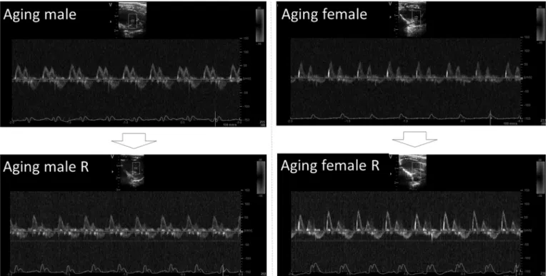
Figure 2.Representative Doppler echocardiographic images of transmitral inflow from listed groups. R, running.
Diastolic function of the left ventricle was slightly improved in both running groups, but the changes were again more prominent in running male rats (Fig. 2).
Transmitral A wave velocity decreased significantly in running male rats in comparison to aging males (P = 0.0002), thus E/A ratio increased (P= 0.0004). A wave ve- locity trended to decrease in running females (P= 0.0470) and E/A ratio failed to improve in comparison to aging female controls. E wave deceleration time (DecT) was rela- tively lengthened in aging males, but significantly short- ened in running males (P < 0.0001, compared to aging males). Value of DecT remained in the normal range in aging females and was unchanged in running females. Left ventricle outflow tract velocities and pressure gradients also remained unchanged. Tissue Doppler echocardiography revealed that TDI e0wave increased in both running male and running female rats in comparison to nonrunning counterparts (P = 0.0067 and P = 0.0209, respectively). The ratio of e0/a0 increased in running females compared to aging females (P= 0.0051) and showed trend toward increase in running males versus nonrunning animals (P = 0.0632).
The ratio of E/e0(indicative for left ventriclefilling pres- sure) improved significantly in both running male and
running female groups in comparison to matched aging controls (P = 0.0090 and P = 0.0087, respectively).
Isovolumic relaxation time (IVRT) was relatively long in aging males, but significantly shortened in running males (P = 0.0007). This parameter failed to improve in female rats. Tei-index decreased in both running females (P = 0.0170) and running males (P= 0.0226) compared to aging controls. In relation to right ventricle function, TAPSE pa- rameters remained unchanged among groups; however, a slight improvement could be detected in right ventricle dia- stolic function expressed as RV E/A ratio in running female animals compared to aging female controls (P= 0.0192).
Determination of Gene Expression and Protein Concentrations
Gene expression alterations are represented relative to aging control groups; 12-wk voluntary exercise–caused changes are depicted.
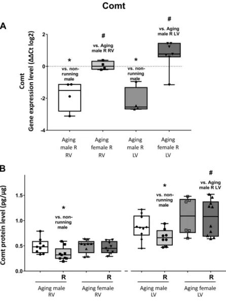
Comt.
As shown in Fig. 3A, a significant repression was found in the expression level of Comt in both ventricles of male
Figure 3.A: the effects of a 12-wk-long voluntary exercise on Comt gene expression in the right and left ventricles of the heart, evaluated by RT-PCR (Comt; change in relative gene expression ratios are shown asDDCT values (log2)).
Data represented as minimum to maximum;n= 4–7/group (individual data points are visible on the graph). Shapiro- Wilk method was used to estimate data distribution, and two-tailed Student’sttest was performed to estimate differ- ences between corresponding running and nonrunning () and between matching male or female (#) groups.B: the effects of a 12-wk-long voluntary exercise on Comt protein expression in the right and left ventricles of the heart, meas- ured by ELISA method. Comt protein expression is shown for each group. After Shapiro-Wilk test, two-tailed Student’s ttest was performed to estimate differences between corre- sponding running and nonrunning () and between match- ing male or female (#) groups. (Comt; expressed as pg/mg protein). Result shown as minimum to maximum;n= 6–9/
group (individual data points are shown for each group).
P<0.05: statistical significance between running and non- running counterparts; #P < 0.05: statistical significance between matching female and male groups. Comt, cate- chol-O-methyltransferase; RV, right ventricle; LV, left ventri- cle; R, running.
animals as a result of physical exercise. However, no signifi- cant change was observed in the case of running female group. Figure 3B shows that a significant decrease was observed in the level of cardiac Comt protein in the right and left ventricles of male rats as a result of exercise. Similarly to gene expression, no significant changes were observed in heart of female animals. Moreover, sex-dependent changes were also observed in the cardiac gene and protein expres- sion of Comt.
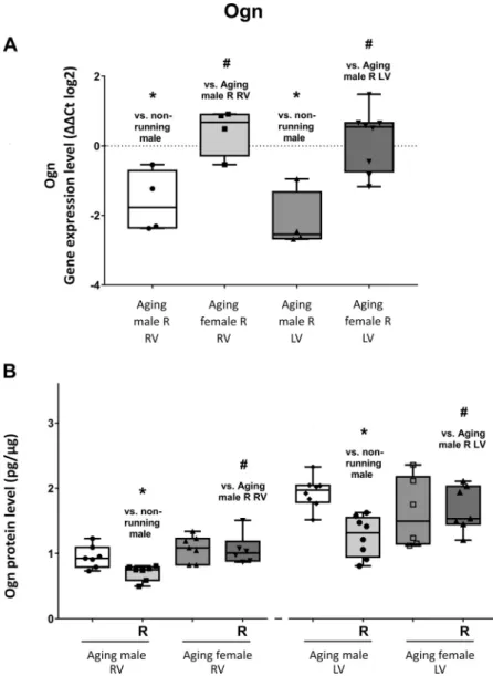
Ogn.
Figure 4A shows that cardiac Ogn expression was signifi- cantly suppressed in male running group due to the 12-wk- long exercise training. In contrast, in the heart of running female animals, no substantial change was noted at gene expression level. In line with gene expression, Ogn protein levels were also attenuated in running male rats. Moreover, a further decrease was found in the RV of females as a result of physical activity, as shown inFig. 4B. Regarding Ogn gene and protein expression, we also observed sexual dimorphism which was considered to be significant.
Pcp4.
Voluntary exercise training resulted in a significant overex- pression of Pcp4 gene in the LV of both running male and female rats. Moreover, Pcp4 expression displayed an eleva- tion in the RV of both groups, but this change could not be considered significant. In the case of protein expression, an increase was noted in both ventricles of the two sexes, and changes in the female LVs were considered to be significant.
In terms of cardiac Pcp4 protein and gene expression, a sex- based difference was also obtained. Data are presented in Fig. 5,AandB.
Esm1.
A stable overexpression of cardiac Esm1 gene was noted as a result of wheel-running exercise in both female and male rats compared to nonrunning counterparts, in addition changes in the LVs of both sexes were found to be signifi- cant. Voluntary exercise caused an elevation in the concen- tration of Esm1 protein in the RV of male rats, and in the heart of female rats, which alteration was found to be signifi- cant. Furthermore, significant sex-dependent changes were
Figure 4.A: the effects of a 12-wk-long voluntary exercise on Ogn gene expression in the right and left ventricles of the heart, evaluated by RT-PCR (Ogn; change in relative gene expression ratios are shown asDDCT values (log2)).
Data represented as minimum to maximum;n= 4–8/group (individual data points are visible on the graph). Shapiro- Wilk method was used to estimate data distribution, and two-tailed Student’sttest was performed to estimate differ- ences between corresponding running and nonrunning () and between matching male and female (#) groups.B: the effects of a 12-wk-long voluntary exercise on Ogn protein expression in the right and left ventricles of the heart, meas- ured by ELISA method. Ogn protein expression is shown for each group. After Shapiro-Wilk test, two-tailed Student’st test was performed to estimate differences between corre- sponding running and nonrunning () and between match- ing male or female (#) groups. (Ogn; expressed as pg/mg protein). Result shown as minimum to maximum;n= 6–8/
group (individual data points are shown for each group).
P<0.05: statistical significance between running and non- running counterparts; #P < 0.05: statistical significance between matching female and male groups. Ogn, osteogly- cin; RV, right ventricle; LV, left ventricle; R, running.
also observed in the cardiac gene and protein expression of Esm1. Data are shown inFig. 6,AandB.
Measurement of Myocardial Infarct Size
According toFig. 7, aging animals have shown a higher rate of infarct size in comparison with running counterparts and male rats represented worse outcomes. Nevertheless, as a result of the 12-wk voluntary exercise, we observed a pro- nounced decrease in the infarcted area of running females.
In addition, a significant attenuation was ascertained in the necrotic extension of the heart in running males.
DISCUSSION
In our present study, we clearly demonstrated the promi- nent role of 12-wk voluntary exercise in improvement of age- related cardiovascular dysfunctionality in aged female and male rats. Our results revealed that a maintained physical ac- tivity improved cardiovascular performance particularly in aged males by affecting functional parameters and moderat- ing the nonfunctional area in the infarcted myocardium.
Moreover, it greatly affected the gene expression in both
ventricles. Obviously, it has already revealed that female sex tend to be protected against ischemia and other pathological heart diseases or even aging (26). In our recent study, cardiac function of male animals deteriorated more dramatically when compared to the females. Hence, improvement of sys- tolic and diastolic cardiac function was more prominent in aged male animals after exercise training, along with a decreased infarcted area in hearts subjected to ischemic injury. Our main finding is that these favorable changes were accompanied by a decreased expression Comt and Ogn genes in the myocardium, whereas Pcp4 expression was found to be significantly increased.
The outcomes of echocardiographic measurements revealed the beneficial effects of exercise training on aged male and female rats. In comparison to nonrunning counterparts, left ventricular EF and FS measured in run- ning male and running female animals were significantly increased. Moreover, MAPSE values of running groups were significantly increased compared to aged controls.
These parameters together show a marked improvement in left ventricular systolic function in both male and female rats receiving exercise training. Data obtained Figure 5.A: the effects of a 12-wk-long voluntary exercise on Pcp4 gene expression in the right and left ventricles of the heart, evaluated by RT-PCR (Pcp4; change in relative gene expression ratios are shown asDDCT values (log2)).
Data represented as minimum to maximum;n= 3–7/group (individual data points are visible on the graph). Shapiro- Wilk method was used to estimate data distribution, and two-tailed Student’sttest was performed to estimate differ- ences between corresponding running and nonrunning () and between matching male or female (#) groups.B: the effects of a 12-wk-long voluntary exercise on Pcp4 protein expression in the right and left ventricles of the heart, meas- ured by ELISA method. Pcp protein expression is shown for each group. After Shapiro-Wilk test, two-tailed Student’st test was performed to estimate differences between corre- sponding running and nonrunning () and between match- ing male or female (#) groups. (Pcp; expressed as pg/mg protein). Result shown as minimum to maximum;n= 5–9/
group (individual data points are shown for each group).
P<0.05: statistical significance between running and non- running counterparts; #P < 0.05: statistical significance between matching female and male groups. Pcp4, Purkinje cell protein-4; RV, right ventricle; LV, left ventricle; R, running.
from Doppler imaging expose that diastolic function of the left ventricle was significantly improved in running male rats, illustrated by increased E/A ratio (decreased atrial wave velocity), normalized DecT, and IVRT. Since prolonged DecT and IVRT indicate impaired myocardial relaxation, relatively shorter durations of these parame- ters in running male group point out an improved dia- stolic function associated with exercise training (27).
These changes were absent in the running female group in comparison to aging female controls; however, it needs to be indicated that diastolic function of the aging female rats was relatively preserved. Tissue Doppler echocardi- ography also revealed that e0 wave velocity increased, while E/e0 ratio (indicative for LV filling pressure) decreased in both male and female groups as a result of exercise training in comparison to nonrunning counter- parts, and e0/a0ratio slightly improved in running female
rats, indicating better myocardial motility. In conclusion, aging male animals showed mild diastolic dysfunction (impaired relaxation and elevated LV filling pressures) which was absent in male counterparts received exercise training. Differences in diastolic parameters were less marked in female rats, as echocardiographic parameters of aging female group only showed mild impairment.
Myocardial performance index is a valuable parameter expressing both systolic and diastolic ventricle function (28). Elevated values of MPI were found in aging female group, which was significantly counteracted by exercise, showing beneficial effects of training on global heart functions. Male animals only showed trends toward decrease in Tei-index. Right ventricle E/A ratios were sig- nificantly increased in running female animals in com- parison to aged females, and no differences were found in TAPSE values among groups. These observations are in Figure 6.A: the effects of a 12-wk-long voluntary exercise
on Esm1 gene expression in the right and left ventricles of the heart, evaluated by RT-PCR (Esm1; change in relative gene expression ratios are shown asDDCT values (log2)).
Data represented as minimum to maximum;n= 4–8/group (individual data points are visible on the graph). Shapiro- Wilk method was used to estimate data distribution, and two-tailed Student’sttest was performed to estimate differ- ences between corresponding running and nonrunning () and between matching male or female (#) groups. B: the effects of a 12-wk-long voluntary exercise on Esm1 protein expression in the right and left ventricles of the heart, meas- ured by ELISA method. Esm1 protein expression is shown for each group. After Shapiro-Wilk test, two-tailed Student’s ttest was performed to estimate differences between corre- sponding running and nonrunning () and between match- ing male or female (#) groups. (Esm1; expressed as pg/mg protein). Result shown as minimum to maximum;n= 5–9/
group (individual data points are shown for each group).
P<0.05: statistical significance between running and non- running counterparts; #P < 0.05: statistical significance between matching female and male groups. Esm1, endothe- lin 1; RV, right ventricle; LV, left ventricle; R, running.
line with several human clinical studies, which corrobo- rate the cardiac-improving effects of exercise (29–31).
Here we showed that aging affects both systolic and dia- stolic parameters of the rat heart, but diastolic impair- ment is more significant in males. Voluntary exercise training successfully improved systolic function in both groups (EF, FS, MAPSE, Tei-index), but its effects were more prominent in male rats, since all signs of the early di- astolic dysfunction (decreased E/A ratio, lengthened DecT and IVRT, and increased E/e0) were absent in the running male group, in contrast to sedentary aging controls.
Sex-dependent changes were also observed in the expres- sion of genes (Comt, Ogn, Pcp4, Esm1) in the myocardial tis- sue in response to aging and exercise training. Comt is a key enzyme in the degradation of catecholamines and catechol estrogen, hence its role in the cardiovascular regulation is substantial. A number of studies support that elevated cate- cholamine levels implicate cardiac dysfunction and induced intracellular calcium overload in cardiomyocytes (11). Long- term exercise training improves heart function by lowering blood pressure and heart rate, which is considered to be an evidence of decreased catecholamine levels. Moreover, low
activity of the Comt gene is associated with a decreased risk of myocardial infarction in human subjects (32). It is note- worthy that Graham et al. (33) discussed that MAO-B inhibitors and COMT inhibitors do not increase mortality compared to placebo; however, the link between the COMT inhibitors and acute myocardial infarction remains to be proven.
We verified that 12-wk exercise resulted in a significant suppression of Comt gene expression in the heart of male rats; however, the aforementioned change was not observed in female animals. Similar to gene expression, protein levels of cardiac Comt were decreased as a result of voluntary exer- cise, which was seen in both sexes. Carneiro et al. (13)first reported the plausible connection between exercise therapy and decreased Comt expression which is consistent with our results. Thesefindings suggest that the attenuated presence of Comt can be proportional to the reduced level of catechol- amines, thus improved cardiac function in response to exer- cise training.
Ogn (osteoglycin or mimecan) is reportedly expressed in fibroblasts, vascular smooth muscle cells, and cardiomyo- cytes, however, its precise role in subcellular signaling is yet to be determined. Ogn expression was found to be increased in the infarcted myocardium, where it may play a role in col- lagen maturation (34). Also, Ogn expression was increased in patients with coronary heart disease (CHD) and correlated to the severity of coronary lesions (9). Here, we found decreased expression of the Ogn gene and protein in the ven- tricles of the running male group, in line with the improved echocardiographic markers and reduced infarcted area.
Although our knowledge of Ogn function in the trained heart is limited, our results are further corroborated by Feng and colleagues (35), who reported a similar decrease in Ogn expression after a 6-wk exercise training in the aortic wall of hypertensive rats. Additionally, injection of irisin, a recently described pro-myogenic factor (exercise-induced myokine) also promotes the down-regulation of the Ogn gene in the skeletal muscle (36). Regarding translational aspects, our knowledge is limited about Ogn function; furthermore, the expression of Ogn may be not only tissue-dependent, but also species-dependent. In contrast to data obtained in rodent models, proteoglycan genes (biglycan, osteoglycin, serglycin) in human striated muscles are rather increased in response to exercise, indicating extracellular matrix remod- eling (37). The picture is further complicated considering that Ogn is posttranscriptionally regulated by micro-RNAs (miR-22) for the control of proliferation and differentiation of muscle cells during myogenesis (38). Additionally, Ogn was recently identified as a cardiac hypertrophy marker, and its increased expression was associated with elevated left ventricle mass (8). As regards our data, we did notfind signif- icant differences in left ventricle mass of running and seden- tary groups. Notwithstanding, this result was expected, as according to previous studies, a 12-wk-long voluntary exer- cise (unlike forced training or swimming) is not sufficient to cause significant cardiac hypertrophy and elevated LV mass (39). Physiological stimuli (voluntary exercise) may rather decrease cardiac hypertrophy and remodeling along with elevated Ogn levels (8). Although demonstration of the Ogn mechanism causing a hypertrophic response requires fur- ther investigation, it is proved to be a decisive factor in this Figure 7.A: the effects of a 12-wk-long voluntary exercise on the exten-
sion of the infarct size, evaluated by TTC staining after I/R injury. Infarct size was lower in the aged females, and voluntary exercise decreased infarcted area in the aged male rats compared to the nonrunning counter- parts. Shapiro-Wilk method was used to estimate data distribution, and two-tailed Student’sttest was performed to estimate differences between corresponding running and nonrunning () and between matching male or female (#) groups. Infarct size was calculated as the percentage of the area at risk. Results are shown as minimum to maximum and mean;n= 6/
group.P<0.05: statistical significance between running and nonrunning counterparts; #P<0.05: statistical significance between matching female and male groups. R, running.B: representative images of TTC-stained rat heart sections from listed groups, 24 hours after I/R injury. Images are positioned below the corresponding groups.
process. As Ogn expression was found to be decreased in the myocardium of the running male animals, we conclude that in terms of exercise, Ogn is rather a marker of susceptibility to MI than cardiac hypertrophy in the aged rat. These results suggest that exercise training–caused Ogn suppression may ameliorate aging-associated adverse changes of the heart.
Pcp4 (or PEP-19) encodes a small IQ-motif protein that is a Ca2þ/calmodulin-dependent protein kinase II (CaMKII) reg- ulator, thus it plays a major role in the intracellular calcium homeostasis (40). This system has recently been proposed to counteract cardiac hypertrophy and apoptosis in disease models (10). Kim et al. previously observed ventricular arrhythmias in a Pcp4-null model in vivo and proved the car- diac excitability modulator properties of Pcp4 (41). In addi- tion, they showed that Pcp4 is down-regulated in disease models (aortic banding, phenylephrine infusion), along with marked echocardiographic changes showing impaired car- diac function and hypertrophy. Consistently, Xie et al. (10) showed that Pcp4 has antihypertrophic effects through inhi- bition of CaMKII and calcineurin (CaN) activation during de- velopment of cardiomyocyte hypertrophy. Pcp4 transfection inhibited apoptosis and protected HEK293T cells against death due to Ca2þ overload. In addition, Pcp4 reduced Ca2þ release from the sarcoplasmic reticulum of cardiomyocytes and also inhibited angiotensin-induced overload of calcium ions. As Ca2þoverload is a major cause of arrhythmias, hy- pertrophy, and increased susceptibility to ischemia-reperfu- sion (I/R) injury, it is proposed that Pcp4 plays an important role in cardioprotection via normalizing Ca handling of myo- cytes through the CaMKII system (42). Our current results show for thefirst time that a 12-wk-long exercise period was able to enhance the expression of Pcp4 gene in the heart of both male and female rats. Aligned with gene expression, Pcp4 protein levels were also elevated in the right ventricles of both sexes. These results suggest that the exercise- induced Pcp4 elevation may contribute to cardioprotection in the aged heart. Aging-associated calcium mishandling may directly affect echocardiographic diastolic function (IVRT, E/e0) through the SERCA/PLB pathway, a major
regulator of cardiac relaxation (43, 44). Consistent to gene expression data, IVRT and E/e0parameters were significantly improved in running animals, further suggesting the normal- ized Ca2þhandling in cardiomyocytes, as a result of exercise.
Pcp4 overexpression may contribute to intracellular calcium balance, normalized cardiac cell excitability; and counteract Ca2þ overload, a significant contributor of hypertrophy and increased susceptibility to I/R injury. Notwithstanding, as li- mitation of this study, these factors were not investigated and may be addressed in future investigations.
Esm1 has recently been associated with vascularization processes. By connecting to the VEGF signaling, it is sus- pected to play an important role in neoangiogenesis (45). It is well known that exercise is able to enhance VEGF produc- tion, which is one of the main inducers of Esm1 (21). In this study, we observed a strong overexpression of cardiac Esm1 in all groups as a result of the 12-wk voluntary exercise com- pared to nonrunning counterparts. In line with genetic mod- ifications, Esm1 protein levels were also elevated in the heart of female rats and in the right ventricle of male animals. Our findings demonstrate that a long-term exercise training may promote angiogenesis through the induction of Esm1; how- ever, the exact underlying molecular mechanisms need to be investigated in the future. As a limitation of this study, we did not measure VEGF levels or other angiogenesis markers, thus evaluating the role of Esm1 in cardiac aging and response to exercise training would be speculative at the moment.
In addition to the beneficial effects of sport on general car- diac function, it also has a protective effect in response to I/
R-induced injury. The ligation of LAD via Langendorff sys- tem imitates the occurrence of myocardial infarction– related pathophysiological aspects. Therefore, the ratio of necrotic area followed by I/R injury has been clearly eval- uated in both running and nonrunning groups. In a previous work of ours, we have already demonstrated that voluntary exercise reduced myocardial infarct size, and thus provided protection against I/R injury (46). In addition, several studies revealed that exercise was able to modify favorably the Figure 8.Summary of the study. Exercise
was associated with decreased Ogn and Comt expression; the presence of Pcp4 and Esm1 was enhanced in aged rats. Sex- dependent changes were also observed in the expression of the cardiovascular key proteins. Comt, catechol-O-methyltransfer- ase; Esm1, endothelin-1; Pcp4, Purkinje cell protein-4; Ogn, osteoglycin. Thisfigure was created using Servier Medical Art tem- plates, licensed under a Creative Commons Attribution 3.0 License;https://smart.servier.
com
adverse changes after myocardial infarction (30,47). Similarly, in this study we observed a marked improvement in the exten- sion of infarct size as a result of 12-wk voluntary exercise. In support of echocardiographic results, it can be stated that long-term exercise training has a protective effect even in the case of acute injury.
These results are consistent with exercise-induced gene expression changes observed in the heart. Although the cen- tral role of maintained physical activity is already cleared, the underlying molecular and genetic mechanisms affecting cardiac functions are still being extensively analyzed. Our results clearly highlight that long-term exercise can pro- foundly induce beneficial changes in the cardiovascular per- formance by affecting gene expression (Fig. 8).
CONCLUSIONS
Over the years, average life expectancy has more than doubled; therefore, understanding and reversing the aging- associated processes have never been more important. Gene- targeted investigations may contribute to the understanding of overall physiological changes influenced by exercise and exercise-related therapies in the future. In addition, a bet- ter insight into cardiac aging may unravel molecular path- ways related to cardiac pathophysiology and contribute to improved prevention of cardiovascular diseases.
GRANTS
This work was supported by the European Union, the State of Hungary, and the University of Debrecen Grants GINOP-2.3.4–15- 2016-00002 and ED_18-1-2019-0028 (Excelling Research Areas).
This work was also supported by the New National Excellence Program of the Ministry for Innovation and Technology from the source of the National Research, Development and Innovation Fund ÚNKP-20-4 (to R. Szabo) and ÚNKP-20-3 (to D. B€orzsei) and by 2019-1.1.1-PIACI-KFI-2019-00390. The research was financed by the Higher Education Institutional Excellence Program (NKFIH- 1150-6/2019) of the Ministry of Innovation and Technology in Hungary, within the framework of the thematic program of the University of Debrecen.
DISCLOSURES
No conflicts of interest,financial or otherwise, are declared by the authors.
AUTHOR CONTRIBUTIONS
A.P. conceived and designed research; D.B., D. P., R.S., M.B., Z.K., Z.R., K.K., A.M.B., and A.P. performed experiments; D.B., D.P., R.S., L.Z.F., B.J., and A.P. analyzed data; R.S., M.B., L.G.P., A.M.B., and B.J. interpreted results of experiments; D.B., D.P., and Z.R. prepared figures; D.B., D.P., Z.K., and L.Z.F. drafted manu- script; K.K., C.V., B.J., and A.P. edited and revised manuscript; C.V.
and A.P. approvedfinal version of manuscript.
REFERENCES
1. Haines DD,Tosaki A.Role of heme oxygenases in cardiovascular syndromes and co-morbidities. Curr Pharm Des24: 2322–2325, 2018. doi:10.2174/1381612824666180727110353.
2. Tosaki A.Pharmacological mechanisms and interventions in ische- mia/reperfusion-induced injury. Curr Pharm Des 19: 6839–6841, 2013. doi:10.2174/138161281939131127103545.
3. Biragyn A, Ferrucci L. Gut dysbiosis: a potential link between increased cancer risk in ageing and inflammaging.Lancet Oncol19:
e295–e304, 2018. doi:10.1016/S1470-2045(18)30095-0.
4. Haines DD,Juhasz B,Tosaki A.Management of multicellular senes- cence and oxidative stress.J Cell Mol Med 17: 936–957, 2013.
doi:10.1111/jcmm.12074.
5. Szabo R,Karacsonyi Z,Borzsei D,Juhasz B,Al-Awar A,Torok S, Berko AM,Takacs I,Kupai K,Varga C,Posa A.Role of exercise- induced cardiac remodeling in ovariectomized female rats.Oxid Med Cell Longev2018: 6709742, 2018. doi:10.1155/2018/6709742.
6. Ungvari Z,Valcarcel-Ares MN,Tarantini S,Yabluchanskiy A,Fulop GA,Kiss T,Csiszar A.Connective tissue growth factor (CTGF) in age-related vascular pathologies.Geroscience39: 491–498, 2017.
doi:10.1007/s11357-017-9995-5.
7. Steenman M, Lande G. Cardiac aging and heart disease in humans. Biophys Rev 9: 131–137, 2017. doi:10.1007/s12551-017- 0255-9.
8. Petretto E,Sarwar R,Grieve I,Lu H,Kumaran MK,Muckett PJ, Mangion J,Schroen B,Benson M,Punjabi PP,Prasad SK,Pennell DJ,Kiesewetter C,Tasheva ES,Corpuz LM,Webb MD,Conrad GW,Kurtz TW,Kren V,Fischer J,Hubner N,Pinto YM,Pravenec M, Aitman TJ, Cook SA. Integrated genomic approaches implicate osteoglycin (Ogn) in the regulation of left ventricular mass. Nat Genet40: 546–552, 2008. doi:10.1038/ng.134.
9. Hu Y,Liu J, Zhao Q,Xu P,Chen Y,Zhou H, Li X.Correlation between mimecan expression and coronary artery stenosis in patients with coronary heart disease.Int J Clin Exp Med8: 21641– 21646, 2015.
10. Xie YY,Sun MM,Lou XF,Zhang C,Han F,Zhang BY,Wang P,Lu YM.Overexpression of PEP-19 suppresses angiotensin II-induced cardiomyocyte hypertrophy.J Pharmacol Sci125: 274–282, 2014.
doi:10.1254/jphs.13208fp.
11. Adameova A, Abdellatif Y, Dhalla NS. Role of the excessive amounts of circulating catecholamines and glucocorticoids in stress- induced heart disease.Can J Physiol Pharmacol87: 493–514, 2009.
doi:10.1139/y09-042.
12. Almas A,Forsell Y,Millischer V,M€oller J,Lavebratt C.Association of catechol-O-methyltransferase (COMT Val(158)Met) with future risk of cardiovascular disease in depressed individuals—a Swedish pop- ulation-based cohort study. BMC Med Genet 19: 126, 2018.
doi:10.1186/s12881-018-0645-2.
13. Carneiro LS, Fonseca AM, Serrão P, Mota MP, Vasconcelos- Raposo J,Vieira-Coelho MA.Impact of physical exercise on cate- chol-O-methyltransferase activity in depressive patients: a prelimi- nary communication.J Affect Disord193: 117–122, 2016. doi:10.1016/j.
jad.2015.12.035.
14. Agarwal SK.Cardiovascular benefits of exercise.Int J Gen Med5:
541–545, 2012. doi:10.2147/IJGM.S30113.
15. Nystoriak MA,Bhatnagar A.Cardiovascular effects and benefits of exercise. Front Cardiovasc Med 5: 135, 2018. doi:10.3389/
fcvm.2018.00135.
16. Lee IM,Paffenbarger RS, Jr,Hennekens CH.Physical activity, physical fitness and longevity. Aging (Milano) 9: 2–11, 1997.
doi:10.1007/BF03340123.
17. Blair SN,Jackson AS.Physicalfitness and activity as separate heart disease risk factors: a meta-analysis.Med Sci Sports Exerc33: 762– 764, 2001. doi:10.1097/00005768-200105000-00013.
18. Pinckard K, Baskin KK, Stanford KI. Effects of exercise to improve cardiovascular health.Front Cardiovasc Med6: 69, 2019.
doi:10.3389/fcvm.2019.00069.
19. Simioni C,Zauli G,Martelli AM,Vitale M,Sacchetti G,Gonelli A, Neri LM.Oxidative stress: role of physical exercise and antioxidant nutraceuticals in adulthood and aging.Oncotarget9: 17181–17198, 2018. doi:10.18632/oncotarget.24729.
20. Kissane RWP,Wright O,Al’Joboori YD,Marczak P,Ichiyama RM, Egginton S.Effects of treadmill training on microvascular remodel- ing in the rat after spinal cord injury.Muscle Nerve59: 370–379, 2019. doi:10.1002/mus.26379.
21. Rocha SF,Schiller M,Jing D,Li H,Butz S,Vestweber D,Biljes D, Drexler HC,Nieminen-Kelh€a M,Vajkoczy P,Adams S,Benedito R, Adams RH.Esm1 modulates endothelial tip cell behavior and vascu- lar permeability by enhancing VEGF bioavailability.Circ Res 115:
581–590, 2014. doi:10.1161/CIRCRESAHA.115.304718.